Submitted Successfully!
To reward your contribution, here is a gift for you: A free trial for our video production service.
Thank you for your contribution! You can also upload a video entry or images related to this topic.
Version Summary Created by Modification Content Size Created at Operation
1 -- 3276 2023-06-18 16:16:37 |
2 Reference format revised. Meta information modification 3276 2023-06-19 03:33:43 |

Video Upload Options

Do you have a full video?

Confirm

Are you sure to Delete?
Cite
If you have any further questions, please contact Encyclopedia Editorial Office.
Rojas-Pirela, M.; Kemmerling, U.; Quiñones, W.; Michels, P.A.M.; Rojas, V. Antimicrobial Peptides as Potential Therapeutic Strategy against Trypanosomiases. Encyclopedia. Available online: https://encyclopedia.pub/entry/45759 (accessed on 27 July 2024).
Rojas-Pirela M, Kemmerling U, Quiñones W, Michels PAM, Rojas V. Antimicrobial Peptides as Potential Therapeutic Strategy against Trypanosomiases. Encyclopedia. Available at: https://encyclopedia.pub/entry/45759. Accessed July 27, 2024.
Rojas-Pirela, Maura, Ulrike Kemmerling, Wilfredo Quiñones, Paul A. M. Michels, Verónica Rojas. "Antimicrobial Peptides as Potential Therapeutic Strategy against Trypanosomiases" Encyclopedia, https://encyclopedia.pub/entry/45759 (accessed July 27, 2024).
Rojas-Pirela, M., Kemmerling, U., Quiñones, W., Michels, P.A.M., & Rojas, V. (2023, June 18). Antimicrobial Peptides as Potential Therapeutic Strategy against Trypanosomiases. In Encyclopedia. https://encyclopedia.pub/entry/45759
Rojas-Pirela, Maura, et al. "Antimicrobial Peptides as Potential Therapeutic Strategy against Trypanosomiases." Encyclopedia. Web. 18 June, 2023.
Antimicrobial Peptides as Potential Therapeutic Strategy against Trypanosomiases
Edit

Trypanosomiases are a group of tropical diseases that have devastating health and socio-economic effects worldwide. In humans, these diseases are caused by the pathogenic kinetoplastids Trypanosoma brucei, causing African trypanosomiasis or sleeping sickness, and Trypanosoma cruzi, causing American trypanosomiasis or Chagas disease. Antimicrobial peptides (AMPs) are small peptides synthesized by both prokaryotes and (unicellular and multicellular) eukaryotes, where they fulfill functions related to competition strategy with other organisms and immune defense. These AMPs can bind and induce perturbation in cell membranes, leading to permeation of molecules, alteration of morphology, disruption of cellular homeostasis, and activation of cell death. These peptides have activity against various pathogenic microorganisms, including parasitic protists. 

trypanosomiases human sleeping sickness Chagas disease antimicrobial peptides anti-Trypanosoma activity

1. Introduction

Kinetoplastids are a group of globally distributed flagellated protists which include both free-living and parasitic species responsible for serious diseases in animals and humans. These protists are distinguished by the presence of a large DNA network-containing region, known as “kinetoplast”, in their single large mitochondrion [1]. Many of the organisms that make up this group have other common characteristics such as (I) the presence of a single flagellum that originates near the kinetoplast of the mitochondrion and emanates from a pocket in the cell membrane (except for the intracellular form of Trypanosoma cruzi); (II) the presence of essential organelles called glycosomes, which are modified peroxisomes in which the first seven steps of glycolysis and several other metabolic processes are carried out; (III) a complex life cycle that involves multiple morphological stages with dramatic changes in their protein expression, metabolism, and membrane composition; (IV) the species-specific production of molecules that are critical for their survival and immune evasion of host; and (V) the presence of 6000 orthologous genes in common between different species that cause different diseases [2][3][4].
Within this group of organisms are included species that cause human diseases such as African trypanosomiasis (HAT or sleeping sickness), which is caused by two infective subspecies of Trypanosoma brucei, and Chagas disease (CD), which is caused by T. cruzi, both are considered Neglected Diseases by the World Health Organization (WHO) [4][5][6]. These kinetoplastid diseases affect millions of people in low- and middle-income countries, located mainly in tropical and subtropical regions, causing around 30,000 deaths per year and inducing disabling morbidities in millions more [2][5][7]. The use of drugs for the treatment of these diseases has important limitations since, in addition to many available drugs date from the early and middle of the 20th century, they have limited efficacy in advanced stages of the disease, are non-specific, and/or are highly toxic [4][7]. Additionally, in the case of CD, T. cruzi can adopt quiescent and phenotypically drug-resistant forms. For its part, T. brucei can reside in the skin and other organs and remain undetected for a long time, even in the absence of detectable parasitemia. All this could contribute to refraction to drug treatment and, in turn, would imply the need for the development of new drugs and therapeutic alternatives for the treatment of these diseases [8][9][10][11].

2. Antimicrobial Peptides (AMPs)

AMPs are a class of small peptides synthesized by pro- and eukaryotic organisms, used as a strategy for competition and defense during invasion by foreign organisms. They are encoded by specific genes and expressed constitutively or in response to specific environmental stimuli [12]. In some insects, AMPs are key for vector–microorganism interaction and are effective against both quiescent and actively proliferating pathogenic organisms [13][14][15].
These peptides are synthesized through three pathways, which include classical ribosomal synthesis, non-ribosomal synthesis, and proteolytic digestion of proteins. Ribosomally synthesized AMPs (RS-AMPs) are those encoded by genes and produced by ribosomal translation of specific mRNAs into the biologically active amino acids sequences. These AMPs are widely distributed in nature, produced by various organisms (such vertebrates, insects, plants, and bacteria) [16][17]. Among the RS-AMPs are the mammalian defensins and amphibian dermaceptins [16]. Non-ribosomally synthesized AMPs (NR-AMPs) are produced by enzymes known as non-ribosomal peptide synthases (NRPSs), which incorporate non-proteinogenic amino acids into the sequence and are found mainly in filamentous fungi and bacteria [17][18]. So far, hundreds of AMPs synthesized in a NRPS-dependent manner have been described, among which are gramicidin S and isopenicillin [19]. Other AMPs are produced via the proteolytic digestion pathway (peptides also known as cryptides) by proteases-mediated cleavage of precursor proteins or larger proteins with other functions, to yield matured bioactive factors [17][20]. During these processes, various fragmented peptides are also produced that can vary in their biological activity [20]. Buforin II is one of the most studied cryptid peptides [21].
Although natural AMPs are molecules with considerable diversity in their structural properties, origins, and mechanisms of action, they have certain characteristics in common. Generally, they are short molecules (≈10–100 amino acids) of a cationic nature at neutral pH (generally ranging from +2 to +11), which facilitates their interaction with charged cell membranes through electrostatic interaction [22][23]. Additionally, most AMPs have a considerable proportion of hydrophobic residues (close to 50%) and an amphipathic structure [23][24]. This latter property is responsible for their structural flexibility and solubility in aqueous environments. [23]. The overall positive net charge and amphipathicity are the two characteristics that contribute to the high affinity of AMPs for membranes [25]. Structurally, AMPs are commonly classified into four groups based on their secondary structure, which include linear α-helical peptides, β-sheet peptides (usually stabilized with one or more disulfide bonds), linear extension or loop (devoid of α- or β-elements) structure, and mixed (α-helical/β-sheet) peptides [22][23][25] (Figure 1). However, some peptides with cyclic structures and unusual complete topologies have also been documented [25]. Most studied among the groups of AMPs are the peptides with an α-helix structure [26].
Figure 1. Structural classification common of naturally antimicrobial peptides (AMPs). Representative examples of common structural classes of AMPs. (A). α-Helical: structure of human cathelicidin LL-37 (PDB ID:2k60). (B). β-Sheet: polyphemusin I (PDB ID:1RKK). (C). Extended or loop: indolicidin (PDB ID:1G89). (D). Mixed (contain both α-helical and β-sheet elements): Defensin A (PDB ID: 1ICA). Created with BioRender.com (accessed on 20 February 2023).

3. Current Treatment of Trypanosomiases

Although standard therapies are available for treatment of trypanosomiases, these are mainly based on synthetic drugs mostly developed more than 40–50 years ago, several of them highly toxic, and their use depends on the stage of the disease and/or trypanosome species causing the infection [27][28].
Treatments for HAT involve five synthetic drugs, pentamidine, suramin sulfate, melarsoprol, nifurtimox/eflornithine combination (NECT), and fexinidazole. The mechanism of action of these drugs is mainly based on causing DNA damage and the inhibition of enzymes involved in various cellular processes of the parasite (DNA replication, glutathione metabolism, trypanothione biosynthesis, NADH/NAD+ balance maintenance, mitochondrial mRNA editing, and glycolysis) [28][29]. Most of these drugs are specific for treating infections caused by either T. b. rhodesiense or T. b. gambiense, except for suramin sulfate, used to treat infections caused by both parasites [28]. The efficacy of these drugs depends on the stage of the disease. Pentamidine and suramin are used during the initial stage of HAT (hemolymphatic), whereas melarsoprol, eflornithine, and NECT are used during the advanced stage of the disease, when parasites have migrated to the central nervous system. All these drugs require prolonged use, intravenous infusion, and are highly costly, often resulting in non-compliance and abandonment of treatment [28]. Also, the administration of these drugs generally has associated side effects that in some cases can be fatal and appear in the first days of treatment [28][30][31]. Another disadvantage is the development of resistance to these drugs that is mainly associated with the loss of function of the parasite’s transporters that mediate their internalization [28][32].
Regarding Chagas disease, currently only two drugs, benznidazole (BZN) and nifurtimox (NF) are licensed for the treatment of this disease. The mechanism of action of both drugs involves intracellular activation of a mitochondrial NADH-dependent type-I nitroreductase (TcNTR), which gives rise to intermediates (free radicals and/or electrophilic metabolites) that bind to intracellular macromolecules and inhibit several vital biological processes of the parasite (DNA synthesis, DNA and RNA metabolism, protein synthesis, and energy metabolism) [27][33]. The efficiency of NF and BZN depends on the stage of the disease. These drugs tend to be less effective in the chronic phase, where the cure figure hardly reaches 20–30% [34][35]. Although both compounds are administered orally in two or three doses, treatment is discontinued in 9–75% of patients due to severe side effects [27][36]. Additionally, the use of these drugs is not recommended during pregnancy and lactation, and in the case of NF, it is only approved for newborns over 2.5 kg [37][38], meaning a limitation for the prevention of vertical transmission of the parasite and timely treatment of congenital CD. The occurrence of resistance in strains, mediated by various mechanisms (e.g., loss/mutations/polymorphism of TcNTR) [39][40] are other limitations of the clinical use of BNZ and NF. Notably, these drugs cannot prevent or reverse the damage caused, especially in the heart, by inflammation in response to T. cruzi infection, even in conditions where a decrease in parasitic load has been observed [41][42].
Most of the drugs currently available for the treatment of trypanosomiases have an ancient origin and high toxicity. Others, despite being specific and efficient for the early and advanced stages of infection, depend on an enzyme or membrane transporter of the parasite for their activation. The latter generally implies a probability of resistance development over time. For all these reasons, there is a need to develop or search compounds that will overcome these limitations observed in anti-trypanosome drugs available on the market. Although this search may seem very demanding, these “ideal drug candidates” could be found in various natural sources. This will be discussed in more depth in the next section.

4. AMPs with Antiparasitic Activity

Several studies have shown the antiparasitic effect of some AMPs [43][44][45], including activity against parasites that cause important tropical diseases [46] (Figure 1). Many of these AMPs have been isolated from various vertebrate and invertebrate hosts of these parasites [44][47][48].
Plasmodium is the parasite on which most studies with AMPs have been carried out [49]. In this protist, many natural AMPs act primarily by disrupting the integrity of cell membranes [50][51][52][53][54][55]. However, some others can interfere with other important cellular processes of the parasite. In Plasmodium berghei, some fungal AMPs have an inhibitory effect on histone deacetylase (HDA), thus inducing histone hypermethylation and subsequent alteration of gene expression in the parasite [56]. Other AMPs derived from Gram-positive bacteria, such as epoxomicin and derivatives of the natural cyclic oligopeptide thiostrepton, have an inhibitory effect on protein synthesis and turnover, due to their binding to and inhibition of catalytic activity of proteasome β subunits (20S) [57][58]. Additionally, thiostrepton can inhibit mRNA translation in the apicoplast through its binding to the plasmodial organellar rRNA promoting structural alterations that prevent its function during protein synthesis [58][59]. Importantly, antimalarial activities have been attributed to some AMPs with semi-synthetic and synthetic origin. Synthetic AMPs inhibit the plasmodial cysteine protease falcipain and aspartic proteases plasmepsin I and plasmepsin II, involved in hemoglobin hydrolysis and hemozoin formation, thus interfering with parasite metabolism and growth [60][61][62]. Notably, some synthetic peptides have also shown an effect on some enzymes such as topoisomerase I, affecting the parasite’s DNA metabolism [63]. Several of these AMPs not only have antiplasmodial activity against different developmental stages of some Plasmodium species (P. falciparum, P. berghei, and P. yoelii nigeriensis) in vitro conditions [54][57][64], but are also effective at high parasitemia in an animal model [55].
In helminths, studies have focused primarily on Schistosoma and Brugia. In these parasites, AMPs have effects on motility, development, egg deposition, and the integumentary surface [65][66][67][68][69]. In Brugia pahangi, synthetic cecropins A/B, AMPs from insect hemolymph, attenuate microfilariae mobility and larval development in adult female Aedes aegypti [65]. In Schistosoma, dermaseptin, a peptide isolated from frogs, can synergistically interact with other natural compounds and contribute to parasite killing and infection control. In combination with piplartine, an amide alkaloid of Piper longum L. (long piper), dermaseptin not only exerts activity against the Schistosoma mansoni (S. mansoni) stages (schistosomula and adult) and affects the reproductive fitness of adult worms, but also induces structural alterations of the tegument and extensive destruction of the tubercles [66][67]. Although the anthelmintic mechanism of AMPs has not been elucidated, it has been proposed that disruption of cell structure by pore formation by direct interaction with the lipid bilayer seems to be the most likely [65][67][70][71]. It should be noted that the integument is essential for the survival of the helminth parasites, since it is involved in nutrient absorption and in the interaction with the host [72][73][74][75]. In both Brugia and Schistosoma, divalent metal transporter 1 (DMT1) molecules are present in the integument and are essential for the absorption of iron, an essential ion for the development and reproduction of these parasites [73][75][76]. In this sense, directing AMPs against the tegument of these parasites could be a good anthelmintic strategy.
Unlike in apicomplexan parasites, the AMPs tested so far on helminths exert their antiparasitic action at micromolar concentrations [65][66][67]. Some AMPs with antimicrobial properties have been discovered in helminths [71][77][78]. In S. mansoni, an AMP called schistocins has been obtained from the protein SmKI-1, a key protein for the survival of this nematode, which has activity against Schistosoma itself [71]. Likewise, putative neuropeptides derived from this parasite alter the behavior of the cercariae stage, therefore their use has been proposed as strategy for the control of the infection [69].
In trypanosomes such as Trypanosoma evansi and Trypanosoma equiperdum, causing surra and dourine in animals, some AMPs have been shown to exert an trypanocidal effect; hence, they have been proposed for use in new treatment strategies of trypanosomiasis in animals [79][80]. Furthermore, AMPs isolated from triatomine hemolymph have been shown to have trypanolytic activity against different strains of Trypanosoma rangeli, an infectious but non-pathogenic human parasite [81]. In these trypanosomes, AMPs exert their action through different mechanisms, including plasma membrane permeabilization, mitochondrial alteration, and parasite lysis [79][80][81].

5. Antimicrobial Peptides against Kinetoplastids Causing Neglected Tropical Diseases

5.1. AMPs against T. brucei

Many of the AMPs that are active against T. brucei are produced by a wide variety of organisms, including mammals and the insect vector [82][83]. These can carry out their action extracellularly, by plasma membrane disturbance, or intracellularly, by altering the function of some intracellular compartments [82][82][83][84] (Figure 2).
Figure 2. Mechanisms of action of antimicrobial peptides (AMPs) against T. brucei. (A). AMPs derived from the vertebrate host and insect vector. Peptides isolated from both vectors and hosts exert their trypanocidal effect through membrane perturbation and induction of cell lysis. (B). Mechanism of action of neuropeptides (NPs). The killing of trypanosomes by NPs requires the NPs to be endocytosed through the flagellar pocket and transported from the endosomes to the acidified lysosome, where they break the lysosomal bilayer membrane and accumulate in the cytoplasm. Once in the cytoplasm, their interference in various cellular processes contributes to morphological alterations and disturbance of organelles (glycosomes and mitochondrion), which ultimately lead to depletion of ATP and failure of the energy metabolism. (C). AMPs isolated from natural sources (bacteria, fungi, and insects). (C.1,C.2). AMPs derived from fungi. The lipopeptide amphomycin, inhibits the biosynthesis of the glycolipid precursor of glycosylphosphatidylinositol (GPI) by which the variant surface glycoproteins (VSGs) are anchored to the plasma membrane of these parasites (C.1). Leucinostatins (A and B) and alamethicin act as ionophores and pore formers in the membranes, causing alteration of cellular homeostasis, ultimately leading to the death of the parasite. (C.3). AMPs isolated from bacteria. Bacteriocin AS-48 targets intracellular compartments without plasma membrane permeabilization. AS-48 may interact at the surface with VSGs and then promotes its internalization through a clathrin-mediated endocytic process. In the cytoplasm, it induces structural alterations and autophagy-like cell death. (C.4). AMPs derived from bee venom. Melittin induces an increased influx of Ca2+ through the plasma membrane or increased release from acidocalcisomes. Excess Ca2+ accumulated intracellularly is stored in the mitochondrion, causing a reduced mitochondrial membrane potential, disorganization of kinetoplast DNA, autophagy, and cell death. (D). Synthetic AMPs. (D.1). SHPs intercalate and insert deeply into the plasma membrane, resulting in changes in the distribution of membrane components, increased membrane stiffness, loss of cell motility, and cell death. (D.2). For their part, the CPPs cross the membrane, accumulate in the cytoplasm and interfere with various cellular processes (such as inhibition of metabolic enzymes and RNA/DNA synthesis). Created with BioRender.com (accessed on 23 November 2022).

5.2. AMPs against T. cruzi

The antiparasitic activity of AMPs has also been evaluated on T. cruzi, using some peptides obtained from a variety of natural sources and others synthetically prepared [15][85][86][87][88][89][90] (Figure 3).
Figure 3. Mechanisms of action of antimicrobial peptides (AMPs) against T. cruzi. (A). AMPs derived from the insect vector. These AMPs carry out their activities by disturbing the plasma membrane and forming pores in it. (A.1). Trialysin induces cell lysis. (A.2). Def1.3 promotes morphological alterations, reduced viability, and inhibits growth of the parasites. (A.3). Cecropin A perforates the plasma membrane, causing cell lysis. (B). AMPs derived from the human host. (B.1). Def-α-1 exerts its trypanocidal effect through membrane pore formation, cytoplasmic vacuolization, and the induction of nuclear and mitochondrial DNA fragmentation, and detachment and release of the flagellum, leading to parasite destruction. Preincubation of parasites with this peptide inhibits their infective ability and causes reduction of the parasitemia. (B.2). The neuropeptide VIP modulates the inflammatory response to T. cruzi, reducing cardiac damage. (C). AMPs derived from other natural sources (insects, reptiles, and amphibians). (C.1) Melittin induces structural changes (including disruption of the plasma membrane, structural changes in the mitochondrion, kinetoplast disorganization, and structural alterations of the flagellum), alteration of Ca2+ homeostasis, and activation of different cell death pathways in the parasite. (C.2). Polybia-CP and MP carry out their trypanocidal effect through the promotion of ROS, mitochondrial dysfunction and apoptosis-like cell death. Additionally, MP can inhibit the glycolytic enzyme GAPDH. (C.3). BatxC and Ctn induce the formation of pores in the plasma membrane, promoting the formation of ROS, loss of the mitochondrial membrane potential, and cell death by necrosis. (C.4). Peptides dermaseptins 1/4 and phylloseptins 7/8 have trypanocidal activity through disruption of the plasma membrane and effect on several intracellular targets (such as protein and nucleic acids synthesis). (D). AMPs derived from aquatic organisms. Peptides isolated from marine organisms (Tach and fragments from hemocyanin) have anti-T. cruzi activity by causing structural alterations in the plasma membrane and the formation of pores, and subsequent activation of cell death by necrosis. (E). Synthetic AMPs. (E.1). Tempz and Tempz-1 have toxicity against T. cruzi through cytoplasmic alterations in the parasite. These alterations are related to chromatin condensation, mitochondrial cristae disorder, kinetoplast disorganization, and an increased number and degeneration of reservosomes. (E.2). For their part, DC1-3 lytic peptides carry out their trypanocidal activity by perforation of the plasma membrane and subsequent cell lysis. Some of these peptides decrease the infectivity of the parasite, as well as the parasitemia and mortality of mice infected with T. cruzi. Created with BioRender.com.

6. Conclusions

AMPs are small peptides that have been shown to possess activity against different strains of T. cruzi and T. brucei, exerting their specific effect through different mechanisms such as rupture of the plasma membrane, alteration of calcium homeostasis, inhibition of some metabolic pathways, disturbance of organelles, and activation of various cell death pathways. Many of them have been shown to carry out their activity against the different developmental stages of trypanosomes. Some of them may also have activity against other kinetoplastids such as Leishmania spp. Additionally, most of them have no or only low toxicity towards mammalian cells and little anti-inflammatory effects. All these attributes render AMPs promising tools for the design of novel trypanocidal agents. It seems appropriate to consider them as candidates for further investigation and possible application as new therapeutic agents for trypanosomiasis and other diseases caused by kinetoplastids, either as an alternative or administered in complementary strategy to conventional treatments. Likewise, they could be used as a template for the design of analogous molecules with greater trypanocidal potency and/or reduced cytotoxicity on the host.

References

  1. d’Avila-Levy, C.M.; Boucinha, C.; Kostygov, A.; Santos, H.L.C.; Morelli, K.A.; Grybchuk-Ieremenko, A.; Duval, L.; Votýpka, J.; Yurchenko, V.; Grellier, P.; et al. Exploring the Environmental Diversity of Kinetoplastid Flagellates in the High-Throughput DNA Sequencing Era. Memórias Inst. Oswaldo Cruz 2015, 110, 956–965.
  2. Stuart, K.; Brun, R.; Croft, S.; Fairlamb, A.; Gürtler, R.E.; McKerrow, J.; Reed, S.; Tarleton, R. Kinetoplastids: Related Protozoan Pathogens, Different Diseases. J. Clin. Investig. 2008, 118, 1301–1310.
  3. Crowe, L.P.; Morris, M.T. Glycosome Heterogeneity in Kinetoplastids. Biochem. Soc. Trans. 2021, 49, 29–39.
  4. Kourbeli, V.; Chontzopoulou, E.; Moschovou, K.; Pavlos, D.; Mavromoustakos, T.; Papanastasiou, I.P. An Overview on Target-Based Drug Design against Kinetoplastid Protozoan Infections: Human African Trypanosomiasis, Chagas Disease and Leishmaniases. Molecules 2021, 26, 4629.
  5. Filardy, A.A.; Guimarães-Pinto, K.; Nunes, M.P.; Zukeram, K.; Fliess, L.; Pereira, L.; Oliveira Nascimento, D.; Conde, L.; Morrot, A. Human Kinetoplastid Protozoan Infections: Where Are We Going Next? Front. Immunol. 2018, 9, 1493.
  6. Trypanosomiasis, Human African (Sleeping Sickness). Available online: https://www.who.int/news-room/fact-sheets/detail/trypanosomiasis-human-african-(sleeping-sickness) (accessed on 3 December 2022).
  7. Rao, S.P.S.; Barrett, M.P.; Dranoff, G.; Faraday, C.J.; Gimpelewicz, C.R.; Hailu, A.; Jones, C.L.; Kelly, J.M.; Lazdins-Helds, J.K.; Mäser, P.; et al. Drug Discovery for Kinetoplastid Diseases: Future Directions. ACS Infect. Dis. 2019, 5, 152–157.
  8. Barrett, M.P.; Kyle, D.E.; Sibley, L.D.; Radke, J.B.; Tarleton, R.L. Protozoan Persister-like Cells and Drug Treatment Failure. Nat. Rev. Microbiol. 2019, 17, 607–620.
  9. Ward, A.I.; Olmo, F.; Atherton, R.L.; Taylor, M.C.; Kelly, J.M. Trypanosoma Cruzi Amastigotes That Persist in the Colon during Chronic Stage Murine Infections Have a Reduced Replication Rate. Open Biol. 2020, 10, 200261.
  10. Crilly, N.P.; Mugnier, M.R. Thinking Outside the Blood: Perspectives on Tissue-Resident Trypanosoma Brucei. PLoS Pathog. 2021, 17, e1009866.
  11. Sánchez-Valdéz, F.J.; Padilla, A.; Wang, W.; Orr, D.; Tarleton, R.L. Spontaneous Dormancy Protects Trypanosoma Cruzi during Extended Drug Exposure. eLife 2018, 7, e34039.
  12. Lei, J.; Sun, L.; Huang, S.; Zhu, C.; Li, P.; He, J.; Mackey, V.; Coy, D.H.; He, Q. The Antimicrobial Peptides and Their Potential Clinical Applications. Am. J. Transl. Res. 2019, 11, 3919–3931.
  13. Amino, R.; Martins, R.M.; Procopio, J.; Hirata, I.Y.; Juliano, M.A.; Schenkman, S. Trialysin, a Novel Pore-Forming Protein from Saliva of Hematophagous Insects Activated by Limited Proteolysis. J. Biol. Chem. 2002, 277, 6207–6213.
  14. Deslouches, B.; Di, Y.P. Antimicrobial Peptides: A Potential Therapeutic Option for Surgical Site Infections. Clin. Surg. 2017, 2, 1740.
  15. Díaz-Garrido, P.; Cárdenas-Guerra, R.E.; Martínez, I.; Poggio, S.; Rodríguez-Hernández, K.; Rivera-Santiago, L.; Ortega-López, J.; Sánchez-Esquivel, S.; Espinoza, B. Differential Activity on Trypanosomatid Parasites of a Novel Recombinant Defensin Type 1 from the Insect Triatoma (Meccus) Pallidipennis. Insect. Biochem. Mol. Biol. 2021, 139, 103673.
  16. Papagianni, M. Ribosomally Synthesized Peptides with Antimicrobial Properties: Biosynthesis, Structure, Function, and Applications. Biotechnol. Adv. 2003, 21, 465–499.
  17. Buda De Cesare, G.; Cristy, S.A.; Garsin, D.A.; Lorenz, M.C. Antimicrobial Peptides: A New Frontier in Antifungal Therapy. mBio 2020, 11, e02123-20.
  18. Finking, R.; Marahiel, M.A. Biosynthesis of Nonribosomal Peptides1. Annu. Rev. Microbiol. 2004, 58, 453–488.
  19. Marahiel, M.A.; Stachelhaus, T.; Mootz, H.D. Modular Peptide Synthetases Involved in Nonribosomal Peptide Synthesis. Chem. Rev. 1997, 97, 2651–2674.
  20. Ueki, N.; Someya, K.; Matsuo, Y.; Wakamatsu, K.; Mukai, H. Cryptides: Functional Cryptic Peptides Hidden in Protein Structures. Biopolymers 2007, 88, 190–198.
  21. Park, C.B.; Yi, K.-S.; Matsuzaki, K.; Kim, M.S.; Kim, S.C. Structure–Activity Analysis of Buforin II, a Histone H2A-Derived Antimicrobial Peptide: The Proline Hinge Is Responsible for the Cell-Penetrating Ability of Buforin II. Proc. Natl. Acad. Sci. USA 2000, 97, 8245–8250.
  22. Huan, Y.; Kong, Q.; Mou, H.; Yi, H. Antimicrobial Peptides: Classification, Design, Application and Research Progress in Multiple Fields. Front. Microbiol. 2020, 11, 582779.
  23. Moretta, A.; Scieuzo, C.; Petrone, A.M.; Salvia, R.; Manniello, M.D.; Franco, A.; Lucchetti, D.; Vassallo, A.; Vogel, H.; Sgambato, A.; et al. Antimicrobial Peptides: A New Hope in Biomedical and Pharmaceutical Fields. Front. Cell. Infect. Microbiol. 2021, 11, 453.
  24. Ma, R.; Wong, S.W.; Ge, L.; Shaw, C.; Siu, S.W.; Kwok, H.F. In Vitro and MD Simulation Study to Explore Physicochemical Parameters for Antibacterial Peptide to Become Potent Anticancer Peptide. Mol. Ther. Oncolytics 2019, 16, 7–19.
  25. Koehbach, J.; Craik, D.J. The Vast Structural Diversity of Antimicrobial Peptides. Trends Pharmacol. Sci. 2019, 40, 517–528.
  26. Erdem Büyükkiraz, M.; Kesmen, Z. Antimicrobial Peptides (AMPs): A Promising Class of Antimicrobial Compounds. J. Appl. Microbiol. 2022, 132, 1573–1596.
  27. Lascano, F.; García Bournissen, F.; Altcheh, J. Review of Pharmacological Options for the Treatment of Chagas Disease. Br. J. Clin. Pharmacol. 2022, 88, 383–402.
  28. Venturelli, A.; Tagliazucchi, L.; Lima, C.; Venuti, F.; Malpezzi, G.; Magoulas, G.E.; Santarem, N.; Calogeropoulou, T.; Cordeiro-da-Silva, A.; Costi, M.P. Current Treatments to Control African Trypanosomiasis and One Health Perspective. Microorganisms 2022, 10, 1298.
  29. Dickie, E.A.; Giordani, F.; Gould, M.K.; Mäser, P.; Burri, C.; Mottram, J.C.; Rao, S.P.S.; Barrett, M.P. New Drugs for Human African Trypanosomiasis: A Twenty First Century Success Story. Trop. Med. Infect. Dis. 2020, 5, 29.
  30. Fairlamb, A.H.; Horn, D. Melarsoprol Resistance in African Trypanosomiasis. Trends Parasitol. 2018, 34, 481–492.
  31. Hidalgo, J.; Ortiz, J.F.; Fabara, S.P.; Eissa-Garcés, A.; Reddy, D.; Collins, K.D.; Tirupathi, R. Efficacy and Toxicity of Fexinidazole and Nifurtimox Plus Eflornithine in the Treatment of African Trypanosomiasis: A Systematic Review. Cureus 2021, 13, e16881.
  32. Unciti-Broceta, J.D.; Arias, J.L.; Maceira, J.; Soriano, M.; Ortiz-González, M.; Hernández-Quero, J.; Muñóz-Torres, M.; de Koning, H.P.; Magez, S.; Garcia-Salcedo, J.A. Specific Cell Targeting Therapy Bypasses Drug Resistance Mechanisms in African Trypanosomiasis. PLoS Pathog. 2015, 11, e1004942.
  33. Pérez-Molina, J.A.; Molina, I. Chagas Disease. Lancet 2018, 391, 82–94.
  34. Apt, W.; Zulantay, I. Update on the treatment of Chagas’ disease. Rev. Med. Chil. 2011, 139, 247–257.
  35. Ribeiro, V.; Dias, N.; Paiva, T.; Hagström-Bex, L.; Nitz, N.; Pratesi, R.; Hecht, M. Current Trends in the Pharmacological Management of Chagas Disease. Int. J. Parasitol. Drugs Drug Resist. 2019, 12, 7–17.
  36. Jackson, Y.; Wyssa, B.; Chappuis, F. Tolerance to Nifurtimox and Benznidazole in Adult Patients with Chronic Chagas’ Disease. J. Antimicrob. Chemother. 2020, 75, 690–696.
  37. Vázquez, C.; García-Vázquez, E.; Carrilero, B.; Simón, M.; Franco, F.; Iborra, M.A.; Gil-Gallardo, L.J.; Segovia, M. Pregnancy and Chagas Disease: Benznidazole’s Impact on Pregnancy and Newborns: A Report of Four Cases. Am. J. Trop. Med. Hyg. 2020, 102, 1075–1077.
  38. Edwards, M.S.; Montgomery, S.P. Chagas Disease: Implementation of Screening to Benefit Mother and Infant. Clin. Perinatol. 2021, 48, 331–342.
  39. Campos, M.C.O.; Leon, L.L.; Taylor, M.C.; Kelly, J.M. Benznidazole-Resistance in Trypanosoma Cruzi: Evidence That Distinct Mechanisms Can Act in Concert. Mol. Biochem. Parasitol. 2014, 193, 17–19.
  40. Revollo, S.; Oury, B.; Vela, A.; Tibayrenc, M.; Sereno, D. In Vitro Benznidazole and Nifurtimox Susceptibility Profile of Trypanosoma Cruzi Strains Belonging to Discrete Typing Units TcI, TcII, and TcV. Pathogens 2019, 8, 197.
  41. Morillo, C.A.; Marin-Neto, J.A.; Avezum, A.; Sosa-Estani, S.; Rassi, A.; Rosas, F.; Villena, E.; Quiroz, R.; Bonilla, R.; Britto, C.; et al. Randomized Trial of Benznidazole for Chronic Chagas’ Cardiomyopathy. N. Engl. J. Med. 2015, 373, 1295–1306.
  42. Carrillo, I.; Rabelo, R.A.N.; Barbosa, C.; Rates, M.; Fuentes-Retamal, S.; González-Herrera, F.; Guzmán-Rivera, D.; Quintero, H.; Kemmerling, U.; Castillo, C.; et al. Aspirin-Triggered Resolvin D1 Reduces Parasitic Cardiac Load by Decreasing Inflammation in a Murine Model of Early Chronic Chagas Disease. PLoS Negl. Trop. Dis. 2021, 15, e0009978.
  43. Giovati, L.; Ciociola, T.; Magliani, W.; Conti, S. Antimicrobial Peptides with Antiprotozoal Activity: Current State and Future Perspectives. Future Med. Chem. 2018, 10, 2569–2572.
  44. Pretzel, J.; Mohring, F.; Rahlfs, S.; Becker, K. Antiparasitic Peptides. Adv. Biochem. Eng. Biotechnol. 2013, 135, 157–192.
  45. de Moura, G.A.; de Oliveira, J.R.; Rocha, Y.M.; de Oliveira Freitas, J.; Rodrigues, J.P.V.; Ferreira, V.P.G.; Nicolete, R. Antitumor and Antiparasitic Activity of Antimicrobial Peptides Derived from Snake Venom: A Systematic Review Approach. Curr. Med. Chem. 2022, 29, 5358–5368.
  46. Ramazi, S.; Mohammadi, N.; Allahverdi, A.; Khalili, E.; Abdolmaleki, P. A Review on Antimicrobial Peptides Databases and the Computational Tools. Database 2022, 2022, baac011.
  47. Bell, A. Antimalarial Peptides: The Long and the Short of It. Curr. Pharm. Des. 2011, 17, 2719–2731.
  48. Lacerda, A.F.; Pelegrini, P.B.; de Oliveira, D.M.; Vasconcelos, É.A.R.; Grossi-de-Sá, M.F. Anti-Parasitic Peptides from Arthropods and Their Application in Drug Therapy. Front. Microbiol. 2016, 7, 91.
  49. Parapep ParaPep-Database of Anti-Parasitic Peptides. Available online: http://crdd.osdd.net/raghava/parapep/ (accessed on 20 October 2022).
  50. Jaynes, J.M.; Burton, C.A.; Barr, S.B.; Jeffers, G.W.; Julian, G.R.; White, K.L.; Enright, F.M.; Klei, T.R.; Laine, R.A. In Vitro Cytocidal Effect of Novel Lytic Peptides on Plasmodium Falciparum and Trypanosoma Cruzi1. FASEB J. 1988, 2, 2878–2883.
  51. Gumila, C.; Ancelin, M.L.; Jeminet, G.; Delort, A.M.; Miquel, G.; Vial, H.J. Differential in Vitro Activities of Ionophore Compounds against Plasmodium Falciparum and Mammalian Cells. Antimicrob. Agents Chemother. 1996, 40, 602–608.
  52. Ghosh, J.K.; Shaool, D.; Guillaud, P.; Cicéron, L.; Mazier, D.; Kustanovich, I.; Shai, Y.; Mor, A. Selective Cytotoxicity of Dermaseptin S3 toward Intraerythrocytic Plasmodium Falciparum and the Underlying Molecular Basis. J. Biol. Chem. 1997, 272, 31609–31616.
  53. Krugliak, M.; Feder, R.; Zolotarev, V.Y.; Gaidukov, L.; Dagan, A.; Ginsburg, H.; Mor, A. Antimalarial Activities of Dermaseptin S4 Derivatives. Antimicrob. Agents Chemother. 2000, 44, 2442–2451.
  54. Moreira, C.K.; Rodrigues, F.G.; Ghosh, A.; Varotti, F.d.P.; Miranda, A.; Daffre, S.; Jacobs-Lorena, M.; Moreira, L.A. Effect of the Antimicrobial Peptide Gomesin against Different Life Stages of Plasmodium spp. Exp. Parasitol. 2007, 116, 346–353.
  55. Couto, J.; Tonk, M.; Ferrolho, J.; Antunes, S.; Vilcinskas, A.; de la Fuente, J.; Domingos, A.; Cabezas-Cruz, A. Antiplasmodial Activity of Tick Defensins in a Mouse Model of Malaria. Ticks Tick Borne Dis. 2018, 9, 844–849.
  56. Darkin-Rattray, S.J.; Gurnett, A.M.; Myers, R.W.; Dulski, P.M.; Crumley, T.M.; Allocco, J.J.; Cannova, C.; Meinke, P.T.; Colletti, S.L.; Bednarek, M.A.; et al. Apicidin: A Novel Antiprotozoal Agent That Inhibits Parasite Histone Deacetylase. Proc. Natl. Acad. Sci. USA 1996, 93, 13143–13147.
  57. Kreidenweiss, A.; Kremsner, P.G.; Mordmüller, B. Comprehensive Study of Proteasome Inhibitors against Plasmodium Falciparum Laboratory Strains and Field Isolates from Gabon. Malar. J 2008, 7, 187.
  58. Schoof, S.; Pradel, G.; Aminake, M.N.; Ellinger, B.; Baumann, S.; Potowski, M.; Najajreh, Y.; Kirschner, M.; Arndt, H.-D. Antiplasmodial Thiostrepton Derivatives: Proteasome Inhibitors with a Dual Mode of Action. Angew. Chem. Int. Ed. Engl. 2010, 49, 3317–3321.
  59. Rogers, M.J.; Bukhman, Y.V.; McCutchan, T.F.; Draper, D.E. Interaction of Thiostrepton with an RNA Fragment Derived from the Plastid-Encoded Ribosomal RNA of the Malaria Parasite. RNA 1997, 3, 815–820.
  60. Rosenthal, P.J.; Wollish, W.S.; Palmer, J.T.; Rasnick, D. Antimalarial Effects of Peptide Inhibitors of a Plasmodium Falciparum Cysteine Proteinase. J. Clin. Investig. 1991, 88, 1467–1472.
  61. Pandey, A.V.; Joshi, R.; Tekwani, B.L.; Singh, R.L.; Chauhan, V.S. Synthetic Peptides Corresponding to a Repetitive Sequence of Malarial Histidine Rich Protein Bind Haem and Inhibit Haemozoin Formation in Vitro. Mol. Biochem. Parasitol. 1997, 90, 281–287.
  62. Semenov, A.; Olson, J.E.; Rosenthal, P.J. Antimalarial Synergy of Cysteine and Aspartic Protease Inhibitors. Antimicrob. Agents Chemother. 1998, 42, 2254–2258.
  63. Roy, A.; D’Annessa, I.; Nielsen, C.J.F.; Tordrup, D.; Laursen, R.R.; Knudsen, B.R.; Desideri, A.; Andersen, F.F. Peptide Inhibition of Topoisomerase IB from Plasmodium Falciparum. Mol. Biol. Int. 2011, 2011, 854626.
  64. Arrighi, R.B.G.; Nakamura, C.; Miyake, J.; Hurd, H.; Burgess, J.G. Design and Activity of Antimicrobial Peptides against Sporogonic-Stage Parasites Causing Murine Malarias. Antimicrob. Agents Chemother. 2002, 46, 2104–2110.
  65. Chalk, R.; Townson, H.; Ham, P.J. Brugia Pahangi: The Effects of Cecropins on Microfilariae in Vitro and in Aedes Aegypti. Exp. Parasitol. 1995, 80, 401–406.
  66. de Moraes, J.; Nascimento, C.; Miura, L.M.C.V.; Leite, J.R.S.A.; Nakano, E.; Kawano, T. Evaluation of the in Vitro Activity of Dermaseptin 01, a Cationic Antimicrobial Peptide, against Schistosoma Mansoni. Chem. Biodivers 2011, 8, 548–558.
  67. de Moraes, J.; Keiser, J.; Ingram, K.; Nascimento, C.; Yamaguchi, L.F.; Bittencourt, C.R.; Bemquerer, M.P.; Leite, J.R.; Kato, M.J.; Nakano, E. In Vitro Synergistic Interaction between Amide Piplartine and Antimicrobial Peptide Dermaseptin against Schistosoma Mansoni Schistosomula and Adult Worms. Curr. Med. Chem. 2013, 20, 301–309.
  68. Aruleba, R.T.; Tincho, M.B.; Pretorius, A.; Kappo, A.P. In Silico Prediction of New Antimicrobial Peptides and Proteins as Druggable Targets towards Alternative Anti-Schistosomal Therapy. Sci. Afr. 2021, 12, e00804.
  69. Fogarty, C.E.; Suwansa-ard, S.; Phan, P.; McManus, D.P.; Duke, M.G.; Wyeth, R.C.; Cummins, S.F.; Wang, T. Identification of Putative Neuropeptides That Alter the Behaviour of Schistosoma Mansoni Cercariae. Biology 2022, 11, 1344.
  70. Park, Y.; Jang, S.-H.; Lee, D.G.; Hahm, K.-S. Antinematodal Effect of Antimicrobial Peptide, PMAP-23, Isolated from Porcine Myeloid against Caenorhabditis Elegans. J. Pept. Sci. 2004, 10, 304–311.
  71. Santos, B.P.O.; Alves, E.S.F.; Ferreira, C.S.; Ferreira-Silva, A.; Góes-Neto, A.; Verly, R.M.; Lião, L.M.; Oliveira, S.C.; de Magalhães, M.T.Q. Schistocins: Novel Antimicrobial Peptides Encrypted in the Schistosoma Mansoni Kunitz Inhibitor SmKI-1. Biochim. Biophys. Acta Gen. Subj. 2021, 1865, 129989.
  72. Castro, G.A. Helminths: Structure, Classification, Growth, and Development. In Medical Microbiology; Baron, S., Ed.; University of Texas Medical Branch at Galveston: Galveston, TX, USA, 1996; ISBN 978-0-9631172-1-2.
  73. Smyth, D.J.; Glanfield, A.; McManus, D.P.; Hacker, E.; Blair, D.; Anderson, G.J.; Jones, M.K. Two Isoforms of a Divalent Metal Transporter (DMT1) in Schistosoma Mansoni Suggest a Surface-Associated Pathway for Iron Absorption in Schistosomes. J. Biol. Chem. 2006, 281, 2242–2248.
  74. Retra, K.; deWalick, S.; Schmitz, M.; Yazdanbakhsh, M.; Tielens, A.G.M.; Brouwers, J.F.H.M.; van Hellemond, J.J. The Tegumental Surface Membranes of Schistosoma Mansoni Are Enriched in Parasite-Specific Phospholipid Species. Int. J. Parasitol. 2015, 45, 629–636.
  75. Ballesteros, C.; Geary, J.F.; Mackenzie, C.D.; Geary, T.G. Characterization of Divalent Metal Transporter 1 (DMT1) in Brugia Malayi Suggests an Intestinal-Associated Pathway for Iron Absorption. Int. J. Parasitol. Drugs Drug Resist. 2018, 8, 341–349.
  76. Glanfield, A.; McManus, D.P.; Anderson, G.J.; Jones, M.K. Pumping Iron: A Potential Target for Novel Therapeutics against Schistosomes. Trends Parasitol. 2007, 23, 583–588.
  77. Hoeckendorf, A.; Leippe, M. SPP-3, a Saposin-like Protein of Caenorhabditis Elegans, Displays Antimicrobial and Pore-Forming Activity and Is Located in the Intestine and in One Head Neuron. Dev. Comp. Immunol. 2012, 38, 181–186.
  78. Bruno, R.; Maresca, M.; Canaan, S.; Cavalier, J.-F.; Mabrouk, K.; Boidin-Wichlacz, C.; Olleik, H.; Zeppilli, D.; Brodin, P.; Massol, F.; et al. Worms’ Antimicrobial Peptides. Mar. Drugs 2019, 17, 512.
  79. Deshwal, S.; Mallon, E.B. Antimicrobial Peptides Play a Functional Role in Bumblebee Anti-Trypanosome Defense. Dev. Comp. Immunol. 2014, 42, 240–243.
  80. Cauchard, S.; Van Reet, N.; Büscher, P.; Goux, D.; Grötzinger, J.; Leippe, M.; Cattoir, V.; Laugier, C.; Cauchard, J. Killing of Trypanozoon Parasites by the Equine Cathelicidin ECATH1. Antimicrob. Agents Chemother. 2016, 60, 2610–2619.
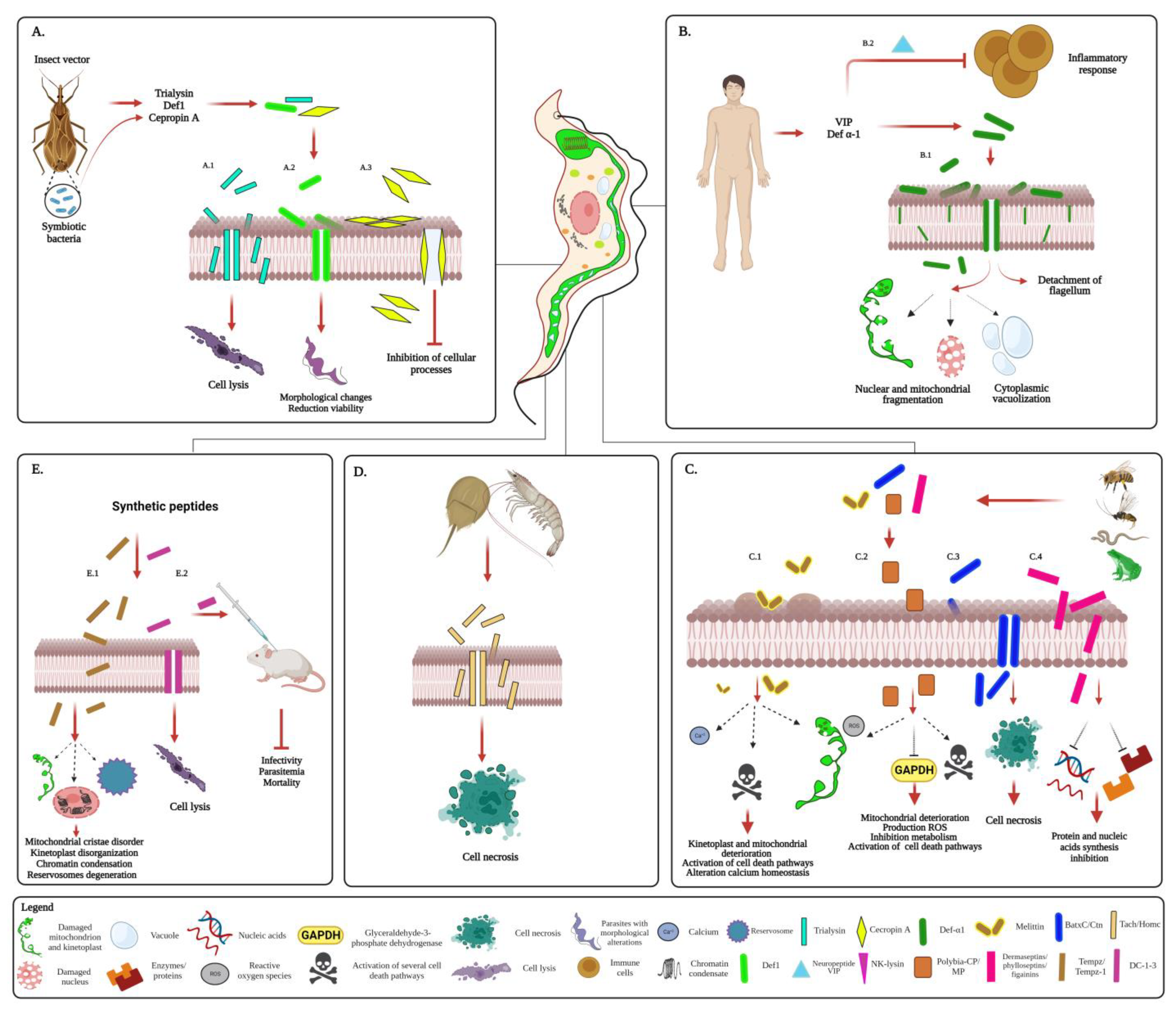
  81. Suárez-Quevedo, Y.; Barbosa-Vinasco, H.J.; Gutiérrez-Garnizo, S.A.; Olaya-Morales, J.L.; Zabala-González, D.; Carranza-Martínez, J.C.; Guhl-Nannetti, F.; Cantillo-Barraza, O.; Vallejo, G.A. Innate Trypanolytic Factors in Triatomine Hemolymph against Trypanosoma Rangeli and T. Cruzi: A Comparative Study in Eight Chagas Disease Vectors. Rev. Acad. Colomb. Cienc. Exactas Físicas Nat. 2020, 44, 88–104.
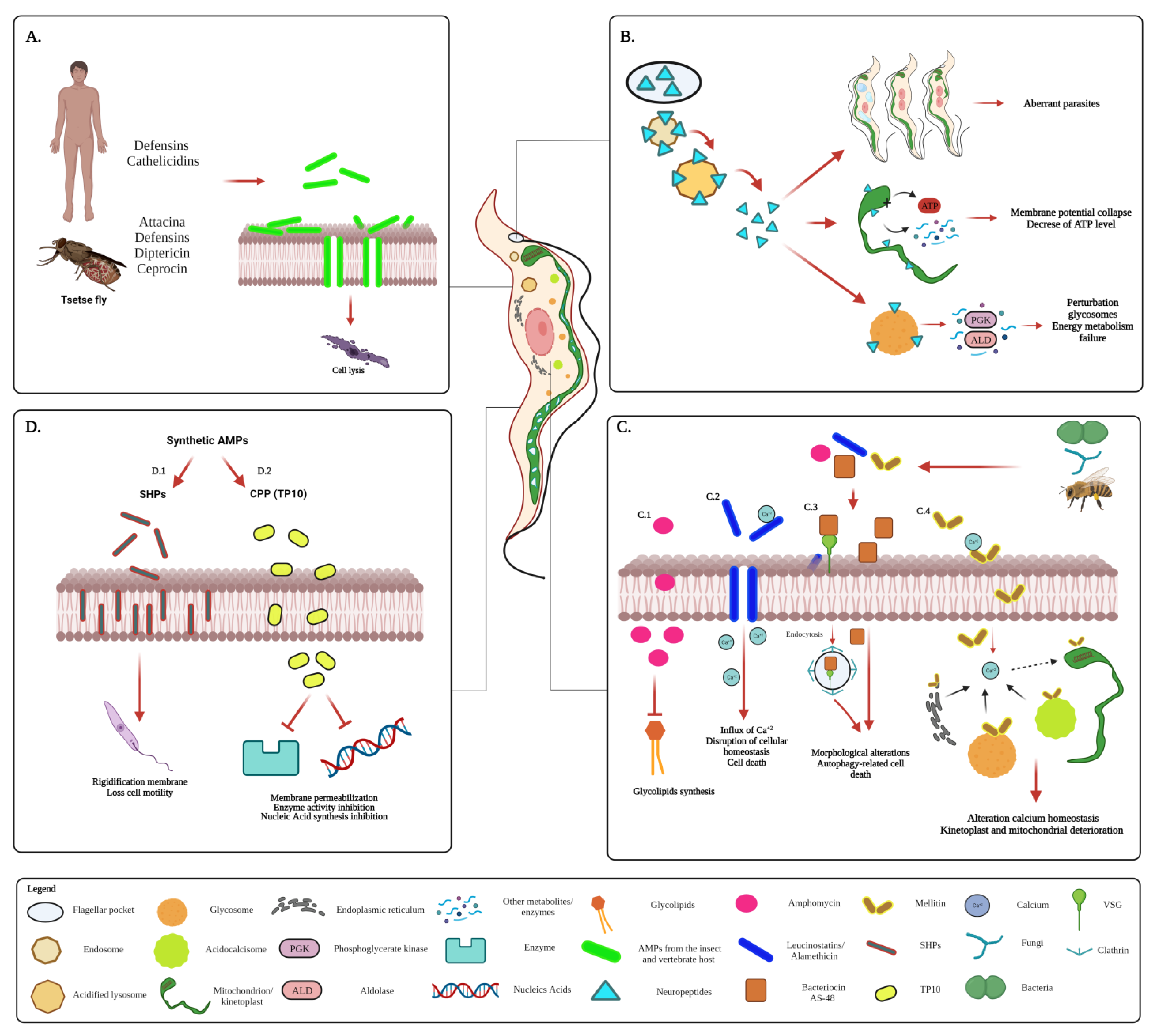
  82. Harrington, J.M. Antimicrobial Peptide Killing of African Trypanosomes. Parasite Immunol. 2011, 33, 461–469.
  83. Hu, Y.; Aksoy, S. An Antimicrobial Peptide with Trypanocidal Activity Characterized from Glossina Morsitans Morsitans. Insect Biochem. Mol. Biol. 2005, 35, 105–115.
  84. Delgado, M.; Anderson, P.; Garcia-Salcedo, J.A.; Caro, M.; Gonzalez-Rey, E. Neuropeptides Kill African Trypanosomes by Targeting Intracellular Compartments and Inducing Autophagic-like Cell Death. Cell Death Differ. 2009, 16, 406–416.
  85. Souza, A.L.A.; Faria, R.X.; Calabrese, K.S.; Hardoim, D.J.; Taniwaki, N.; Alves, L.A.; De Simone, S.G. Temporizin and Temporizin-1 Peptides as Novel Candidates for Eliminating Trypanosoma Cruzi. PLoS ONE 2016, 11, e0157673.
  86. Monteiro, M.L.; Lima, D.B.; de Menezes, R.R.P.P.B.; Sampaio, T.L.; Silva, B.P.; Serra Nunes, J.V.; Cavalcanti, M.M.; Morlighem, J.-E.; Martins, A.M.C. Antichagasic Effect of Hemocyanin Derived from Antimicrobial Peptides of Penaeus Monodon Shrimp. Exp. Parasitol. 2020, 215, 107930.
  87. Pinto, E.G.; Pimenta, D.C.; Antoniazzi, M.M.; Jared, C.; Tempone, A.G. Antimicrobial Peptides Isolated from Phyllomedusa Nordestina (Amphibia) Alter the Permeability of Plasma Membrane of Leishmania and Trypanosoma Cruzi. Exp. Parasitol. 2013, 135, 655–660.
  88. Memariani, H.; Memariani, M. Melittin as a Promising Anti-Protozoan Peptide: Current Knowledge and Future Prospects. AMB Express 2021, 11, 69.
  89. Barr, S.C.; Rose, D.; Jaynes, J.M. Activity of Lytic Peptides against Intracellular Trypanosoma Cruzi Amastigotes in Vitro and Parasitemias in Mice. J. Parasitol. 1995, 81, 974–978.
  90. Löfgren, S.E.; Miletti, L.C.; Steindel, M.; Bachère, E.; Barracco, M.A. Trypanocidal and Leishmanicidal Activities of Different Antimicrobial Peptides (AMPs) Isolated from Aquatic Animals. Exp. Parasitol. 2008, 118, 197–202.
More
Information
Contributors MDPI registered users' name will be linked to their SciProfiles pages. To register with us, please refer to https://encyclopedia.pub/register : , , , ,
View Times: 297
Revisions: 2 times (View History)
Update Date: 19 Jun 2023
1000/1000
Video Production Service