Submitted Successfully!
To reward your contribution, here is a gift for you: A free trial for our video production service.
Thank you for your contribution! You can also upload a video entry or images related to this topic.
Version Summary Created by Modification Content Size Created at Operation
1 -- 2948 2023-06-14 16:32:20 |
2 format change Meta information modification 2948 2023-06-15 04:47:30 |

Video Upload Options

Do you have a full video?

Confirm

Are you sure to Delete?
Cite
If you have any further questions, please contact Encyclopedia Editorial Office.
Kloc, M. Seahorse Male Pregnancy. Encyclopedia. Available online: https://encyclopedia.pub/entry/45595 (accessed on 14 September 2024).
Kloc M. Seahorse Male Pregnancy. Encyclopedia. Available at: https://encyclopedia.pub/entry/45595. Accessed September 14, 2024.
Kloc, Malgorzata. "Seahorse Male Pregnancy" Encyclopedia, https://encyclopedia.pub/entry/45595 (accessed September 14, 2024).
Kloc, M. (2023, June 14). Seahorse Male Pregnancy. In Encyclopedia. https://encyclopedia.pub/entry/45595
Kloc, Malgorzata. "Seahorse Male Pregnancy." Encyclopedia. Web. 14 June, 2023.
Seahorse Male Pregnancy
Edit

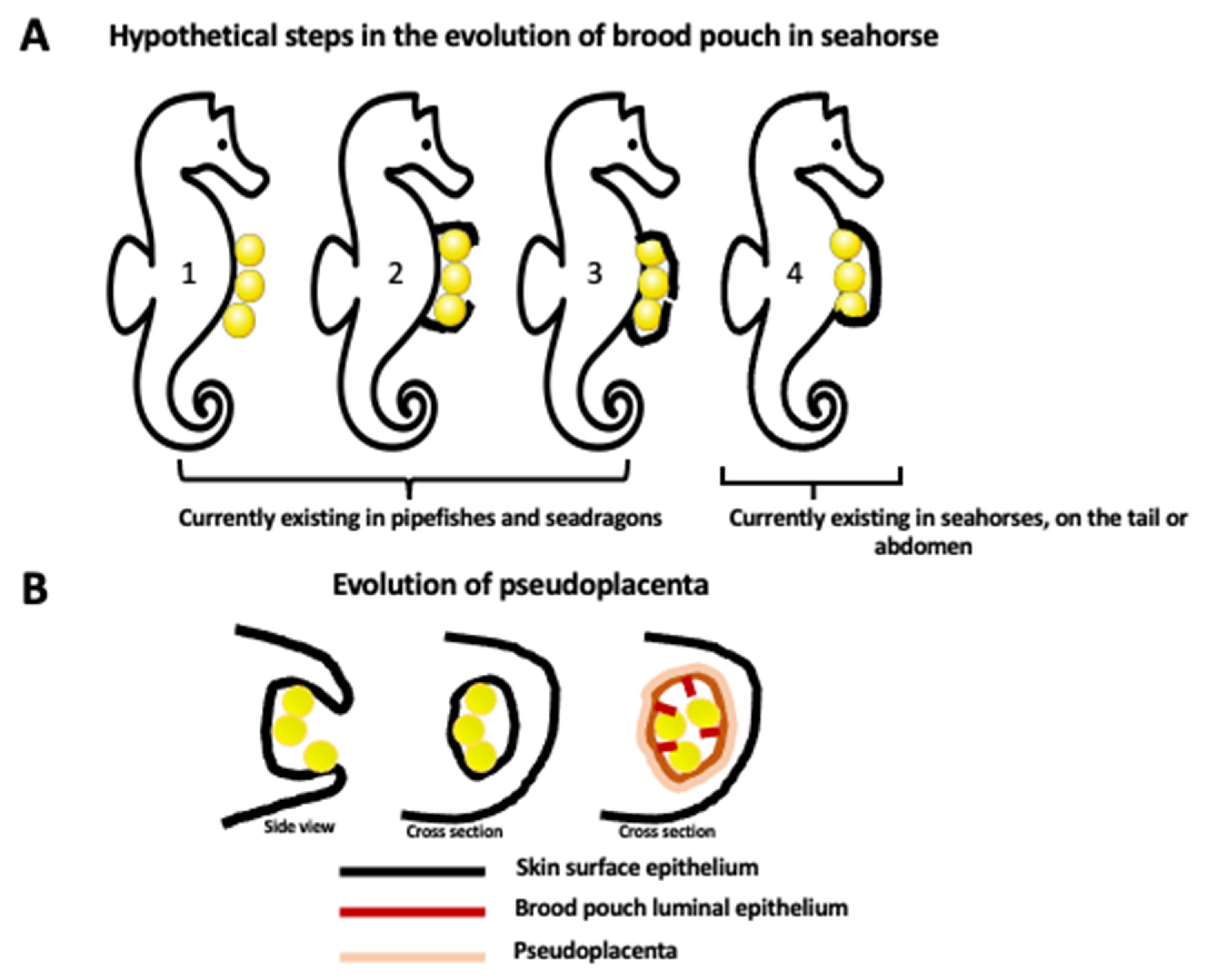
Seahorses, together with sea dragons and pipefishes, belong to the Syngnathidae family of teleost fishes. Seahorses and other Syngnathidae species have a very peculiar feature: male pregnancy. Among different species, there is a gradation of paternal involvement in carrying for the offspring, from a simple attachment of the eggs to the skin surface, through various degrees of egg coverage by skin flaps, to the internal pregnancy within a brood pouch, which resembles mammalian uterus with the placenta. Because of the gradation of parental involvement and similarities to mammalian pregnancy, seahorses are a great model to study the evolution of pregnancy and the immunologic, metabolic, cellular, and molecular processes of pregnancy and embryo development. Seahorses are also very useful for studying the effects of pollutants and environmental changes on pregnancy, embryo development, and offspring fitness. 

seahorse male pregnancy brood pouch placenta MHC pollution

1. What Is a Seahorse, and Why Is It Used as a Model System?

The seahorse is a fish from the Hippocampus genus. The name Hippocampus derives from the Greek word hippo, meaning ‘horse’, and campus, meaning ‘sea creature’ or ‘monster’. Seahorses, together with seadragons and pipefishes, form a family of Syngnathidae [1]. They are evolutionarily very old; the genus Syngnathus (pipefishes) is known from early Oligocene (33.9 million–28.1 million years ago) fossils [1]. Seahorses (genus Hippocampus) evolved from the pipefishes in the late Oligocene period [2]. Currently, there are 47 known species of seahorses. They live in shallow marine habitats, and their population is in constant decline because of pollution and overfishing for use in traditional medicine, as aquarium pets, and, in some countries, as a food delicacy. The biggest known seahorse is the big-belly (potbelly) seahorse, which reaches 35 cm in length and weighs around 35 g. The smallest are pigmy seahorses, of roughly 1.4–2.7 cm in length. One species of them, the Bargibant’s pygmy seahorses (discovered in 1969 by marine biologist George Bargibant), represents the best-camouflaged species in the world. They inhabit gorgonian coral, and their color and tubercles (nodules) covering the body (purple with pink tubercles or yellow with orange tubercles) mimic the color and shape of the host coral [3].

2. Male Pregnancy

Pregnancy is an extremely pricy investment requiring many physiological, metabolic, and anatomical changes in the parental organism, which promotes all the progeny’s well-being and survival. Pregnancy has evolved independently around 150 times in different vertebrate lineages [4][5]. One of the curious evolutionary inventions is male pregnancy in seahorses and other members of Syngnathidae fishes. Although there is no scientific consensus on the advantages of male versus female pregnancy, one of them can be a division of reproduction costs between two parents, and another is an increase in the offspring number; while the male carries babies in the brood pouch, the mother can produce another batch of eggs.

2.1. Brood Pouch Development

Different types of male pregnancy in various Syngnathidae fishes exemplify evolutionary gradation and progress in paternal involvement, from the simple attachment of eggs to the body surface to a fully developed internal pregnancy within the uterus/placenta-like pouch. In the subfamily Nerophinae, eggs are just attached to the skin surface without any protection. In Oosthethus, Doryrhamphus, and some Solegnathiinae, the attached eggs are protected by the special flaps of the skin. Finally, Syngnathus and Hippocampus have a completely closed brood pouch where developing embryos are integrated in paternal tissue and fully supplied through the vascularized placenta-like structure (Figure 1 and Figure 2) [4][6][7][8][9][10][11]. Harada et al. [7] studied in detail the brood pouch morphology and histology in five species of the Syngnathidae family and described five types of brood pouches. The alligator pipefish has a completely open pouch without skinfolds (type I), the messmate pipefish has an open pouch with skinfolds (type II), the seaweed pipefish has a closed pouch with skinfolds (type III), the pot-bellied seahorse has a closed pouch on the tail (type IV), and the Japanese pygmy seahorse has a closed pouch within a body cavity (type V) [7]. Interestingly, the type V pouch is positioned in the seahorse body cavity, and the embryos are located between the kidney and intestine [7]. The juvenile seahorse does not have a brood pouch. It develops during the post-juvenile stage [9]. While the female seahorse is laying eggs in a male brood pouch, the male fertilizes them at the pouch entry. Developing embryos are gradually enclosed within individual compartments of the placenta-like tissue of the pouch. This pseudo-placenta is vascularized and allows for the exchange of gases, nutrients, and waste removal through the epithelium that lines the pouch lumen [9]. The luminal epithelium of the pouch derives from the surface epithelium covering the seahorse’s body. Thus, at some point in pouch development, there must be a change in the epithelium properties. Kawaguchi et al. [9] studied successive stages of pouch development in the pot-bellied seahorse Hippocampus abdominalis as well as the molecules involved in the transition of the surface epithelium of the dermis into the luminal epithelium of the pouch. Pouch development lasts several months and starts from the long projections of the dermis (epithelium with underlying collagenous fibers) on both ventral sides of the body, which eventually fuse in the ventral midline, forming the pouch. During this stage, the pouch consists of only two layers of the dermis (epithelial layers with underlying collagenous fibers) and still lacks placenta-like features. The final step involves the formation of the pseudo-placenta. The smooth muscles and blood vessels form, and the properties of the epithelium change. The epithelium of the dermis and developing pouch express two types of C-lectins, haCTL I and haCTL II. In contrast, the luminal epithelium of a fully developed pouch with a pseudo-placenta specifically expresses haCTL IV lectin, which is never expressed in skin epithelium (Figure 2) [9]. By analogy to the muscle contraction during mammalian birth, the smooth muscles of the brood pouch were thought to be needed for pouch contraction during the expulsion of neonates. However, recent micro-computed tomography studies of the seahorse brood pouch showed that the muscles of the brood pouch are limited to the scattered small bundles, which cannot produce enough contraction during labor. Instead, male seahorses, in contrast to females, have large muscle bundles attached to the anal fin bones at the opening of the brood pouch, and contraction of those fin muscles, together with the body movements, opens the pouch and expels neonates [6].
Figure 1. Evolution of brood pouch. (A) Hypothetical steps in the evolution of closed brood pouch in seahorses. Currently, pipefishes and seadragons have an external or open brood pouch (step 1, 2, 3) while seahorses have a fully closed brood pouch placed in the abdomen or in the tail (step 4). Brood pouch evolution in the Syngnathidae family of teleost fishes starts from the simple attachment of eggs to the surface of the skin (1) and progresses through the different degrees of enclosure of eggs by the skin flaps (2, 3), ending up with the development of a fully internal brood pouch (4). (B) The evolution of the internal brood pouch is accompanied by the modification of the external skin epithelium to the internal (luminal) epithelium of the pouch, and the formation of vascularized pseudoplacenta. The first drawing represents the side view, and the last two drawings represent the cross section.
Figure 2. The transition of the skin surface epithelium into the luminal epithelium of the brood pouch. Surface epithelium of the skin is underlined by the collagenous fibers and expresses two forms of lectin: haCTL I and haCTL II. During development of the closed brood pouch, the external epithelium of the skin changes into the luminal (internal) epithelium of the brood pouch. Epithelial transformation correlates with the expression of haCTL IV lectin exclusively in the luminal epithelium. Epithelial transformation is accompanied by the aggregation of collagenous fibers, smooth muscle cells, and the development of capillaries below the epithelium, which all together form the pseudoplacenta that supports egg/embryo development. Development of the brood pouch and pseudoplacenta is regulated by many different nonhormonal and hormonal factors and pathways, including gonadotropin-inhibitory hormone GnIH, 11-ketotestosterone, and 17α-hydroxy-20β-dihydroprogesterone (see text for the details).

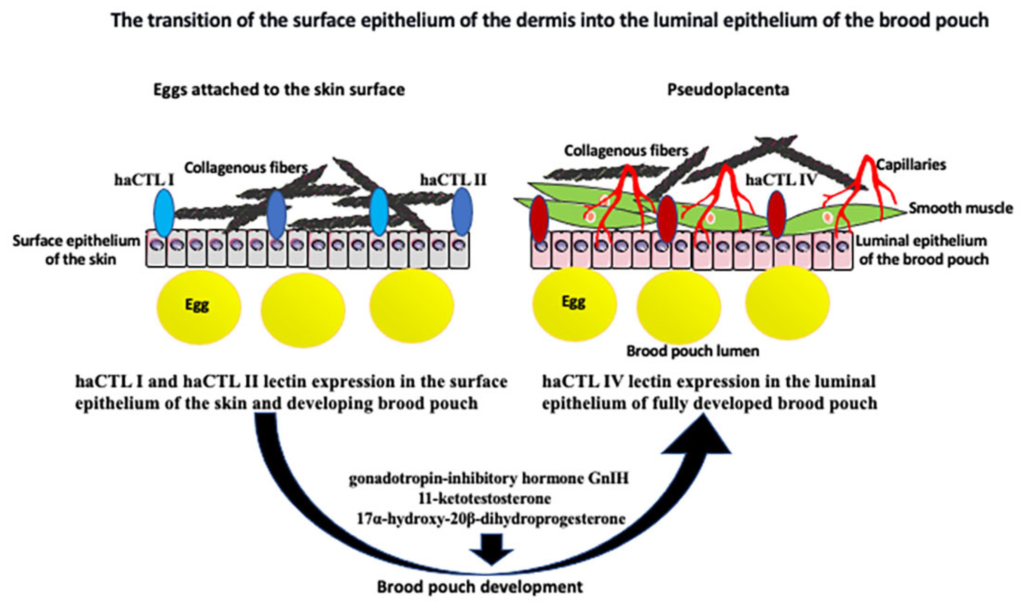
2.2. Hormonal Regulation of Seahorse Male Pregnancy

Like in most vertebrates, reproduction stages and cycles in fishes are regulated by the hypothalamic–pituitary–gonadal (HPG) axis. In females and males, the gonadotropin-releasing hormone (GnRH) produced by the hypothalamus induces the pituitary gland to release the gonadotropins, follicle-stimulating hormone (FSH) and luteinizing hormone (LH), into the bloodstream [12], while the gonadotropin-inhibitory hormone (GnIH) inhibits gonadotropin secretion [13]. In females, gonadotropins stimulate steroid (testosterone and estradiol) production by the ovary, which, in turn, promotes secondary sexual features and sex-dependent behavior, follicle development, and vitellogenin production. Oocyte maturation is stimulated by the LH-induced production of gamete maturation-inducing steroids (MISs). Ovulation in fish is stimulated by an increase in LH followed by the release of ovarian prostaglandins, which regulate spawning behavior [14][15][16]. In males, FSH induces spermatogenesis, and LH stimulates sperm maturation, and both these gonadotropins stimulate the male gonad to produce androgens, such as testosterone and 11-ketotestosterone, in fishes, which, in turn, regulate sexual dimorphism [16][17]. Gonadotropins also induce testes to produce MISs, which regulate spermiogenesis and the final maturation of spermatozoa [16][18].

2.3. Retinoic Acid

Another molecule involved in seahorse male pregnancy regulation is retinoic acid (RA), a metabolite of vitamin A1 (all-trans-retinol). Among various retinoic acid isomers, such as 13-cis- and 9-cis-retinoic acid, the all-trans-retinoic acid (retinoic acid) is most abundant and required for growth, embryonic development, differentiation, cancer, and immunity [19][20][21][22][23][24]. During early development, RA signaling through the homeobox (Hox) and POU genes establishes the anterior–posterior axis and patterning of the embryo [22][23]. Comparative transcriptomic and metabolomic analyses of the lined seahorse Hippocampus erectus in different stages of brood pouch formation (unformed, newly formed, and pregnant pouch) identified 141 genes and 73 metabolites related to pouch formation and 2533 and 121 metabolites related to pregnancy. Additionally, integrative omics showed that retinoic acid (RA) synthesis and signaling were involved in brood pouch formation and seahorse pregnancy and in regulating antioxidant defenses [25]. These studies also showed that in H. erectus and H. abdominalis, RA functions upstream of testosterone and progesterone, directly regulating pouch formation via G protein-coupled receptor FSHR and cholesterol 7alpha-hydroxylase CYP7A1, a member of the cytochrome P450 superfamily of enzymes involved in the synthesis of cholesterol, steroids, and other lipids [25][26][27].

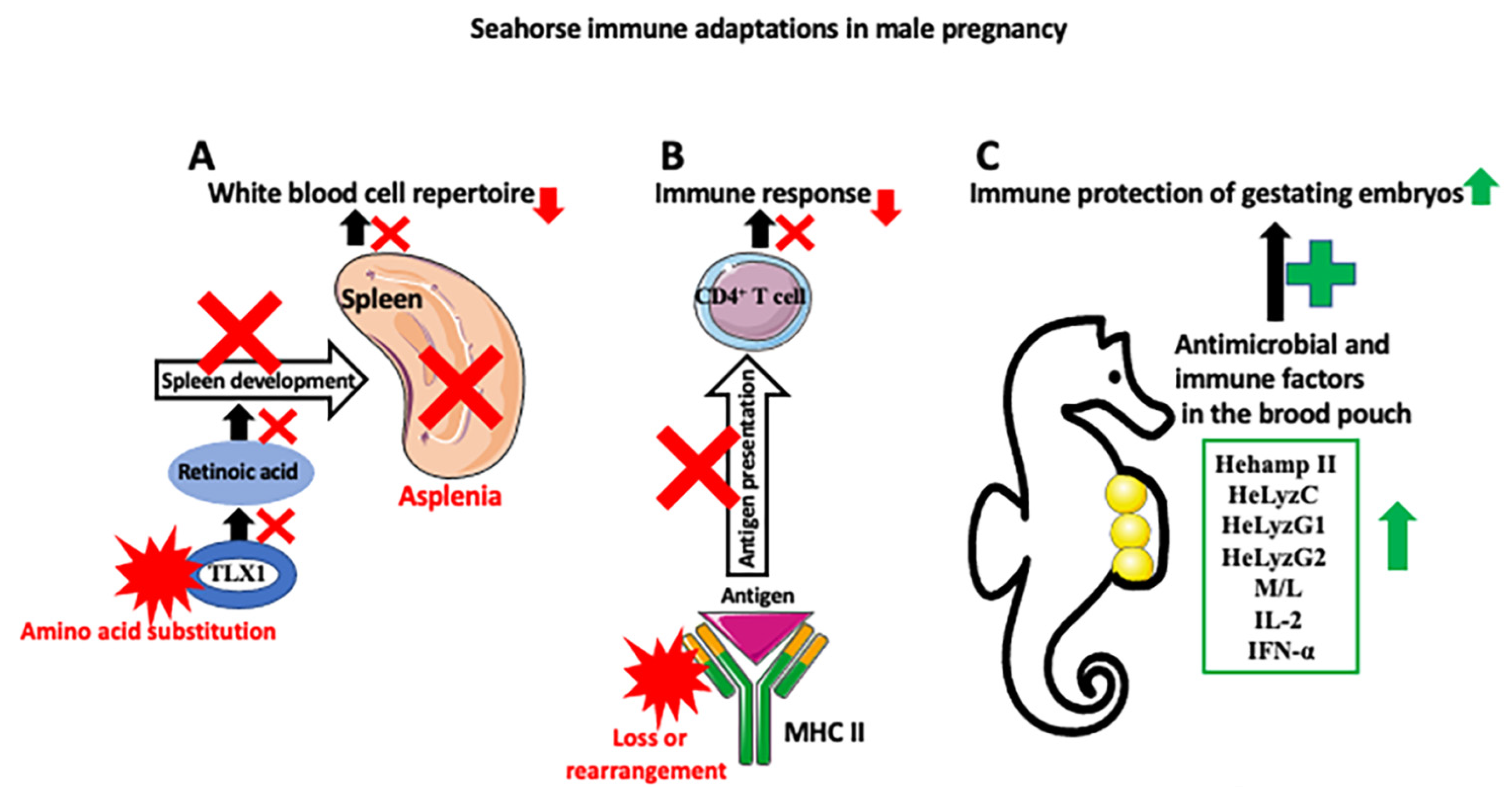
3. Adaptations of the Immune System to Male Pregnancy

The biggest challenge in pregnancy is how not to reject an allogeneic (nonself) embryo [28]. In mammalian pregnancy, immune tolerance toward the semi-allogenic embryo occurs through the downregulation of the major histocompatibility genes MHC I and MHC II. Paradoxically, pregnancy starts from the inflammation of the endometrium necessary for the implantation of the embryo. Chavan et al. [29] and Griffith et al. [30] called this “the inflammation paradox”. They believe that, early in evolution, acute endometrial inflammation (still occurring in some marsupials) was a natural maternal immune reaction toward the attaching embryo. Subsequently, during evolution, by suppressing the most damaging parts of the inflammatory response, the acute inflammation was transformed into the embryo-friendly process of implantation and placental pregnancy [29][30]. In mammals, several mechanisms based on the extensive crosstalk between the embryonic trophoblast layer and maternal uterine immune cells prevent the rejection of the allogeneic embryo by the maternal immune system. First, trophoblast does not express MHC II, which otherwise would present embryonic antigens to maternal T-helper cells (Th) and initiate an immune response [31][32]. Additionally, the MHC I genes, which present antigens to maternal cytotoxic T cells, are downregulated in the trophoblast [32][33]. Additionally, regulatory T cells (Tregs) recognize fetal antigens via maternal antigen-presenting cells (APCs) and induce tolerance toward the embryo [34].
Thus, the fascinating question is how, during evolution, did the immune system adjust to tolerating the nonself embryo. Because most mammals, except for egg-laying monotremata, such as echidna and platypus, have internal pregnancies, they are useless for reconstructing evolutionary progress in parental immunotolerance. In contrast, male seahorses, with their gradation of pregnancy, from a simple attachment of eggs to the skin, through a different degree of coverage by the skin flaps, to internal gestation within the brood pouch, are perfect for reconstructing evolutionary progress in the development of immune tolerance toward the nonself embryo (Figure 3). Recently, using this approach, Roth et al. [4] studied the immune gene repertoire across male pregnancy gradients in 12 species of seahorses and pipefishes. These detailed studies showed that the evolution of pregnancy coincided with either a complete loss or rearrangement of MHC II pathway genes and correlated with the expansion of the MHC I gene repertoire. MHC II molecules are found on professional antigen-presenting cells such as dendritic cells, mononuclear phagocytes, some endothelial cells, thymic epithelial cells, and B cells, which are crucial for initiating immune response. The antigens presented by the MHC II genes derive from extracellular proteins, while antigens presented by the MHC I genes mainly derive from cytosolic proteins. MHC II binds peptides, which are derived from the proteolysis of self and non-self proteins, and presents them to antigen-specific CD4+ T cells. On the same theme, studies from another laboratory compared transcriptome-wide gene expression in four syngnathid species with different degrees of paternal involvement at four pregnancy stages (nonpregnant, early, late, and at birth) [35]. They found that the loss or downregulation of MHC pathway gene expression occurs only in species with a brood pouch pregnancy, and that a decrease in MHC pathway gene expression is limited to the early and middle stages of pregnancy. In late pregnancy and at birth, the expression of immune genes was elevated, suggesting that the late embryos, in contrast to the early embryos, are no longer in direct contact with the paternal immune system [35][36].
Figure 3. Seahorse immune adaptations in male pregnancy. Three main mechanisms are responsible for the development of seahorse immune tolerance against allogeneic embryos. (A) Seahorses are asplenic (spleen loss). Asplenia is caused by the amino acid substitution in the transcription factor TLX1, which, through the retinoic acid pathway, regulates spleen development. Asplenia affects the white blood cell repertoire (causes partial loss of red blood cells, platelets, various subsets of T and B cells, dendritic cells (DCs), and macrophages) and decreases immune response. (B) There is a downregulation of the MHC II pathway, either through a loss or rearrangement of MHC II genes. This, in turn, decreases self- and non-self-antigen presentation to the effector immune cells, such as CD4+ T cells, and lowers the immune response against allogenic embryos. (C) Despite the weakened immune responses of the parent against embryos, gestating embryos are protected against pathogens abundant in a nonsterile seawater-filled pouch by the increase in antibacterial peptides and immune factors, such as hepcidin (Hehamp II), lysozymes (HeLyzC, HeLyzG1, and HeLyzG2), monocytes and leukocytes, interleukin 2 (IL-2), and interferon alfa (IFN-α) in the pouch. Red X symbol depicts inhibition of disruption of the process, and red star depicts changes in molecule composition or structure.

4. Effects of Male Pregnancy on the Microbiome

One of the most fascinating changes occurring during Syngnathidae male pregnancy is a profound change in their microbiome. Although there is not much information on this subject in seahorses, there are recent, very detailed studies on pipefish pregnancy [37]. In animals with maternal pregnancy, some microorganisms are transferred to the offspring from the mother, and some colonize newborns (through the mouth) from the environment. In maternal pregnancy, it is very hard to establish if there is any input of microorganisms from the father. Thus, animals with male pregnancy, such as pipefishes or seahorses, are very well suited to study this phenomenon and answer the question of how pregnancy influences the parental microbiome. Recent studies on pipefish Syngnathus typhle sequenced microbial 16S rRNA from maternal gonads and the brood pouches of non-pregnant and pregnant males [37]. These studies also assessed the effect of the parental immune system on the complexity of the microbiome. These analyses showed that maternal gonads and male brood pouches contain different microbial species (listed in the entry) and contain different species in early and late pregnancy brood pouches [37]. The most abundant bacteria in the maternal gonads and in the paternal brood pouch were Marinomonas (28.0%), Halomonas (10.3%), Aeribacillus (6.3%), Ruegeria (5.8%), Bacteroidetes (4.5%), and Nesterenkonia (3.8%). Additionally, the paternal immune system changes the bacterial composition to a higher abundance of Kiloniella, Aquimarina, Ulvibacter, and Marinomonas. These are commensal bacteria that help fight pathogenic bacteria and possibly boost the immune response in the offspring [37].

5. Effects of Environmental Changes and Pollutants on Seahorses

Many species of seahorses are endangered by overfishing, degradation of their habitats, and pollution. Even under the best circumstances, seahorse numbers are low because of the demanding and restrictive lifestyle. Seahorses sparsely inhabit an intricately structured niche, and their survival depends on the elaborate camouflage specific to a particular surrounding. Because seahorses are poor swimmers, nonmigratory, monogamous, and form a lifelong pair bond, finding a new partner when one dies is very difficult. Seahorse reproductive rate, even under optimal conditions, is low because of the small brood size and lengthy parental care. Consequently, seahorses are on the local, international, and IUCN Red List of Threatened Species [38], becoming a bioindicator of crude oil exposure [39]. Studies of the effects of ocean warming and acidification on the physiology and behavior of the long-snouted seahorse Hippocampus guttulatus, which has been recently added to list of threatened and/or declining species by the Oslo and Paris (OSPAR) commission, showed that although the adults are quite resistant to ocean warming, the combination of warming and acidification (caused by CO2 uptake from the atmosphere) causes lethargy and reduction in feeding and gill ventilation rates [40]. In contrast, the seahorse newborns are much more sensitive to warming, which causes heat-induced hypometabolism [41].
Besides factors related to climate warming, seahorses, like other aquatic animals, are constantly exposed to various types of pollutants. For example, studies of seahorse species inhabiting coastal waters of the Black Sea and China showed a high accumulation of heavy metals (including Cu, Pb, Cd, Cr, and Hg), benzo(a)pyrene (B[a]P), organochlorine pesticides, polycyclic aromatic hydrocarbons (PAHs), and microplastics [42][43][44][45][46].

References

  1. Wilson, A.B.; Orr, J.W. The evolutionary origins of Syngnathidae: Pipefishes and seahorses. J. Fish. Biol. 2011, 78, 1603–1623.
  2. Teske, P.R.; Beheregaray, L.B. Evolution of seahorses’ upright posture was linked to Oligocene expansion of seagrass habitats. Biol. Lett. 2009, 5, 521–523.
  3. Froese, R.; Pauly, D. (Eds.) Fish Base; World Wide Web Electronic Publication: 2013. Available online: www.fishbase.org (accessed on 25 May 2023).
  4. Roth, O.; Solbakken, M.H.; Tørresen, O.K.; Bayer, T.; Matschiner, M.; Baalsrud, H.T.; Hoff, S.N.K.; Brieuc, M.S.O.; Haase, D.; Hanel, R.; et al. Evolution of male pregnancy associated with remodeling of canonical vertebrate immunity in seahorses and pipefishes. Proc. Natl. Acad. Sci. USA 2020, 117, 9431–9439.
  5. Long, X.; Charlesworth, D.; Qi, J.; Wu, R.; Chen, M.; Wang, Z.; Xu, L.; Fu, H.; Zhang, X.; Chen, X.; et al. Independent Evolution of Sex Chromosomes and Male Pregnancy-Related Genes in Two Seahorse Species. Mol. Biol. Evol. 2023, 40, msac279.
  6. Dudley, J.S.; Paul, J.W.; Teh, V.; Mackenzie, T.E.; Butler, T.A.; Tolosa, J.M.; Smith, R.; Foley, M.; Dowland, S.; Thompson, M.B.; et al. Seahorse brood pouch morphology and control of male parturition in Hippocampus abdominalis. Placenta 2022, 127, 88–94.
  7. Harada, A.; Shiota, R.; Okubo, R.; Yorifuji, M.; Sogabe, A.; Motomura, H.; Hiroi, J.; Yasumasu, S.; Kawaguchi, M. Brood pouch evolution in pipefish and seahorse based on histological observation. Placenta 2022, 120, 88–96.
  8. Herald, E.S. From pipefish to seahorse—A study of phylogenetic relationships. Proc. Calif. Acad. Sci. 1959, 29, 465–473.
  9. Kawaguchi, M.; Okubo, R.; Harada, A.; Miyasaka, K.; Takada, K.; Hiroi, J.; Yasumasu, S. Morphology of brood pouch formation in the pot-bellied seahorse Hippocampus abdominalis. Zool. Lett. 2017, 3, 19.
  10. Vincent, A.; Ahnesjö, I.; Berglund, A.; Rosenqvist, G. Pipefishes and seahorses: Are they all sex role reversed? Trends Ecol. Evol. 1992, 7, 237–241.
  11. Wilson, A.B.; Vincent, A.; Ahnesjö, I.; Meyer, A. Male pregnancy in seahorses and pipefishes (family Syngnathidae): Rapid diversification of paternal brood pouch morphology inferred from a molecular phylogeny. J. Hered. 2001, 92, 159–166.
  12. Marques, P.; Skorupskaite, K.; Rozario, K.S.; Anderson, R.A.; George, J.T. Physiology of GnRH and Gonadotropin Secretion. In Endotext ; Feingold, K.R., Anawalt, B., Blackman, M.R., Boyce, A., Chrousos, G., Corpas, E., de Herder, W.W., Dhatariya, K., Dungan, K., Hofland, J., Eds.; MDText.com, Inc.: South Dartmouth, MA, USA, 2000.
  13. Zhang, H.; Chen, L.; Zhang, B.; Lin, Q. Molecular identification of GnIH and its potential role in reproductive physiology and male pregnancy of the lined seahorse (Hippocampus erectus). Gen. Comp. Endocrinol. 2019, 279, 196–202.
  14. Cole, K.S.; Stacey, N.E. Prostaglandin induction of spawning behavior in Cichlasoma bimaculatum (Pisces cichlidae). Horm. Behav. 1984, 18, 235–248.
  15. Omony, J.B.; Biran, J.; Kahwa, D.; Aizen, J.; Golan, M.; Nyatia, E.; Levavi-Sivan, B.; Rutaisire, J. Cloning of gonadotropin Gph-alpha, FSH-beta and LH-beta subunits and seasonal profiles of steroid hormones in wild-caught Nile perch, Lates niloticus. Gen. Comp. Endocrinol. 2022, 323–324, 114035.
  16. Scobell, S.K.; Mackenzie, D.S. Reproductive endocrinology of Syngnathidae. J. Fish. Biol. 2011, 78, 1662–1680.
  17. Yazawa, T.; Uesaka, M.; Inaoka, Y.; Mizutani, T.; Sekiguchi, T.; Kajitani, T.; Kitano, T.; Umezawa, A.; Miyamoto, K. Cyp11b1 is induced in the murine gonad by luteinizing hormone/human chorionic gonadotropin and involved in the production of 11-ketotestosterone, a major fish androgen: Conservation and evolution of the androgen metabolic pathway. Endocrinology 2008, 149, 1786–1792.
  18. Barannikova, I.A.; Dyubin, V.P.; Bayunova, L.V.; Semenkova, T.B. Steroids in the control of reproductive function in fish. Neurosci. Behav. Physiol. 2002, 32, 141–148.
  19. Lovat, P.E.; Annicchiarico-Petruzzelli, M.; Corazzari, M.; Dobson, M.G.; Malcolm, A.J.; Pearson, A.D.; Melino, G.; Redfern, C.P. Differential effects of retinoic acid isomers on the expression of nuclear receptor co-regulators in neuroblastoma. FEBS Lett. 1999, 445, 415–419.
  20. Brown, G. Targeting the Retinoic Acid Pathway to Eradicate Cancer Stem Cells. Int. J. Mol. Sci. 2023, 24, 2373.
  21. Heeg, M.; Goldrath, A.W. License to kill: Retinoic acid programs T cells for tissue residency. J. Exp. Med. 2023, 220, e20230161.
  22. Koop, D.; Holland, N.D.; Sémon, M.; Alvarez, S.; de Lera, A.R.; Laudet, V.; Holland, L.Z.; Schubert, M. Retinoic acid signaling targets Hox genes during the amphioxus gastrula stage: Insights into early anterior-posterior patterning of the chordate body plan. Dev. Biol. 2010, 338, 98–106.
  23. Bozzo, M.; Bellitto, D.; Amaroli, A.; Ferrando, S.; Schubert, M.; Candiani, S. Retinoic Acid and POU Genes in Developing Amphioxus: A Focus on Neural Development. Cells 2023, 12, 614.
  24. Yeung, K.W.Y.; Lai, R.W.S.; Zhou, G.J.; Leung, K.M.Y. Concentration-response of six marine species to all-trans-retinoic acid and its ecological risk to the marine environment. Ecotoxicol. Environ. Saf. 2022, 235, 113455.
  25. Li, C.; Li, Y.; Qin, G.; Chen, Z.; Qu, M.; Zhang, B.; Han, X.; Wang, X.; Qian, P.Y.; Lin, Q. Regulatory Role of Retinoic Acid in Male Pregnancy of the Seahorse. Innovation 2020, 1, 100052.
  26. Powell, J.R.; Kim, D.H.; Ausubel, F.M. The G protein-coupled receptor FSHR-1 is required for the Caenorhabditis elegans innate immune response. Proc. Natl. Acad. Sci. USA 2009, 106, 2782–2787.
  27. Zhang, J.; Wang, X.; Jiang, H.; Yang, F.; Du, Y.; Wang, L.; Hong, B. MicroRNA-185 modulates CYP7A1 mediated cholesterol-bile acid metabolism through post-transcriptional and post-translational regulation of FoxO1. Atherosclerosis 2022, 348, 56–67.
  28. Monteiro, N. Mom and dad are not that different after all: Immune modulation as a prerequisite for the evolution of pregnancy. Mol. Ecol. 2023, 32, 753–755.
  29. Chavan, A.R.; Griffith, O.W.; Wagner, G.P. The inflammation paradox in the evolution of mammalian pregnancy: Turning a foe into a friend. Curr. Opin. Genet. Dev. 2017, 47, 24–32.
  30. Griffith, O.W.; Chavan, A.R.; Protopapas, S.; Maziarz, J.; Romero, R.; Wagner, G.P. Embryo implantation evolved from an ancestral inflammatory attachment reaction. Proc. Natl. Acad. Sci. USA 2017, 114, E6566–E6575.
  31. Murphy, S.P.; Choi, J.C.; Holtz, R. Regulation of major histocompatibility complex class II gene expression in trophoblast cells. Reprod. Biol. Endocrinol. 2004, 2, 52.
  32. Rock, K.L.; Reits, E.; Neefjes, J. Present Yourself! By MHC Class I and MHC Class II Molecules. Trends Immunol. 2016, 37, 724–737.
  33. Rutigliano, H.M.; Thomas, A.J.; Wilhelm, A.; Sessions, B.R.; Hicks, B.A.; Schlafer, D.H.; White, K.L.; Davies, C.J. Trophoblast Major Histocompatibility Complex Class I Expression Is Associated with Immune-Mediated Rejection of Bovine Fetuses Produced by Cloning. Biol. Reprod. 2016, 95, 39.
  34. Tsuda, S.; Nakashima, A.; Shima, T.; Saito, S. New Paradigm in the Role of Regulatory T Cells during Pregnancy. Front. Immunol. 2019, 10, 573.
  35. Parker, J.; Dubin, A.; Schneider, R.; Wagner, K.S.; Jentoft, S.; Böhne, A.; Bayer, T.; Roth, O. Immunological tolerance in the evolution of male pregnancy. Mol. Ecol. 2023, 32, 819–840.
  36. Parker, J.; Dubin, A.; Roth, O. Genome rearrangements, male pregnancy and immunological tolerance—The curious case of the syngnathid immune system. Front. Mar. Sci. 2023, 10, 1099231.
  37. Beemelmanns, A.; Poirier, M.; Bayer, T.; Kuenzel, S.; Roth, O. Microbial embryonal colonization during pipefish male pregnancy. Sci. Rep. 2019, 9, 3.
  38. Vincent, A.C.; Foster, S.J.; Koldewey, H.J. Conservation and management of seahorses and other Syngnathidae. J. Fish. Biol. 2011, 78, 1681–1724.
  39. Delunardo, F.A.; de Carvalho, L.R.; da Silva, B.F.; Galão, M.; Val, A.L.; Chippari-Gomes, A.R. Seahorse (Hippocampus reidi) as a bioindicator of crude oil exposure. Ecotoxicol. Environ. Saf. 2015, 117, 28–33.
  40. Faleiro, F.; Baptista, M.; Santos, C.; Aurélio, M.L.; Pimentel, M.; Pegado, M.R.; Paula, J.R.; Calado, R.; Repolho, T.; Rosa, R. Seahorses under a changing ocean: The impact of warming and acidification on the behaviour and physiology of a poor-swimming bony-armoured fish. Conserv. Physiol. 2015, 3, cov009.
  41. Aurélio, M.; Faleiro, F.; Lopes, V.M.; Pires, V.; Lopes, A.R.; Pimentel, M.S.; Repolho, T.; Baptista, M.; Narciso, L.; Rosa, R. Physiological and behavioral responses of temperate seahorses (Hippocampus guttulatus) to environmental warming. Mar. Biol. 2013, 160, 2663–2670.
  42. Liu, Y.; Shang, D.; Yang, Y.; Cui, P.; Sun, J. Bioaccumulation of contaminants in wild seahorses collected from coastal China. Front. Mar. Sci. 2022, 9, 1021170.
  43. Liu, Y.; Shang, D.; Yang, Y.; Cui, P.; Sun, J. Transcriptomic Analysis Provides Insights into Microplastic and Heavy Metal Challenges in the Line Seahorse (Hippocampus erectus). Fishes 2022, 7, 338.
  44. Nenciu, M.-I.; Coatu, V.; Oros, A.; Rosiorua, D.; Tiganus, D.; Rosoiu, N. Pollutant bioaccumulation in the long-snouted seahorse at the Romanian coast. J. Environ. Prot. Ecol. 2014, 15, 1650–1659.
  45. Zhang, W.; Zhang, Y.; Zhang, L.; Lin, Q. Bioaccumulation of Metals in Tissues of Seahorses Collected from Coastal China. Bull. Environ. Contam. Toxicol. 2016, 96, 281–288.
  46. Wright, S.L.; Thompson, R.C.; Galloway, T.S. The Physical Impacts of Microplastics on Marine Organisms: A Review. Environ. Pollut. 2013, 178, 483–492.
More
Information
Subjects: Biology
Contributor MDPI registered users' name will be linked to their SciProfiles pages. To register with us, please refer to https://encyclopedia.pub/register :
View Times: 365
Revisions: 2 times (View History)
Update Date: 15 Jun 2023
1000/1000
ScholarVision Creations