Submitted Successfully!
To reward your contribution, here is a gift for you: A free trial for our video production service.
Thank you for your contribution! You can also upload a video entry or images related to this topic.
Version Summary Created by Modification Content Size Created at Operation
1 -- 3429 2023-06-13 17:27:29 |
2 Reference format revised. Meta information modification 3429 2023-06-15 03:25:04 |

Video Upload Options

Do you have a full video?

Confirm

Are you sure to Delete?
Cite
If you have any further questions, please contact Encyclopedia Editorial Office.
Triposkiadis, F.; Xanthopoulos, A.; Lampropoulos, K.; Briasoulis, A.; Sarafidis, P.; Skoularigis, J.; Boudoulas, H. Aortic Stiffness as a Risk Factor for Multimorbidity. Encyclopedia. Available online: https://encyclopedia.pub/entry/45520 (accessed on 27 July 2024).
Triposkiadis F, Xanthopoulos A, Lampropoulos K, Briasoulis A, Sarafidis P, Skoularigis J, et al. Aortic Stiffness as a Risk Factor for Multimorbidity. Encyclopedia. Available at: https://encyclopedia.pub/entry/45520. Accessed July 27, 2024.
Triposkiadis, Filippos, Andrew Xanthopoulos, Konstantinos Lampropoulos, Alexandros Briasoulis, Pantelis Sarafidis, John Skoularigis, Harisios Boudoulas. "Aortic Stiffness as a Risk Factor for Multimorbidity" Encyclopedia, https://encyclopedia.pub/entry/45520 (accessed July 27, 2024).
Triposkiadis, F., Xanthopoulos, A., Lampropoulos, K., Briasoulis, A., Sarafidis, P., Skoularigis, J., & Boudoulas, H. (2023, June 13). Aortic Stiffness as a Risk Factor for Multimorbidity. In Encyclopedia. https://encyclopedia.pub/entry/45520
Triposkiadis, Filippos, et al. "Aortic Stiffness as a Risk Factor for Multimorbidity." Encyclopedia. Web. 13 June, 2023.
Aortic Stiffness as a Risk Factor for Multimorbidity
Edit

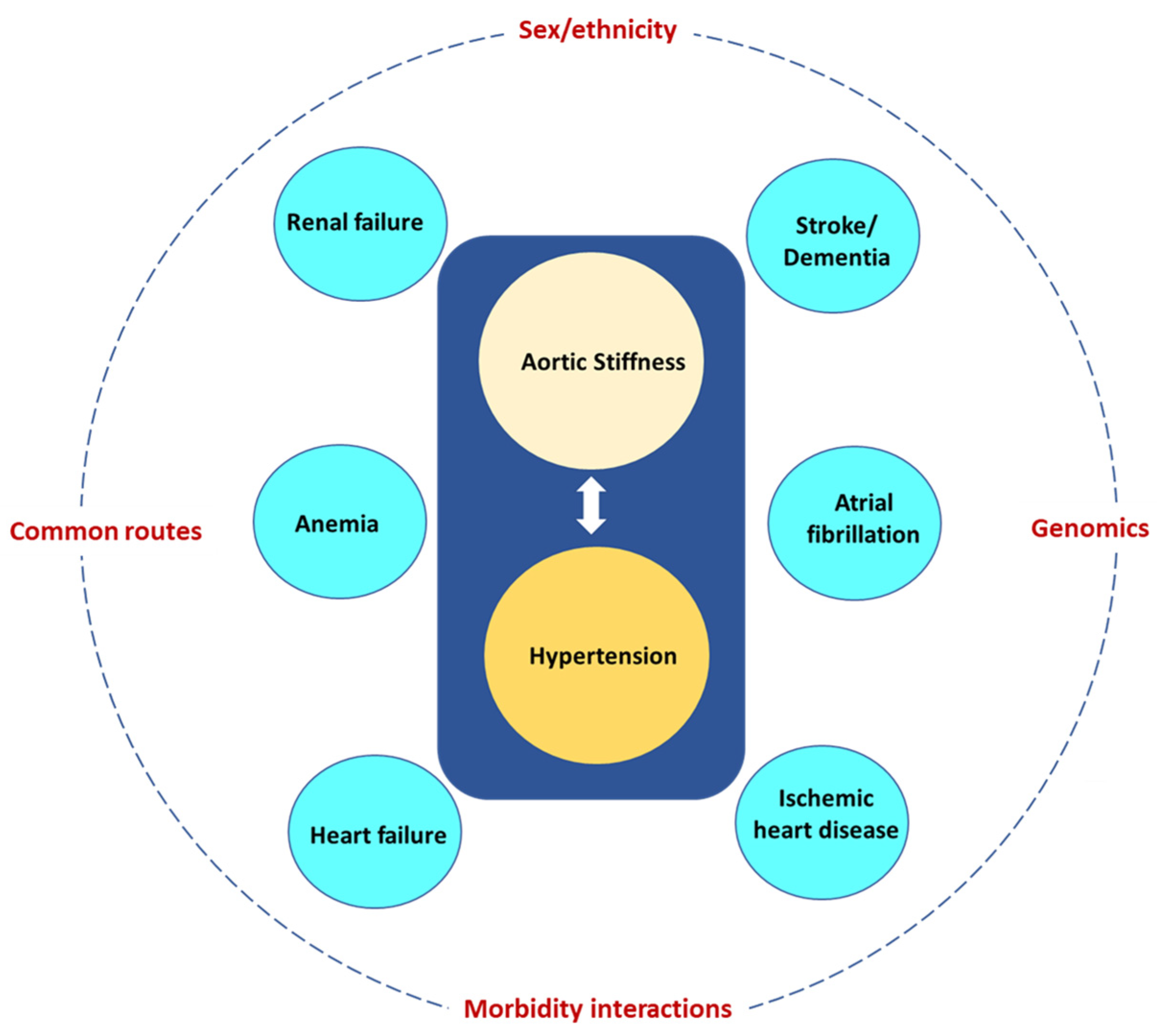
Multimorbidity, the coexistence of multiple health conditions in an individual, has emerged as one of the greatest challenges facing health services, and this crisis is partly driven by the aging population. Aging is associated with increased aortic stiffness (AoStiff), which in turn is linked with several morbidities frequently affecting and having disastrous consequences for the elderly. These include hypertension, ischemic heart disease, heart failure, atrial fibrillation, chronic kidney disease, anemia, ischemic stroke, and dementia. Two or more of these disorders (multimorbidity) often coexist in the same elderly patient and the specific multimorbidity pattern depends on several factors including sex, ethnicity, common morbidity routes, morbidity interactions, and genomics.

multimorbidity elderly aortic stiffness

1. Introduction

Multimorbidity, the coexistence of multiple health conditions in an individual [1], has emerged as one of the biggest challenges facing health services, and this crisis is partly driven by the aging population [2]. In one study including older US patients on Medicare, the prevalence of multimorbidity was 62% at ages 65 to 74 years and 82% at ages 85 years and older [3]. This problem will significantly worsen in the future. In 2020, there were an estimated 727 million people aged 65 years or over worldwide, and this number is projected to increase by 2050, reaching over 1.5 billion people [4].
Multimorbidity adversely affects health outcomes in older adults [5]. Further, there is compelling epidemiological evidence linking aortic stiffness (AoStiff) with several morbidities [6].

2. The Normal Young Aorta

The aorta serves not only as a conduit, but also has important roles in affecting left ventricular (LV) performance, myocardial perfusion, central hemodynamics, and arterial function throughout the entire cardiovascular system with all these actions of the aorta globally influencing the circulation [7].
The arterial wall is made of matrix proteins (collagen fibers oriented in different directions and elastic lamellae), smooth muscle cells, and glycosaminoglycans, together with endothelial cells in the intimal layer [8]. The composition of the aortic wall changes from the central aorta towards the periphery; the media of the human ascending aorta is composed of concentric elastic lamellae (up to 60–80), interdigitated by connective tissue layers containing smooth muscle cells. This microstructure gradually fades and in medium sized, and particularly smaller vessels (such as arterioles) the number of elastic lamellae is decreased, whereas smooth muscle cells contents are increased [9].
AoStiff is the deformation resistance provided by the aorta. Due to the composition of the aortic wall, the ascending aorta is less stiff (more elastic or compliant) than the abdominal aorta (stiffness gradient) [10]. As a result, the aorta and some of the proximal large vessels act as an elastic buffering chamber in series with the LV (the Windkessel function) [11]. However, the aorta exhibits by far the largest buffering capacity in the human arterial system, with the ascending aorta and aortic arch providing approximately one-third of the total pressure buffering capacity [12].
In normal conditions, during diastole, the elastic forces of the central aortic wall forward 50% of the volume to the peripheral circulation, creating a nearly continuous peripheral blood flow. Further, pulse waves generated during the LV systole travel down the aorta to the peripheral mid-sized arteries, where they incur increased resistance due to branch points and increased arterial tone. This results in reflection of the incident waves, which subsequently travel back toward the central aorta from the periphery in phase with diastole, augmenting diastolic coronary perfusion [10].

3. The Aging Aorta

3.1. Biology

Aging is characterized by complex physiological, cellular and molecular changes leading to a progressive loss of structural and physiological integrity, organ dysfunction, and increased vulnerability to death [13]. Cellular senescence is linked with aging and presents as stable cell cycle arrest in correlation with typical morphological cellular changes and a unique secretome, the so-called senescence-associated secretory phenotype (SASP). The SASP involves pro-inflammatory factors, such as cytokines, chemokines, matrix remodeling proteases, growth factors, and lipids, of potential importance in the pathobiology of chronic diseases [14].
Classical hallmarks of aging at the molecular level have included genomic instability, loss of proteostasis, telomere attrition, deregulated nutrient sensing, epigenetic alterations, mitochondrial dysfunction, cellular senescence, and altered intercellular communication [13]. Recently, new hallmarks of aging were formulated, namely compromised autophagy, microbiome disturbance, altered mechanical properties, splicing dysregulation, and inflammation, among other emerging indicators [15].
At the level of the arterial wall, aging is associated with inflammation and oxidative stress leading to the reduced bioavailability of nitric oxide (NO), increased production of elastase and collagenase, hyperplasia, and cross-differentiation of vascular smooth muscle cells that result in functional (i.e., endothelial dysfunction) and structural changes in the arterial wall (i.e., disruption of normal elastin and collagen and substitution with altered proteins) and arterial stiffening [16].

3.2. Stiffness

AoStiff increases with advancing age. Invasive measurements of input impedance in humans demonstrated that AoStiff increased by 137% over the age range of 20 to 60 years [17]. Further, cfPWV increases with age, particularly after 50 years of age, when the age slope increases fourfold [18].
The aging-induced increase in AoStiff impacts blood pressure (BP) levels. Cross-sectional and longitudinal population studies have demonstrated that both peripheral systolic BP (SBP) and diastolic BP (DBP) trajectories increase progressively between adolescence and adulthood [19][20]. Before 50 years of age, the increase in both brachial DBP and SBP can be attributed to the increase in peripheral vascular resistance. DBP plateaus around the age of 50 years, and then reduces. On the contrary, SBP continues to increase even after the age of 50 in response to the continuous age-induced augmentation in AoStiff. After the age of 60 years, the different trajectories of SBP and DBP explain why PP begins to rise after age 50. The rapid widening of pulse pressure (PP) is considered to be the result of aortic stiffening. Therefore, isolated systolic hypertension is considered the most frequent subtype of hypertension after age 60  [20].

4. Cardiovascular Disorders

4.1. Hypertension

The relative timing of aortic stiffening and incident hypertension has been debated, as AoStiff is sensitive to the distending mean blood pressure (MBP) [21]. In humans, however, aortic stiffening precedes hypertension [22][23]. At the same time, high blood pressure may harm large and small arteries, resulting in further endothelial dysfunction, increased vascular stiffness, reduced lumen diameter, and formation of atherosclerotic plaques [24]. From a clinical perspective, hypertension is clearly associated with alterations in vascular function and structure that are more pronounced than the changes that would be expected as part of a normal aging process [25].
Regarding the pathogenetic mechanisms of hypertension in the setting of increased AoStiff, it is currently widely accepted that the increased AoStiff results in a more rapid propagation of the reflected waves from the periphery, which return in phase with cardiac systole, augmenting central SBP (systolic hypertension), increasing the hemodynamic load on the LV (and consequently on the left atrium (LA)), and decreasing the DBP, and consequently the diastolic coronary perfusion (ischemia) [26][27]. In accordance with the aforementioned points, AoStiff is an independent risk factor for major cardiovascular (CV) events in hypertensive patients.

4.2. Atrial Fibrillation

Hypertension is considered to be the major independent risk factor for AF [28]. Hypertension, which in the elderly is predominantly due to the increased AoStiff, provokes excessive fibroblast proliferation, increased collagen accumulation, and induces cardiomyocyte apoptosis and inflammation, leading to diffused fibrosis and LV development [29]. Hypertension, in turn, further increases AoStiff with subsequent systolic and diastolic function loss, resulting in additional heart muscle remodeling. Indeed, a sub-analysis of the MESA (Multiethnic Study of Atherosclerosis) reported that PP is associated with a higher incidence of AF, even after adjusting for LA diameter and LV mass index [30].

4.3. Ischemic Heart Disease

AoStiff is independently associated with the progression and severity of coronary atherosclerosis [31]. In a Korean population, a CAVI >8 predicted obstructive IHD [32], whereas in a recent study that included individuals referred for coronary artery calcium (CAC) scoring and Coronary Computed Tomography Angiography (CCTA), the degree of CAC and severe coronary stenosis demonstrated significant correlation with CAVI [33]. It is noteworthy that in patients with stable angina and non-obstructive IHD, a higher aortic PWV is associated with stress-induced myocardial ischemia, and assessment of aortic stiffness was recommended for the diagnostic evaluation of this patient population [34].
The mechanisms underlying the link between AoStiff and IHD have not been delineated. Accepted explanations of the association between increased AoStif and IHD include [35][36]: (A) Increased AoStiff disturbs the oxygen demand–supply balance, causing myocardial ischemia. The LV overload due to AoStiff induced an increase in SBP and PP, resulting in LV hypertrophy and, consequently, increased oxygen demand. Further, the AoStiff-induced LV decrease in DBP is closely linked with impaired coronary perfusion. (B) AoStiff increases with age in parallel with the prevalence of traditional risk factors for IHD (e.g., hypertension, diabetes mellitus, dyslipidemia, and smoking) which also increases with age. (C) The systolic hypertension, which results from increased AoStiff, is tightly linked to endothelial dysfunction, one of the first and most important causes of the processes leading to atherosclerosis and IHD [37]. (D) Atherosclerosis underlying both IHD and increased AoStiff is characterized by the retention of lipids and inflammatory cells such as macrophages, T lymphocytes, and mast cells in the damaged arterial wall [38].

4.4. Heart Failure

HF has been attributed to the interaction between cardiovascular aging and specific risk factors, comorbidities, and disease modifiers [4][39]. Wave reflections in the rigid aorta of the elderly tend to increase mid-to-late systolic LV load and myocardial wall stress [40]. When the LV pump function is preserved, the reflected wave induces a late systolic pressure peak in the pressure waveform, augmenting aortic pressure in mid-to-late systole. Conversely, when the LV pump function is depressed, wave reflection may exert more pronounced effects to decrease flow, with no apparent alteration in the appearance of the pressure waveform. In patients with severe LV systolic dysfunction, wave reflections truncate flow, reduce stroke volume and shorten the duration of LV ejection. In addition, forward waves may also be altered in patients with severely depressed LV function, as indicated by the decrease in the ratio of the first to second systolic peak, compared to individuals with preserved LV function [41].

5. Chronic Kidney Disease

The kidneys are adversely affected by the increased AoStiff because they are high-flow, low resistance organs receiving approximately 20% of the resting cardiac output. Due to the anatomy of renal microcirculation with small number of branches between the aorta and the glomeruli, the main protective mechanism against increased transmission of pulsatile pressure to the glomerulus is the myogenic response of the afferent arteriole, which can normally autoregulate renal blood flow within a wide range of preglomerular perfusion pressures, in co-ordination with the tubulo-glomerular feedback mechanism [42]. In a rigid aorta with lost “windkessel effect” a greater percentage of pulsatile power in the arterial pressure and flow waveforms penetrates deeper into the renal microcirculation, where it may cause microvascular damage in the preglomerular vessels [43]. In people with compromised renal blood flow autoregulation (i.e., in the elderly or patients with diabetes mellitus) [44][45], this arterial stiffness-related pressure pulsatility is also transmitted to a greater extent in the glomerular capillary. As such, it can be a major contributor to glomerular hypertension and hyperfiltration, and the development of typical albuminuric chronic kidney disease [46].
The association between AoStiff and renal dysfunction has been repeatedly demonstrated. Out of the 3666 participants (mean age = 65 years old; 58% women) from the Rotterdam Study, 601 participants with incident CKD were recognized during a median follow-up of 11 years [47]. In the model adjusted for age, sex, mean arterial pressure, heart rate, and baseline GFR, each standard deviation (SD) higher PP was associated with 0.15-mL/min per 1.73 m2 steeper annual eGFR decline (95% confidence interval [95% CI], 0.10 to 0.20) and 11% higher risk of incident CKD (95% CI, 1.05 to 1.18), whereas each SD higher PWV was associated with 7% higher risk of incident CKD (95% CI, 1.00 to 1.14). Similar findings of a prospective study [48] including individuals aged ≥40 years without overt kidney disease reported that PWV, age, and eGFR were independent predictors of renal function decline. In the same study, baseline eGFR did not determine the annual change in PWV, suggesting a unidirectional association between arterial AoStiff and eGFR.

6. Anemia

Anemia is quite commonly diagnosed in older adults and is an important indicator of several reactive and clonal conditions. In a retrospective study of more than 19,000 hospital patients, the incidence of anemia increased from 15% at the ages of 64–69 to 37% in those over aged 90 [49]. The etiology of anemia in the elderly is multifactorial and ranges from bone marrow failure syndromes to CKD and HF, and from nutritional deficiencies to inflammatory processes, including inflammaging in immunosenescence. In several cases, though, no clear-cut etiology is identified. These patients are referred to as unexplained anemia in the elderly (UAE) [50].
The mechanisms linking AoStiff with UAE are diverse and complex. There is evidence to suggest that AoStiff is inversely associated with red blood cell volume [51] and erythropoietin (EPO) levels [52] in healthy individuals. It is reasonable, therefore, to assume that AoStiff is a contributing factor to the typical low EPO concentration observed in UAE, impairing the endocrine feedback pathways governing the basal regulation of kidney EPO synthesis, as well as renal perfusion. In this regard, it is plausible that an increase in AoStiff may lead to baroreceptor dysfunction, hindering the release of blood volume-regulating hormones that directly stimulate EPO synthesis [53]. Further, increased AoStiff is strongly associated with a reduction in renal perfusion contributing to impaired hormone delivery to EPO-producing cells. Admittedly, however, the potential impact of AoStiff on UAE etiology deserves further investigation.

7. Ischemic Stroke

The association between AoStiff and ischemic stroke has been attributed to several mechanisms which are not mutually exclusive. Hemodynamic alterations secondary to AoStiff may come into play. Raised PP promotes arterial remodeling, increases arterial wall thickness, and induces the development of atherosclerotic plaques, which may rupture or ulcerate leading to intravascular thrombosis [54]. Further, the rigid aorta facilitates the penetration of excessive pulsatile energy into the cerebral microcirculation, resulting in its damage [55]. Undoubtedly, however, the fact that the measured high aortic AoStiff may reflect parallel structural changes in the intracerebral vasculature, such as breaking of elastic fibers, fibrosis, calcifications, medial smooth muscle necrosis, and diffusion of macromolecules into the arterial wall, cannot be excluded [56]. Finally, the classical cardiovascular risk factors and diseases (e.g., hypertension, atherosclerosis, coronary artery disease, and CKD), which are promoted by AoStiff, are also risk factors for ischemic stroke [57].

8. Dementia

Neurodegenerative dementias such as Alzheimer’s disease are the most frequent, followed by microangiopathies such as vascular dementia (VaD) and Lewy body dementia (LBD), with mixed pathology often seen.
Dementia is a syndrome defined by the deterioration of cognitive function beyond that expected as a result of biological aging, which presents as a disturbance of multiple higher cortical functions including learning and memory, complex attention, executive function, language, motor perception, and social cognition, affecting the patient’s ability to perform daily activities independently [58]. Dementia is an umbrella term for a range of conditions characterized by progressive cognitive impairment and behavioral changes that interact with daily functioning [59][60].
Different diagnostic criteria have been proposed for VaD, all consisting of the same three core elements: established (1) acquired cognitive impairment, (2) vascular damage in the brain, and (3) a causal association between the two [61]. Factors characterizing subtypes of VaD encompass the nature and extent of vascular pathologies, the degree of involvement of extra and intracranial vessels, and the anatomical location of tissue changes, as well as the time after the initial vascular event. Cerebral small vessel disease (CSVD), characterized by arteriolosclerosis, lacunar infarcts and cortical and subcortical microinfarcts, and diffuse white matter changes, which involve myelin loss and axonal abnormalities, has gained prominence as an important substrate of cognitive impairment [62].
The brain, like the kidney, is a high-flow organ receiving approximately 20% of resting cardiac output [43]. Increased AoStiff exposes the small vessels in the brain to abnormal flow pulsations and, as such, may contribute to the pathogenesis of CSVD and cognitive impairment [63].

9. Frailty

Frailty is defined as increased vulnerability to poor resolution of homoeostasis after a stressor event (i.e., infection, surgery, or new medical treatment), which increases the risk of adverse outcomes (i.e., death or hospitalization), and it may present as unexplained weight loss, severe fatigue, falls, delirium, or fluctuating disability (i.e., “good and bad days”) [64]. Its prevalence increases with ageing and has been reported to range between 12–24% among people aged ≥50 across 62 countries [65]. Frailty and CVD share common risk factors, namely hypertension, diabetes mellitus, and obesity [66]. The potential common underlying pathophysiologic mechanism is inflammation (“inflammageing”) [66][67]. Arterial factors, especially AoStiff, have been suggested to be a potential linking mechanism between frailty and CVD. Frailty has been associated with an increased risk of unfavorable outcomes in patients with peripheral artery disease [68], valvular disease [69][70], as well as in adults with [71] and without coronary artery disease [72]. A recent systematic review and meta-analysis of five cross-sectional studies and 7575 participants, aged above 60 years, revealed an association between frailty (defined as a clinical syndrome in which three or more of the following criteria were present: (a) unintentional weight loss (10 lbs in past year), (b) self-reported exhaustion, (c) weakness, (d) slow walking speed, and (e) low physical activity or defined as Simple Frailty score = 2) and prefrailty (defined as Simple Frailty score = 1) and greater AoStiff, compared to non-frail patients [73].

10. Determinants of Multimorbidity Pattern in the Elderly

1. Sex and ethnicity. Women are more often affected by multimorbidity than men. In the Rotterdam study (n = 6094, median years of follow-up 9.2) two-thirds of people over 45 developed multimorbidity in their remaining lifetime, with women manifesting nearly double the risk of multimorbidity compared to men [74]. MicroRNAs (miRNAs) are small, non-coding endogenous RNA molecules that regulate gene expression either by mRNA cleavage/destabilization or inhibition of translation. miR-181a, which acts as an inflamma-miRNA playing important roles in the aging process, has been identified as multimorbidity-associated miRNA [75].

2. Common pathogenic morbidity routes. In aging populations many patients have multiple diseases characterized by acceleration of the normal aging process. Common signaling pathways and cellular events have been identified in the pathogenesis of increased AoStiff, CVD, CKD, and neurodegenerative disease. These include cellular senescence with telomere shortening, activation of PI3K–AKT–mTOR signaling, impaired autophagy, mitochondrial dysfunction, stem cell exhaustion, epigenetic changes, abnormal microRNA profiles, immunosenescence, and low-grade chronic inflammation (“inflammaging”). Many of these pathways are driven by chronic oxidative stress. There is also a reduction in anti-aging molecules, such as sirtuins and Klotho, which further accelerates the aging process [76][77][78].
3. Morbidity interactions. Increased AoStiff and the tightly linked hypertension give rise to morbidities, which in turn lead to other morbidities and further increase AoStiff. A common scenario is illustrated in Figure 1. Increased AoStiff causes hypertension, which in turn further increases AoStiff and may give rise to HF and renal failure, further increasing AoStiff and the severity of hypertension and leading to a vicious cycle. To complicate things further, hypertension is a powerful risk factor for AF, IHD, stroke, and dementia, whereas both renal failure and HF are powerful risk factors for the development of anemia. The above considerations are in accordance with a prospective analysis using the data from the Survey of Health, Ageing and Retirement in Europe in both 2013 and 2015 [79].
/media/item_content/202306/648a681be7ed9jcm-12-02321-g006.png
Figure 1. Aortic stiffness and the tightly linked hypertension give rise to several morbidities (blue circles) most of them being powerful risk factors for other morbidities. The eventual multimorbidity pattern depends on modifiers, such as sex and ethnicity, common routes, morbidity interactions, and genomics.
4. Genomics. Genetic data can be used to identify genetic overlaps between multiple diseases, including those that might not be known to have shared genetic pathways [80]. Using the results of published GWAS (genome wide associated studies), Melzer and colleagues uncovered 22 genetic variants that were linked with multiple age-related diseases, 12 of which were linked and mostly inherited together [81]. One of these twelve genetic variants was found in apolipoprotein E (APOE) and was linked with Alzheimer’s and coronary artery disease, whereas two other DNA variants were located in the genes LPA (lipoprotein a) and LDLR (low-density lipoprotein receptor) and were linked to altered blood lipids and cardiovascular traits. The remaining nine associations between genetic variants that tended to be inherited together and age-associated diseases included links to three or more of the diseases, including Alzheimer’s disease, stroke, coronary artery disease, type 2 diabetes, kidney disease, osteoarthritis, and several cancers. The phenomenon in which a single genetic variant or gene affects multiple biological pathways or diseases is called “pleiotropy”.

11. Conclusions

AoStiff contributes to the development of pathologies frequently affecting the elderly, such as cardiovascular disorders (hypertension, AF, IHD, HF, CKD, anemia, stroke, and dementia). The coexistence of the aforementioned morbidities in groups of two or more (multimorbidity) is very frequent in the elderly. The appearance of specific multimorbidity patterns depends on several factors, including sex, ethnicity, common morbidity pathogenic routes, morbidity interactions, and genomics. Hypertension is probably the most important morbidity, as it further increases AoStiff, is causally related to most of the other pathologies, and is present in most multimorbidity patterns. Prevention and treatment of increased AoStiff may be one of the most important strategies for the management of multimorbidity in the elderly.

References

  1. Johnston, M.C.; Crilly, M.; Black, C.; Prescott, G.J.; Mercer, S.W. Defining and measuring multimorbidity: A systematic review of systematic reviews. Eur. J. Public Health 2019, 29, 182–189.
  2. Pearson-Stuttard, J.; Ezzati, M.; Gregg, E.W. Multimorbidity-a defining challenge for health systems. Lancet Public Health 2019, 4, e599–e600.
  3. Salive, M.E. Multimorbidity in older adults. Epidemiol. Rev. 2013, 35, 75–83.
  4. Triposkiadis, F.; Xanthopoulos, A.; Butler, J. Cardiovascular Aging and Heart Failure: JACC Review Topic of the Week. J. Am. Coll. Cardiol. 2019, 74, 804–813.
  5. Rodrigues, L.P.; de Oliveira Rezende, A.T.; Delpino, F.M.; Mendonca, C.R.; Noll, M.; Nunes, B.P.; de Oliviera, C.; Silveira, E.A. Association between multimorbidity and hospitalization in older adults: Systematic review and meta-analysis. Age Ageing 2022, 51, afac155.
  6. Vasan, R.S.; Pan, S.; Xanthakis, V.; Beiser, A.; Larson, M.G.; Seshadri, S.; Mitchell, G.F. Arterial Stiffness and Long-Term Risk of Health Outcomes: The Framingham Heart Study. Hypertension 2022, 79, 1045–1056.
  7. Boudoulas, K.D.; Vlachopoulos, C.; Raman, S.V.; Sparks, E.A.; Triposciadis, F.; Stefanadis, C.; Boudoulas, H. Aortic function: From the research laboratory to the clinic. Cardiology 2012, 121, 31–42.
  8. Fukamachi, K.; Horvath, D.J.; Karimov, J.H.; Kado, Y.; Miyamoto, T.; Kuban, B.D.; Starling, R.C. Left atrial assist device to treat patients with heart failure with preserved ejection fraction: Initial in vitro study. J. Thorac. Cardiovasc. Surg. 2021, 162, 120–126.
  9. Lacolley, P.; Regnault, V.; Segers, P.; Laurent, S. Vascular Smooth Muscle Cells and Arterial Stiffening: Relevance in Development, Aging, and Disease. Physiol. Rev. 2017, 97, 1555–1617.
  10. Briet, M.; Boutouyrie, P.; Laurent, S.; London, G.M. Arterial stiffness and pulse pressure in CKD and ESRD. Kidney Int. 2012, 82, 388–400.
  11. Belz, G.G. Elastic properties and Windkessel function of the human aorta. Cardiovasc. Drugs Ther. 1995, 9, 73–83.
  12. Wilkinson, I.B.; Maki-Petaja, K.M.; Mitchell, G.F. Uses of Arterial Stiffness in Clinical Practice. Arter. Thromb. Vasc. Biol. 2020, 40, 1063–1067.
  13. Lopez-Otin, C.; Blasco, M.A.; Partridge, L.; Serrano, M.; Kroemer, G. The hallmarks of aging. Cell 2013, 153, 1194–1217.
  14. Mehdizadeh, M.; Aguilar, M.; Thorin, E.; Ferbeyre, G.; Nattel, S. The role of cellular senescence in cardiac disease: Basic biology and clinical relevance. Nat. Rev. Cardiol. 2022, 19, 250–264.
  15. Schmauck-Medina, T.; Moliere, A.; Lautrup, S.; Zhang, J.; Chlopicki, S.; Madsen, H.B.; Cao, S.; Soendenbroe, C.; Mansell, E.; Vestergaard, M.B.; et al. New hallmarks of ageing: A 2022 Copenhagen ageing meeting summary. Aging 2022, 14, 6829–6839.
  16. Zanoli, L.; Briet, M.; Empana, J.P.; Cunha, P.G.; Maki-Petaja, K.M.; Protogerou, A.D.; Tedgui, A.; Touyz, R.M.; Schiffrin, E.L.; Spronck, B.; et al. Vascular consequences of inflammation: A position statement from the ESH Working Group on Vascular Structure and Function and the ARTERY Society. J. Hypertens. 2020, 38, 1682–1698.
  17. Nichols, W.W.; O’Rourke, M.F.; Avolio, A.P.; Yaginuma, T.; Murgo, J.P.; Pepine, C.J.; Conti, C.R. Effects of age on ventricular-vascular coupling. Am. J. Cardiol. 1985, 55, 1179–1184.
  18. Mitchell, G.F.; Wang, N.; Palmisano, J.N.; Larson, M.G.; Hamburg, N.M.; Vita, J.A.; Levy, D.; Benjamin, E.J.; Vasan, R.S. Hemodynamic correlates of blood pressure across the adult age spectrum: Noninvasive evaluation in the Framingham Heart Study. Circulation 2010, 122, 1379–1386.
  19. Burt, V.L.; Whelton, P.; Roccella, E.J.; Brown, C.; Cutler, J.A.; Higgins, M.; Horan, M.J.; Labarthe, D. Prevalence of hypertension in the US adult population. Results from the Third National Health and Nutrition Examination Survey, 1988-1991. Hypertension 1995, 25, 305–313.
  20. Franklin, S.S.; Gustin, W.t.; Wong, N.D.; Larson, M.G.; Weber, M.A.; Kannel, W.B.; Levy, D. Hemodynamic patterns of age-related changes in blood pressure. The Framingham Heart Study. Circulation 1997, 96, 308–315.
  21. Mitchell, G.F. Arterial stiffness and hypertension: Chicken or egg? Hypertension 2014, 64, 210–214.
  22. Mitchell, G.F.; Guo, C.Y.; Benjamin, E.J.; Larson, M.G.; Keyes, M.J.; Vita, J.A.; Vasan, R.S.; Levy, D. Cross-sectional correlates of increased aortic stiffness in the community: The Framingham Heart Study. Circulation 2007, 115, 2628–2636.
  23. Kaess, B.M.; Rong, J.; Larson, M.G.; Hamburg, N.M.; Vita, J.A.; Levy, D.; Benjamin, E.J.; Vasan, R.S.; Mitchell, G.F. Aortic stiffness, blood pressure progression, and incident hypertension. JAMA 2012, 308, 875–881.
  24. Laurent, S.; Boutouyrie, P. The structural factor of hypertension: Large and small artery alterations. Circ. Res. 2015, 116, 1007–1021.
  25. Olsen, M.H.; Angell, S.Y.; Asma, S.; Boutouyrie, P.; Burger, D.; Chirinos, J.A.; Damasceno, A.; Delles, C.; Gimenez-Roqueplo, A.P.; Hering, D.; et al. A call to action and a lifecourse strategy to address the global burden of raised blood pressure on current and future generations: The Lancet Commission on hypertension. Lancet 2016, 388, 2665–2712.
  26. Angoff, R.; Mosarla, R.C.; Tsao, C.W. Aortic Stiffness: Epidemiology, Risk Factors, and Relevant Biomarkers. Front. Cardiovasc. Med. 2021, 8, 709396.
  27. Mitchell, G.F. Arterial Stiffness in Aging: Does It Have a Place in Clinical Practice?: Recent Advances in Hypertension. Hypertension 2021, 77, 768–780.
  28. Verdecchia, P.; Angeli, F.; Reboldi, G. Hypertension and Atrial Fibrillation: Doubts and Certainties From Basic and Clinical Studies. Circ. Res. 2018, 122, 352–368.
  29. Gumprecht, J.; Domek, M.; Lip, G.Y.H.; Shantsila, A. Invited review: Hypertension and atrial fibrillation: Epidemiology, pathophysiology, and implications for management. J. Hum. Hypertens. 2019, 33, 824–836.
  30. Roetker, N.S.; Chen, L.Y.; Heckbert, S.R.; Nazarian, S.; Soliman, E.Z.; Bluemke, D.A.; Lima, J.A.; Alonso, A. Relation of systolic, diastolic, and pulse pressures and aortic distensibility with atrial fibrillation (from the Multi-Ethnic Study of Atherosclerosis). Am. J. Cardiol. 2014, 114, 587–592.
  31. Izuhara, M.; Shioji, K.; Kadota, S.; Baba, O.; Takeuchi, Y.; Uegaito, T.; Mutsuo, S.; Matsuda, M. Relationship of cardio-ankle vascular index (CAVI) to carotid and coronary arteriosclerosis. Circ. J. 2008, 72, 1762–1767.
  32. Park, J.B.; Park, H.E.; Choi, S.Y.; Kim, M.K.; Oh, B.H. Relation between cardio-ankle vascular index and coronary artery calcification or stenosis in asymptomatic subjects. J. Atheroscler. Thromb. 2013, 20, 557–567.
  33. Birudaraju, D.; Cherukuri, L.; Kinninger, A.; Chaganti, B.T.; Haroun, P.; Pidikiti, S.; Lakshmanan, S.; Hamal, S.; Flores, F.; Dailing, C.; et al. Relationship between cardio-ankle vascular index and obstructive coronary artery disease. Coron. Artery Dis. 2020, 31, 550–555.
  34. Lonnebakken, M.T.; Eskerud, I.; Larsen, T.H.; Midtbo, H.B.; Kokorina, M.V.; Gerdts, E. Impact of aortic stiffness on myocardial ischaemia in non-obstructive coronary artery disease. Open Heart 2019, 6, e000981.
  35. Frak, W.; Wojtasinska, A.; Lisinska, W.; Mlynarska, E.; Franczyk, B.; Rysz, J. Pathophysiology of Cardiovascular Diseases: New Insights into Molecular Mechanisms of Atherosclerosis, Arterial Hypertension, and Coronary Artery Disease. Biomedicines 2022, 10, 1938.
  36. Kim, H.-L. Arterial stiffness and coronary artery disease. EMJ Cardiol. 2016, 4, 84–89.
  37. Medina-Leyte, D.J.; Zepeda-Garcia, O.; Dominguez-Perez, M.; Gonzalez-Garrido, A.; Villarreal-Molina, T.; Jacobo-Albavera, L. Endothelial Dysfunction, Inflammation and Coronary Artery Disease: Potential Biomarkers and Promising Therapeutical Approaches. Int. J. Mol. Sci. 2021, 22, 3850.
  38. Back, M.; Yurdagul, A., Jr.; Tabas, I.; Oorni, K.; Kovanen, P.T. Inflammation and its resolution in atherosclerosis: Mediators and therapeutic opportunities. Nat. Rev. Cardiol. 2019, 16, 389–406.
  39. Triposkiadis, F.; Xanthopoulos, A.; Parissis, J.; Butler, J.; Farmakis, D. Pathogenesis of chronic heart failure: Cardiovascular aging, risk factors, comorbidities, and disease modifiers. Heart Fail. Rev. 2022, 27, 337–344.
  40. Weber, T.; Chirinos, J.A. Pulsatile arterial haemodynamics in heart failure. Eur. Heart J. 2018, 39, 3847–3854.
  41. Curtis, S.L.; Zambanini, A.; Mayet, J.; Mc, G.T.S.A.; Foale, R.; Parker, K.H.; Hughes, A.D. Reduced systolic wave generation and increased peripheral wave reflection in chronic heart failure. Am. J. Physiol. Circ. Physiol. 2007, 293, H557–H562.
  42. Bidani, A.K.; Griffin, K.A.; Williamson, G.; Wang, X.; Loutzenhiser, R. Protective importance of the myogenic response in the renal circulation. Hypertension 2009, 54, 393–398.
  43. Mitchell, G.F. Aortic stiffness, pressure and flow pulsatility, and target organ damage. J. Appl. Physiol. (1985) 2018, 125, 1871–1880.
  44. Christensen, P.K.; Hansen, H.P.; Parving, H.H. Impaired autoregulation of GFR in hypertensive non-insulin dependent diabetic patients. Kidney Int. 1997, 52, 1369–1374.
  45. Hill, G.S.; Heudes, D.; Bariety, J. Morphometric study of arterioles and glomeruli in the aging kidney suggests focal loss of autoregulation. Kidney Int. 2003, 63, 1027–1036.
  46. Georgianos, P.I.; Sarafidis, P.A.; Liakopoulos, V. Arterial Stiffness: A Novel Risk Factor for Kidney Injury Progression? Am. J. Hypertens. 2015, 28, 958–965.
  47. Sedaghat, S.; Mattace-Raso, F.U.; Hoorn, E.J.; Uitterlinden, A.G.; Hofman, A.; Ikram, M.A.; Franco, O.H.; Dehghan, A. Arterial Stiffness and Decline in Kidney Function. Clin. J. Am. Soc. Nephrol. 2015, 10, 2190–2197.
  48. van Varik, B.J.; Vossen, L.M.; Rennenberg, R.J.; Stoffers, H.E.; Kessels, A.G.; de Leeuw, P.W.; Kroon, A.A. Arterial stiffness and decline of renal function in a primary care population. Hypertens. Res. 2017, 40, 73–78.
  49. Bach, V.; Schruckmayer, G.; Sam, I.; Kemmler, G.; Stauder, R. Prevalence and possible causes of anemia in the elderly: A cross-sectional analysis of a large European university hospital cohort. Clin. Interv. Aging 2014, 9, 1187–1196.
  50. Stauder, R.; Valent, P.; Theurl, I. Anemia at older age: Etiologies, clinical implications, and management. Blood 2018, 131, 505–514.
  51. Montero, D.; Diaz-Canestro, C.; Keiser, S.; Lundby, C. Arterial stiffness is strongly and negatively associated with the total volume of red blood cells. Int. J. Cardiol. 2016, 221, 77–80.
  52. Tiwaskar, M.; Kalra, S.; Bantwal, G.; Bhattacharya, A.; Sahay, M.; Jadhav, U.; Joshi, A.; Das, A.K.; Khullar, D.; Baruah, M.; et al. SGLT2-inhibition and Vascular Euphoria a Reconciliation of Vascular Health and Disease Homeostasis. J. Assoc. Physicians India 2021, 69, 11–12.
  53. Montero, D.; Diaz-Canestro, C.; Flammer, A.; Lundby, C. Unexplained Anemia in the Elderly: Potential Role of Arterial Stiffness. Front. Physiol. 2016, 7, 485.
  54. Chen, Y.; Shen, F.; Liu, J.; Yang, G.Y. Arterial stiffness and stroke: De-stiffening strategy, a therapeutic target for stroke. Stroke Vasc. Neurol. 2017, 2, 65–72.
  55. Fico, B.G.; Miller, K.B.; Rivera-Rivera, L.A.; Corkery, A.T.; Pearson, A.G.; Eisenmann, N.A.; Howery, A.J.; Rowley, H.A.; Johnson, K.M.; Johnson, S.C.; et al. The Impact of Aging on the Association Between Aortic Stiffness and Cerebral Pulsatility Index. Front. Cardiovasc. Med. 2022, 9, 821151.
  56. Jaminon, A.; Reesink, K.; Kroon, A.; Schurgers, L. The Role of Vascular Smooth Muscle Cells in Arterial Remodeling: Focus on Calcification-Related Processes. Int. J. Mol. Sci. 2019, 20, 5694.
  57. Boehme, A.K.; Esenwa, C.; Elkind, M.S. Stroke Risk Factors, Genetics, and Prevention. Circ. Res. 2017, 120, 472–495.
  58. Am Psychiatric Association. Diagnostic and Statistical Manual of Mental Disorders, 5th ed.; (DSM-5); Am Psychiatric Association: Washington, DC, USA, 2013.
  59. Decourt, B.; Noorda, K.; Noorda, K.; Shi, J.; Sabbagh, M.N. Review of Advanced Drug Trials Focusing on the Reduction of Brain Beta-Amyloid to Prevent and Treat Dementia. J. Exp. Pharm. 2022, 14, 331–352.
  60. Niotis, K.; Akiyoshi, K.; Carlton, C.; Isaacson, R. Dementia Prevention in Clinical Practice. Semin. Neurol. 2022, 42, 525–548.
  61. Biessels, G.J. Diagnosis and treatment of vascular damage in dementia. Biochim. Biophys. Acta. 2016, 1862, 869–877.
  62. Kalaria, R.N. The pathology and pathophysiology of vascular dementia. Neuropharmacology 2018, 134, 226–239.
  63. Iulita, M.F.; Noriega de la Colina, A.; Girouard, H. Arterial stiffness, cognitive impairment and dementia: Confounding factor or real risk? J. Neurochem. 2018, 144, 527–548.
  64. Clegg, A.; Young, J.; Iliffe, S.; Rikkert, M.O.; Rockwood, K. Frailty in elderly people. Lancet 2013, 381, 752–762.
  65. O’Caoimh, R.; Sezgin, D.; O’Donovan, M.R.; Molloy, D.W.; Clegg, A.; Rockwood, K.; Liew, A. Prevalence of frailty in 62 countries across the world: A systematic review and meta-analysis of population-level studies. Age Ageing 2021, 50, 96–104.
  66. Piotrowicz, K.; Gasowski, J. Risk Factors for Frailty and Cardiovascular Diseases: Are They the Same? Adv. Exp. Med. Biol. 2020, 1216, 39–50.
  67. Soysal, P.; Arik, F.; Smith, L.; Jackson, S.E.; Isik, A.T. Inflammation, Frailty and Cardiovascular Disease. Adv. Exp. Med. Biol. 2020, 1216, 55–64.
  68. Schaller, M.S.; Ramirez, J.L.; Gasper, W.J.; Zahner, G.J.; Hills, N.K.; Grenon, S.M. Frailty Is Associated with an Increased Risk of Major Adverse Cardiac Events in Patients with Stable Claudication. Ann. Vasc. Surg. 2018, 50, 38–45.
  69. Ewe, S.H.; Ajmone Marsan, N.; Pepi, M.; Delgado, V.; Tamborini, G.; Muratori, M.; Ng, A.C.; van der Kley, F.; de Weger, A.; Schalij, M.J.; et al. Impact of left ventricular systolic function on clinical and echocardiographic outcomes following transcatheter aortic valve implantation for severe aortic stenosis. Am. Heart J. 2010, 160, 1113–1120.
  70. Arnold, S.V.; Zhao, Y.; Leon, M.B.; Sathananthan, J.; Alu, M.; Thourani, V.H.; Smith, C.R.; Mack, M.J.; Cohen, D.J. Impact of Frailty and Prefrailty on Outcomes of Transcatheter or Surgical Aortic Valve Replacement. Circ. Cardiovasc. Interv. 2022, 15, e011375.
  71. Campo, G.; Maietti, E.; Tonet, E.; Biscaglia, S.; Ariza-Sole, A.; Pavasini, R.; Tebaldi, M.; Cimaglia, P.; Bugani, G.; Serenelli, M.; et al. The Assessment of Scales of Frailty and Physical Performance Improves Prediction of Major Adverse Cardiac Events in Older Adults with Acute Coronary Syndrome. J. Gerontol. A Biol. Sci. Med. Sci. 2020, 75, 1113–1119.
  72. Damluji, A.A.; Chung, S.E.; Xue, Q.L.; Hasan, R.K.; Moscucci, M.; Forman, D.E.; Bandeen-Roche, K.; Batchelor, W.; Walston, J.D.; Resar, J.R.; et al. Frailty and cardiovascular outcomes in the National Health and Aging Trends Study. Eur. Heart J. 2021, 42, 3856–3865.
  73. Piotrowicz, K.; Gryglewska, B.; Grodzicki, T.; Gasowski, J. Arterial stiffness and frailty—A systematic review and metaanalysis. Exp. Gerontol. 2021, 153, 111480.
  74. Velek, P.; Luik, A.I.; Brusselle, G.G.O.; Stricker, B.C.; Bindels, P.J.E.; Kavousi, M.; Kieboom, B.C.T.; Voortman, T.; Ruiter, R.; Ikram, M.A.; et al. Sex-specific patterns and lifetime risk of multimorbidity in the general population: A 23-year prospective cohort study. BMC Med. 2022, 20, 304.
  75. Rippo, M.R.; Olivieri, F.; Monsurro, V.; Prattichizzo, F.; Albertini, M.C.; Procopio, A.D. MitomiRs in human inflamm-aging: A hypothesis involving miR-181a, miR-34a and miR-146a. Exp. Gerontol. 2014, 56, 154–163.
  76. Barnes, P.J. Mechanisms of development of multimorbidity in the elderly. Eur. Respir. J. 2015, 45, 790–806.
  77. O’Sullivan, E.D.; Hughes, J.; Ferenbach, D.A. Renal Aging: Causes and Consequences. J. Am. Soc. Nephrol. 2017, 28, 407–420.
  78. Lacolley, P.; Regnault, V.; Laurent, S. Mechanisms of Arterial Stiffening: From Mechanotransduction to Epigenetics. Arter. Thromb. Vasc. Biol. 2020, 40, 1055–1062.
  79. Sheridan, P.E.; Mair, C.A.; Quinones, A.R. Associations between prevalent multimorbidity combinations and prospective disability and self-rated health among older adults in Europe. BMC Geriatr. 2019, 19, 198.
  80. Masoli, J.A.H.; Pilling, L.C.; Frayling, T.M. Genomics and multimorbidity. Age Ageing 2022, 51, afac285.
  81. Melzer, D.; Pilling, L.C.; Ferrucci, L. The genetics of human ageing. Nat. Rev. Genet. 2020, 21, 88–101.
More
Information
Contributors MDPI registered users' name will be linked to their SciProfiles pages. To register with us, please refer to https://encyclopedia.pub/register : , , , , , ,
View Times: 299
Revisions: 2 times (View History)
Update Date: 15 Jun 2023
1000/1000
Video Production Service