Submitted Successfully!
To reward your contribution, here is a gift for you: A free trial for our video production service.
Thank you for your contribution! You can also upload a video entry or images related to this topic.
Version Summary Created by Modification Content Size Created at Operation
1 -- 3089 2023-06-06 17:10:35 |
2 Reference format revised. Meta information modification 3089 2023-06-08 05:22:55 |

Video Upload Options

Do you have a full video?

Confirm

Are you sure to Delete?
Cite
If you have any further questions, please contact Encyclopedia Editorial Office.
Daglish, S.C.D.; Fennell, E.M.J.; Graves, L.M. POLRMT Inhibition as an Anti-Cancer Strategy. Encyclopedia. Available online: https://encyclopedia.pub/entry/45251 (accessed on 22 July 2024).
Daglish SCD, Fennell EMJ, Graves LM. POLRMT Inhibition as an Anti-Cancer Strategy. Encyclopedia. Available at: https://encyclopedia.pub/entry/45251. Accessed July 22, 2024.
Daglish, Sabrina C. D., Emily M. J. Fennell, Lee M. Graves. "POLRMT Inhibition as an Anti-Cancer Strategy" Encyclopedia, https://encyclopedia.pub/entry/45251 (accessed July 22, 2024).
Daglish, S.C.D., Fennell, E.M.J., & Graves, L.M. (2023, June 06). POLRMT Inhibition as an Anti-Cancer Strategy. In Encyclopedia. https://encyclopedia.pub/entry/45251
Daglish, Sabrina C. D., et al. "POLRMT Inhibition as an Anti-Cancer Strategy." Encyclopedia. Web. 06 June, 2023.
POLRMT Inhibition as an Anti-Cancer Strategy
Edit

Transcription of the mitochondrial genome is essential for the maintenance of oxidative phosphorylation (OXPHOS) and other functions directly related to this unique genome. Considerable evidence suggests that mitochondrial transcription is dysregulated in cancer and cancer metastasis and contributes significantly to cancer cell metabolism. The inhibitors of the mitochondrial DNA-dependent RNA polymerase (POLRMT) were identified as potentially attractive new anti-cancer compounds. These molecules (IMT1, IMT1B) inactivate cancer cell metabolism through reduced transcription of mitochondrially-encoded OXPHOS subunits such as ND1-5 (Complex I) and COI-IV (Complex IV). 

mitochondria mitochondrial transcription oxidative phosphorylation POLRMT ClpP mitochondrial DNA

1. Mitochondrial Transcription and Metabolism as Targets for New Anti-Cancer Approaches

There is growing interest in the role of mitochondria in cancer due to their involvement in cellular metabolism, apoptotic regulation, redox balance maintenance, and activation of the integrated stress response and related immune responses [1][2][3][4][5][6][7]. Mitochondria are unique in that they have their own DNA (mtDNA) which must be transcribed and translated to make RNAs and proteins necessary for mitochondrial function [8]. The mitochondrial genome is composed of circular double stranded DNA containing a light and heavy strand that encodes genes for 2 ribosomal RNAs, 22 transfer RNAs, and 13 proteins essential for oxidative phosphorylation (OXPHOS) [8]. Transcription of these genes is mediated by the light strand promotor (LSP) and two heavy strand promotors (HSP1, HSP2) [9]. mtDNA-encoded genes are involved in mitochondrial translation, OXPHOS, neuroprotection, and gene expression regulation [8][10][11][12]. There are numerous pharmacological tools that target these mitochondrial processes, including electron transport chain inhibitors (i.e., metformin, rotenone, and oligomycin), TCA cycle inhibitors (i.e., CPI-613 and AGI-519), and antibiotics that inhibit mitochondrial translation (i.e., doxycycline and tetracycline) [13][14][15][16][17]. Some of these compounds have shown clinical potential for the treatment of cancer and other diseases, and new ways to target mitochondria are continually being discovered [7][18].
Inhibition of mitochondrial transcription has recently been pursued as a novel anti-cancer approach due to the significant effects observed on cancer cell viability [19][20]. While little is known about the direct importance of mitochondrial transcription in cancer, a number of studies have examined the relationship between mtDNA copy number and cancer [21][22][23][24], and dysregulation of mitochondrial transcription has been linked to many of the “hallmarks” of cancer [25][26][27]. However, it is currently unclear whether copy number changes in cancer are due to rapid division or selection for advantageous mutations, since some cancers display increases or decreases in mtDNA copy number compared to normal cells [21][28][29]. Many mtDNA mutations have been discovered in cancer, but it is unclear if these mutations are beneficial to the cancer cells [24][30][31]. Regardless, cancer-dependent differences in mtDNA copy number or mitochondrial transcription could provide a unique opportunity to selectively arrest cancer cells while avoiding normal cells. In support of this, small molecules targeting the mitochondrial DNA-dependent RNA polymerase (POLRMT) have shown promise in pre-clinical studies. These compounds (IMT1, IMT1B) appear to inhibit cell proliferation by disrupting mitochondrial function and inducing an ATP-depleted energy crisis in breast and ovarian cancer [20][32].
Another evolving approach to targeting mitochondrial transcription includes the recently discovered small molecule activators of the mitochondrial protease ClpP (herein referred to as ClpP agonists). These compounds include the imipridones ONC201 and ONC206 (currently in clinical trials), the more potent TR compounds, and other recently discovered molecules [33][34][35][36][37]. Studies show that ClpP agonists rapidly deplete POLRMT protein and other key proteins involved in mtDNA transcription [33][34][35][38]. Despite an incomplete understanding of the mechanism of action, ClpP agonists are currently under investigation as potential anti-cancer compounds due to their significant growth inhibitory effects on many different cancer cell lines and cancer models [35][39][40][41][42].

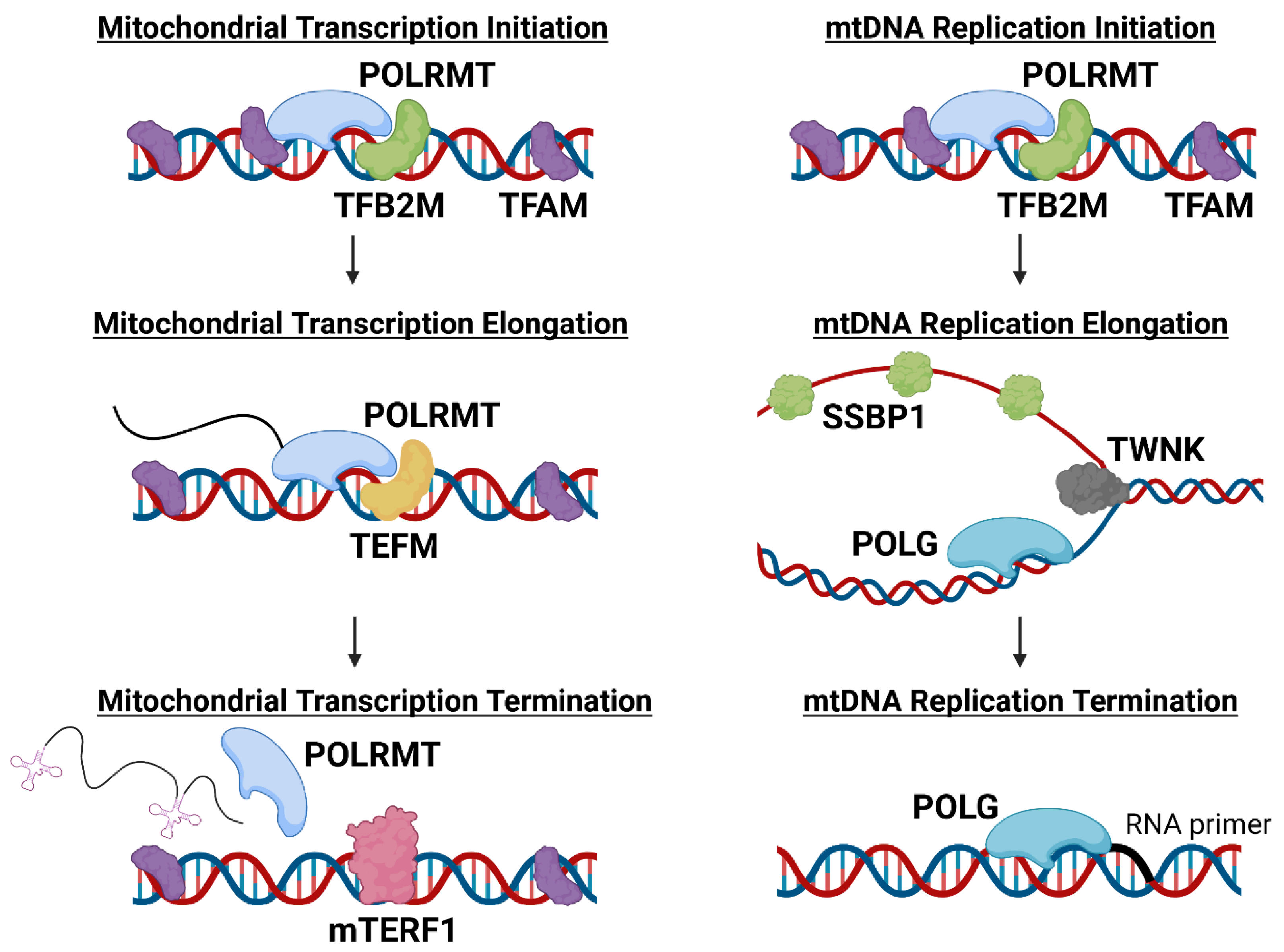
2. Functional Roles of POLRMT in Mitochondria

POLRMT, the only RNA polymerase present in the mitochondria, transcribes and facilitates the replication of mtDNA [43]. POLRMT is therefore a pivotal enzyme for the expression of the genes encoded by mtDNA and for the maintenance of functional mitochondria and related metabolic processes [44]. During mitochondrial transcription, POLRMT works in concert with mitochondrial transcriptional factor A (TFAM) and mitochondrial dimethyladenosine transferase 2 (TFB2M) to initiate mitochondrial transcription. TFAM binds to mtDNA promoters to direct POLRMT to initiate transcription, while TFB2M aids POLRMT in melting and stabilizing the mtDNA strands to enable transcription (Figure 1) [9][43]. TFAM has an additional role in mtDNA maintenance. TFAM binds nonspecifically to mtDNA, causing the DNA to bend and allowing the packaging of genetic material into nucleoids [9][45]. TFAM typically binds to mtDNA every 15–20 kb, and the abundance of TFAM bound to mtDNA is critically important in mtDNA transcriptional regulation [46][47]. Overexpression of TFAM has been shown to reduce transcription and expression of mitochondrially-encoded genes, potentially due to TFAM blocking POLRMT access to mtDNA [47][48]. However, some tissues have been observed to upregulate expression of POLRMT, potentially as a mechanism to overcome high TFAM levels and promote increased transcription [47]. For example, transcription at LSP, HSP1, or HSP2 can be regulated by the TFAM protein level because each promotor is transcribed optimally at a different TFAM:mtDNA ratio [47][48]. These data suggest both TFAM and POLRMT play critical roles in regulating mitochondrial transcription and function.
Figure 1. The role of mitochondrial proteins in initiation, elongation, and termination of mitochondrial transcription and mtDNA replication. The above schematic depicts mitochondrial transcription, mtDNA replication, and the respective role of mitochondrial DNA-dependent RNA polymerase (POLRMT) in each. Mitochondrial DNA (mtDNA) replication starts at the light strand promoter (LSP), whereas mitochondrial transcription can start from the LSP, the heavy strand promotor 1, or 2 (HSP1, HSP2). After mitochondrial transcription is initiated, mitochondrial transcription factor A (TFAM) and mitochondrial dimethyladenosine transferase 2 (TFB2M) dissociate from POLRMT and mitochondrial transcription elongation factor (TEFM) binds to stabilize POLRMT and mtDNA during elongation. After the polycistronic transcript has been created, POLRMT is sterically blocked by mitochondrial transcriptional termination factor 1 (mTERF1) (during LSP transcription), or by the 7S DNA region (during HSP2 transcription). Once POLRMT, TFAM, and TFB2M initiate mtDNA replication, the initiation complex dissociates and DNA polymerase γ (POLG) begins replication. Twinkle mtDNA helicase (TWNK) anneals the DNA strands while mitochondrial single-stranded DNA binding protein (SSBP1) binds to and stabilizes the single-stranded DNA strand. When POLG reaches the RNA primer again, replication is terminated.

3. POLRMT Inhibition as an Anti-Cancer Strategy

POLRMT is overexpressed at both the mRNA and protein level in several cancers, including breast, skin, lung, endometrial cancer, and osteosarcomas [32][49][50][51][52][53]. In both primary tumors and cancer cell lines, POLRMT overexpression promoted cell proliferation, migration, and invasion [32][49][50][51][52][53]. Knockout of POLRMT also impaired multiple cancer-related processes such as cell proliferation, migration, invasion, and angiogenesis [32][49][51][52][53]. Additional studies found that depletion of POLRMT induced apoptosis, as measured by caspase-3 and PARP cleavage, and increased TUNEL staining [51][52]. Mitochondrial membrane depolarization was also observed following POLRMT knockout, indicating that POLRMT activity is essential for mitochondrial function [52]. Despite this knowledge, the potential for targeting POLRMT and associated processes in cancer has only recently been explored.
Small molecule antiviral ribonucleosides (AVRNs) were first introduced in clinical trials for Hepatitis C as a strategy to inhibit the HCVRNA-dependent RNA polymerase [54]. An off-target effect of AVRNs was inhibition of POLRMT, leading to increased interest in identifying POLRMT inhibitors [55]. To screen for novel inhibitors of POLRMT, Bergbrede et al. developed a cell-free assay which identified the small molecule IMT1 as an inhibitor of POLRMT and mitochondrial transcription [19]. Further characterization determined that IMT1 selectively binds and inhibits POLRMT over other RNA polymerases found in humans, yeast, bacteria, and viruses [20]. IMT1 and its analogs (i.e., IMT1B), were shown to exert anti-cancer properties, most notably inhibiting cell proliferation and inducing cell death [20][32]. Consistent with POLRMT knockout experiments, IMT1-mediated inhibition of POLRMT impaired mitochondrial function through mtDNA depletion, OXPHOS inhibition [20], mitochondria depolarization, and mitochondrial reactive oxygen species (ROS) level induction [32].

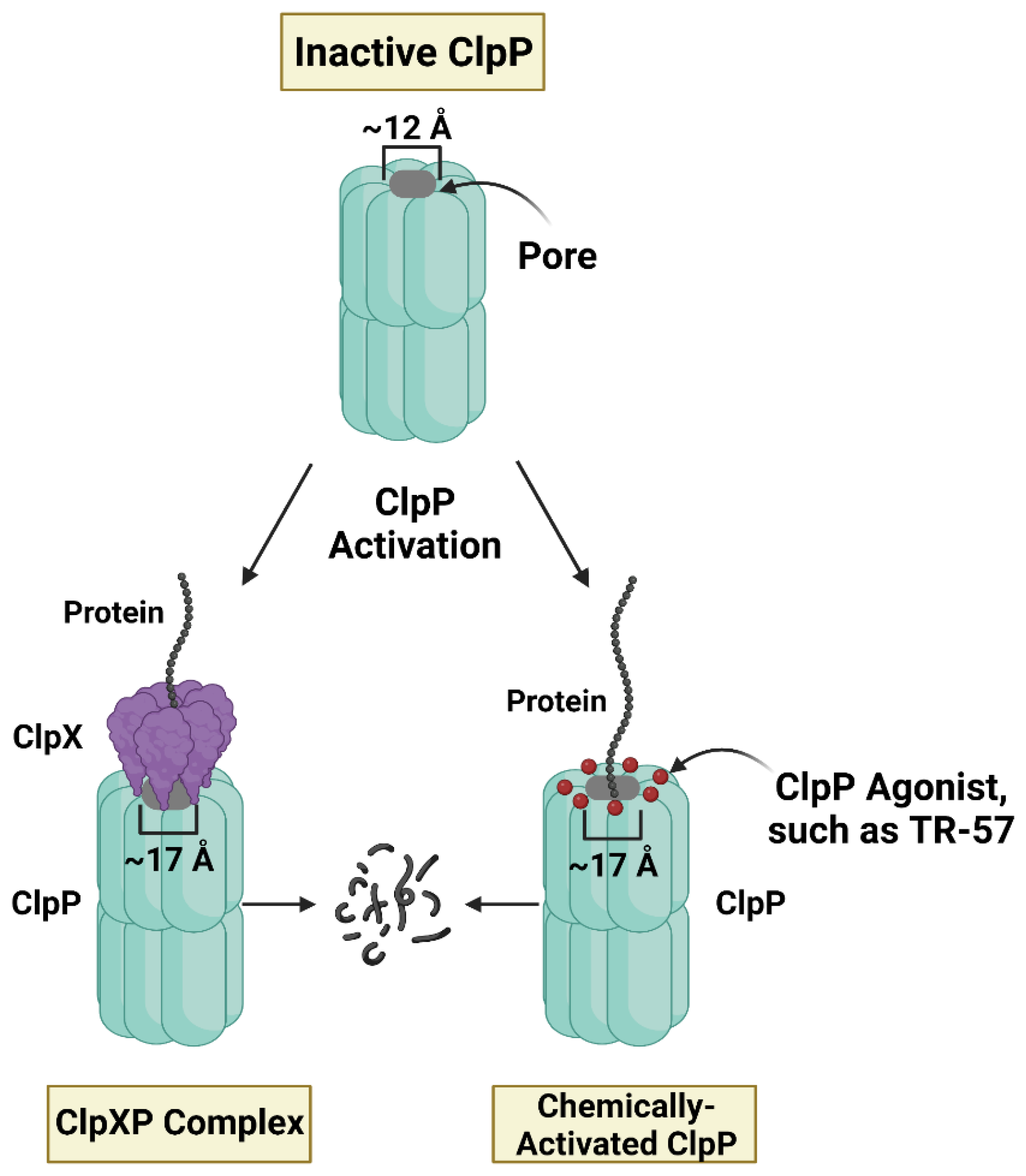
4. Small Molecule ClpP Agonists as Anti-Cancer Compounds

Recently, a novel class of mitochondria-targeting drugs were discovered based on their highly unusual property of activating the mitochondrial protease ClpP. ONC201 was first identified as a potential anti-cancer compound in a screen for small molecule inducers of tumor necrosis factor-related apoptosis-inducing ligand (TRAIL) [37]. ONC201 (Dordaviprone) and ONC206 have now progressed to phase I, II, and III clinical trials for gliomas, acute myeloid leukemia (AML), endometrial, breast, and other cancers [56][57][58]. More recently, Madera Therapeutics LLC developed scaffold analogs of ONC201 (TR compounds) which are highly potent and selective agonists of ClpP [33]. Studies confirmed that these compounds bind to ClpP, the proteolytic barrel of the ClpXP mitochondrial matrix protease. The ClpXP protease is comprised of two components, ClpP and the unfoldase ClpX, which plays an important role in protein quality control and mitochondrial proteostasis (Figure 2) [59][60].
Figure 2. Regulated and unregulated activity of mitochondrial caseinolytic protease ClpP. The above figure depicts methods of ClpP activation. The protease ClpP is composed of 14 ClpP subunits, assembled into 2 heptameric rings. ClpP is typically bound to ClpX, an ATP-dependent unfoldase that contributes to ClpP proteolytic specificity. ClpP agonists bind to ClpP in the same location as ClpX, causing the barrel-shaped pore in ClpP to open. The closed pore is ~12 Å and the opened pore is ~17 Å, as determined by Ishizawa et al. [61]. With the pore open, ClpP is active and can degrade proteins, so ClpP agonists are able to increase proteolysis in the absence of ClpX.
Though ClpP activation results in the widespread loss of mitochondrial matrix proteins [38], identification of specific ClpP substrates is still being pursued. Recent studies identified multiple ClpP-interacting proteins and putative substrates through proximity ligation (Bio-ID) and N-terminome (HYTANE) profiling methods [35][62]. Proteomics profiling in conjunction with transcriptomics and metabolomics analyses has provided further insight into the cellular response to ClpP activation [38]
POLRMT protein was shown to be strongly reduced in triple-negative breast cancer (TNBC) cells following ClpP activation by TR-57 and TR-107 [34][38]. Immunoblot and proteomic analysis revealed that POLRMT was significantly depleted after 24 h of treatment with TR-57 or TR-107, but was unaffected in ClpP knockout TNBC cell lines following treatment [34][38]. POLRMT subsequently was identified as a ClpP-interacting protein by Bio-ID, indicating that the loss of this protein may be due to direct degradation by ClpP [62]. TFAM, a known POLRMT interactor, was also strongly reduced at the protein level following ClpP activation in TNBC cells in a time- and dose-dependent manner [33][34][63]. TFAM was also identified as a potential ClpP interactor by Bio-ID [62], and determined to be a direct ClpP substrate in response to activation by TR-65 [35].

5. Mechanistic Similarities between ClpP Agonists and POLRMT Inhibitors

5.1. Inhibition of Cell Proliferation

ClpP agonists and POLRMT inhibitors have been shown to impair cellular proliferation in multiple cancer cell lines and models [20][32][33][64][65][66]. Some evidence suggests that ClpP agonists and POLRMT inhibitors can induce cell death, though these effects are generally minimal compared to the cytostatic effects observed on cell proliferation [20][32][63][65][67][68][69]. However, these results may depend on the specific ClpP agonist studied. For example, ONC201 treatment increased apoptosis in mantle cell lymphoma, AML, pancreatic ductal adenocarcinoma, multiple myeloma, and ovarian cancer cell lines, whereas there have been no reports of significant increases in apoptosis after TR compound treatment [65][68][69][70][71]. ONC201 was previously reported to be both a ClpP agonist and a dopamine receptor D2 antagonist, while the TR compounds are selective ClpP agonists [33][35][72], which may explain these differences.

5.2. Cytostatic to Cancer Cells, Harmless to Normal Cells

One of the unusual properties of ClpP agonists is that they appear to adversely affect cancer cells but have little or no effect on normal cells. Several groups have tested ClpP agonists on immortalized cell lines (i.e., HFF) or primary cells and observed no significant effects on cell proliferation, viability, or apoptosis as compared to the cancer cell models [42][61][63][73]. Additionally, ClpP agonists were reported to be well tolerated clinically, with few side effects observed in patients with refractory solid tumors and neuroendocrine tumors [57][58]. In contrast, other mitochondria-targeting drugs such as metformin and CPI-631 have shown negative side effects such as gastrointestinal tract disorders, nausea, and vomiting; however, these toxicities were not reported in ONC201 clinical trials [57][58][74][75].

5.3. Dysregulation of Cancer Cell Metabolic Programs

Both ClpP agonists and POLRMT inhibitors induce major metabolic alterations in cancer cells, most prominently observed with the inhibition of OXPHOS [20][34][63][66][72]. Both classes of compounds reduce the mitochondrially-encoded protein components required for OXPHOS [20][34]. However, the mechanism by which OXPHOS is impaired in response to ClpP activation is less well understood than the proposed mechanism of POLRMT inhibitors. Loss of POLRMT activity results in the direct inhibition of mitochondrial transcription, which in turn prevents the expression of the 13 protein-coding genes essential for respiration [20]. Significant decreases in mRNA levels of several of these genes have been shown following IMT1 treatment [20][32], confirming inhibition of mitochondrial transcription following POLRMT inhibition. This combined with the loss of their cognate proteins (i.e., COI) could explain the observed inhibition of OXPHOS in these cells [20].
Another common metabolic alteration observed following ClpP activation or POLRMT inhibition is AMPK activation [20][63]. Greer et al. (2018) observed a decline in ATP levels and an increase in AMPK phosphorylation (Thr172) following ONC201 treatment [63]. Similar effects were observed with IMT1 treatment [20]. AMPK activation is mediated by a high cellular AMP/ATP ratio [76], further indicating that inhibition of OXPHOS and/or other mitochondrial processes required for ATP generation is occurring in response to these two different mitochondrial targeting approaches.
As an expected consequence of OXPHOS inhibition, ClpP agonists have been shown to cause a compensatory increase in glycolysis [34][63][66]. A similar compensatory response has not been demonstrated following POLRMT inhibition, although it may be predicted.

5.4. Loss of mtDNA Content

An important similarity between ClpP agonists and POLRMT inhibitors is the corresponding reduction in mtDNA content, despite observations that the mtDNA copy number appears to change at different rates. IMT1 treatment reportedly took ~96 h to reduce the mtDNA copy number to ~25% of its original number [20], whereas ONC201 treatment required less time (~48 h) to reduce the mtDNA copy number to the same level [63]. This may in part be determined by the rate of TFAM protein loss. TFAM expression is closely correlated to the mtDNA copy number, likely because it binds to and packages mtDNA [77]. ClpP activation results in a time- and dose-dependent decline in TFAM protein that correlates with observed decreases in mtDNA content [33][34][63][66], as do POLRMT inhibitors (Daglish, unpublished observations (IMT1)).

5.5. Inhibition of Mitochondrial Transcription

It has been shown that POLRMT and TFAM protein expression decreases after treatment with ClpP agonists, but it has not been established that mitochondrial transcription is inhibited. The scholars predicted that mitochondrial transcription would be impaired by ClpP agonists due to their ability to deplete POLRMT, TFAM, and TEFM proteins which are all essential for mitochondrial transcription (Figure 1). Both TR-57 and IMT1 inhibited mitochondrial transcription in the tumorigenic immortalized HEK293T cells line (Figure 3) [78][79]. mRNA transcript levels of two mitochondrially-encoded protein genes required for OXPHOS (ND1 and ND6) were measured by quantitative PCR to monitor mitochondrial transcription from the heavy and light strands of mtDNA, respectively. Both treatments significantly decreased ND1 and ND6 transcript levels after 3 and 6 h, although there was a greater decrease observed following IMT1 treatment (Figure 3).
Figure 3. TR-57 and IMT1 both inhibit mitochondrial transcription from the heavy and light strand. HEK293T cells were plated at 4 × 10⁵ cells/well in 6 well plates, allowed to adhere overnight, and treated with TR-57 (150 nM) and IMT1 (10 µM) for indicated times. Cells were washed 3 times with cold DPBS, scraped, and pelleted. RNA was isolated using the RNeasy Plus Mini Kit (Qiagen) and residual DNA was removed using the RNase-Free DNase Set (Qiagen). cDNA was synthesized from 2 μg of extracted RNA in 20 μL total volume using the High-Capacity cDNA Reverse Transcription Kit (Applied Biosystems) and the Bio-Rad T100 thermal cycler. Quantitative PCR was performed using iTaq Universal SYBR Green Supermix (Bio-Rad). The nuclear reference gene 18S, mitochondrial heavy strand gene, ND1, and mitochondrial light strand gene, ND6, were quantified using the CFX96 Touch Real-Time System. A one-way ANOVA analysis was performed to determine significance; p-value < 0.05 (*), 0.01 (**), 0.001 (***). N = 2 biological replicates.

6. Mechanistic Differences between ClpP Agonists and POLRMT Inhibitors

6.1. Differences in Treatment Response Times

Despite the many mechanistic similarities between ClpP agonists and POLRMT inhibitors, there are several notable differences. Primarily, ClpP agonists inhibit cell proliferation much more robustly than POLRMT inhibitors. TR-57 strongly inhibits HEK293T cell proliferation following 72 h treatment, whereas IMT1 treatment requires a minimum of 120 h to show a similar inhibition of cell proliferation (Figure 4). In terms of potency, the TR compounds are more potent inhibitors of HEK293T growth; the IC50 of TR-57 in HEK293T cells is ~15 nM after 72 h, whereas the POLRMT inhibitor IMT1 has an IC50 of ~190 nM after 120 h of treatment (Figure 4).
Figure 4. The ClpP agonist TR-57 inhibits cell proliferation more potently and rapidly than the POLRMT inhibitor IMT1. HEK293T cells were cultured in DMEM media supplemented with 10% fetal bovine serum and 1% antibiotic/antimycotic (Gibco). Cells were either plated in 96 well plates at 5 × 10³ cells/well, allowed to adhere overnight, and treated for 72 h with indicated concentrations of TR-57 and IMT1, or cells were plated at 1.5 × 10³ cells/well, allowed to adhere overnight, and treated with indicated concentrations of IMT1 for 120 h. After drug treatment for 72 (A) or 120 h (B), cells were stained with 2.5 µg/mL of Hoechst 33,342 and incubated for 20 min at 37 °C and 5% CO2. Total cell number was counted using the Celígo S imager (Nexcelom Biosciences). N = 2 biological replicates. The reported IC50 values are averaged from the 2 replicates.

6.2. Known and Unknown Mechanisms of Resistance

There are a few known cancer cell mechanisms of resistance to ClpP agonists, including p0 cells, fumarate hydratase knockout cell lines, and ClpP knockout or ClpP mutant cell lines [80]. The specific ClpP mutation, D190A, is known to confer resistance to ClpP agonists [61]. Comparatively, there are specific POLRMT mutations that confer resistance to IMT1. Bonekamp et al. (2020) reported six mutations in POLRMT that lead to IMT1-resistance [20]. A CRISPR screen aimed at identifying mechanisms of resistance against IMT1 found that loss of von Hippel-Landau protein (VHL) and mammalian target of rapamycin complex 1 (mTORC1) expression produced IMT1-resistance in RKO cells [81]

6.3. Inhibiting One Protein versus Degrading Many Proteins

Major differences distinguishing the effects of POLRMT inhibitors from ClpP agonists may result from IMT1 selectively inhibiting mitochondrial transcription [20], whereas ClpP agonists dysregulate multiple mitochondrial processes [35][39][61][62][68]. As such, elucidating the specific mechanisms of action of ClpP agonists may prove to be more difficult. While efforts continue to identify specific ClpP substrates and related metabolic processes, the connection between loss of a specific ClpP substrate and its effects on cell proliferation remains to be established. Comparatively, a potential drawback to POLRMT inhibition is that these inhibitors only primarily impact one process or cancer cell vulnerability (mitochondrial transcription) [20]. Since pharmacological activation of ClpP proteolysis affects multiple growth-related cellular processes, this intuits an overall more disruptive mechanism than POLRMT inhibition. In the context of cancer cell growth, there may be an advantage to eliminating multiple events required for cancer cell growth to prevent the development of resistance more effectively.

References

  1. Chakrabarty, R.P.; Chandel, N.S. Beyond ATP, New Roles of Mitochondria. Biochemist 2022, 44, 2–8.
  2. Haynes, C.M.; Petrova, K.; Benedetti, C.; Yang, Y.; Ron, D. ClpP Mediates Activation of a Mitochondrial Unfolded Protein Response in C. Elegans. Dev. Cell 2007, 13, 467–480.
  3. Giampazolias, E.; Zunino, B.; Dhayade, S.; Bock, F.; Cloix, C.; Cao, K.; Roca, A.; Lopez, J.; Ichim, G.; Proïcs, E.; et al. Mitochondrial Permeabilization Engages NF-ΚB-Dependent Anti-Tumour Activity under Caspase Deficiency. Nat. Cell Biol. 2017, 19, 1116–1129.
  4. Collins, Y.; Chouchani, E.T.; James, A.M.; Menger, K.E.; Cochemé, H.M.; Murphy, M.P. Mitochondrial Redox Signalling at a Glance. J. Cell Sci. 2012, 125, 801–806.
  5. Riley, J.S.; Tait, S.W. Mitochondrial DNA in Inflammation and Immunity. EMBO Rep. 2020, 21, e49799.
  6. Qureshi, M.A.; Haynes, C.M.; Pellegrino, M.W. The Mitochondrial Unfolded Protein Response: Signaling from the Powerhouse. J. Biol. Chem. 2017, 292, 13500–13506.
  7. Bueno, M.J.; Ruiz-Sepulveda, J.L.; Quintela-Fandino, M. Mitochondrial Inhibition: A Treatment Strategy in Cancer? Curr. Oncol. Rep. 2021, 23, 49.
  8. Rackham, O.; Filipovska, A. Organization and Expression of the Mammalian Mitochondrial Genome. Nat. Rev. Genet. 2022, 23, 606–623.
  9. Hillen, H.S.; Temiakov, D.; Cramer, P. Structural Basis of Mitochondrial Transcription. Nat. Struct. Mol. Biol. 2018, 25, 754–765.
  10. Hashimoto, Y.; Kurita, M.; Aiso, S.; Nishimoto, I.; Matsuoka, M. Humanin Inhibits Neuronal Cell Death by Interacting with a Cytokine Receptor Complex or Complexes Involving CNTF Receptor Alpha/WSX-1/Gp130. Mol. Biol. Cell 2009, 20, 2864–2873.
  11. Ro, S.; Ma, H.-Y.; Park, C.; Ortogero, N.; Song, R.; Hennig, G.W.; Zheng, H.; Lin, Y.-M.; Moro, L.; Hsieh, J.-T.; et al. The Mitochondrial Genome Encodes Abundant Small Noncoding RNAs. Cell Res. 2013, 23, 759–774.
  12. Kummer, E.; Ban, N. Mechanisms and Regulation of Protein Synthesis in Mitochondria. Nat. Rev. Mol. Cell Biol. 2021, 22, 307–325.
  13. Sainero-Alcolado, L.; Liaño-Pons, J.; Ruiz-Pérez, M.V.; Arsenian-Henriksson, M. Targeting Mitochondrial Metabolism for Precision Medicine in Cancer. Cell Death Differ. 2022, 29, 1304–1317.
  14. Wheaton, W.W.; Weinberg, S.E.; Hamanaka, R.B.; Soberanes, S.; Sullivan, L.B.; Anso, E.; Glasauer, A.; Dufour, E.; Mutlu, G.M.; Budigner, G.S.; et al. Metformin Inhibits Mitochondrial Complex I of Cancer Cells to Reduce Tumorigenesis. eLife 2014, 3, e02242.
  15. Moullan, N.; Mouchiroud, L.; Wang, X.; Ryu, D.; Williams, E.G.; Mottis, A.; Jovaisaite, V.; Frochaux, M.V.; Quiros, P.M.; Deplancke, B.; et al. Tetracyclines Disturb Mitochondrial Function across Eukaryotic Models: A Call for Caution in Biomedical Research. Cell Rep. 2015, 10, 1681–1691.
  16. Dijk, S.N.; Protasoni, M.; Elpidorou, M.; Kroon, A.M.; Taanman, J.-W. Mitochondria as Target to Inhibit Proliferation and Induce Apoptosis of Cancer Cells: The Effects of Doxycycline and Gemcitabine. Sci. Rep. 2020, 10, 4363.
  17. Rivas, M.O.G.; Stuart, S.D.; Thach, D.; Dahan, M.; Shorr, R.; Zachar, Z.; Bingham, P.M. Evidence for a Novel, Effective Approach to Targeting Carcinoma Catabolism Exploiting the First-in-Class, Anti-Cancer Mitochondrial Drug, CPI-613. PLoS ONE 2022, 17, e0269620.
  18. Delaunay, S.; Pascual, G.; Feng, B.; Klann, K.; Behm, M.; Hotz-Wagenblatt, A.; Richter, K.; Zaoui, K.; Herpel, E.; Münch, C.; et al. Mitochondrial RNA Modifications Shape Metabolic Plasticity in Metastasis. Nature 2022, 607, 593–603.
  19. Bergbrede, T.; Hoberg, E.; Larsson, N.-G.; Falkenberg, M.; Gustafsson, C.M. An Adaptable High-Throughput Technology Enabling the Identification of Specific Transcription Modulators. SLAS Discov. 2017, 22, 378–386.
  20. Bonekamp, N.A.; Peter, B.; Hillen, H.S.; Felser, A.; Bergbrede, T.; Choidas, A.; Horn, M.; Unger, A.; Di Lucrezia, R.; Atanassov, I.; et al. Small-Molecule Inhibitors of Human Mitochondrial DNA Transcription. Nature 2020, 588, 712–716.
  21. Reznik, E.; Miller, M.L.; Şenbabaoğlu, Y.; Riaz, N.; Sarungbam, J.; Tickoo, S.K.; Al-Ahmadie, H.A.; Lee, W.; Seshan, V.E.; Hakimi, A.A.; et al. Mitochondrial DNA Copy Number Variation across Human Cancers. eLife 2016, 5, e10769.
  22. van Osch, F.H.M.; Voets, A.M.; Schouten, L.J.; Gottschalk, R.W.H.; Simons, C.C.J.M.; van Engeland, M.; Lentjes, M.H.F.M.; van den Brandt, P.A.; Smeets, H.J.M.; Weijenberg, M.P. Mitochondrial DNA Copy Number in Colorectal Cancer: Between Tissue Comparisons, Clinicopathological Characteristics and Survival. Carcinogenesis 2015, 36, 1502–1510.
  23. Yuan, Y.; Ju, Y.S.; Kim, Y.; Li, J.; Wang, Y.; Yoon, C.J.; Yang, Y.; Martincorena, I.; Creighton, C.J.; Weinstein, J.N.; et al. Comprehensive Molecular Characterization of Mitochondrial Genomes in Human Cancers. Nat. Genet. 2020, 52, 342–352.
  24. Tseng, L.-M.; Yin, P.-H.; Chi, C.-W.; Hsu, C.-Y.; Wu, C.-W.; Lee, L.-M.; Wei, Y.-H.; Lee, H.-C. Mitochondrial DNA Mutations and Mitochondrial DNA Depletion in Breast Cancer. Genes Chromosom. Cancer 2006, 45, 629–638.
  25. Wallace, D.C. Mitochondria and Cancer. Nat. Rev. Cancer 2012, 12, 685–698.
  26. Vyas, S.; Zaganjor, E.; Haigis, M.C. Mitochondria and Cancer. Cell 2016, 166, 555–566.
  27. Zong, W.-X.; Rabinowitz, J.D.; White, E. Mitochondria and Cancer. Mol. Cell 2016, 61, 667–676.
  28. Filograna, R.; Mennuni, M.; Alsina, D.; Larsson, N.-G. Mitochondrial DNA Copy Number in Human Disease: The More the Better? FEBS Lett. 2021, 595, 976–1002.
  29. Grandhi, S.; Bosworth, C.; Maddox, W.; Sensiba, C.; Akhavanfard, S.; Ni, Y.; LaFramboise, T. Heteroplasmic Shifts in Tumor Mitochondrial Genomes Reveal Tissue-Specific Signals of Relaxed and Positive Selection. Hum. Mol. Genet. 2017, 26, 2912–2922.
  30. Lax, N.Z.; Turnbull, D.M.; Reeve, A.K. Mitochondrial Mutations. Neuroscientist 2011, 17, 645–658.
  31. Van Gisbergen, M.W.; Voets, A.M.; Starmans, M.H.W.; De Coo, I.F.M.; Yadak, R.; Hoffmann, R.F.; Boutros, P.C.; Smeets, H.J.M.; Dubois, L.; Lambin, P. How Do Changes in the MtDNA and Mitochondrial Dysfunction Influence Cancer and Cancer Therapy? Challenges, Opportunities and Models. Mutat. Res. Rev. Mutat. Res. 2015, 764, 16–30.
  32. Li, S.; Ou, L.; Zhang, Y.; Shen, F.; Chen, Y. A First-in-Class POLRMT Specific Inhibitor IMT1 Suppresses Endometrial Carcinoma Cell Growth. Cell Death Dis. 2023, 14, 152.
  33. Graves, P.R.; Aponte-Collazo, L.J.; Fennell, E.M.J.; Graves, A.C.; Hale, A.E.; Dicheva, N.; Herring, L.E.; Gilbert, T.S.K.; East, M.P.; McDonald, I.M.; et al. Mitochondrial Protease ClpP Is a Target for the Anticancer Compounds ONC201 and Related Analogues. ACS Chem. Biol. 2019, 14, 1020–1029.
  34. Fennell, E.M.J.; Aponte-Collazo, L.J.; Wynn, J.D.; Drizyte-Miller, K.; Leung, E.; Greer, Y.E.; Graves, P.R.; Iwanowicz, A.A.; Ashamalla, H.; Holmuhamedov, E.; et al. Characterization of TR-107, a Novel Chemical Activator of the Human Mitochondrial Protease ClpP. Pharmacol. Res. Perspect. 2022, 10, e00993.
  35. Mabanglo, M.F.; Wong, K.S.; Barghash, M.M.; Leung, E.; Chuang, S.H.W.; Ardalan, A.; Majaesic, E.M.; Wong, C.J.; Zhang, S.; Lang, H.; et al. Potent ClpP Agonists with Anticancer Properties Bind with Improved Structural Complementarity and Alter the Mitochondrial N-Terminome. Structure 2022, 31, 185–200.
  36. Wang, P.; Zhang, T.; Wang, X.; Xiao, H.; Li, H.; Zhou, L.-L.; Yang, T.; Wei, B.; Zhu, Z.; Zhou, L.; et al. Aberrant Human ClpP Activation Disturbs Mitochondrial Proteome Homeostasis to Suppress Pancreatic Ductal Adenocarcinoma. Cell Chem. Biol. 2022, 29, 1396–1408.e8.
  37. Allen, J.E.; Krigsfeld, G.; Patel, L.; Mayes, P.A.; Dicker, D.T.; Wu, G.S.; El-Deiry, W.S. Identification of TRAIL-Inducing Compounds Highlights Small Molecule ONC201/TIC10 as a Unique Anti-Cancer Agent That Activates the TRAIL Pathway. Mol. Cancer 2015, 14, 99.
  38. Fennell, E.M.J.; Aponte-Collazo, L.J.; Pathmasiri, W.; Rushing, B.R.; Barker, N.K.; Partridge, M.C.; Li, Y.-Y.; White, C.A.; Greer, Y.E.; Herring, L.E.; et al. Multi-Omics Analyses Reveal ClpP Activators Disrupt Essential Mitochondrial Pathways in Triple-Negative Breast Cancer. Front. Pharmacol. 2023, 14, 1136317.
  39. Kline, C.L.B.; Van den Heuvel, A.P.J.; Allen, J.E.; Prabhu, V.V.; Dicker, D.T.; El-Deiry, W.S. ONC201 Kills Solid Tumor Cells by Triggering an Integrated Stress Response Dependent on ATF4 Activation by Specific EIF2α Kinases. Sci. Signal. 2016, 9, ra18.
  40. Ray, J.E.; Ralff, M.D.; Jhaveri, A.; Zhou, L.; Dicker, D.T.; Ross, E.A.; El-Deiry, W.S. Antitumorigenic Effect of Combination Treatment with ONC201 and TRAIL in Endometrial Cancer in Vitro and in Vivo. Cancer Biol. Ther. 2021, 22, 554–563.
  41. Yuan, X.; Kho, D.; Xu, J.; Gajan, A.; Wu, K.; Wu, G.S. ONC201 Activates ER Stress to Inhibit the Growth of Triple-Negative Breast Cancer Cells. Oncotarget 2017, 8, 21626–21638.
  42. Allen, J.E.; Crowder, R.; El-Deiry, W.S. First-in-Class Small Molecule ONC201 Induces DR5 and Cell Death in Tumor but Not Normal Cells to Provide a Wide Therapeutic Index as an Anti-Cancer Agent. PLoS ONE 2015, 10, e0143082.
  43. Hillen, H.S.; Morozov, Y.I.; Sarfallah, A.; Temiakov, D.; Cramer, P. Structural Basis of Mitochondrial Transcription Initiation. Cell 2017, 171, 1072–1081.e10.
  44. Shutt, T.E.; Bestwick, M.; Shadel, G.S. The Core Human Mitochondrial Transcription Initiation Complex. Transcription 2011, 2, 55–59.
  45. Kaufman, B.A.; Durisic, N.; Mativetsky, J.M.; Costantino, S.; Hancock, M.A.; Grutter, P.; Shoubridge, E.A. The Mitochondrial Transcription Factor TFAM Coordinates the Assembly of Multiple DNA Molecules into Nucleoid-like Structures. Mol. Biol. Cell 2007, 18, 3225–3236.
  46. Kukat, C.; Wurm, C.A.; Spåhr, H.; Falkenberg, M.; Larsson, N.-G.; Jakobs, S. Super-Resolution Microscopy Reveals That Mammalian Mitochondrial Nucleoids Have a Uniform Size and Frequently Contain a Single Copy of MtDNA. Proc. Natl. Acad. Sci. USA 2011, 108, 13534–13539.
  47. Bonekamp, N.A.; Jiang, M.; Motori, E.; Villegas, R.G.; Koolmeister, C.; Atanassov, I.; Mesaros, A.; Park, C.B.; Larsson, N.-G. High Levels of TFAM Repress Mammalian Mitochondrial DNA Transcription in Vivo. Life Sci. Alliance 2021, 4, 11.
  48. Lodeiro, M.F.; Uchida, A.; Bestwick, M.; Moustafa, I.M.; Arnold, J.J.; Shadel, G.S.; Cameron, C.E. Transcription from the Second Heavy-Strand Promoter of Human MtDNA Is Repressed by Transcription Factor A in Vitro. Proc. Natl. Acad. Sci. USA 2012, 109, 6513–6518.
  49. Salem, A.F.; Whitaker-Menezes, D.; Howell, A.; Sotgia, F.; Lisanti, M.P. Mitochondrial Biogenesis in Epithelial Cancer Cells Promotes Breast Cancer Tumor Growth and Confers Autophagy Resistance. Cell Cycle 2012, 11, 4174–4180.
  50. Sotgia, F.; Whitaker-Menezes, D.; Martinez-Outschoorn, U.E.; Salem, A.F.; Tsirigos, A.; Lamb, R.; Sneddon, S.; Hulit, J.; Howell, A.; Lisanti, M.P. Mitochondria “Fuel” Breast Cancer Metabolism: Fifteen Markers of Mitochondrial Biogenesis Label Epithelial Cancer Cells, but Are Excluded from Adjacent Stromal Cells. Cell Cycle 2012, 11, 4390–4401.
  51. Han, Q.-C.; Zhang, X.-Y.; Yan, P.-H.; Chen, S.-F.; Liu, F.-F.; Zhu, Y.-R.; Tian, Q. Identification of Mitochondrial RNA Polymerase as a Potential Therapeutic Target of Osteosarcoma. Cell Death Discov. 2021, 7, 393.
  52. Zhou, T.; Sang, Y.-H.; Cai, S.; Xu, C.; Shi, M. The Requirement of Mitochondrial RNA Polymerase for Non-Small Cell Lung Cancer Cell Growth. Cell Death Dis. 2021, 12, 751.
  53. Wang, Y.; Ou, L.; Li, X.; Zheng, T.; Zhu, W.; Li, P.; Wu, L.; Zhao, T. The Mitochondrial RNA Polymerase POLRMT Promotes Skin Squamous Cell Carcinoma Cell Growth. Cell Death Discov. 2022, 8, 347.
  54. Sarrazin, C.; Hézode, C.; Zeuzem, S.; Pawlotsky, J.-M. Antiviral Strategies in Hepatitis C Virus Infection. J. Hepatol. 2012, 56, S88–S100.
  55. Arnold, J.J.; Smidansky, E.D.; Moustafa, I.M.; Cameron, C.E. Human Mitochondrial RNA Polymerase: Structure–Function, Mechanism and Inhibition. Biochim. Et Biophys. Acta (BBA)—Gene Regul. Mech. 2012, 1819, 948–960.
  56. Home—ClinicalTrials.Gov. Available online: https://clinicaltrials.gov/ (accessed on 27 January 2023).
  57. Arrillaga-Romany, I.; Odia, Y.; Prabhu, V.V.; Tarapore, R.S.; Merdinger, K.; Stogniew, M.; Oster, W.; Allen, J.E.; Mehta, M.; Batchelor, T.T.; et al. Biological Activity of Weekly ONC201 in Adult Recurrent Glioblastoma Patients. Neuro Oncol. 2020, 22, 94–102.
  58. Stein, M.N.; Bertino, J.R.; Kaufman, H.L.; Mayer, T.; Moss, R.; Silk, A.; Chan, N.; Malhotra, J.; Rodriguez, L.; Aisner, J.; et al. First-in-Human Clinical Trial of Oral ONC201 in Patients with Refractory Solid Tumors. Clin. Cancer Res. 2017, 23, 4163–4169.
  59. Fei, X.; Bell, T.A.; Jenni, S.; Stinson, B.M.; Baker, T.A.; Harrison, S.C.; Sauer, R.T. Structures of the ATP-Fueled ClpXP Proteolytic Machine Bound to Protein Substrate. eLife 2020, 9, e52774.
  60. Szczepanowska, K.; Trifunovic, A. Mitochondrial Matrix Proteases: Quality Control and Beyond. FEBS J. 2022, 289, 7128–7146.
  61. Ishizawa, J.; Zarabi, S.F.; Davis, R.E.; Halgas, O.; Nii, T.; Jitkova, Y.; Zhao, R.; St-Germain, J.; Heese, L.E.; Egan, G.; et al. Mitochondrial ClpP-Mediated Proteolysis Induces Selective Cancer Cell Lethality. Cancer Cell 2019, 35, 721–737.e9.
  62. Mabanglo, M.F.; Bhandari, V.; Houry, W.A. Substrates and Interactors of the ClpP Protease in the Mitochondria. Curr. Opin. Chem. Biol. 2022, 66, 102078.
  63. Greer, Y.E.; Porat-Shliom, N.; Nagashima, K.; Stuelten, C.; Crooks, D.; Koparde, V.N.; Gilbert, S.F.; Islam, C.; Ubaldini, A.; Ji, Y.; et al. ONC201 Kills Breast Cancer Cells in Vitro by Targeting Mitochondria. Oncotarget 2018, 9, 18454–18479.
  64. Zhang, J.; Luo, B.; Sui, J.; Qiu, Z.; Huang, J.; Yang, T.; Luo, Y. IMP075 Targeting ClpP for Colon Cancer Therapy in Vivo and in Vitro. Biochem. Pharmacol. 2022, 204, 115232.
  65. Fan, Y.; Wang, J.; Fang, Z.; Pierce, S.R.; West, L.; Staley, A.; Tucker, K.; Yin, Y.; Sun, W.; Kong, W.; et al. Anti-Tumor and Anti-Invasive Effects of ONC201 on Ovarian Cancer Cells and a Transgenic Mouse Model of Serous Ovarian Cancer. Front. Oncol. 2022, 12, 789450.
  66. Greer, Y.E.; Hernandez, L.; Fennell, E.M.J.; Kundu, M.; Voeller, D.; Chari, R.; Gilbert, S.F.; Gilbert, T.S.K.; Ratnayake, S.; Tang, B.; et al. Mitochondrial Matrix Protease ClpP Agonists Inhibit Cancer Stem Cell Function in Breast Cancer Cells by Disrupting Mitochondrial Homeostasis. Cancer Res. Commun. 2022, 2, 1144–1161.
  67. Mishukov, A.; Odinokova, I.; Mndlyan, E.; Kobyakova, M.; Abdullaev, S.; Zhalimov, V.; Glukhova, X.; Galat, V.; Galat, Y.; Senotov, A.; et al. ONC201-Induced Mitochondrial Dysfunction, Senescence-like Phenotype, and Sensitization of Cultured BT474 Human Breast Cancer Cells to TRAIL. Int. J. Mol. Sci. 2022, 23, 15551.
  68. Ishizawa, J.; Kojima, K.; Chachad, D.; Ruvolo, P.; Ruvolo, V.; Jacamo, R.O.; Borthakur, G.; Mu, H.; Zeng, Z.; Tabe, Y.; et al. ATF4 Induction through an Atypical Integrated Stress Response to ONC201 Triggers P53-Independent Apoptosis in Hematological Malignancies. Sci. Signal. 2016, 9, ra17.
  69. Jhaveri, A.V.; Zhou, L.; Ralff, M.D.; Lee, Y.S.; Navaraj, A.; Carneiro, B.A.; Safran, H.; Prabhu, V.V.; Ross, E.A.; Lee, S.; et al. Combination of ONC201 and TLY012 Induces Selective, Synergistic Apoptosis in Vitro and Significantly Delays PDAC Xenograft Growth in Vivo. Cancer Biol. 2021, 22, 607–618.
  70. Rumman, M.; Buck, S.; Polin, L.; Dzinic, S.; Boerner, J.; Winer, I.S. ONC201 Induces the Unfolded Protein Response (UPR) in High- and Low-Grade Ovarian Carcinoma Cell Lines and Leads to Cell Death Regardless of Platinum Sensitivity. Cancer Med. 2021, 10, 3373–3387.
  71. Tu, Y.; He, J.; Liu, H.; Lee, H.C.; Wang, H.; Ishizawa, J.; Allen, J.E.; Andreeff, M.; Orlowski, R.Z.; Davis, R.E.; et al. The Imipridone ONC201 Induces Apoptosis and Overcomes Chemotherapy Resistance by Up-Regulation of Bim in Multiple Myeloma. Neoplasia 2017, 19, 772–780.
  72. Prabhu, V.V.; Morrow, S.; Rahman Kawakibi, A.; Zhou, L.; Ralff, M.; Ray, J.; Jhaveri, A.; Ferrarini, I.; Lee, Y.; Parker, C.; et al. ONC201 and Imipridones: Anti-Cancer Compounds with Clinical Efficacy. Neoplasia 2020, 22, 725–744.
  73. Cole, A.; Wang, Z.; Coyaud, E.; Voisin, V.; Gronda, M.; Jitkova, Y.; Mattson, R.; Hurren, R.; Babovic, S.; Maclean, N.; et al. Inhibition of the Mitochondrial Protease ClpP as a Therapeutic Strategy for Human Acute Myeloid Leukemia. Cancer Cell 2015, 27, 864–876.
  74. Lycan, T.; Pardee, T.; Petty, W.; Bonomi, M.; Alistar, A.; Lamar, Z.; Isom, S.; Chan, M.; Miller, A.; Ruiz, J. A Phase II Clinical Trial of CPI-613 in Patients with Relapsed or Refractory Small Cell Lung Carcinoma. PLoS ONE 2016, 11, e0164244.
  75. Brown, J.R.; Chan, D.K.; Shank, J.J.; Griffith, K.A.; Fan, H.; Szulawski, R.; Yang, K.; Reynolds, R.K.; Johnston, C.; McLean, K.; et al. Phase II Clinical Trial of Metformin as a Cancer Stem Cell–Targeting Agent in Ovarian Cancer. JCI Insight 2020, 5, e133247.
  76. Mihaylova, M.M.; Shaw, R.J. The AMP-Activated Protein Kinase (AMPK) Signaling Pathway Coordinates Cell Growth, Autophagy, & Metabolism. Nat. Cell Biol. 2011, 13, 1016–1023.
  77. Ekstrand, M.I.; Falkenberg, M.; Rantanen, A.; Park, C.B.; Gaspari, M.; Hultenby, K.; Rustin, P.; Gustafsson, C.M.; Larsson, N.-G. Mitochondrial Transcription Factor A Regulates MtDNA Copy Number in Mammals. Hum. Mol. Genet. 2004, 13, 935–944.
  78. Stepanenko, A.A.; Dmitrenko, V.V. HEK293 in Cell Biology and Cancer Research: Phenotype, Karyotype, Tumorigenicity, and Stress-Induced Genome-Phenotype Evolution. Gene 2015, 569, 182–190.
  79. Shen, C.; Gu, M.; Song, C.; Miao, L.; Hu, L.; Liang, D.; Zheng, C. The Tumorigenicity Diversification in Human Embryonic Kidney 293 Cell Line Cultured in Vitro. Biologicals 2008, 36, 263–268.
  80. Wedam, R.; Greer, Y.E.; Wisniewski, D.J.; Weltz, S.; Kundu, M.; Voeller, D.; Lipkowitz, S. Targeting Mitochondria with ClpP Agonists as a Novel Therapeutic Opportunity in Breast Cancer. Cancers 2023, 15, 1936.
  81. Mennuni, M.; Filograna, R.; Felser, A.; Bonekamp, N.A.; Giavalisco, P.; Lytovchenko, O.; Larsson, N.-G. Metabolic Resistance to the Inhibition of Mitochondrial Transcription Revealed by CRISPR-Cas9 Screen. EMBO Rep. 2022, 23, e53054.
More
Information
Subjects: Cell Biology
Contributors MDPI registered users' name will be linked to their SciProfiles pages. To register with us, please refer to https://encyclopedia.pub/register : , ,
View Times: 348
Revisions: 2 times (View History)
Update Date: 08 Jun 2023
1000/1000
Video Production Service