Submitted Successfully!
To reward your contribution, here is a gift for you: A free trial for our video production service.
Thank you for your contribution! You can also upload a video entry or images related to this topic.
Version Summary Created by Modification Content Size Created at Operation
1 -- 4022 2023-06-01 20:06:47 |
2 references update Meta information modification 4022 2023-06-02 04:33:10 |

Video Upload Options

Do you have a full video?

Confirm

Are you sure to Delete?
Cite
If you have any further questions, please contact Encyclopedia Editorial Office.
Montanari, M.; Imbriani, P.; Bonsi, P.; Martella, G.; Peppe, A. Role of Enteric Nervous System in Parkinson’s Disease. Encyclopedia. Available online: https://encyclopedia.pub/entry/45115 (accessed on 01 July 2024).
Montanari M, Imbriani P, Bonsi P, Martella G, Peppe A. Role of Enteric Nervous System in Parkinson’s Disease. Encyclopedia. Available at: https://encyclopedia.pub/entry/45115. Accessed July 01, 2024.
Montanari, Martina, Paola Imbriani, Paola Bonsi, Giuseppina Martella, Antonella Peppe. "Role of Enteric Nervous System in Parkinson’s Disease" Encyclopedia, https://encyclopedia.pub/entry/45115 (accessed July 01, 2024).
Montanari, M., Imbriani, P., Bonsi, P., Martella, G., & Peppe, A. (2023, June 01). Role of Enteric Nervous System in Parkinson’s Disease. In Encyclopedia. https://encyclopedia.pub/entry/45115
Montanari, Martina, et al. "Role of Enteric Nervous System in Parkinson’s Disease." Encyclopedia. Web. 01 June, 2023.
Role of Enteric Nervous System in Parkinson’s Disease
Edit

The enteric nervous system (ENS) is a nerve network composed of neurons and glial cells that regulates the motor and secretory functions of the gastrointestinal (GI) tract. There is abundant evidence of mutual communication between the brain and the GI tract. Dysfunction of these connections appears to be involved in the pathophysiology of Parkinson’s disease (PD). Alterations in the ENS have been shown to occur very early in PD, even before central nervous system (CNS) involvement. Post-mortem studies of PD patients have shown aggregation of α-synuclein (αS) in specific subtypes of neurons in the ENS. Subsequently, αS spreads retrogradely in the CNS through preganglionic vagal fibers to this nerve’s dorsal motor nucleus (DMV) and other central nervous structures.

Parkinson’s disease enteric nervous system neurons

1. Introduction

The gastrointestinal (GI) tract is a long tubular structure that harbors highly diverse and complex communities of microorganisms, including bacteria, archaea, microeukaryotes, and viruses that readily vary with diet, pharmacological intervention, and disease [1].
Numerous articles have described differences in the composition and function of the gut microbiome of healthy individuals and patients of metabolic, autoimmune, and neurodegenerative diseases [2][3][4]. In the pathogenesis of brain disorders, the possible involvement of peripheral organs has always been marginal. However, it is now well established that the environment of the GI tract and distant organs, such as the brain, is affected by the homeostasis of the gut microbiota and the host’s health [4][5][6]. Early colonization of the gut microbiota is vital for brain function and behavior, considering that its absence results in impairment of the blood–brain barrier [7].
The enteric nervous system (ENS), the part of the nervous system closest to the microbiome, has recently become the subject of in-depth investigations [5][8][9]. It is now known that the microbiome affects the development and functioning of the ENS, modulating it throughout life [4]. Since a wide range of neuropathies are associated with ENS dysfunction, researchers believe it is worth taking a closer look at it [5][8][9].
The ENS is derived from pre-enteric (rhombencephalon) and sacral neural crest cells and includes efferent and afferent neurons, interneurons, and glial cells.
This well-organized and integrated network of plexuses is relatively independent because it can control gut function independently of CNS sympathetic and parasympathetic innervations [10][11]. However, the ENS is not autonomous, since several CNS structures monitor and regulate what is happening in the GI tract through biochemical signals [12][13]. The gut–brain axis (GBA) consists of a bidirectional communication between the CNS and ENS, linking the emotional and cognitive centers of the brain with peripheral gut functions [14][15][16]. The GBA connects CNS cognitive centers with gut centers, regulating immune activation, enteric reflex, entero-endocrine signaling, and intestinal permeability [5][17][18]. The bidirectional communication between the gut and the brain implies a vital role for the gut microbiome through regulating host metabolism and immune and vascular systems [19]. In addition, the gut microbiome can also influence the CNS through the vagus nerve by transmitting signals from the gut microbiome to the brain and vice versa in both health and disease through neuro-immuno-endocrine mediators [17][18][20][21].
Disruption of GBA results in alterations in intestinal motility and secretion causes visceral hypersensitivity and leads to cellular changes in the entero-endocrine and immune systems [20].
Considering this complexity, the GI tract can be affected by aging, irritable bowel syndrome, severe inflammatory conditions (Crohn’s disease and ulcerative colitis), and even neurodegenerative diseases such as Parkinson’s and Alzheimer’s [19][21][22][23][24].
Parkinson’s disease (PD) is a neurodegenerative disease characterized by the loss of dopaminergic cells in the Substantia Nigra pars compacta (SNpc) and brain accumulation of Lewy bodies (LB), which are abnormal aggregates of α-synuclein (αS) [25][26][27]. PD results from a synergistic interaction between genetic factors and environmental stressors in most patients, a condition termed “double-strike theory” [25][26][28][29].
Therefore, exploring the potential interaction between distinct genetic and environmental factors is essential to identify convergent pathways and potential molecular targets for neuroprotection [30]. PD patients are a heterogeneous group, varying in the age of disease onset, speed of progression, the severity of motor and non-motor symptoms, and the extent of central and peripheral inflammation [31][32][33][34][35][36]. Indeed, PD is characterized by motor features and numerous non-motor symptoms that include sensory abnormalities, fatigue, sleep disturbances, autonomic dysfunction, psychiatric disorders (depression, anxiety, and apathy), and others [32][36][37][38]. Orthostatic hypotension, urogenital system disorders, hypersalivation, swallowing impairment, delayed gastric emptying, and constipation are the common manifestations related to autonomic dysfunction in PD [39][40][41][42][43][44][45]. Constipation is one of the most frequent non-motor symptoms, affecting up to 80% of PD patients, and may precede the onset of motor symptoms by years [46][47][48][49][50]. In the premotor phase, idiopathic constipation is one of the most critical risk factors for the onset of PD and is associated with neurodegenerative changes in the ENS [12][51].
According to Braak’s classic hypothesis [52], neurodegenerative diseases, particularly PD, may recognize a peripheral origin when putative pathogens enter the mucosa of the GI tract, inducing misfolding and aggregation of the hallmark αS in specific subtypes of CNS neurons, then spreading retrogradely to the CNS via preganglionic vagal fibers to the dorsal motor nucleus (DMV) and, finally, to other central nerve structures [12][51][53][54].
Recently, two categories of PD patients have been identified: a brain-first (top-down) type, in which the αS pathology arises initially in the CNS and then in the peripheral autonomic nervous system, and a body-first (bottom-up) type, in which the pathology originates in the ENS and then spreads to the CNS [55].
As pointed out earlier, PD is now considered a systemic disorder despite its typical neurological manifestations. Several autonomic changes in peripheral organs have been described as symptoms and prodromal markers [43][56][57][58]. The GI tract is primarily affected, hence the importance of assessing early changes occurring in the ENS and interpreting their role in the pathogenesis of PD [43][57][58]. This could help to understand the relationship between α-synucleinopathy, inflammation, neuroprotection, and neurotoxicity, which characterize patients with PD [4][6][7][59].
Data from patients and animal models suggest that PD affects distinct subsets of neurons and glia in the ENS and that the latter may participate in the pathogenesis of this disorder [10][12][56]. Moreover, numerous publications have pointed out the highly complex gut–brain link in PD, laying the foundation for developing new biomarkers and therapies [60]. However, the microbiome appears strongly influenced by environment and socioeconomic background, thus presenting extreme heterogeneity among individuals and little uniqueness [60][61].

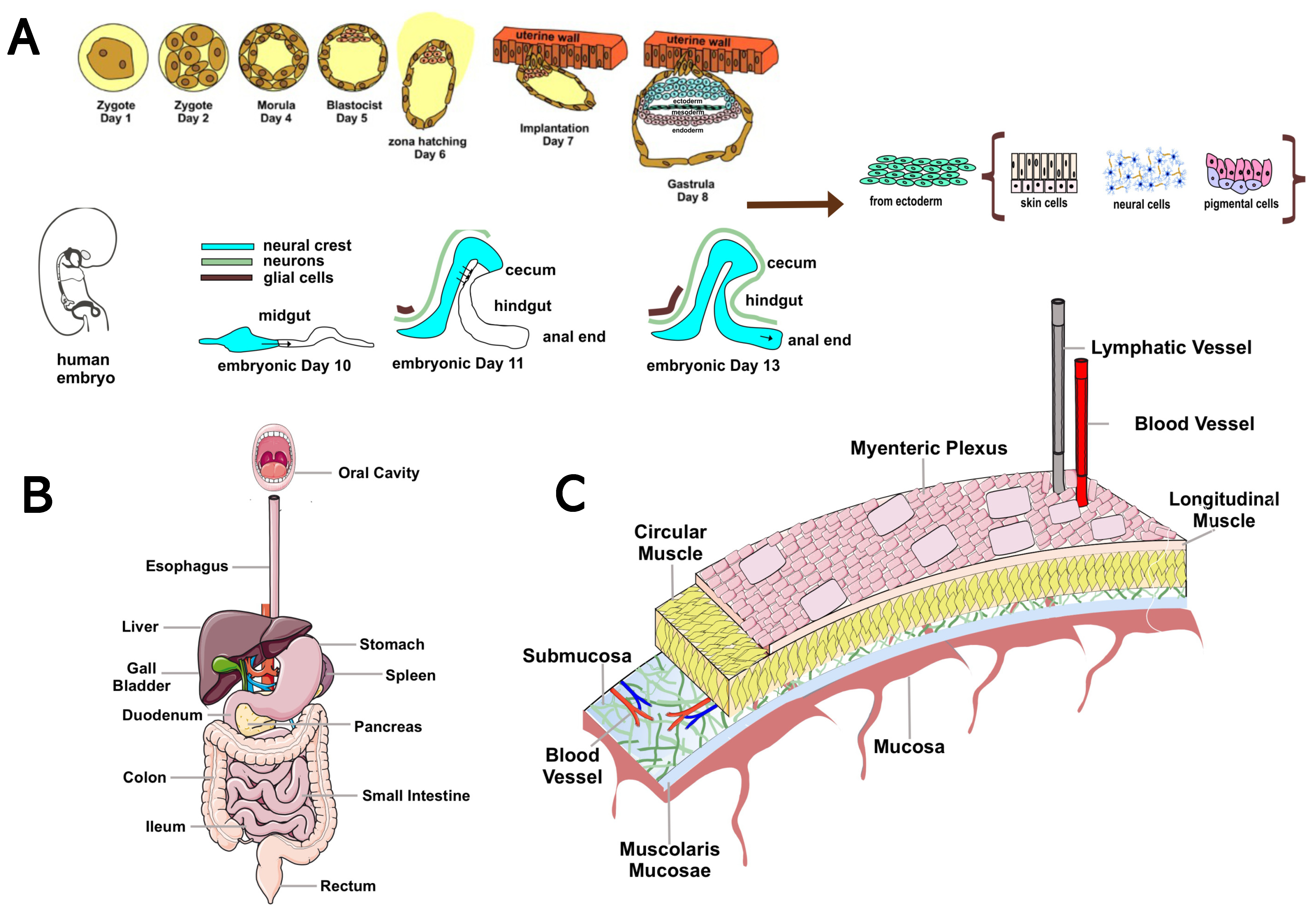
2. Overview of the Enteric Nervous System: Anatomy and Function

The ENS, the intrinsic innervation of the GI tract, is the largest and most complex division of vertebrates’ peripheral and autonomic nervous systems. In humans, the ENS contains 400–600 million neurons and an array of neurotransmitters and neuromodulators similar to those found in the CNS [11]. Unlike the CNS, in which efferent pathways are characterized by pre-ganglionic and post-ganglionic neurons [62], the axons of gut neurons in the ENS project to the sympathetic ganglia, brainstem, spinal cord, pancreas, gallbladder, and trachea [10]. The anatomy and physiology of the ENS have been studied since the 19th century, going so far as to demonstrate early in the last century how the peristaltic reflex (i.e., the pressure-induced propulsive activity of the intestines) is a local nervous mechanism that occurs in the absence of external nerve input [8]. Because of this autonomy and its complexity, Michael D. Gershon likened the ENS to a second brain [11]. Two-way communications between the ENS and the CNS are always active: the CNS can regulate or alter the normal functioning of the ENS and vice versa. For example, certain gut disorders impair the production of psychoactive substances such as serotonin (5-HT, 5-hydroxytryptamine), dopamine (DA), and opiates, which can affect mood [63]. Conversely, emotional states, such as intense anxiety, can cause colitis, constipation, irritable colon, or mucosal ulcers by stimulating peristalsis and hyperproduction of neurotransmitters [63]. The ENS originates around the eighth day of embryonic life from neural crest progenitor cells, endowed with stem-like properties, which migrate through the forming GI tract and colonize it within five days [11]. They subsequently differentiate into neurons and glia by integrating predetermined instructions with information from the microenvironment [9]. In humans, the ENS becomes functional in the last trimester of gestation and continues to develop after birth [9]. The ENS comprises small aggregations of nerve cells, the enteric ganglia, the neural connections between these ganglia, and the nerve fibers that supply effector tissues, including gut wall muscle, epithelial lining, intrinsic blood vessels, and gastroenteropancreatic endocrine cells [8][10][11][64]. Enteric neurons (NEs) are organized into ganglionic plexuses: the myenteric (Auerbach’s) plexus and the submucosal (Meissner’s) plexus. Ganglionic plexuses are enveloped by glial cells, such as CNS astrocytes, which form a proper blood–enteric barrier. Glial cells release enterocyte differentiation factors, participate in GI functions, and are involved in the pathogenesis of inflammatory disorders of the GI tract. Auerbach’s myenteric plexus, located in the muscle tonaca between the layers of longitudinal and circular muscles, consists of linear chains of numerous interconnected neurons that span the length of the GI tract and regulate its movements. Meissner’s submucosal plexus, located in the submucosa of the small and large intestines but absent in the esophagus and stomach, consists of ganglia stratified at different levels. It integrates sensory signals from the intestinal epithelium and contributes to the local control of secretion, intestinal absorption, blood flow, and submucosal muscle contraction [8][10][64] (Figure 1).
Figure 1. Overview of the anatomy and organization of the ENS. (A) Time course of ENS development. The ENS originates around the eighth day of embryonic life from neural crest progenitor cells (ENCDCs) with stem-like properties, which migrate through the GI tract and colonize it within five days. After invading the anterior intestine, these pre-ENCDCs migrate rostro-caudally, proliferating and differentiating into neurons and glia. During this process, the intestine elongates, changing shape from a straight line to a single curve, with the middle and small intestine closely adjacent. The cecal appendix grows and the entire intestine elongates further. At embryonic days 11 and 13, ENCDCs invade the colon by crossing the mesentery and transiting into the cecum. The cecal and trans mesenteric populations then fuse to form the ENS in the rostral colon. In humans, the ENS becomes functional in the last trimester of gestation and continues to develop after birth. (B) Schematic diagram of the human GI tract. (C) Organization of the ENS. NEs are organized into ganglionic plexuses: the myenteric plexus and the submucosal plexus. The ganglionic plexuses are enveloped by glial cells, such as CNS astrocytes, which form a proper blood–enteric barrier. The myenteric plexus is in the muscle tonaca between the layers of longitudinal and circular muscles. It consists of linear chains of numerous interconnected neurons that span the length of the GI and regulate its movements.
Twenty types of NEs characterized by different morphological, neurochemical, and electrophysiological aspects, connections, and functional roles have been identified [9][65][66]. Based on intracellular electrophysiological recordings, two types of NEs were detected: S and AH neurons. S neurons are characterized by high excitability and can exhibit rapid excitatory postsynaptic potentials, followed by a short-lived hyperpolarizing current (20–100 ms), rapidly restoring the membrane potential [65][67]. On the other hand, AH neurons exhibit large action potentials followed by a slow hyperpolarizing current (2–30 s) that makes them less excitable. NEs use more than 50 neurotransmitters in synaptic communications, from small neurotransmitters (e.g., ACh, acetylcholine, 5-HT) to neuropeptides (e.g., CGRP, calcitonin gene-related peptide, somatostatin, substance P, and VIP, vasoactive intestinal peptide) to gases (e.g., NO, nitric oxide) [66][67]. NEs are grouped into three functional classes: intrinsic sensory neurons called IPANs, muscle motor neurons, and interneurons. IPANs are large and equipped with numerous axons; they can sense mechanical, chemical, and thermal stimuli and transmit information about muscle tension state and endoluminal content to motor neurons [68], triggering reflexes that regulate motility, secretion, and blood flow. They make up about 10–30% of the neurons located in the submucosal and myenteric plexus of the small and large intestines; they are not present in the esophagus (whose motility is controlled by fibers originating from the CNS) and stomach (whose motility is under the control of vagal fibers) [68]. Motor neurons are divided into muscular and secretomotor-vasodilatory. The former (Dogiel’s type I) innervate the circular and longitudinal musculature and the muscular mucosae, determining their contraction or relaxation; they have an elongated cell body, numerous dendrites, and a single slender axon; electrophysiologically, they correspond to type S. Neurons innervating circular and longitudinal musculature have their cell bodies in the myenteric plexus and are excitatory (using ACh and TK, tachykinin, and projecting orally) or inhibitory (using NO and VIP and projecting anally) [68]. Muscle motor neurons generate, following regional stimulation, coordinated and polarized muscle responses that allow the progression of intestinal contents, i.e., induce contraction in the oral direction and relaxation in the anal direction [68]. On the other hand, secretomotor-vasodilator neurons are located mainly in the submucosal ganglia, controlling both the secretion of ions and water via ACh and the vasodilation of submucosal arterioles via VIP [65][66]. Some influence glucose transport across the mucosa of the small intestine [69], a process also regulated by vagal-like reflexes; others modulate acid secretion in the stomach [69]. Interneurons integrate sensory afferents and organize effector responses [66][67]. In the myenteric plexus, they form chains that run in ascending and descending directions. They resemble type I neurons and are S-type [67]. In the course of life, the ENS undergoes plastic changes as a spatiotemporal adaptive response to external stimuli, which arrive through sensory afferents, and to internal stimuli that come from autonomic innervation [8]. In the complex microenvironment of the gut wall lodge, different types of cells (neurons, glia, Cajal cells, muscle cells, and immune cells) can communicate with each other in synaptic or paracrine ways. This interactive plurality modulates the functional state of NEs by influencing the digestive and secretory functions of the GI tract [70]. Changes in diet and perturbations in the gut microbiome, with its metabolites and neuroactive compounds, affect the functioning of the NE and its connections with the CNS, since they alter mucosal permeability and the secretion of hormones and immune cells. In addition, NEs are vulnerable to aging-related degeneration [70].

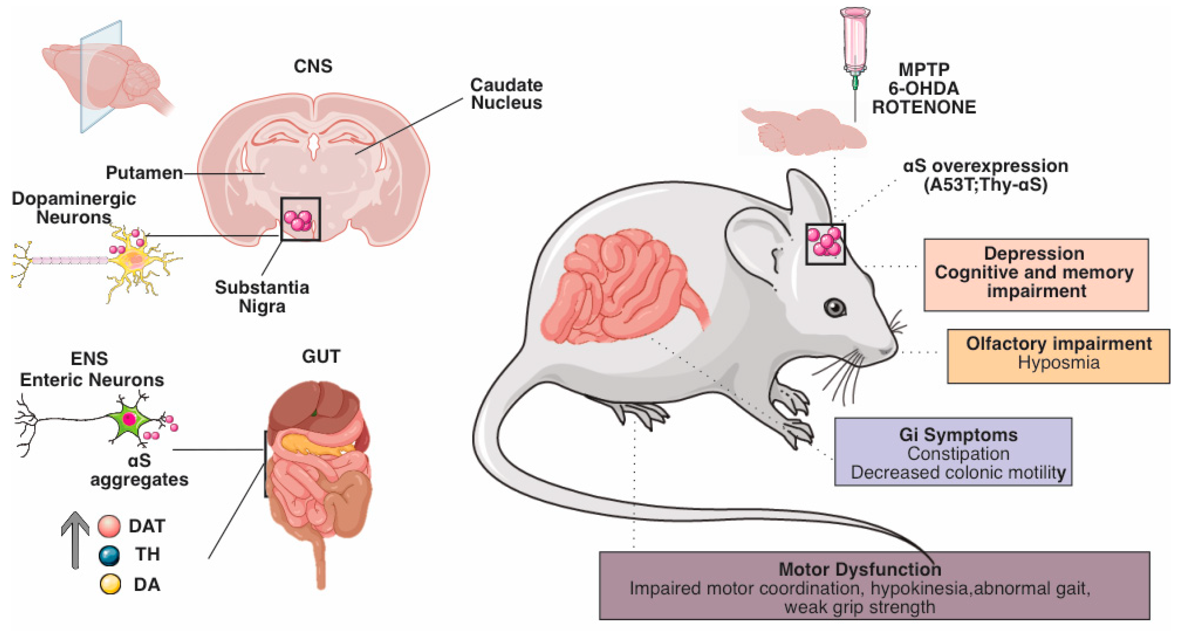
3. Evidence of the Role of the Enteric Nervous System in Animal Models of Parkinson’s Disease

GI dysfunction is a common non-motor symptom of PD. While, in PD patients, it is present in 80–90% of cases and has been associated with αS aggregation and neuronal loss in the CNS, reports of GI symptoms in animal models of PD are known to vary, and the degree to which pathology in the CNS contributes to GI symptoms remains unclear [71].
PD benefits from a wide range of animal models whose diverse pharmacological, toxin, and genetic features are essential to study its etiology and neurobiology [72]. Animal models of PD rely on pharmacological or genetic approaches to simulate nigrostriatal neurodegeneration and disease pathogenesis [72]. However, much remains to be discovered and requires continuous questioning by the research community.
The most commonly used pharmacological models are based on neurotoxins administered to mice, rats, and nonhuman primates [73] (Figure 2).
Figure 2. Schematic representation of the main physiological and behavioral changes in CNS and ENS of preclinical models of PD. PD is a heterogeneous disorder with varying ages of onset, symptoms, and progression rates. This heterogeneity requires the use of a variety of animal models to study different aspects of the disease. (Right) Neurotoxin-based approaches include exposure of rodents or nonhuman primates to 6-OHDA, MPTP, and agrochemicals such as the pesticide rotenone. Acute neurotoxin exposure induces motor deficits and rapid nigrostriatal dopaminergic cell death by disrupting mitochondrial function and increasing oxidative stress. Chronic neurotoxin administration induces progressive patterns that may include αS aggregates. Genetics-based approaches to modeling PD include transgenic and viral-vector-mediated models based on genes linked to monogenic PD. Among these, overexpression and introduction of preformed α-S fibrils induce toxic protein aggregates, nigrostriatal neurodegeneration, and variable motor deficits, depending on the specific model. (Left) GI dysfunction is the most common non-motor symptom of PD. Symptoms of GI dysmotility in PD include premature satiety and weight loss due to delayed gastric emptying and constipation due to altered colonic transit. Researchers can find numerous alterations in the ENS in preclinical models of PD: neurodegeneration of NEs, which is the leading cause of behavioral and electrophysiological alterations in mouse models.
Both neurotoxins, 1-methyl-4-phenyl-1,2,3,6-tetrahydropyridine (MPTP) and 6-hydroxydopamine (6-OHDA), consistently affect nigrostriatal dopaminergic pathways [73]. However, their impact on gut function and the CNS varies, depending on the agent, mode of administration, and assays used [74][75][76][77]. Systemic administration of MPTP in mice causes loss of dopaminergic neurons in the myenteric plexus but does not cause severe defects in GI motility [75][76]. Peripheral administration of MPTP in rats does not significantly affect the number of dopaminergic neurons and the expression of dopaminergic markers in the SNpc [78]. However, it significantly reduces tyrosine hydroxylase-immunoreactive (TH-IR) neurons in the GI tract, suggesting that the degeneration of dopaminergic neurons might start earlier than in the SNpc [48][75][78]. Parenteral administration of MPTP in mice simultaneously induces dopaminergic neurodegeneration in the ENS, which is associated with behavioral and electrophysiological alterations. Following MPTP intoxication, acceleration of motility (increased contraction) and decreased colonic relaxation are observed in response to electric field stimulation of the NE [79][80]. These complementary findings point to the altered function of enteric DA neurons. Several articles have shown that exogenous DA antagonizes colonic muscle contractility in a receptor-dependent manner [80].
Furthermore, confirming that MPTP is selectively toxic to dopaminergic neurons in the ENS, just as in the CNS, TH-positive neurons in the myenteric ganglia are reduced [81][82][83]. Most TH-positive neurons with cell bodies in the myenteric plexus can be considered dopaminergic, since adrenergic and noradrenergic inputs to the GI tract are mainly extrinsic [84].
Considering the neuropathological and electrophysiological findings, it is likely that dysfunction and death of dopaminergic neurons cause the transient increase in colonic motility observed after MPTP intoxication. Decreased dopaminergic inhibitory tone results in faster colonic transit due to the relative abundance of stimulatory neuronal input [79][80].
Neurotransmitters related to the GI dysfunction of PD could be involved in the intestinal dopaminergic, cholinergic, and oxidergic nitric systems [35]. To investigate the relationship between the GI dysfunction of PD and the alteration of GI neurotransmitters, 6-OHDA was microinjected into one side of the nigrostriatal system of the brain to generate an animal model of PD through the impairment of rat dopaminergic neurons, and the effect of neurotransmitter alterations in the CNS on GI function was observed [74].
GI dysfunction and changes in dopaminergic, nitric oxide synthase (NOS), and cholinergic neurons in the myenteric plexus were analyzed. Compared with control samples, 6-OHDA rats had delayed gastric emptying and constipation, which could be related to increased GI TH and decreased NOS. These symptoms were not associated with alterations in cholinergic transmitters [77].
Unfortunately, some of these studies did not analyze the submucosal plexus, making a direct comparison with more robust findings in complex PD patients [56]. Rats treated with 6-OHDA show elevated protein levels of TH and dopamine transporter (DAT) (dopaminergic markers) in both the epithelium and neurons of the GI tract, resulting in increased DA content in the gut and delayed gastric emptying [78]. In the epithelium and neurons of the GI tract, neurodegeneration of the SN by 6-OHDA increases the expression of TH and DAT proteins. It is hypothesized that the number of enteric dopaminergic neurons and cells may increase to compensate for the loss of DA in the SN in PD patients [78].
In contrast, the increased protein expression of TH and DAT in 6-OHDA-treated rats may increase the concentration of DA in the colon and the loss of DA in the SN, which may cause constipation [78].
Alterations in the monoaminergic system and decreased colonic motility were observed in rats microinjected with 6-OHDA in the bilateral SN [74].
DA, NE, and 5-HT play essential roles in regulating colonic motility: increased DA content, upregulation of β3-ARs, and decreased 5-HT4 receptors could contribute to the decreased spontaneous colonic contraction and constipation observed in rats with 6-OHDA [74].
Rats with lesions of SN dopaminergic neurons manifest GI dysmotility [85][86], including gastroparesis and constipation [86][87].
Animal models do not yet allow for an adequate study of how PD prodromal constipation occurs [88]. To date, there is a paucity of relevant experimental models of GI dysfunction associated with αS pathology; αS deposition in the ENS of PD patients has been reported in the myenteric and submucosal plexuses of GI tracts [89][90]. Transgenic mouse lines expressing a mutant form of human αS (A53T or A30P) under its promoter show colonic disorders similar to constipation and pathology characteristic of αS [91]. In a transgenic mouse model in which mutant human αS (A53T) was expressed under the control of the prion promoter [92], aggregates of αS were observed in the ENS prior to changes in the CNS [91]. This finding suggests that αS pathology may be initiated from the ENS and propagate to the CNS via the vagus nerve [52]. In support of this, in a transgenic mouse model, the accumulation of αS aggregates in the ENS precedes changes in the CNS [91].
Expression of human αS in the DMV, a region of the brain severely affected by PD, causes an age-related slowing in A53T mice of GI motility reminiscent of that observed in patients with PD [52][93]. The symptoms coincide with the disruption of efferent vagal processes that project from the DMV to the GI tract. This pattern parallels the pathology of postmortem specimens of PD patients and implicates the DMV as a possible mediator of GI neuropathology and symptomatology in PD [94].
However, αS mutations are only responsible for rare cases of PD [30]. Mice overexpressing wild-type human αS under the Thy-1 promoter (Thy1-αS) show increased transit time and colonic content compared with wild-type (WT) pups when tested at 12–14 months of age [95]. However, striatal dopamine loss occurs only after 14 months in Thy1-αS mice, manifesting motor and non-motor deficits, such as olfactory disturbances, as early as 2–3 months of age [96][97].
The mechanisms underlying colonic motor impairments may be related to αS overexpression in the colonic myenteric nervous system [95]. The reduced response to defecation stimuli in Thy1-αS could be related to the accumulation of αS in colonic myenteric plexuses [95].
The GI system is one of the most susceptible to environmental stressors, since it is in direct contact with environmental agents [98][99][100]. In a recent study, intra-gastric administration of rotenone in mice caused progressive αS deposition in both the ENS and CNS neurons affected by PD, such as neurons in the myenteric plexus, the vagus DMV, the spinal cord, and the sympathetic nervous system (SNS) [101]. These studies suggested that environmental stressors to the GI system could lead to αS pathology in the CNS.
Numerous preclinical pieces of evidence associate GI symptoms in toxic models of PD based on oral administration of rotenone [98]. Previous studies have shown that orally administered rotenone exposure induces PD-like changes in the ENS and triggers PD progression throughout the nervous system to the SN [99][101]. Interestingly, the latter changes appear as early as the first moments after rotenone administration (2 months) before the onset of motor symptoms (which occur after three months of exposure in this animal model), thus mimicking the pattern of progression observed in PD patients.
In two recent studies, rotenone exposure reduced sympathetic noradrenergic [102] and vagal cholinergic gut innervation [103].
The mechanism by which environmental agents induce αS aggregation is unknown. However, a recent study showed that αS expression in the ENS could be upregulated by agents that cause depolarization and increase cyclic AMP levels [104].
An emerging concept in gastroenterology is that a wide range of diseases, such as motility disorders, can be partially considered enteric neuropathies. In particular, aging is associated with various motility or gut disorders, including delayed gastric emptying and longer intestinal transit time [105]. Aged rats show neuronal loss and changes in neurochemical phenotype in the ENS, which may result in motility disorders [106]. Surprisingly, along with neuronal loss, these rats exhibit dystrophic NEs that contain αS aggregates reminiscent of Lewy pathology [107].
Braak et al. hypothesized that PD originated in the gut and subsequently progressed up, as if along a ladder, along the nerves connecting the gut to the brain [90].
Using double transgenic mice expressing mutant αS, it is possible to observe how early alterations in ENS can be identified as early disease markers. These animal models expressing mutant αS provide an opportunity to investigate the potential role of ENS as an early marker of disease [91]. Early ENS dysfunction would not only trigger disease but facilitate the entry of deleterious factors that cause progression and spread to the CNS [12][91].

References

  1. Endres, K.; Schäfer, K.-H. Influence of commensal microbiota on the enteric nervous system and its role in neurodegenerative diseases. J. Innate Immun. 2018, 10, 172–180.
  2. Clemente, J.C.; Ursell, L.K.; Parfrey, L.W.; Knight, R. The impact of the gut microbiota on human health: An integrative view. Cell 2012, 148, 1258–1270.
  3. Bercik, P.; Denou, E.; Collins, J.; Jackson, W.; Lu, J.; Jury, J.; Deng, Y.; Blennerhassett, P.; Macri, J.; McCoy, K.D.; et al. The intestinal microbiota affect central levels of brain-derived neurotropic factor and behavior in mice. Gastroenterology 2011, 141, 599–609.e3.
  4. Sampson, T.R.; Mazmanian, S.K. Control of brain development, function, and behavior by the microbiome. Cell Host Microbe 2015, 17, 565–576.
  5. Carabotti, M.; Scirocco, A.; Maselli, M.A.; Severi, C. The gut-brain axis: Interactions between enteric microbiota, central and enteric nervous systems. Ann. Gastroenterol. 2015, 28, 203–209.
  6. Kabouridis, P.S.; Lasrado, R.; McCallum, S.; Chng, S.H.; Snippert, H.J.; Clevers, H.; Pettersson, S.; Pachnis, V. Microbiota controls the homeostasis of glial cells in the gut lamina propria. Neuron 2015, 85, 289–295.
  7. Belkaid, Y.; Hand, T.W. Role of the microbiota in immunity and inflammation. Cell 2014, 157, 121–141.
  8. Furness, J.B. The Enteric Nervous System; John Wiley & Sons: Hoboken, NJ, USA, 2008; ISBN 9781405173445.
  9. Sasselli, V.; Pachnis, V.; Burns, A.J. The enteric nervous system. Dev. Biol. 2012, 366, 64–73.
  10. Furness, J.B. The enteric nervous system and neurogastroenterology. Nat. Rev. Gastroenterol. Hepatol. 2012, 9, 286–294.
  11. Gershon, M.D. The enteric nervous system: A second brain. Hosp. Pract. 1999, 34, 31–52.
  12. Natale, G.; Pasquali, L.; Paparelli, A.; Fornai, F. Parallel manifestations of neuropathologies in the enteric and central nervous systems. Neurogastroenterol. Motil. 2011, 23, 1056–1065.
  13. Furness, J.B. The organisation of the autonomic nervous system: Peripheral connections. Auton. Neurosci. 2006, 130, 1–5.
  14. Cryan, J.F.; O’Riordan, K.J.; Cowan, C.S.M.; Sandhu, K.V.; Bastiaanssen, T.F.S.; Boehme, M.; Codagnone, M.G.; Cussotto, S.; Fulling, C.; Golubeva, A.V.; et al. The Microbiota-Gut-Brain Axis. Physiol. Rev. 2019, 99, 1877–2013.
  15. Wang, H.X.; Wang, Y.P. Gut Microbiota-brain Axis. Chin. Med. J. 2016, 129, 2373–2380.
  16. Jaggar, M.; Rea, K.; Spichak, S.; Dinan, T.G.; Cryan, J.F. You’ve got male: Sex and the microbiota-gut-brain axis across the lifespan. Front. Neuroendocrinol. 2020, 56, 100815.
  17. Bauer, P.V.; Hamr, S.C.; Duca, F.A. Regulation of energy balance by a gut-brain axis and involvement of the gut microbiota. Cell. Mol. Life Sci. 2016, 73, 737–755.
  18. Margolis, K.G.; Cryan, J.F.; Mayer, E.A. The Microbiota-Gut-Brain Axis: From Motility to Mood. Gastroenterology 2021, 160, 1486–1501.
  19. Rhee, S.H.; Pothoulakis, C.; Mayer, E.A. Principles and clinical implications of the brain-gut-enteric microbiota axis. Nat. Rev. Gastroenterol. Hepatol. 2009, 6, 306–314.
  20. Kasarello, K.; Cudnoch-Jedrzejewska, A.; Czarzasta, K. Communication of gut microbiota and brain via immune and neuroendocrine signaling. Front. Microbiol. 2023, 14, 1118529.
  21. Mayer, E.A.; Savidge, T.; Shulman, R.J. Brain-gut microbiome interactions and functional bowel disorders. Gastroenterology 2014, 146, 1500–1512.
  22. Varesi, A.; Pierella, E.; Romeo, M.; Piccini, G.B.; Alfano, C.; Bjørklund, G.; Oppong, A.; Ricevuti, G.; Esposito, C.; Chirumbolo, S.; et al. The potential role of gut microbiota in alzheimer’s disease: From diagnosis to treatment. Nutrients 2022, 14, 668.
  23. Quigley, E.M.M. Microbiota-Brain-Gut Axis and Neurodegenerative Diseases. Curr. Neurol. Neurosci. Rep. 2017, 17, 94.
  24. Sidransky, E.; Lopez, G. The link between the GBA gene and parkinsonism. Lancet Neurol. 2012, 11, 986–998.
  25. Schirinzi, T.; Martella, G.; Pisani, A. Double hit mouse model of Parkinson’s disease. Oncotarget 2016, 7, 80109–80110.
  26. Martella, G.; Madeo, G.; Maltese, M.; Vanni, V.; Puglisi, F.; Ferraro, E.; Schirinzi, T.; Valente, E.M.; Bonanni, L.; Shen, J.; et al. Exposure to low-dose rotenone precipitates synaptic plasticity alterations in PINK1 heterozygous knockout mice. Neurobiol. Dis. 2016, 91, 21–36.
  27. Dickson, D.W. Parkinson’s disease and parkinsonism: Neuropathology. Cold Spring Harb. Perspect. Med. 2012, 2, a009258.
  28. Hawkes, C.H.; Del Tredici, K.; Braak, H. Parkinson’s disease: A dual-hit hypothesis. Neuropathol. Appl. Neurobiol. 2007, 33, 599–614.
  29. Tanner, C.M.; Goldman, S.M. Epidemiology of Parkinson’s disease. Neurol. Clin. 1996, 14, 317–335.
  30. Vance, J.M.; Ali, S.; Bradley, W.G.; Singer, C.; Di Monte, D.A. Gene-environment interactions in Parkinson’s disease and other forms of parkinsonism. Neurotoxicology 2010, 31, 598–602.
  31. Kline, E.M.; Houser, M.C.; Herrick, M.K.; Seibler, P.; Klein, C.; West, A.; Tansey, M.G. Genetic and environmental factors in parkinson’s disease converge on immune function and inflammation. Mov. Disord. 2021, 36, 25–36.
  32. Poewe, W. Non-motor symptoms in Parkinson’s disease. Eur. J. Neurol. 2008, 15 (Suppl. S1), 14–20.
  33. Noyce, A.J.; Bestwick, J.P.; Silveira-Moriyama, L.; Hawkes, C.H.; Giovannoni, G.; Lees, A.J.; Schrag, A. Meta-analysis of early nonmotor features and risk factors for Parkinson disease. Ann. Neurol. 2012, 72, 893–901.
  34. Amara, A.W.; Memon, A.A. Effects of Exercise on Non-motor Symptoms in Parkinson’s Disease. Clin. Ther. 2018, 40, 8–15.
  35. Postuma, R.B.; Aarsland, D.; Barone, P.; Burn, D.J.; Hawkes, C.H.; Oertel, W.; Ziemssen, T. Identifying prodromal Parkinson’s disease: Pre-motor disorders in Parkinson’s disease. Mov. Disord. 2012, 27, 617–626.
  36. Martinez-Martin, P.; Rodriguez-Blazquez, C.; Kurtis, M.M.; Chaudhuri, K.R. NMSS Validation Group The impact of non-motor symptoms on health-related quality of life of patients with Parkinson’s disease. Mov. Disord. 2011, 26, 399–406.
  37. Battaglia, S.; Nazzi, C.; Thayer, J.F. Fear-induced bradycardia in mental disorders: Foundations, current advances, future perspectives. Neurosci. Biobehav. Rev. 2023, 149, 105163.
  38. Battaglia, S.; Di Fazio, C.; Vicario, C.M.; Avenanti, A. Neuropharmacological Modulation of N-methyl-D-aspartate, Noradrenaline and Endocannabinoid Receptors in Fear Extinction Learning: Synaptic Transmission and Plasticity. Int. J. Mol. Sci. 2023, 24, 5926.
  39. Tan, A.H.; Lim, S.Y.; Lang, A.E. The microbiome-gut-brain axis in Parkinson disease—From basic research to the clinic. Nat. Rev. Neurol. 2022, 18, 476–495.
  40. Mukherjee, A.; Biswas, A.; Das, S.K. Gut dysfunction in Parkinson’s disease. World J. Gastroenterol. 2016, 22, 5742–5752.
  41. Zeng, J.; Wang, X.; Pan, F.; Mao, Z. The relationship between Parkinson’s disease and gastrointestinal diseases. Front. Aging Neurosci. 2022, 14, 955919.
  42. Bhidayasiri, R.; Phuenpathom, W.; Tan, A.H.; Leta, V.; Phumphid, S.; Chaudhuri, K.R.; Pal, P.K. Management of dysphagia and gastroparesis in Parkinson’s disease in real-world clinical practice—Balancing pharmacological and non-pharmacological approaches. Front. Aging Neurosci. 2022, 14, 979826.
  43. Chen, Z.; Li, G.; Liu, J. Autonomic dysfunction in Parkinson’s disease: Implications for pathophysiology, diagnosis, and treatment. Neurobiol. Dis. 2020, 134, 104700.
  44. Chiang, H.-L.; Lin, C.-H. Altered gut microbiome and intestinal pathology in parkinson’s disease. J. Mov. Disord. 2019, 12, 67–83.
  45. Devos, D.; Lebouvier, T.; Lardeux, B.; Biraud, M.; Rouaud, T.; Pouclet, H.; Coron, E.; Bruley des Varannes, S.; Naveilhan, P.; Nguyen, J.-M.; et al. Colonic inflammation in Parkinson’s disease. Neurobiol. Dis. 2013, 50, 42–48.
  46. Cersosimo, M.G.; Benarroch, E.E. Pathological correlates of gastrointestinal dysfunction in Parkinson’s disease. Neurobiol. Dis. 2012, 46, 559–564.
  47. Stocchi, F.; Torti, M. Constipation in parkinson’s disease. Int. Rev. Neurobiol. 2017, 134, 811–826.
  48. Singaram, C.; Ashraf, W.; Gaumnitz, E.A.; Torbey, C.; Sengupta, A.; Pfeiffer, R.; Quigley, E.M. Dopaminergic defect of enteric nervous system in Parkinson’s disease patients with chronic constipation. Lancet 1995, 346, 861–864.
  49. Pfeiffer, R.F.; Isaacson, S.H.; Pahwa, R. Clinical implications of gastric complications on levodopa treatment in Parkinson’s disease. Park. Relat. Disord. 2020, 76, 63–71.
  50. Lebouvier, T.; Neunlist, M.; Bruley des Varannes, S.; Coron, E.; Drouard, A.; N’Guyen, J.-M.; Chaumette, T.; Tasselli, M.; Paillusson, S.; Flamand, M.; et al. Colonic biopsies to assess the neuropathology of Parkinson’s disease and its relationship with symptoms. PLoS ONE 2010, 5, e12728.
  51. Zheng, H.; Shi, C.; Luo, H.; Fan, L.; Yang, Z.; Hu, X.; Zhang, Z.; Zhang, S.; Hu, Z.; Fan, Y.; et al. α-Synuclein in Parkinson’s Disease: Does a Prion-like Mechanism of Propagation from Periphery to the Brain Play a Role? Neuroscientist 2021, 27, 367–387.
  52. Braak, H.; Del Tredici, K.; Rüb, U.; de Vos, R.A.I.; Jansen Steur, E.N.H.; Braak, E. Staging of brain pathology related to sporadic Parkinson’s disease. Neurobiol. Aging 2003, 24, 197–211.
  53. Arotcarena, M.-L.; Dovero, S.; Prigent, A.; Bourdenx, M.; Camus, S.; Porras, G.; Thiolat, M.-L.; Tasselli, M.; Aubert, P.; Kruse, N.; et al. Bidirectional gut-to-brain and brain-to-gut propagation of synucleinopathy in non-human primates. Brain 2020, 143, 1462–1475.
  54. Natale, G.; Pasquali, L.; Ruggieri, S.; Paparelli, A.; Fornai, F. Parkinson’s disease and the gut: A well known clinical association in need of an effective cure and explanation. Neurogastroenterol. Motil. 2008, 20, 741–749.
  55. Leclair-Visonneau, L.; Neunlist, M.; Derkinderen, P.; Lebouvier, T. The gut in Parkinson’s disease: Bottom-up, top-down, or neither? Neurogastroenterol. Motil. 2020, 32, e13777.
  56. Chalazonitis, A.; Rao, M. Enteric nervous system manifestations of neurodegenerative disease. Brain Res. 2018, 1693, 207–213.
  57. Menozzi, E.; Macnaughtan, J.; Schapira, A.H.V. The gut-brain axis and Parkinson disease: Clinical and pathogenetic relevance. Ann. Med. 2021, 53, 611–625.
  58. Berg, D.; Borghammer, P.; Fereshtehnejad, S.-M.; Heinzel, S.; Horsager, J.; Schaeffer, E.; Postuma, R.B. Prodromal Parkinson disease subtypes—Key to understanding heterogeneity. Nat. Rev. Neurol. 2021, 17, 349–361.
  59. Elfil, M.; Kamel, S.; Kandil, M.; Koo, B.B.; Schaefer, S.M. Implications of the gut microbiome in parkinson’s disease. Mov. Disord. 2020, 35, 921–933.
  60. Klann, E.M.; Dissanayake, U.; Gurrala, A.; Farrer, M.; Shukla, A.W.; Ramirez-Zamora, A.; Mai, V.; Vedam-Mai, V. The Gut-Brain Axis and Its Relation to Parkinson’s Disease: A Review. Front. Aging Neurosci. 2021, 13, 782082.
  61. Ma, Z.S. Heterogeneity-disease relationship in the human microbiome-associated diseases. FEMS Microbiol. Ecol. 2020, 96, fiaa093.
  62. Brodal, P. The Central Nervous System: Structure and Function; Oxford University Press: Oxford, UK, 2004; ISBN 9780195165609.
  63. Cussotto, S.; Strain, C.R.; Fouhy, F.; Strain, R.G.; Peterson, V.L.; Clarke, G.; Stanton, C.; Dinan, T.G.; Cryan, J.F. Differential effects of psychotropic drugs on microbiome composition and gastrointestinal function. Psychopharmacology 2019, 236, 1671–1685.
  64. The Enteric Nervous System and Regulation of Intestinal Motility—ProQuest. Available online: https://www.proquest.com/docview/222539969?pq-origsite=gscholar&fromopenview=true (accessed on 10 August 2022).
  65. Brehmer, A. Structure of Enteric Neurons; Springer Science & Business Media: Berlin/Heidelberg, Germany, 2006; ISBN 9783540328742.
  66. Costa, M.; Furness, J.B.; Gibbins, I.L. Chapter 15 Chemical coding of enteric neurons. In Progress in Brain Research; Elsevier: Amsterdam, The Netherlands, 1986; Volume 68, pp. 217–239. ISBN 9780444807625.
  67. Furness, J.B.; Costa, M. Types of nerves in the enteric nervous system. In Commentaries in the Neurosciences; Elsevier: Amsterdam, The Netherlands, 1980; pp. 235–252. ISBN 9780080255019.
  68. Furness, J.B.; Callaghan, B.P.; Rivera, L.R.; Cho, H.-J. The enteric nervous system and gastrointestinal innervation: Integrated local and central control. Adv. Exp. Med. Biol. 2014, 817, 39–71.
  69. Shirazi-Beechey, S.P.; Moran, A.W.; Batchelor, D.J.; Daly, K.; Al-Rammahi, M. Glucose sensing and signalling; regulation of intestinal glucose transport. Proc. Nutr. Soc. 2011, 70, 185–193.
  70. Saffrey, M.J. Cellular changes in the enteric nervous system during ageing. Dev. Biol. 2013, 382, 344–355.
  71. McQuade, R.M.; Singleton, L.M.; Wu, H.; Lee, S.; Constable, R.; Di Natale, M.; Ringuet, M.T.; Berger, J.P.; Kauhausen, J.; Parish, C.L.; et al. The association of enteric neuropathy with gut phenotypes in acute and progressive models of Parkinson’s disease. Sci. Rep. 2021, 11, 7934.
  72. Lama, J.; Buhidma, Y.; Fletcher, E.J.R.; Duty, S. Animal models of Parkinson’s disease: A guide to selecting the optimal model for your research. Neuronal Signal. 2021, 5, NS20210026.
  73. Tieu, K. A guide to neurotoxic animal models of Parkinson’s disease. Cold Spring Harb. Perspect. Med. 2011, 1, a009316.
  74. Zhang, X.; Li, Y.; Liu, C.; Fan, R.; Wang, P.; Zheng, L.; Hong, F.; Feng, X.; Zhang, Y.; Li, L.; et al. Alteration of enteric monoamines with monoamine receptors and colonic dysmotility in 6-hydroxydopamine-induced Parkinson’s disease rats. Transl. Res. 2015, 166, 152–162.
  75. Anderson, G.; Noorian, A.R.; Taylor, G.; Anitha, M.; Bernhard, D.; Srinivasan, S.; Greene, J.G. Loss of enteric dopaminergic neurons and associated changes in colon motility in an MPTP mouse model of Parkinson’s disease. Exp. Neurol. 2007, 207, 4–12.
  76. Chaumette, T.; Lebouvier, T.; Aubert, P.; Lardeux, B.; Qin, C.; Li, Q.; Accary, D.; Bézard, E.; Bruley des Varannes, S.; Derkinderen, P.; et al. Neurochemical plasticity in the enteric nervous system of a primate animal model of experimental Parkinsonism. Neurogastroenterol. Motil. 2009, 21, 215–222.
  77. Zhu, H.C.; Zhao, J.; Luo, C.Y.; Li, Q.Q. Gastrointestinal dysfunction in a Parkinson’s disease rat model and the changes of dopaminergic, nitric oxidergic, and cholinergic neurotransmitters in myenteric plexus. J. Mol. Neurosci. 2012, 47, 15–25.
  78. Tian, Y.M.; Chen, X.; Luo, D.Z.; Zhang, X.H.; Xue, H.; Zheng, L.F.; Yang, N.; Wang, X.M.; Zhu, J.X. Alteration of dopaminergic markers in gastrointestinal tract of different rodent models of Parkinson’s disease. Neuroscience 2008, 153, 634–644.
  79. Li, Z.S.; Schmauss, C.; Cuenca, A.; Ratcliffe, E.; Gershon, M.D. Physiological modulation of intestinal motility by enteric dopaminergic neurons and the D2 receptor: Analysis of dopamine receptor expression, location, development, and function in wild-type and knock-out mice. J. Neurosci. 2006, 26, 2798–2807.
  80. Walker, J.K.; Gainetdinov, R.R.; Mangel, A.W.; Caron, M.G.; Shetzline, M.A. Mice lacking the dopamine transporter display altered regulation of distal colonic motility. Am. J. Physiol. Gastrointest. Liver Physiol. 2000, 279, G311–G318.
  81. Bové, J.; Prou, D.; Perier, C.; Przedborski, S. Toxin-induced models of Parkinson’s disease. NeuroRx 2005, 2, 484–494.
  82. Jackson-Lewis, V.; Jakowec, M.; Burke, R.E.; Przedborski, S. Time course and morphology of dopaminergic neuronal death caused by the neurotoxin 1-methyl-4-phenyl-1,2,3,6-tetrahydropyridine. Neurodegeneration 1995, 4, 257–269.
  83. Heikkila, R.E.; Hess, A.; Duvoisin, R.C. Dopaminergic neurotoxicity of 1-methyl-4-phenyl-1,2,5,6-tetrahydropyridine in mice. Science 1984, 224, 1451–1453.
  84. Li, Z.S.; Pham, T.D.; Tamir, H.; Chen, J.J.; Gershon, M.D. Enteric dopaminergic neurons: Definition, developmental lineage, and effects of extrinsic denervation. J. Neurosci. 2004, 24, 1330–1339.
  85. Wakabayashi, K.; Takahashi, H.; Ohama, E.; Ikuta, F. Parkinson’s disease: An immunohistochemical study of Lewy body-containing neurons in the enteric nervous system. Acta Neuropathol. 1990, 79, 581–583.
  86. Colucci, M.; Cervio, M.; Faniglione, M.; De Angelis, S.; Pajoro, M.; Levandis, G.; Tassorelli, C.; Blandini, F.; Feletti, F.; De Giorgio, R.; et al. Intestinal dysmotility and enteric neurochemical changes in a Parkinson’s disease rat model. Auton. Neurosci. 2012, 169, 77–86.
  87. Zheng, L.F.; Song, J.; Fan, R.F.; Chen, C.L.; Ren, Q.Z.; Zhang, X.L.; Feng, X.Y.; Zhang, Y.; Li, L.S.; De Giorgio, R.; et al. The role of the vagal pathway and gastric dopamine in the gastroparesis of rats after a 6-hydroxydopamine microinjection in the substantia nigra. Acta Physiol. 2014, 211, 434–446.
  88. Rota, L.; Pellegrini, C.; Benvenuti, L.; Antonioli, L.; Fornai, M.; Blandizzi, C.; Cattaneo, A.; Colla, E. Constipation, deficit in colon contractions and alpha-synuclein inclusions within the colon precede motor abnormalities and neurodegeneration in the central nervous system in a mouse model of alpha-synucleinopathy. Transl. Neurodegener. 2019, 8, 5.
  89. Qualman, S.J.; Haupt, H.M.; Yang, P.; Hamilton, S.R. Esophageal Lewy bodies associated with ganglion cell loss in achalasia. Gastroenterology 1984, 87, 848–856.
  90. Braak, H.; de Vos, R.A.I.; Bohl, J.; Del Tredici, K. Gastric alpha-synuclein immunoreactive inclusions in Meissner’s and Auerbach’s plexuses in cases staged for Parkinson’s disease-related brain pathology. Neurosci. Lett. 2006, 396, 67–72.
  91. Kuo, Y.-M.; Li, Z.; Jiao, Y.; Gaborit, N.; Pani, A.K.; Orrison, B.M.; Bruneau, B.G.; Giasson, B.I.; Smeyne, R.J.; Gershon, M.D.; et al. Extensive enteric nervous system abnormalities in mice transgenic for artificial chromosomes containing Parkinson disease-associated alpha-synuclein gene mutations precede central nervous system changes. Hum. Mol. Genet. 2010, 19, 1633–1650.
  92. Gispert, S.; Del Turco, D.; Garrett, L.; Chen, A.; Bernard, D.J.; Hamm-Clement, J.; Korf, H.-W.; Deller, T.; Braak, H.; Auburger, G.; et al. Transgenic mice expressing mutant A53T human alpha-synuclein show neuronal dysfunction in the absence of aggregate formation. Mol. Cell. Neurosci. 2003, 24, 419–429.
  93. Pfeiffer, R.F. Gastrointestinal dysfunction in Parkinson’s disease. Park. Relat. Disord. 2011, 17, 10–15.
  94. Noorian, A.R.; Rha, J.; Annerino, D.M.; Bernhard, D.; Taylor, G.M.; Greene, J.G. Alpha-synuclein transgenic mice display age-related slowing of gastrointestinal motility associated with transgene expression in the vagal system. Neurobiol. Dis. 2012, 48, 9–19.
  95. Wang, L.; Fleming, S.M.; Chesselet, M.-F.; Taché, Y. Abnormal colonic motility in mice overexpressing human wild-type alpha-synuclein. Neuroreport 2008, 19, 873–876.
  96. Lam, H.A.; Wu, N.; Cely, I.; Kelly, R.L.; Hean, S.; Richter, F.; Magen, I.; Cepeda, C.; Ackerson, L.C.; Walwyn, W.; et al. Elevated tonic extracellular dopamine concentration and altered dopamine modulation of synaptic activity precede dopamine loss in the striatum of mice overexpressing human α-synuclein. J. Neurosci. Res. 2011, 89, 1091–1102.
  97. Chesselet, M.-F.; Richter, F. Modelling of Parkinson’s disease in mice. Lancet Neurol. 2011, 10, 1108–1118.
  98. Schaffernicht, G.; Shang, Q.; Stievenard, A.; Bötzel, K.; Dening, Y.; Kempe, R.; Toussaint, M.; Gündel, D.; Kranz, M.; Reichmann, H.; et al. Pathophysiological Changes in the Enteric Nervous System of Rotenone-Exposed Mice as Early Radiological Markers for Parkinson’s Disease. Front. Neurol. 2021, 12, 642604.
  99. Pan-Montojo, F.; Schwarz, M.; Winkler, C.; Arnhold, M.; O’Sullivan, G.A.; Pal, A.; Said, J.; Marsico, G.; Verbavatz, J.-M.; Rodrigo-Angulo, M.; et al. Environmental toxins trigger PD-like progression via increased alpha-synuclein release from enteric neurons in mice. Sci. Rep. 2012, 2, 898.
  100. Klingelhoefer, L.; Reichmann, H. Pathogenesis of Parkinson disease--the gut-brain axis and environmental factors. Nat. Rev. Neurol. 2015, 11, 625–636.
  101. Pan-Montojo, F.J.; Funk, R.H.W. Oral administration of rotenone using a gavage and image analysis of alpha-synuclein inclusions in the enteric nervous system. J. Vis. Exp. 2010, 44, e2123.
  102. Arnhold, M.; Dening, Y.; Chopin, M.; Arévalo, E.; Schwarz, M.; Reichmann, H.; Gille, G.; Funk, R.H.W.; Pan-Montojo, F. Changes in the sympathetic innervation of the gut in rotenone treated mice as possible early biomarker for Parkinson’s disease. Clin. Auton. Res. 2016, 26, 211–222.
  103. Sharrad, D.F.; Chen, B.N.; Gai, W.P.; Vaikath, N.; El-Agnaf, O.M.; Brookes, S.J.H. Rotenone and elevated extracellular potassium concentration induce cell-specific fibrillation of α-synuclein in axons of cholinergic enteric neurons in the guinea-pig ileum. Neurogastroenterol. Motil. 2017, 29, e12985.
  104. Paillusson, S.; Tasselli, M.; Lebouvier, T.; Mahé, M.M.; Chevalier, J.; Biraud, M.; Cario-Toumaniantz, C.; Neunlist, M.; Derkinderen, P. α-Synuclein expression is induced by depolarization and cyclic AMP in enteric neurons. J. Neurochem. 2010, 115, 694–706.
  105. Camilleri, M.; Cowen, T.; Koch, T.R. Enteric neurodegeneration in ageing. Neurogastroenterol. Motil. 2008, 20, 185–196.
  106. Phillips, R.J.; Powley, T.L. Innervation of the gastrointestinal tract: Patterns of aging. Auton. Neurosci. 2007, 136, 1–19.
  107. Phillips, R.J.; Walter, G.C.; Ringer, B.E.; Higgs, K.M.; Powley, T.L. Alpha-synuclein immunopositive aggregates in the myenteric plexus of the aging Fischer 344 rat. Exp. Neurol. 2009, 220, 109–119.
More
Information
Subjects: Biology
Contributors MDPI registered users' name will be linked to their SciProfiles pages. To register with us, please refer to https://encyclopedia.pub/register : , , , ,
View Times: 654
Entry Collection: Neurodegeneration
Revisions: 2 times (View History)
Update Date: 02 Jun 2023
1000/1000
Video Production Service