Submitted Successfully!
To reward your contribution, here is a gift for you: A free trial for our video production service.
Thank you for your contribution! You can also upload a video entry or images related to this topic.
Version Summary Created by Modification Content Size Created at Operation
1 -- 2915 2023-05-31 12:14:44 |
2 format correct Meta information modification 2915 2023-06-01 04:39:38 |

Video Upload Options

Do you have a full video?

Confirm

Are you sure to Delete?
Cite
If you have any further questions, please contact Encyclopedia Editorial Office.
El Hage, R.; Al-Arawe, N.; Hinterseher, I. Gut Microbiome and Trimethylamine Oxide in Atherosclerosis. Encyclopedia. Available online: https://encyclopedia.pub/entry/45052 (accessed on 27 July 2024).
El Hage R, Al-Arawe N, Hinterseher I. Gut Microbiome and Trimethylamine Oxide in Atherosclerosis. Encyclopedia. Available at: https://encyclopedia.pub/entry/45052. Accessed July 27, 2024.
El Hage, Racha, Nada Al-Arawe, Irene Hinterseher. "Gut Microbiome and Trimethylamine Oxide in Atherosclerosis" Encyclopedia, https://encyclopedia.pub/entry/45052 (accessed July 27, 2024).
El Hage, R., Al-Arawe, N., & Hinterseher, I. (2023, May 31). Gut Microbiome and Trimethylamine Oxide in Atherosclerosis. In Encyclopedia. https://encyclopedia.pub/entry/45052
El Hage, Racha, et al. "Gut Microbiome and Trimethylamine Oxide in Atherosclerosis." Encyclopedia. Web. 31 May, 2023.
Gut Microbiome and Trimethylamine Oxide in Atherosclerosis
Edit

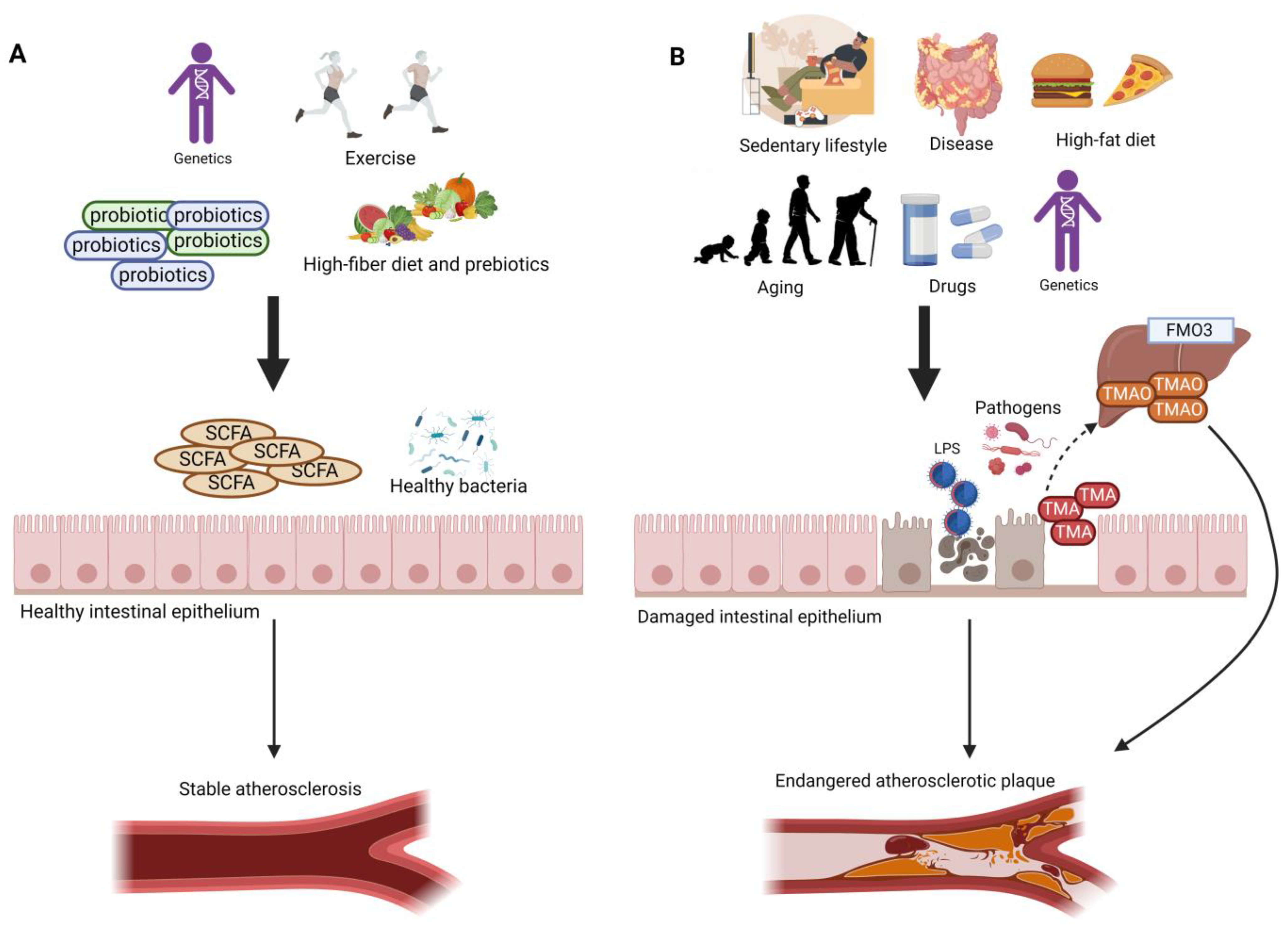
The gut microbiome plays a major role in human health, and gut microbial imbalance or dysbiosis is associated with disease development. Modulation in the gut microbiome can be used to treat or prevent different diseases. Gut dysbiosis increases with aging, and it has been associated with the impairment of gut barrier function leading to the leakage of harmful metabolites such as trimethylamine (TMA). TMA is a gut metabolite resulting from dietary amines that originate from animal-based foods. TMA enters the portal circulation and is oxidized by the hepatic enzyme into trimethylamine oxide (TMAO). Increased TMAO levels have been reported in elderly people. High TMAO levels are linked to peripheral artery disease (PAD), endothelial senescence, and vascular aging.

gut microbiome atherosclerosis TMAO

1. Introduction

Atherosclerosis is a chronic disease that affects medium and large arteries of the body through a major increase in the lipoproteins of their intimal layer [1]. Risk factors associated with atherosclerosis include diabetes mellitus, hypertension, dyslipidemia, obesity, and smoking [2]. Diabetes mellitus is a major public health problem that has been a leading cause for mortalities worldwide. Diabetes is characterized by elevated levels of blood glucose, which leads over time to serious damage to the heart, blood vessels, eyes, kidneys, and nerves [3]. As a result, diabetes mellitus is associated with accelerated atherosclerosis, leading to vascular lesions that include cardiovascular disease (CVD), coronary artery disease, cerebrovascular disease, and peripheral arterial disease (PAD), with CVD being the major cause of premature death in diabetes. Another consequence of diabetes mellitus is microangiopathy that occurs in the colon, and which has been reported to be more common in diabetics than non-diabetics [4]. Microangiopathy can also occur in the retina, skin (specifically foot skin/diabetic foot ulcer), nerve, kidney, muscle, and heart of diabetic patients and is associated with the thickening of the capillary basement membrane [5]. Other mediators of diabetes that cause vascular complications include dyslipidemia, chronic hyperglycemia, and insulin resistance. Dyslipidemia and chronic inflammation are among the major causes of the development of atherosclerosis, which causes chronic accumulation of lipid-rich plaque in the arteries in diabetic patients [6][7]. Therefore, the regulation of these chronic diseases is crucial.
The human gut microbiome is a huge microbial community that plays a vital role in human health. With the development in research, the influence of intestinal flora on human diseases has been gradually revealed. Dysbiosis in the gut microbiota (GM) has been reported to have adverse health effects on the human body that lead to a variety of chronic diseases. Regulation of the GM can provide a potential target for the prevention and treatment of disease. The fermentation products of the gut microbiota are by far the most well studied, and they have been described to have a key role in the maintenance of the gut microbial ecology and the modulation of host immunity and metabolic disease [8][9][10][11][12]. The major fermentation products of the GM that result from dietary fibers are short chain fatty acids (SCFAs), with the most abundant metabolites being acetate, propionate, and butyrate. [12][13]. SCFAs can function as a macronutrient energy source and hormone-like signaling molecules that enter the portal circulation to signal through specific host receptor systems in order to regulate the innate immunity and host metabolism. Most studies linking the GM to disease designate SCFAs as potential disease-moderating or prevention factors in metabolic disease, intestinal immunity, cancer, and liver disease [12][13]. Recent reports have indicated that dysbiosis is increased with aging, and that the GM of elderly people is enriched with pro-inflammatory commensals and fewer beneficial microbes [14]. Dysbiosis is presumed to be the primary cause of age-associated morbidities, and, consequently, the premature death of elderly people [14]. Gut dysbiosis leads to a disruption of the microbial metabolites, impaired function of the gastrointestinal tract, and increased leakage of the gut [14]. These events enhance systemic inflammation which is associated with aging, termed inflammaging, and they, consequently, result in aging-associated pathologies [14].
Trimethylamine (TMA) is a byproduct generated from the gut microbial metabolism of dietary amines such as choline, betaine, and carnitine that originate from animal-based foods [15][16]. TMA is absorbed into the portal circulation and is oxidized by the liver into trimethylamine-N-oxide (TMAO) using the flavin monooxygenase enzyme (Figure 1) [16][17]. More attention has been directed upon circulating TMAO due to its pro-inflammatory, pro-atherogenic, and pro-thrombotic properties [18][19][20][21][22]. Several factors, such as diet, gut microbial flora, drug administration, and liver flavin monooxygenase activity, influence the plasma TMAO level [16]. TMAO has been described as vital for lipid balance and for the increase in scavenger receptors, such as CD36 and scavenger receptor class A type 1 (SR-A1), that contribute to the surge of fat accumulation in foam cells, which in turn play a major part in atherosclerotic plaque progress [16][18][23]. In addition, the hepatic enzyme flavin-containing monooxygenase 3 (FMO3) is considered the most active in converting TMA into TMAO, leading to higher plasma TMAO levels. High levels of plasma TMAO have been linked to an alteration of reverse cholesterol transport [16][24], hyperlipidemia and hyperglycemia [16][24][25], and the overexpression of inflammatory markers including tumor necrosis factor alpha (TNF-α), interleukin-6 (IL-6), c-reactive protein (CRP) [26][27], and insulin resistance [16][25], which all lead to the promotion of atherosclerosis [16][19][25][28][29]. It has also been reported that high plasma levels of TMAO metabolite are related to the prognosis of 5-year all-cause mortality in stable patients diagnosed with peripheral artery disease [28][30]. Furthermore, Brunt et al. confirmed that circulating TMAO was high in older compared with younger adults, and that elevated TMAO was correlated with a higher carotid–femoral pulse wave velocity (PWV). Their findings in humans represented the first link between the age-related increase in circulating TMAO with higher aortic stiffness and blood pressure (BP). Nevertheless, as both aortic stiffness and BP are aging risk factors, it might be that both outcomes are in fact not causally associated with TMAO [31].
Figure 1. Different factors, such as the gut microbiome, diet, lifestyle, and genetics, play a major role in the development of atherosclerosis. (A) High-fiber diet, active lifestyle, and the intake of probiotics increase the abundance of beneficial gut bacteria and the amount of SCFAs, which support the tight junctions and protect the intestinal epithelium, thus preventing harmful metabolites from entering the circulation. This contributes to a stable atherosclerotic plaque. (B) High-fat diet, sedentary lifestyle, intestinal disease (e.g., Crohn’s disease, irritable bowel disease), the intake of different drugs (e.g., antibiotics), and aging lead to a disruption in the gut microbial profile, resulting in higher abundance of pathogenic bacteria and lower amounts of SCFAs. These effects lead to damage in the gut epithelium; thus, a leaky gut allows the translocation of LPS, TMA, and other damaging metabolites into the circulation. High amounts of plasma LPS and TMAO result in an endangered atherosclerotic plaque.

2. TMAO, TMA-Producing Gut Bacteria, and Atherosclerosis

Previous studies described that many human gut colonizing bacteria are capable of producing TMA, which leads to an increase of TMAO levels in plasma. These gut bacteria include Streptococcus sanguinis, Desulfovibrio alaskensis, Desulfovibrio desulfuricans, Acinetobacter, Serratia, Escherichia coli, Citrobacter, Klebsiella pneumoniae, Providencia, Shigella, Achiomobacter, and Sporosorcine, which belong to the Firmicutes and Actinobacteria phyla. [16][23]. On the other hand, bacteria belonging to phylum Bacteroidetes are not able to produce TMA [16][32]. Several previous studies have investigated the impact of host factors, such as diet and dietary compounds, on TMAO plasma levels, and it was reported that higher plasma TMAO levels have been linked to an animal-based diet [33][34] compared to vegetarians (Figure 1) [19][35]. Mainly, TMA is generated by the enzymes produced by the gut microbiota, and its levels are dependent on the amount of precursors available and the abundance and activity of bacteria catalyzing TMA formation [36], which compete with the host for these precursors that are usually absorbed as essential nutrients [36]. As the quantification of TMA producers in the gut is limited, it is more difficult to understand the distribution of these bacteria in the gut and specify their niches, which is important for designing effective and sustainable treatment strategies to minimize TMAO plasma concentrations [36]. In order to discover the abundance and diversity of TMA-forming gut bacteria, Rath et al. developed assays that targeted key genes encoding enzymes responsible for TMA formation from choline (choline-TMA lyase, CutC), carnitine (carnitine monooxygenase, CntA), and betaine (subunit B of betaine reductase, grdH) [17]. In addition to the association of TMAO with PAD and atherosclerosis, advancing age has also been strongly linked to TMAO levels. As age increases, the host’s physiology and function are altered. For example, the epithelial integrity of the colon, which is needed to promote the influx of bacterial metabolites, including TMAO, might be reduced [37][38]. Rath et al. reported that there was an association between carotid intima-media thickness (IMT) and TMAO only in individuals above 65 years of age, which indicates that aging people are principally affected by this metabolite [38]. Age-related associations between TMAO plasma levels and health parameters have not yet been reported in patients with PAD. Preclinical studies in both animal and in vitro models on human-derived material have emphasized the contribution of TMAO to endothelial senescence and vascular aging [39][40]; however, age-specific effects of TMAO have still not been fully studied. Many studies have investigated the link between diet and TMAO levels, reporting different results; however, an analysis of gut microbiota has only recently been included [38]. Rath et al. were able to provide important information regarding the formation of TMAO in the general population, and they have elaborated on the functional role of the gut microbiota and specific foods, clarifying the increased levels of TMAO with increasing age [38].

3. Gut Dysbiosis, Aging, and TMAO Levels

Gut dysbiosis is the disruption in the gut microbiome that is associated with different diseases. Dysbiosis disturbs the gut barrier function, leading to the leakage of harmful metabolites, such as lipopolysaccharides (LPS), and other bacterial components, such as peptidoglycans, into the circulation, which triggers an inflammatory response leading to atherosclerosis (Figure 1) [41][42][43]. LPS can stimulate the uptake of modified low-density lipoprotein (LDL) and reduce the efflux of cholesterol from foam cells, promoting monocyte recruitment and macrophage foam cell formation [41][43][44]. LPS can induce vascular inflammation directly or by producing pro-inflammatory factors from immune cells [44]. The increased production of pro-inflammatory cytokines promotes oxidative stress and oxidized LDL (oxLDL), increasing the risk of hypertension via nitric oxide synthase inhibition. This effect reduces vasodilator nitric oxide levels and increases levels of vasoconstrictor endothelin-1 [41].

Aging, Gut Dysbiosis, and TMAO

Aging leads to several changes in cells, tissues, and organs [45] and is influenced by an individual’s genetics, lifestyle, and environment [46]. The term “immunoscence” first appeared a few decades ago to refer to impaired or faulty immune responses leading to a decrease in the ability to trigger the immune response and effectively produce antibodies against different pathogens [47][48]. The gut microbiome undergoes dynamic changes through time, and gut dysbiosis is an age-related complication caused by host senescence, changes in nutritional behavior, drug use, and the lifestyle of aged people [48]. The changes in the gut microbiome include shifts in bacterial composition and metabolic function [49]. In humans, age-related gut dysbiosis is characterized by increased inter-individual variation and decreased species diversity; specifically, a loss of Clostridiales and Bifidobacterium, an enrichment of Proteobacteria, Lactobacilli, and an overrepresentation of pathobionts such as Enterobacteriaceae [49][50][51][52]. However, the major gut microbiota aging feature is the decreased ratio of Firmicutes/Bacteroidetes [53]. Schneeberger et al. reported that aged mice showed a decrease in beneficial gut bacteria, such as in Clostridium members of cluster IV that produce SCFAs and Akkermansia muciniphila, and an increase in pro-inflammatory microbes [14][54]. Overall, the decrease in intestinal commensal microbes diversity is associated with increased susceptibility to pathogen infection accompanied by disturbance of the gut mucosal barrier and enrichment in pro-inflammatory cytokines; all these events have a detrimental consequence in aging [14][55]. Recent studies suggested that gut dysbiosis is associated with the development of several chronic diseases including cardiovascular disease and other metabolic disorders [56].
Many studies have reported a close relationship between TMAO levels, aging, and age-related diseases. Several animal models have been used to identify mechanisms that underlie TMAO’s role in senescence [57]. Cell senescence involves many processes including increased production of reactive oxygen species (ROS), mitochondrial dysfunction, and senescence-associated secretory phenotype (SASP) [57]. Ke et al. reported that senescence-accelerated prone mouse strain 8 (SAMP8) and senescence-accelerated mouse resistant 1 (SAMR1) were treated with 1.5% (w/v) TMAO for 16 weeks to induce vascular aging and advanced vascular aging processes, respectively [40]. Many potential mechanisms underlie TMAO’s role in aging, including the inhibition of sirtuin 1 (SIRT1) expression, which increases oxidative stress and results in the activation of the p53/p21/Rb pathway. Increased P53 and P21 acetylation and reduced CDK2, cyclinE1, and Rb phosphorylation are followed by enhanced endothelial cell senescence and vascular aging [58]. In addition, TMAO increases the accumulation of ROS, matrix metalloproteinase 2 (MMP2), and matrix metalloproteinase 9 (MMP9) in vivo and in vitro, which are associated to oxidative stress in cells [59]. Furthermore, high TMAO levels are linked to increased expression of pro-inflammatory cytokines, such as TNF-α and IL-1β, as well as decreased production of anti-inflammatory cytokines such as IL-10 [26].

4. Mechanisms Underlying the Therapeutic Effect of Probiotics in Atherosclerosis

The mechanisms underlying the protective effect of probiotics against atherosclerosis are not fully understood. Nevertheless, the action of probiotics at different steps is becoming clear as more studies are being conducted. To start with, it has been reported in previous studies how probiotics can combat gut dysbiosis through strengthening the epithelial tight junctions, preventing the translocation of damaging metabolites, such as LPS and TMAO, into the circulation, which can lead to a stable atherosclerotic plaque [60]. Several studies have demonstrated the hypocholesterolemic effect of probiotics [61][62]. Liong and Shah have pointed out the ability of Lactobacillus strains to reduce cholesterol in an in vitro model, and this was achieved through various mechanisms such as assimilation of cholesterol during growth, incorporation of cholesterol into the membrane of cells, the binding of cholesterol to the cell surface, and co-precipitation with deconjugated bile [63]. These findings were backed up in another study by Zeng et al., who studied Lactobacillus buchneri P2 and confirmed the cholesterol removal trait of this bacteria through an assimilation mechanism [64]. Another study by Huang and Zheng reported the cholesterol lowering property of a probiotic strain through the inhibition of the gene expression of NPC1L1 in Caco-2 cells [65][66]. The NPC1L1 protein plays a major role in cholesterol absorption, and it is considered to be a promising target for cholesterol-lowering medication [67]. NPC1L1 has been identified by Duval et al. to be a novel target gene for the liver X receptors (LXRs), which support the crucial role of LXRs in intestinal cholesterol homeostasis [68]. LXRs activation has been reported to reduce whole-body cholesterol levels and reduce atherosclerosis [69]. In addition, VSL-3 was found to improve lipid profiles in mice [70], and this was attained by promoting BA deconjugation and fecal excretion, and by increasing hepatic BA synthesis through the downregulation of the FXR/FGF15 axis [71]. BAs can regulate cholesterol balance, and disruption in the circulation of enterohepatic BAs can lead to gall bladder [72] and gastrointestinal diseases [73]. The metabolism of BAs is also associated with obesity, diabetes, and cardiovascular diseases [74][75][76]. BAs are synthesized from hepatic cholesterol, and they are further conjugated with amino acids glycine and taurine to form bile salts that are transferred to the intestine. Bile salts’ amphiphilic combination is crucial for fat absorption in the intestine, yet excessive bile salts are toxic to the gut bacteria [77]. Bile salt hydrolase (BSH), which is present in the gut microbiome, is responsible for the catalysis of the conjugated bile salts into deconjugated BAs in order to maintain the balance of metabolism of BAs. Deconjugated BAs function as signaling molecules to aid in the secretion of GLP-1 hormone [78], activate other receptors, and impact different metabolic processes involved in various diseases [79][80]. The presence of BSH has been identified in different microbial genera such as Lactobacillus, Bifidobacterium, Enterococcus, Clostridium spp., and Bacteroides. It has also been reported that one bacterial strain can possess distinct BSHs that can have different properties [81]. In addition, recent evidence suggested that TMAO can promote atherosclerosis, partially through inhibiting hepatic bile acid synthesis [82].
In addition, probiotics can apply their anti-inflammatory actions through modulating the expression of key transcription factors or microRNAs (miRNAs), which are associated with pro-inflammatory signaling [83][84]. For instance, Chen et al. reported that L. acidophilus ATCC 4356 was able to reduce the levels of TNF-α and oxidative stress markers in addition to its ability of reversing the reduction in IL-10 levels via inhibiting the activation of NF-κB and its translocation to the nucleus [83]. In the case of atherosclerosis, T lymphocytes and macrophages accumulate and proliferate at the atherosclerotic lesions, which leads to the secretion of inflammatory cytokines such as TNF-α and IL-10 [83]. These inflammatory cytokines can, in turn, activate intracellular NF-κB signaling pathways which can stimulate the production of more cytokines, leading to further inflammation [85]. Activated NF-κB exists in the fibrotic thickened intima-media and atheromatous areas of the atherosclerotic lesion, smooth muscle cells, macrophages, and endothelial cells; however, little or no activated NF-κB can be detected in vessels lacking atherosclerosis [86][87]. The activation of NF-κB is associated with the phosphorylation of IκB-α and subsequent degradation of IκB-α, which results in the translocation of NF-κB into the nucleus. It has also been reported that TNF-α is one of the inflammatory markers that can promote atherosclerosis. On the other hand, IL-10 is an anti-inflammatory marker that provides a crucial atheroprotective signal [44]. As for the miRNAs, Lactobacillus acidophilus has been shown to protect against apoptosis and necrosis in human endothelial cells, which is induced by LPS stimulation, and this in turn was associated with a decrease in the expression of pro-inflammatory miR-155 and increased expression of anti-apoptotic mIR-21 [43][84].

References

  1. Hansson, G.K.; Libby, P.; Schönbeck, U.; Yan, Z.Q. Innate and adaptive immunity in the pathogenesis of atherosclerosis. Circ. Res. 2002, 91, 281–291.
  2. Komaroff, A.L. The Microbiome and Risk for Atherosclerosis. JAMA 2018, 319, 2381–2382.
  3. WHO. Diabetes. Available online: https://www.who.int/health-topics/diabetes#tab=tab_1 (accessed on 5 November 2022).
  4. Sasor, A.; Ohlsson, B. Microangiopathy is common in submucosal vessels of the colon in patients with diabetes mellitus. Rev. Diabet. Stud. 2014, 11, 175–180.
  5. Fiordaliso, F.; Clerici, G.; Maggioni, S.; Caminiti, M.; Bisighini, C.; Novelli, D.; Minnella, D.; Corbelli, A.; Morisi, R.; De Iaco, A.; et al. Prospective study on microangiopathy in type 2 diabetic foot ulcer. Diabetologia 2016, 59, 1542–1548.
  6. Hasheminasabgorji, E.; Jha, J.C. Dyslipidemia, Diabetes and Atherosclerosis: Role of Inflammation and ROS-Redox-Sensitive Factors. Biomedicines 2021, 9, 1602.
  7. Xiao, Y.; Guo, Z.; Li, Z.; Ling, H.; Song, C. Role and mechanism of action of butyrate in atherosclerotic diseases: A review. J. Appl. Microbiol. 2021, 131, 543–552.
  8. Koh, A.; De Vadder, F.; Kovatcheva-Datchary, P.; Bäckhed, F. From Dietary Fiber to Host Physiology: Short-Chain Fatty Acids as Key Bacterial Metabolites. Cell 2016, 165, 1332–1345.
  9. Canfora, E.E.; Jocken, J.W.; Blaak, E.E. Short-chain fatty acids in control of body weight and insulin sensitivity. Nat. Rev. Endocrinol. 2015, 11, 577–591.
  10. Miyamoto, J.; Kasubuchi, M.; Nakajima, A.; Irie, J.; Itoh, H.; Kimura, I. The role of short-chain fatty acid on blood pressure regulation. Curr. Opin. Nephrol. Hypertens. 2016, 25, 379–383.
  11. Leung, C.; Rivera, L.; Furness, J.B.; Angus, P.W. The role of the gut microbiota in NAFLD. Nat. Rev. Gastroenterol. Hepatol. 2016, 13, 412–425.
  12. Brown, J.M.; Hazen, S.L. Microbiome series: Microbial modulation of cardiovascular disease. Nat. Rev. Microbiol. 2018, 16, 171–181.
  13. El Hage, R.; Hernandez-Sanabria, E.; Van De Wiele, T. Emerging Trends in “Smart Probiotics”: Functional Consideration for the Development of Novel Health and Industrial Applications. Front. Microbiol. 2017, 8, 01889.
  14. Emeline Ragonnaud, A.B. Gut microbiota as the key controllers of “healthy” aging of elderly people. Immun. Ageing 2021, 18, 2.
  15. Rath, S.; Heidrich, B.; Pieper, D.H.; Vital, M. Uncovering the trimethylamine-producing bacteria of the human gut microbiota. Microbiome 2017, 5, 54.
  16. Janeiro, M.H.; Ramírez, M.J.; Milagro, F.I.; Martínez, J.A.; Solas, M. Implication of Trimethylamine N-Oxide (TMAO) in Disease: Potential Biomarker or New Therapeutic Target. Nutrients 2018, 10, 1398.
  17. Rath, S.; Rud, T.; Pieper, D.H.; Vital, M. Potential TMA-Producing Bacteria Are Ubiquitously Found in Mammalia. Front. Microbiol. 2020, 10, 2966.
  18. Wang, Z.; Klipfell, E.; Bennett, B.J.; Koeth, R.; Levison, B.S.; DuGar, B.; Feldstein, A.E.; Britt, E.B.; Fu, X.; Chung, Y.-M.; et al. Gut flora metabolism of phosphatidylcholine promotes cardiovascular disease. Nature 2011, 472, 57–63.
  19. Koeth, R.A.; Wang, Z.; Levison, B.S.; Buffa, J.A.; Org, E.; Sheehy, B.T.; Britt, E.B.; Fu, X.; Wu, Y.; Li, L.; et al. Intestinal microbiota metabolism of L-carnitine, a nutrient in red meat, promotes atherosclerosis. Nat. Med. 2013, 19, 576–585.
  20. Zhu, W.; Gregory, J.C.; Org, E.; Buffa, J.A.; Gupta, N.; Wang, Z.; Li, L.; Fu, X.; Wu, Y.; Mehrabian, M.; et al. Gut Microbial Metabolite TMAO Enhances Platelet Hyperreactivity and Thrombosis Risk. Cell 2016, 165, 111–124.
  21. Seldin, M.M.; Meng, Y.; Qi, H.; Zhu, W.; Wang, Z.; Hazen, S.L.; Lusis, A.J.; Shih, D.M. Trimethylamine N-Oxide Promotes Vascular Inflammation Through Signaling of Mitogen-Activated Protein Kinase and Nuclear Factor-κB. J. Am. Heart Assoc. 2016, 5, e002767.
  22. Jia, X.; Osborn, L.J.; Wang, Z. Simultaneous Measurement of Urinary Trimethylamine (TMA) and Trimethylamine N-Oxide (TMAO) by Liquid Chromatography–Mass Spectrometry. Molecules 2020, 25, 1862.
  23. Wang, Z.; Roberts, A.B.; Buffa, J.A.; Levison, B.S.; Zhu, W.; Org, E.; Gu, X.; Huang, Y.; Zamanian-Daryoush, M.; Culley, M.K.; et al. Non-lethal Inhibition of Gut Microbial Trimethylamine Production for the Treatment of Atherosclerosis. Cell 2015, 163, 1585–1595.
  24. Warrier, M.; Shih, D.M.; Burrows, A.C.; Ferguson, D.; Gromovsky, A.D.; Brown, A.L.; Marshall, S.; McDaniel, A.; Schugar, R.C.; Wang, Z.; et al. The TMAO Generating Enzyme Flavin Monooxygenase 3 is a Central Regulator of Cholesterol Balance. Cell Rep. 2016, 10, 326–338.
  25. Miao, J.; Morbid Obesity Study Group; Ling, A.V.; Manthena, P.V.; Gearing, M.E.; Graham, M.J.; Crooke, R.M.; Croce, K.J.; Esquejo, R.M.; Clish, C.; et al. Flavin-containing monooxygenase 3 as a potential player in diabetes-associated atherosclerosis. Nat. Commun. 2015, 6, 6498.
  26. Chen, K.; Zheng, X.; Feng, M.; Li, D.; Zhang, H. Gut Microbiota-Dependent Metabolite Trimethylamine N-Oxide Contributes to Cardiac Dysfunction in Western Diet-Induced Obese Mice. Front. Physiol. 2017, 8, 139.
  27. Sun, G.; Yin, Z.; Liu, N.; Bian, X.; Yu, R.; Su, X.; Zhang, B.; Wang, Y. Gut microbial metabolite TMAO contributes to renal dysfunction in a mouse model of diet-induced obesity. Biochem. Biophys. Res. Commun. 2017, 493, 964–970.
  28. Senthong, V.; Wang, Z.; Fan, Y.; Wu, Y.; Hazen, S.L.; Tang, W.W. Trimethylamine N-Oxide and Mortality Risk in Patients With Peripheral Artery Disease. J. Am. Heart Assoc. 2016, 5, e004237.
  29. Shih, D.M.; Wang, Z.; Lee, R.; Meng, Y.; Che, N.; Charugundla, S.; Qi, H.; Wu, J.; Pan, C.; Brown, J.M.; et al. Flavin containing monooxygenase 3 exerts broad effects on glucose and lipid metabolism and atherosclerosis. J. Lipid Res. 2015, 56, 22–37.
  30. Biscetti, F.; Nardella, E.; Cecchini, A.L.; Landolfi, R.; Flex, A. The Role of the Microbiota in the Diabetic Peripheral Artery Disease. Mediat. Inflamm. 2019, 2019, 4128682.
  31. Pierce, G.L.; Roy, S.J.; Gimblet, C.J. The Gut-Arterial Stiffness Axis: Is TMAO a Novel Target to Prevent Age-Related Aortic Stiffening? Hypertension 2021, 78, 512–515.
  32. Hui, D.Y. Intestinal phospholipid and lysophospholipid metabolism in cardiometabolic disease. Curr. Opin. Lipidol. 2016, 27, 507–512.
  33. Tang, W.H.W.; Wang, Z.; Levison, B.S.; Koeth, R.A.; Britt, E.B.; Fu, X.; Wu, Y.; Hazen, S.L. Intestinal microbial metabolism of phosphatidylcholine and cardiovascular risk. N. Engl. J. Med. 2013, 368, 1575–1584.
  34. Wang, Z.; Bergeron, N.; Levison, B.S.; Li, X.S.; Chiu, S.; Jia, X.; Koeth, R.A.; Li, L.; Wu, Y.; Tang, W.H.W.; et al. Impact of chronic dietary red meat, white meat, or non-meat protein on trimethylamine N-oxide metabolism and renal excretion in healthy men and women. Eur. Heart J. 2019, 40, 583–594.
  35. Koeth, R.A.; Lam-Galvez, B.R.; Kirsop, J.; Wang, Z.; Levison, B.S.; Gu, X.; Copeland, M.F.; Bartlett, D.; Cody, D.B.; Dai, H.J.; et al. l-Carnitine in omnivorous diets induces an atherogenic gut microbial pathway in humans. J. Clin. Investig. 2019, 129, 373–387.
  36. Rath, S.; Rud, T.; Karch, A.; Pieper, D.H.; Vital, M. Pathogenic functions of host microbiota. Microbiome 2018, 6, 174.
  37. Buford, T.W. (Dis)Trust your gut: The gut microbiome in age-related inflammation, health, and disease. Microbiome 2017, 5, 80.
  38. Rath, S.; Rox, K.; Kleine Bardenhorst, S.; Schminke, U.; Dörr, M.; Mayerle, J.; Frost, F.; Lerch, M.M.; Karch, A.; Brönstrup, M.; et al. Higher Trimethylamine-N-Oxide Plasma Levels with Increasing Age Are Mediated by Diet and Trimethylamine-Forming Bacteria. mSystems 2021, 6, e00945-21.
  39. Brunt, V.E.; Gioscia-Ryan, R.A.; Richey, J.J.; Zigler, M.C.; Cuevas, L.M.; Gonzalez, A.; Vázquez-Baeza, Y.; Battson, M.L.; Smithson, A.T.; Gilley, A.D.; et al. Suppression of the gut microbiome ameliorates age-related arterial dysfunction and oxidative stress in mice. J. Physiol. 2019, 597, 2361–2378.
  40. Ke, Y.; Li, D.; Zhao, M.; Liu, C.; Liu, J.; Zeng, A.; Shi, X.; Cheng, S.; Pan, B.; Zheng, L.; et al. Gut flora-dependent metabolite Trimethylamine-N-oxide accelerates endothelial cell senescence and vascular aging through oxidative stress. Free. Radic. Biol. Med. 2018, 116, 88–100.
  41. Lau, K.; Srivatsav, V.; Rizwan, A.; Nashed, A.; Liu, R.; Shen, R.; Akhtar, M. Bridging the Gap between Gut Microbial Dysbiosis and Cardiovascular Diseases. Nutrients 2017, 9, 859.
  42. Feng, Q.; Chen, W.D.; Wang, Y.D. Gut Microbiota: An Integral Moderator in Health and Disease. Front. Microbiol. 2018, 9, 151.
  43. O’Morain, V.L.; Ramji, D.P. The Potential of Probiotics in the Prevention and Treatment of Atherosclerosis. Mol. Nutr. Food Res. 2020, 64, e1900797.
  44. Tang, W.H.W.; Bäckhed, F.; Landmesser, U.; Hazen, S.L. Intestinal Microbiota in Cardiovascular Health and Disease: JACC State-of-the-Art Review. J. Am. Coll. Cardiol. 2019, 73, 2089–2105.
  45. Khan, S.S.; Singer, B.D.; Vaughan, D.E. Molecular and physiological manifestations and measurement of aging in humans. Aging Cell 2017, 16, 624–633.
  46. Ferrucci, L.; Gonzalez-Freire, M.; Fabbri, E.; Simonsick, E.; Tanaka, T.; Moore, Z.; Salimi, S.; Sierra, F.; de Cabo, R. Measuring biological aging in humans: A quest. Aging Cell 2020, 19, e13080.
  47. Montecino-Rodriguez, E.; Berent-Maoz, B.; Dorshkind, K. Causes, consequences, and reversal of immune system aging. J. Clin. Investig. 2013, 123, 958–965.
  48. Bosco, N.; Noti, M. The aging gut microbiome and its impact on host immunity. Genes Immun. 2021, 22, 289–303.
  49. Xu, C.; Zhu, H.; Qiu, P. Aging progression of human gut microbiota. BMC Microbiol. 2019, 19, 236.
  50. Woodmansey, E.J. Intestinal bacteria and ageing. J. Appl. Microbiol. 2007, 102, 1178–1186.
  51. Jeffery, I.B.; Lynch, D.B.; O’Toole, P.W. Composition and temporal stability of the gut microbiota in older persons. ISME J. 2016, 10, 170–182.
  52. O’Toole, P.W.; Jeffery, I.B. Gut microbiota and aging. Science 2015, 350, 1214–1215.
  53. Mariat, D.; Firmesse, O.; Levenez, F.; Guimarăes, V.; Sokol, H.; Doré, J.; Corthier, G.; Furet, J.P. The Firmicutes/Bacteroidetes ratio of the human microbiota changes with age. BMC Microbiol. 2009, 9, 123.
  54. Schneeberger, M.; Everard, A.; Gómez-Valadés, A.G.; Matamoros, S.; Ramírez, S.; Delzenne, N.M.; Gomis, R.; Claret, M.; Cani, P.D. Akkermansia muciniphila inversely correlates with the onset of inflammation, altered adipose tissue metabolism and metabolic disorders during obesity in mice. Sci. Rep. 2015, 5, 16643.
  55. Ling, Z.; Liu, X.; Cheng, Y.; Yan, X.; Wu, S. Gut microbiota and aging. Crit. Rev. Food Sci. Nutr. 2022, 62, 3509–3534.
  56. Yang, T.; Santisteban, M.M.; Rodriguez, V.; Li, E.; Ahmari, N.; Carvajal, J.M.; Zadeh, M.; Gong, M.; Qi, Y.; Zubcevic, J.; et al. Gut Dysbiosis Is Linked to Hypertension. Hypertension 2015, 65, 1331–1340.
  57. Zhang, L.; Yu, F.; Xia, J. Trimethylamine N-oxide: Role in cell senescence and age-related diseases. Eur. J. Nutr. 2022.
  58. Huang, R.; Yan, L.; Lei, Y. The Gut Microbial-Derived Metabolite Trimethylamine N-Oxide and Atrial Fibrillation: Relationships, Mechanisms, and Therapeutic Strategies. Clin. Interv. Aging 2021, 16, 1975.
  59. Brunt, V.E.; Gioscia-Ryan, R.A.; Casso, A.G.; VanDongen, N.S.; Ziemba, B.P.; Sapinsley, Z.J.; Richey, J.J.; Zigler, M.C.; Neilson, A.P.; Davy, K.P.; et al. Trimethylamine-N-Oxide Promotes Age-Related Vascular Oxidative Stress and Endothelial Dysfunction in Mice and Healthy Humans. Hypertension 2020, 76, 101–112.
  60. Shen, X.; Li, L.; Sun, Z.; Zang, G.; Zhang, L.; Shao, C.; Wang, Z. Gut Microbiota and Atherosclerosis-Focusing on the Plaque Stability. Front. Cardiovasc. Med. 2021, 8, 668532.
  61. Agerholm-Larsen, L.; Raben, A.; Haulrik, N.; Hansen, A.S.; Manders, M.; Astrup, A. Effect of 8 week intake of probiotic milk products on risk factors for cardiovascular diseases. Eur. J. Clin. Nutr. 2000, 54, 288–297.
  62. Ooi, L.G.; Liong, M.T. Cholesterol-lowering effects of probiotics and prebiotics: A review of in vivo and in vitro findings. Int. J. Mol. Sci. 2010, 11, 2499–2522.
  63. Liong, M.T.; Shah, N.P. Acid and bile tolerance and cholesterol removal ability of lactobacilli strains. J. Dairy Sci. 2005, 88, 55–66.
  64. Zeng, X.Q.; Pan, D.D.; Guo, Y.X. The probiotic properties of Lactobacillus buchneri P2. J. Appl. Microbiol. 2010, 108, 2059–2066.
  65. Hassan, A.; Din, A.U.; Zhu, Y.; Zhang, K.; Li, T.; Wang, Y.; Luo, Y.; Wang, G. Updates in understanding the hypocholesterolemia effect of probiotics on atherosclerosis. Appl. Microbiol. Biotechnol. 2019, 103, 5993–6006.
  66. Huang, Y.; Zheng, Y. The probiotic Lactobacillus acidophilus reduces cholesterol absorption through the down-regulation of Niemann-Pick C1-like 1 in Caco-2 cells. Br. J. Nutr. 2010, 103, 473–478.
  67. Miura, S.; Saku, K. Ezetimibe, a selective inhibitor of the transport of cholesterol. Intern. Med. 2008, 47, 1165–1170.
  68. Duval, C.; Touche, V.; Tailleux, A.; Fruchart, J.C.; Fievet, C.; Clavey, V.; Staels, B.; Lestavel, S. Niemann-Pick C1 like 1 gene expression is down-regulated by LXR activators in the intestine. Biochem. Biophys. Res. Commun. 2006, 340, 1259–1263.
  69. Bradley, M.N.; Hong, C.; Chen, M.; Joseph, S.B.; Wilpitz, D.C.; Wang, X.; Lusis, A.J.; Collins, A.; Hseuh, W.A.; Collins, J.L.; et al. Ligand activation of LXR beta reverses atherosclerosis and cellular cholesterol overload in mice lacking LXR alpha and apoE. J. Clin. Investig. 2007, 117, 2337–2346.
  70. Kriaa, A.; Bourgin, M.; Potiron, A.; Mkaouar, H.; Jablaoui, A.; Gérard, P.; Maguin, E.; Rhimi, M. Microbial impact on cholesterol and bile acid metabolism: Current status and future prospects. J. Lipid Res. 2019, 60, 323–332.
  71. Degirolamo, C.; Rainaldi, S.; Bovenga, F.; Murzilli, S.; Moschetta, A. Microbiota modification with probiotics induces hepatic bile acid synthesis via downregulation of the Fxr-Fgf15 axis in mice. Cell Rep. 2014, 7, 12–18.
  72. Cai, J.S.; Chen, J.H. The mechanism of enterohepatic circulation in the formation of gallstone disease. J. Membr. Biol. 2014, 247, 1067–1082.
  73. Copaci, I.; Micu, L.; Iliescu, L.; Voiculescu, M. New therapeutical indications of ursodeoxycholic acid. Rom. J. Gastroenterol. 2005, 14, 259–266.
  74. Cariou, B.; Chetiveaux, M.; Zaïr, Y.; Pouteau, E.; Disse, E.; Guyomarc’h-Delasalle, B.; Laville, M.; Krempf, M. Fasting plasma chenodeoxycholic acid and cholic acid concentrations are inversely correlated with insulin sensitivity in adults. Nutr. Metab. 2011, 8, 48.
  75. Joyce, S.A.; MacSharry, J.; Casey, P.G.; Kinsella, M.; Murphy, E.F.; Shanahan, F.; Hill, C.; Gahan, C.G. Regulation of host weight gain and lipid metabolism by bacterial bile acid modification in the gut. Proc. Natl. Acad. Sci. USA 2014, 111, 7421–7426.
  76. Charach, G.; Argov, O.; Geiger, K.; Charach, L.; Rogowski, O.; Grosskopf, I. Diminished bile acids excretion is a risk factor for coronary artery disease: 20-year follow up and long-term outcome. Therap. Adv. Gastroenterol. 2018, 11, 1756283x17743420.
  77. Ignacio Barrasa, J.; Olmo, N.; Pérez-Ramos, P.; Santiago-Gómez, A.; Lecona, E.; Turnay, J.; Antonia Lizarbe, M. Deoxycholic and chenodeoxycholic bile acids induce apoptosis via oxidative stress in human colon adenocarcinoma cells. Apoptosis 2011, 16, 1054–1067.
  78. Parker, H.E.; Wallis, K.; le Roux, C.W.; Wong, K.Y.; Reimann, F.; Gribble, F.M. Molecular mechanisms underlying bile acid-stimulated glucagon-like peptide-1 secretion. Br. J. Pharmacol. 2012, 165, 414–423.
  79. Haeusler, R.A.; Astiarraga, B.; Camastra, S.; Accili, D.; Ferrannini, E. Human insulin resistance is associated with increased plasma levels of 12α-hydroxylated bile acids. Diabetes 2013, 62, 4184–4191.
  80. Cipriani, S.; Mencarelli, A.; Chini, M.G.; Distrutti, E.; Renga, B.; Bifulco, G.; Baldelli, F.; Donini, A.; Fiorucci, S. The bile acid receptor GPBAR-1 (TGR5) modulates integrity of intestinal barrier and immune response to experimental colitis. PLoS ONE 2011, 6, e25637.
  81. Song, Z.; Cai, Y.; Lao, X.; Wang, X.; Lin, X.; Cui, Y.; Kalavagunta, P.K.; Liao, J.; Jin, L.; Shang, J. Taxonomic profiling and populational patterns of bacterial bile salt hydrolase (BSH) genes based on worldwide human gut microbiome. Microbiome 2019, 7, 1–16.
  82. Chen, M.-l.; Yi, L.; Zhang, Y.; Zhou, X.; Ran, L.; Yang, J.; Zhu, J.-d.; Zhang, Q.-y.; Mi, M.-t. Resveratrol attenuates trimethylamine-N-oxide (TMAO)-induced atherosclerosis by regulating TMAO synthesis and bile acid metabolism via remodeling of the gut microbiota. MBio 2016, 7, e02210–e02215.
  83. Chen, L.; Liu, W.; Li, Y.; Luo, S.; Liu, Q.; Zhong, Y.; Jian, Z.; Bao, M. Lactobacillus acidophilus ATCC 4356 attenuates the atherosclerotic progression through modulation of oxidative stress and inflammatory process. Int. Immunopharmacol. 2013, 17, 108–115.
  84. Kalani, M.; Hodjati, H.; Sajedi Khanian, M.; Doroudchi, M. Lactobacillus acidophilus Increases the Anti-apoptotic Micro RNA-21 and Decreases the Pro-inflammatory Micro RNA-155 in the LPS-Treated Human Endothelial Cells. Probiotics Antimicrob. Proteins 2016, 8, 61–72.
  85. Amaretti, A.; di Nunzio, M.; Pompei, A.; Raimondi, S.; Rossi, M.; Bordoni, A. Antioxidant properties of potentially probiotic bacteria: In vitro and in vivo activities. Appl. Microbiol. Biotechnol. 2013, 97, 809–817.
  86. Ohira, H.; Tsutsui, W.; Fujioka, Y. Are Short Chain Fatty Acids in Gut Microbiota Defensive Players for Inflammation and Atherosclerosis? J. Atheroscler Thromb. 2017, 24, 660–672.
  87. Bultman, S.J. Bacterial butyrate prevents atherosclerosis. Nat. Microbiol. 2018, 3, 1332–1333.
More
Information
Subjects: Microbiology
Contributors MDPI registered users' name will be linked to their SciProfiles pages. To register with us, please refer to https://encyclopedia.pub/register : , ,
View Times: 476
Revisions: 2 times (View History)
Update Date: 01 Jun 2023
1000/1000
Video Production Service