Submitted Successfully!
To reward your contribution, here is a gift for you: A free trial for our video production service.
Thank you for your contribution! You can also upload a video entry or images related to this topic.
Version Summary Created by Modification Content Size Created at Operation
1 -- 1232 2023-05-30 04:06:10 |
2 references update Meta information modification 1232 2023-05-30 05:44:55 |

Video Upload Options

Do you have a full video?

Confirm

Are you sure to Delete?
Cite
If you have any further questions, please contact Encyclopedia Editorial Office.
Moon, D. The Role of Calcium in Cytosolic Protein-Mediated Apoptosis. Encyclopedia. Available online: https://encyclopedia.pub/entry/44987 (accessed on 06 September 2024).
Moon D. The Role of Calcium in Cytosolic Protein-Mediated Apoptosis. Encyclopedia. Available at: https://encyclopedia.pub/entry/44987. Accessed September 06, 2024.
Moon, Dong-Oh. "The Role of Calcium in Cytosolic Protein-Mediated Apoptosis" Encyclopedia, https://encyclopedia.pub/entry/44987 (accessed September 06, 2024).
Moon, D. (2023, May 30). The Role of Calcium in Cytosolic Protein-Mediated Apoptosis. In Encyclopedia. https://encyclopedia.pub/entry/44987
Moon, Dong-Oh. "The Role of Calcium in Cytosolic Protein-Mediated Apoptosis." Encyclopedia. Web. 30 May, 2023.
The Role of Calcium in Cytosolic Protein-Mediated Apoptosis
Edit

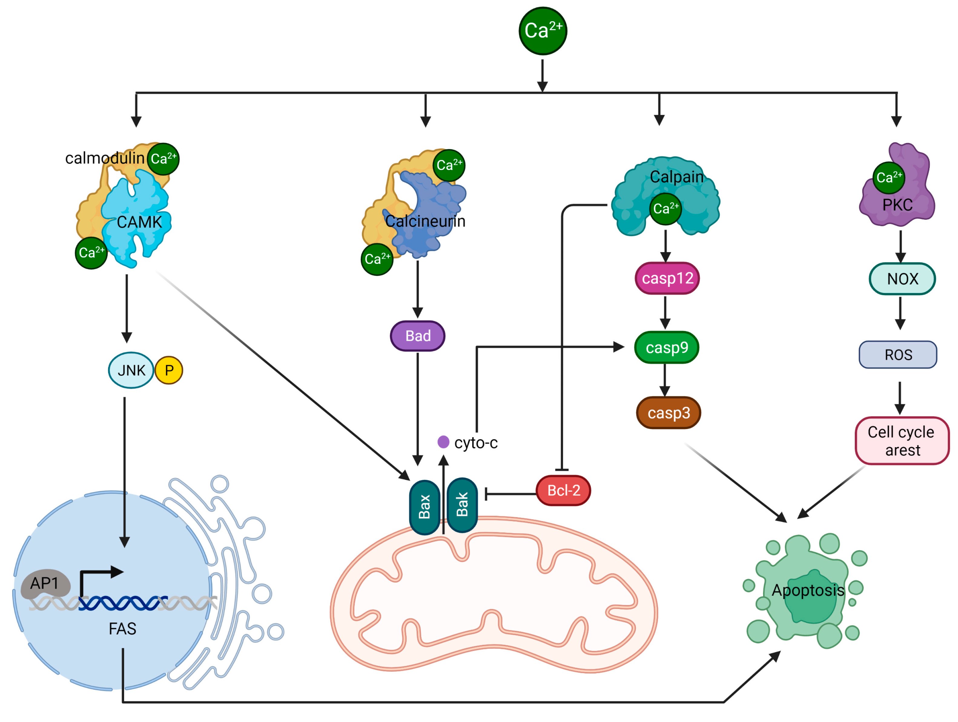
Calcium is an essential intracellular messenger that plays a vital role in controlling a broad range of cellular processes, including apoptosis. Cytosolic calcium levels are tightly regulated, and an imbalance in calcium concentrations can trigger apoptosis through various mechanisms involving cytosolic proteins.

calcium apoptosis mitochondria

1. Introduction

Calcium, an essential second messenger in cells, controls numerous cellular functions, including cell death [1]. The concentration of calcium varies in different cell compartments, with the cytoplasm maintaining relatively low levels to ensure effective cellular signaling and prevent cytotoxic effects. This balance is achieved through various mechanisms, such as calcium pumps, sodium–calcium exchangers, calcium-binding proteins, and the mitochondrial calcium uniporter (MCU), that help regulate cytoplasmic calcium levels [2]. Cancer cells are characterized by modifications in calcium channel, pump, and binding protein function, leading to calcium concentrations that surpass the usual limits seen in healthy cells. This calcium surplus fosters cell growth and malignancy. Elevated intracellular calcium levels in many cancer types can be attributed to heightened activity or irregular control of specific calcium channels and pumps. Examples include the overactivity of certain members of the transient receptor potential (TRP) channel family, like TRPM3, TRPC1, TRPC6, TRPV4, and TRPV6, seen across a variety of tumors [3]. In addition, channels like TRPA1 are notably more active in cancers such as those of the breast and lung [4]. Regulated Cell Death (RCD), which includes apoptosis, autophagy, necrosis, etc., is vital for maintaining homeostasis in living organisms, and calcium signaling plays a pivotal role in controlling it [5]. Calcium’s central role in apoptosis is particularly significant due to its influence on the key molecular events and signaling pathways determining cell fate. Understanding calcium’s function in cell death can potentially uncover new therapeutic targets for illnesses related to dysregulated cell death.

2. The Role of Calcium in Cytosolic Protein-Mediated Apoptosis

Cytosolic calcium levels are tightly regulated, and an imbalance in calcium concentrations can trigger apoptosis through various mechanisms involving cytosolic proteins. Here are some ways calcium contributes to cytosolic protein-mediated apoptosis.

2.1. Calcium/CAMK II/JNK/Fas Pathway

The calcium-regulated CaMKII/JNK/Fas pathway is a critical signaling cascade involved in controlling apoptosis. This process begins with elevated intracellular calcium levels, leading to the activation of CaMKII, a serine/threonine protein kinase. CaMKII is activated when it binds to calmodulin, a calcium-binding protein, in the presence of increased calcium concentrations [6]. Once activated, CaMKII phosphorylates a variety of substrates, such as transcription factors, ion channels, and other kinases, thereby influencing their functions. A key downstream target of CaMKII is the JNK signaling pathway. CaMKII activates the JNK pathway by stimulating upstream kinases like MKK4/7 [6]. Following activation, MKK4/7 phosphorylates and activates JNK, a stress-activated protein kinase involved in several cellular processes, including apoptosis. Activated JNK phosphorylates and activates multiple transcription factors, among them c-Jun, a component of the AP-1 complex. The AP-1 complex is essential for controlling the expression of various genes, including those associated with apoptosis. Fas, an apoptosis-related gene regulated by JNK, is a cell surface receptor that is part of the TNF receptor superfamily. When activated, Fas forms a death-inducing signaling complex (DISC) with FADD and procaspase-8, leading to caspase-8 activation. The subsequent caspase cascade ultimately results in apoptosis. In normal cells, calcium-induced activation of CAMK II can trigger the activation of JNK, leading to apoptosis through different downstream targets, including the Fas receptor.
However, in cancer cells, there can be disruptions in calcium signaling, resulting in abnormal CAMK II activation [7]. Additionally, alterations in the expression and functionality of the Fas in cancer cells can impact its involvement in apoptotic signaling [8]. These differences contribute to distinct apoptotic responses between normal cells and cancer cells.

2.2. Calcium/Calcineurin/Bcl-2 Pathway

Calcineurin, sometimes referred to as protein phosphatase 2B (PP2B), is a serine/threonine protein phosphatase that relies on calcium and calmodulin for activation [9][10][11]. It plays a critical role in various cellular processes, including cell signaling, immune response, and the regulation of apoptosis. The activation of calcineurin occurs when there is an increase in intracellular calcium levels, which causes calcium ions to bind to calmodulin. This calcium/calmodulin complex then interacts with calcineurin, triggering a conformational change that activates its phosphatase activity [10][11]. Once activated, calcineurin dephosphorylates a range of substrates, including transcription factors and other proteins, modulating their functions. One of the targets of activated by calcineurin is the protein BAD. Dephosphorylation of BAD by calcineurin causes it to detach from the 14-3-3 protein and relocate to the outer mitochondrial membrane (OMM) [12]. BAD then forms dimers with anti-apoptotic proteins Bcl-2 and Bcl-xL, suppressing pro-survival signals. This process prompts BAX to translocate to the OMM, initiating the formation of mPTP. The formation of mPTP accelerates the apoptotic cascade by releasing cytochrome c from the mitochondria, ultimately leading to cell death execution by caspases 3, 6, and 7.

2.3. Calpain/Caspases Pathway

Calpain, a family of calcium-dependent cysteine proteases, has important roles in various cellular processes, such as signal transduction, cytoskeletal remodeling, cell differentiation, and apoptosis [13]. These proteases are activated when intracellular calcium levels increase, enabling them to cleave specific target proteins through their proteolytic functions. Caspase-12 is an ER-associated caspase. During ER stress, calpain becomes activated by elevated intracellular calcium levels and cleaves the inactive pro-caspase-12, releasing active caspase-12 from the ER membrane [14]. Active caspase-12 participates in the apoptotic signaling cascade by cleaving and activating caspase-9, which subsequently activates executioner caspases such as caspase-3, caspase-6, and caspase-7. These caspases then cleave various cellular substrates, leading to the characteristic features of apoptosis.
Calpains also cleave pro-apoptotic proteins like Bid. The cleaved Bid (tBid) translocates to the mitochondria, where it interacts with Bax and Bak, resulting in mitochondrial outer membrane permeabilization (MOMP) [15]. MOMP allows cytochrome c and other pro-apoptotic factors to be released into the cytosol. Furthermore, calpains can cleave and inactivate anti-apoptotic proteins such as Bcl-2, shifting the balance in favor of apoptosis [16].

2.4. PKC/PDK/ERK Pathway

Protein kinase C (PKC) consists of a group of serine/threonine kinases activated by various signals, including growth factors, hormones, and neurotransmitters [17]. PKC is typically activated by binding to diacylglycerol (DAG), a lipid-derived secondary messenger, and elevated intracellular calcium levels. A downstream target of PKC is NADPH oxidase, an enzyme complex composed of multiple subunits that produce reactive oxygen species (ROS), such as superoxide anion (O2) and hydrogen peroxide (H2O2). PKC activates NADPH oxidase by phosphorylating its regulatory subunits, causing the enzyme complex to assemble and become active [18]. When NADPH oxidase is activated, it generates ROS, which are capable of damaging cellular components, including proteins, lipids, and DNA. Normally, ROS production is carefully regulated, and ROS serve as signaling molecules that control various cellular processes like cell proliferation, differentiation, and survival. In the context of apoptosis, ROS can induce cell death through multiple mechanisms. For example, ROS can directly damage mitochondrial components, leading to mitochondrial outer membrane permeabilization and the subsequent release of pro-apoptotic factors like cytochrome c, which triggers the intrinsic apoptotic pathway. Additionally, ROS can activate stress kinases such as JNK and p38 MAPK, which can phosphorylate and modulate the activity of pro- and anti-apoptotic proteins, tipping the balance towards apoptosis. The signaling pathways involving calcium-regulated cytoplasmic proteins in apoptosis are depicted in Figure 1.
Figure 1. The regulation of apoptosis by calcium. Elevated calcium levels contribute to apoptosis activation by modulating the activities of several proteins, including CAMK, calcineurin, calpain, and PKC. These proteins participate in various signaling pathways and cellular processes, ultimately leading to cell death when calcium concentrations become excessive.

References

  1. Berridge, M.J.; Bootman, M.D.; Roderick, H.L. Calcium signalling: Dynamics, homeostasis and remodelling. Nat. Rev. Mol. Cell. Biol. 2003, 4, 517–529.
  2. Berridge, M.J. Calcium hypothesis of Alzheimer’s disease. Pflug. Arch. 2010, 459, 441–449.
  3. Prevarskaya, N.; Zhang, L.; Barritt, G. TRP channels in cancer. Biochim. Biophys. Acta (BBA)—Mol. Basis Dis. 2007, 1772, 937–946.
  4. Gautier, M.; Dhennin-Duthille, I.; Ay, A.S.; Rybarczyk, P.; Korichneva, I.; Ouadid-Ahidouch, H. New insights into pharmacological tools to TR(i)P cancer up. Br. J. Pharmacol. 2014, 171, 2582–2592.
  5. Galluzzi, L.; Vitale, I.; Aaronson, S.A.; Abrams, J.M.; Adam, D.; Agostinis, P.; Alnemri, E.S.; Altucci, L.; Amelio, I.; Andrews, D.W.; et al. Molecular mechanisms of cell death: Recommendations of the Nomenclature Committee on Cell Death 2018. Cell Death Differ. 2018, 25, 486–541.
  6. Jiang, S.J.; Wang, W. Research progress on the role of CaMKII in heart disease. Am. J. Transl. Res. 2020, 12, 7625–7639.
  7. Chi, M.; Evans, H.; Gilchrist, J.; Mayhew, J.; Hoffman, A.; Pearsall, E.A.; Jankowski, H.; Brzozowski, J.S.; Skelding, K.A. Phosphorylation of calcium/calmodulin-stimulated protein kinase II at T286 enhances invasion and migration of human breast cancer cells. Sci. Rep. 2016, 6, 33132.
  8. Peshes-Yaloz, N.; Rosen, D.; Sondel, P.M.; Krammer, P.H.; Berke, G. Up-regulation of Fas (CD95) expression in tumour cells in vivo. Immunology 2007, 120, 502–511.
  9. Timmins, J.M.; Ozcan, L.; Seimon, T.A.; Li, G.; Malagelada, C.; Backs, J.; Backs, T.; Bassel-Duby, R.; Olson, E.N.; Anderson, M.E.; et al. Calcium/calmodulin-dependent protein kinase II links ER stress with Fas and mitochondrial apoptosis pathways. J. Clin. Investig. 2009, 119, 2925–2941.
  10. Creamer, T.P. Calcineurin. Cell Commun. Signal. 2020, 18, 137.
  11. Rusnak, F.; Mertz, P. Calcineurin: Form and function. Physiol. Rev. 2000, 80, 1483–1521.
  12. Saraf, J.; Bhattacharya, P.; Kalia, K.; Borah, A.; Sarmah, D.; Kaur, H.; Dave, K.R.; Yavagal, D.R. A Friend or Foe: Calcineurin across the Gamut of Neurological Disorders. ACS Cent. Sci. 2018, 4, 805–819.
  13. Ono, Y.; Saido, T.C.; Sorimachi, H. Calpain research for drug discovery: Challenges and potential. Nat. Rev. Drug. Discov. 2016, 15, 854–876.
  14. Ahn, Y.J.; Kim, M.S.; Chung, S.K. Calpain and Caspase-12 Expression in Lens Epithelial Cells of Diabetic Cataracts. Am. J. Ophthalmol. 2016, 167, 31–37.
  15. Mandic, A.; Viktorsson, K.; Strandberg, L.; Heiden, T.; Hansson, J.; Linder, S.; Shoshan, M.C. Calpain-mediated Bid cleavage and calpain-independent Bak modulation: Two separate pathways in cisplatin-induced apoptosis. Mol. Cell. Biol. 2002, 22, 3003–3013.
  16. Chan, S.L.; Mattson, M.P. Caspase and calpain substrates: Roles in synaptic plasticity and cell death. J. Neurosci. Res. 1999, 58, 167–190.
  17. Isakov, N. Protein kinase C (PKC) isoforms in cancer, tumor promotion and tumor suppression. Semin. Cancer Biol. 2018, 48, 36–52.
  18. Cosentino-Gomes, D.; Rocco-Machado, N.; Meyer-Fernandes, J.R. Cell signaling through protein kinase C oxidation and activation. Int. J. Mol. Sci. 2012, 13, 10697–10721.
More
Information
Subjects: Biology
Contributor MDPI registered users' name will be linked to their SciProfiles pages. To register with us, please refer to https://encyclopedia.pub/register :
View Times: 535
Revisions: 2 times (View History)
Update Date: 30 May 2023
1000/1000
ScholarVision Creations