Your browser does not fully support modern features. Please upgrade for a smoother experience.
Submitted Successfully!
 +1 credit
Thank you for your contribution! You can also upload a video entry or images related to this topic.
For video creation, please contact our Academic Video Service.
| Version | Summary | Created by | Modification | Content Size | Created at | Operation |
|---|---|---|---|---|---|---|
| 1 | Luca Piroddi | -- | 3676 | 2023-05-27 19:20:05 | | | |
| 2 | Fanny Huang | Meta information modification | 3676 | 2023-05-28 03:08:33 | | |
Video Upload Options
We provide professional Academic Video Service to translate complex research into visually appealing presentations. Would you like to try it?
Cite
If you have any further questions, please contact Encyclopedia Editorial Office.
Piroddi, L.; Abu Zeid, N.; Calcina, S.V.; Capizzi, P.; Capozzoli, L.; Catapano, I.; Cozzolino, M.; D’amico, S.; Lasaponara, R.; Tapete, D. Proximal Sensing. Encyclopedia. Available online: https://encyclopedia.pub/entry/44922 (accessed on 28 July 2026).
Piroddi L, Abu Zeid N, Calcina SV, Capizzi P, Capozzoli L, Catapano I, et al. Proximal Sensing. Encyclopedia. Available at: https://encyclopedia.pub/entry/44922. Accessed July 28, 2026.
Piroddi, Luca, Nasser Abu Zeid, Sergio Vincenzo Calcina, Patrizia Capizzi, Luigi Capozzoli, Ilaria Catapano, Marilena Cozzolino, Sebastiano D’amico, Rosa Lasaponara, Deodato Tapete. "Proximal Sensing" Encyclopedia, https://encyclopedia.pub/entry/44922 (accessed July 28, 2026).
Piroddi, L., Abu Zeid, N., Calcina, S.V., Capizzi, P., Capozzoli, L., Catapano, I., Cozzolino, M., D’amico, S., Lasaponara, R., & Tapete, D. (2023, May 27). Proximal Sensing. In Encyclopedia. https://encyclopedia.pub/entry/44922
Piroddi, Luca, et al. "Proximal Sensing." Encyclopedia. Web. 27 May, 2023.
Copy Citation
Proximal sensing techniques denote several non-invasive technologies in which the target objects—in the present context, cultural heritage manufacts—are placed within a short distance of the sensor, detector or camera lens collecting the data. Depending on the technology employed and the study purpose, the sensors/detectors work in different portions of the electromagnetic spectrum, from X-ray to ultraviolet (UV), from visible (VIS) to infrared (IR) and, further, from microwave to radio.
proximal sensing
micro-geophysics
non-destructive diagnostics
imaging techniques
cultural heritage
1. Introduction
Proximal sensing techniques denote several non-invasive technologies in which the target objects—in the present context, cultural heritage manufacts—are placed within a short distance of the sensor, detector or camera lens collecting the data. Depending on the technology employed and the study purpose, the sensors/detectors work in different portions of the electromagnetic spectrum, from X-ray to ultraviolet (UV), from visible (VIS) to infrared (IR) and, further, from microwave to radio (Figure 1). For the sake of simplicity, these techniques may be divided into “spot” and “imaging” methodologies. Examples of spot methodologies include X-ray fluorescence (XRF) spectroscopy, Fourier transform infrared (FT-IR) spectroscopy and fiber-optic reflectance spectroscopy (FORS). On the contrary, ultraviolet-induced fluorescence (UVF), infrared reflectography (IRR) and infrared photography (IRP), X-ray radiography (XRR) [1][2][3][4][5][6], multi-spectral imaging (MSI), hyperspectral imaging (HSI) and time-domain terahertz imaging (THz-TDI) are examples of imaging methodologies, naming only a few of the most-employed ones [7][8][9][10][11][12][13][14][15][16][17][18].
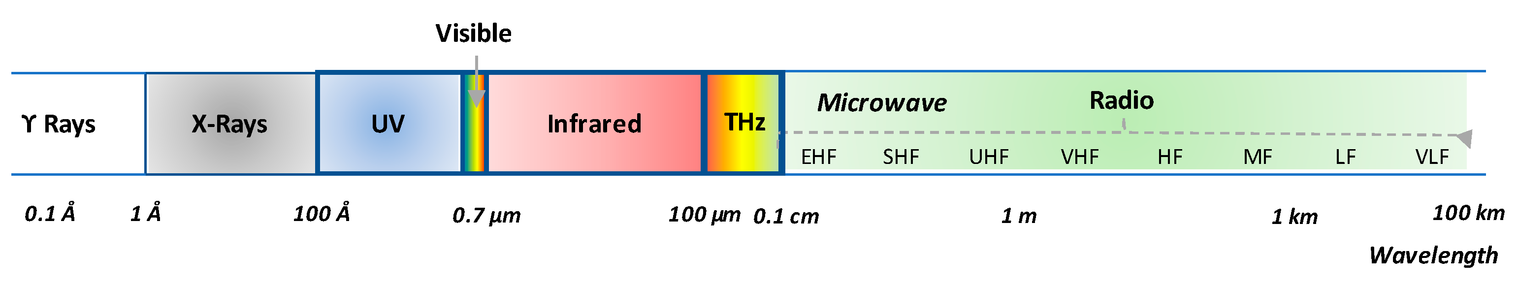
Figure 1. Portions of the electromagnetic spectrum exploited by proximal sensing technology for cultural heritage investigations.
2. X-ray Radiography (XRR)
X-ray Radiography (XRR) is among the oldest and most-standardly employed non-destructive inspection technologies [19]. XRR exploits ionizing electromagnetic signals whose wavelength ranges between 1 and 250 pm (picometer, 10−12 m) and works in transmission mode, with the object under investigation located between the source and the detector. As the transmission of X-rays is inversely proportional to the density of the encountered materials, XRR is employed to achieve an insight into the inner structure of artwork. Moreover, since elements having high atomic weight, such as mercury (atomic number equal to 80), block the X-ray transmission, XRR is useful to discriminate pigments and inks containing such chemical elements.
Historically, XRR detectors have been designed to measure the intensity of the radiation only and thus suffer from internal noise, which constrains the achievable dynamic range, i.e., the number of collectable gray levels. As a consequence, continuous efforts have been made towards the development of novel detectors capable of enhancing XRR performance, so, today, a new generation of devices providing digital, color and high-resolution images are available [20]. Moreover, systems have been developed to scan tridimensional (3D) and/or large-size objects, thanks to the use of mechanical devices which allow for automatic motion of both the object and the detector [21][22].
Concerning the XRR imaging of large 3D objects, an interesting example is offered by the case study of “Doppio corpo”, a wooden writing cabinet (studied by Pietro Piffetti) whose dimensions are 312 cm × 128 cm × 62 cm [23]. A novel prototype of computed tomography (CT) scanning was employed to obtain information about the inner structure of the masterpiece, its building technique, its conservation conditions and the presence of previous repairs and restorations. Specifically, the adopted CT prototype is made up of an X-ray source, a rotating platform—used to locate the object to be investigated—and a digital linear X-ray detector, scanning the projection plane using a high-precision mechanical system. The motion and the synchronization of the two moving parts as well as the data acquisition are regulated by two computers. Such CT devices and configurations overcome the size limits of characterizing the systems developed for medical purposes when used in the frame of CH for investigating objects such as mummies [24]. In this respect, recent advances in CT allow the totally non-invasive 3D reconstruction of the inner parts of inspected precious and delicate ancient goods [25].
3. Ultraviolet-Induced Fluorescence (UVF)
Fluorescence induced by UV radiation (UVF) is a common examination tool in artwork analysis because materials (especially organic materials) exhibit different fluorescence colors according to their chemical nature. UVF is an active imaging technique exploiting a source which emits in the UV region (100–400 nm wavelengths) and special photographic arrangements capable of acquiring the fluorescence signal. This signal is re-emitted from the materials after the molecular absorption of energy, which triggers the transition of the electrons to a higher electronic energy state [1][26].
UVF makes possible the localization of organic and inorganic materials (e.g., binders, colorants, pigments, etc.), the differentiation of materials with similar optical properties but different chemical composition (e.g., retouching, coatings and varnishes, added materials, etc.), the characterization of artwork condition, as well as the discrimination of repainted areas. In this regard, it is taken into account that the intensity of the fluorescence signal increases as the aging of materials proceeds [26][27][28].
One of the relevant examples regarding the employment of UVF for studying artwork deals with the study of Stradivari and Guarneri violins reported in [29]. In this study, UVF was employed to gather information about the varnish thickness of three famous violins, specifically, the Stradivari “Toscano”, the Stradivari “Ford” and the “Principe Doria” by Guarneri del Gesù, which were subject to minor, medium and major restoration actions. Specifically, it was observed that the UV color becomes darker when moving from the thickest to the thinnest varnished areas; thus, the most-worn portions of the violins could be detected. This was successfully verified for the well-preserved Stradivari Toscano violin, where each distinct fluorescent-colored area represented a different varnish thickness range and, therefore, represented a portion of the back plate of the violin with a specific conservation degree. Regarding the Ford violin, the UV imaging led to the hypothesis that a very thin UV-transparent over-polishing varnish was applied to the entire surface of the back plate. Finally, for the Guarneri del Gesù violin, it was found that the five differently UV-colored areas corresponded to three varnish strata of different thicknesses, which is maybe a consequence of the undocumented ordinary and extraordinary maintenance and restoration actions performed on this violin over time.
4. Infrared Reflectography (IRR), Photography (IRP) and Thermography (IRT)
IR radiation is widely used for artwork inspections and the technologies based on their use can be discriminated according to the frequency range at which they work. Indeed, the IR portion of the electromagnetic spectrum ranges from 0.7 µm up to 1 mm wavelengths and it is divided into five sub-regions: near-infrared region (NIR; 0.7–1.1 µm), short-wave infrared (SWIR; 1.1–3 µm), mid-wave infrared (MWIR; 3–6 µm), long-wave infrared (LWIR; 6–15 µm) and extreme infrared region (15–100 µm), as shown in Figure 2.
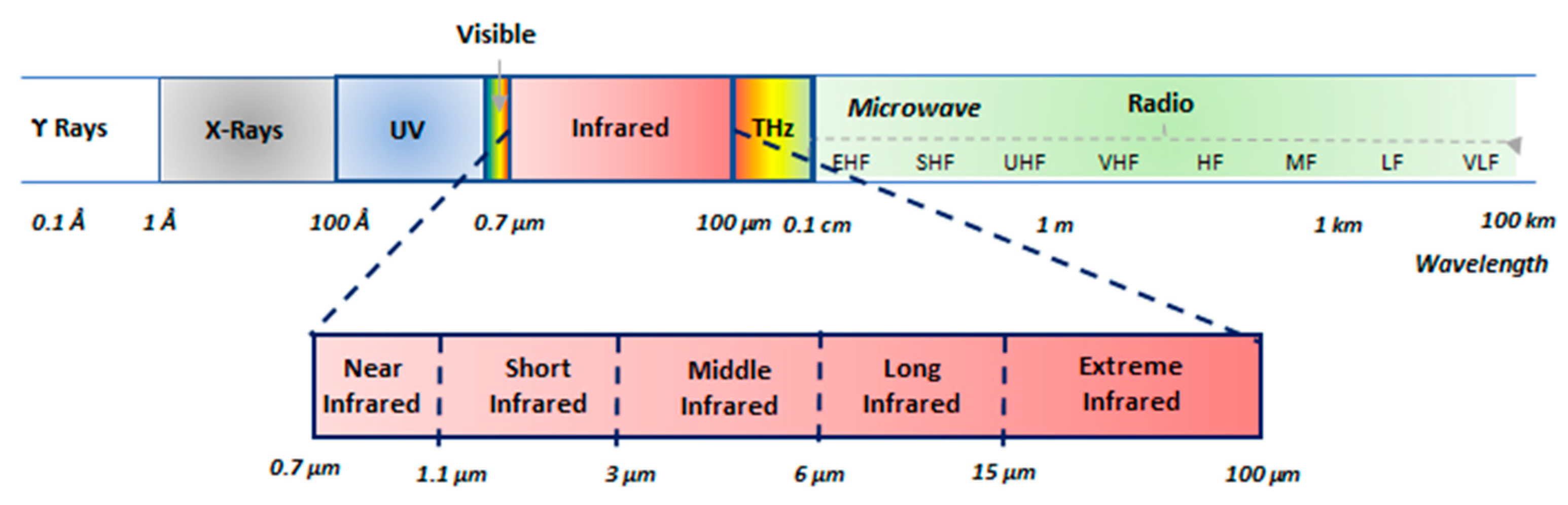
Figure 2. Wavelength partition of the infrared range.
Infrared reflectography (IRR) and infrared photography (IRP) work in the NIR region, typically exploiting 0.7–1.4 µm wavelengths, though there are devices exploiting wavelengths in the 1.5–3 µm range. Both IRR and IRP use an incandescent lamp, such as a halogen float lamp of 1000 W, or a quantum source, such as a light-emitting diode (LED) or laser, as a primary source and record the reflected radiation by means of a camera equipped with an infrared film (IRP) or a sensor sensitive to the infrared part of the spectrum (IRR) and an infrared cut-off filter [2][5][30]. The latter absorbs visible light and lets the infrared radiation pass. The commercially available IR-films are sensitive to radiation with a wavelength of up to 1.1 µm, while modern cameras can record IR radiation of wavelengths up to 2.4 µm. The gathered image, referred to as reflectogram, is not post-processed before interpretation.
In the NIR region, surface pictorial layers have an opacity that varies up to the point of becoming transparent and this depends on two main factors: the absorption of light by the pigment and the scattering of light by pigment particles, as detailed in [31].
Accordingly, IRR and IRP are frequently exploited to reveal the presence or absence of the preparatory design, the so-called “pentimenti” (changes from the initial design made by the artist during the painting process), or retouching and tampering on the work that can be attributed to previous restorations [32][33][34]. It is worth pointing out that revealing the presence of underdrawings and their relation to the final painting is a piece of valuable information to study the artist’s technique and it can help art-historians to distinguish an original picture from a copy. However, the visibility of underdrawings depends on four parameters, two related to the material and the other two dependent on the adopted diagnostic instrumentation, as follows:
-
The difference between the reflectance of the materials used for the preparatory layer and of those used for the design (contrast factor);
-
The transparency of the pictorial material to the infrared radiation;
-
The sensitivity of the sensor;
-
The resolution capability of the detection system.
Active infrared thermography (IRT) accounts for the transient behavior of the artwork response to a thermal excitation; hence, it works mainly with long-wave (6–15 µm) infrared waves, which are thermal. IRT requires a suitable heating of the object, which is produced by the light emitted by flash or DC lamps, lasers or other light sources, and employs an infrared camera capable of recording the thermal radiation coming from the object under investigation. The output is a thermogram, i.e., a map allowing the identification of areas with different thermal diffusivity. Data processing of these maps in time-lapse, by means of properly designed software, enables the extraction of information about subsurface features and material inhomogeneities. In this regard, it is worth recalling that a homogeneous material is characterized by a uniform temperature distribution, while the presence of inhomogeneities, at or beneath the surface, can modify the heat propagation, resulting in localized thermal contrasts.
An issue with IRT is the duration of exposure to the heat source. Materials such as thick woods and metals can be subject to longer exposures and this allows for a longer time to study the heat exchange. Conversely, prolonged heat exposure can damage precious canvas paintings. In this case, a quick powerful flash is often considered to reduce the exposure time, but this allows a small window, 0.1–0.5 s, in which to record the temperature differential [35].
In the field of cultural heritage, IRT is used frequently to investigate both large objects, including monuments [36], portions of historical buildings [37] and ancient bridges [38], and small objects, such as books and documents, archaeological findings and artwork [39]. Regarding the latter category, interesting reviews are given in [40][41][42].
Thermographic inspection of panel paintings can be part of more complex protocols, including other diagnostic techniques [43][44]. Through active IRT, it is possible to collect very detailed evidence of subsurface features. For example, a typical experimental setup of pulsed thermography is shown, where the synchronization of IR flashes’ energization and thermal camera acquisition achieves complete control of the experiment and obtains detailed diagnostic outputs. The algorithm used here, pulsed phase thermography (PPT), is based on the fact that, mathematically, a pulse can be decomposed into a multitude of individual sinusoidal components; when a specimen is pulse heated, thermal waves of various amplitudes and frequencies are launched into the specimen and their properties can be reconstructed by extracting the various frequencies, with discrete one-dimensional Fourier transform, of each pixel (x,y) of the thermogram sequence [45].
When inspecting large surfaces of historical buildings, the energization from the sun is often used to perform active IRT acquisition over a long time interval. In similar configurations, Sfarra et al. [46] proposed a hybrid procedure combining concepts from PPT, thermographic signal reconstruction (TSR) and principal component thermography (PCT) to obtain quantitative information such as the defect depth. The authors tested their approach on the façade of the Santa Maria Collemaggio church (L’Aquila, Italy), wherein quantitative results related to the sub-superficial discontinuities were obtained thanks to the use of such advanced techniques. First, the time-lapse datasets were de-noised through their approximation by the first five to six elements of a logarithmic polynomic regression and then the filtered results were alternatively subject to the two processing steps (PPT or PCT). Introducing a characteristic frequency named fch [Hz] corresponding to the frequency of the first extreme (maximum or minimum) of the phase—or the amplitude—contrast, the z depth of the defect [m] can be retrieved:
where α is the thermal diffusivity parameter [m2/s]. The absolute phase contrast is as follows:
in which Φdefective is the phase value for a defective area and Φdefect-free is the phase value for a defined sound area.
ΔØ = Ødefective − Ødefect-free
Another recent case study is the analysis of La Primavera, a 145 × 220 cm oil on canvas painting, realized by Mario Nuzzi and Filippo Lauri in 1658–59 and preserved in Palazzo Chigi (Ariccia, Italy) [47]. This masterpiece was investigated by means of reflectographic and thermographic techniques, which were used in a complementary way. First, an IR–ITR laser scanner prototype, developed at the ENEA Research Centre of Frascati (Rome, Italy), was employed to perform the preliminary and remote near-IR reflectographic survey of the areas where the canvas was located. Thereafter, the near-IR reflectographic map was used for planning the thermographic and mid-IR reflectographic studies, focusing the analyses on the most interesting areas of one of the paintings. The combination of the three imaging techniques revealed several details that were not otherwise visible by the naked eye; in particular, several pentimenti were detected that corresponded with the human figures.
5. Multispectral Imaging (MSI)
Multispectral imaging can be considered as the extension of white light imaging that captures several images of an object in a series of spectral bands, covering parts of the infrared and ultraviolet regions. This is performed by either using image sensors having multiple photodetectors for each pixel or adding a multispectral wheel which contains multiple optical band-pass filters for selecting different wavelength regions and then connecting to a conventional camera with a broadband response. The first technical solution implies that the image sensor changes with the spectral channels to be considered. Since the photodetectors cover certain spectral regions, the spectral data outside these regions are lost, causing a disadvantage in terms of achievable sensitivities. This drawback becomes more and more significant when the number of spectral channels increases and, thus, it is difficult to reach high performance and at the same time to cover a wide spectral region. The second technical solution offers the advantage of a simple selection of the spectral ranges by continuously rotating the wheel or by using a computer control to select one filter at a time. Once the images are registered and calibrated, they are combined to form a reflectance image cube, where the images are represented by the X- and Y-axes and the Z-dimension denotes the wavelength of each image. Sometimes, Multi Images Stacking algorithms [48][49][50] are used to improve the outline details related to the artifact contents or to the conditions of the observed objects. In the field of artwork investigation, multispectral imaging is used to pursue two main goals. The first one is to obtain high color fidelity mainly of paintings, making them more readable; the second one is image spectroscopy, which allows for gathering information about the employed materials [51].
Multispectral datasets in the form of multispectral cubes can be processed with common image processing and fusion techniques to detect subtle details in painting [52]. In the cited reference, the authors proposed a processing workflow including a 2D wavelet decomposition of spectral—NIR—images, a histogram enhancing their high frequency spatial components and image fusion (removing the lowest frequency in NIR) to the visible datasets. The result is an image where the details (that were partially invisible due to painting deterioration or that are located immediately under the most recent painting layer) are revealed and made more readable.
The ability of NIR radiation to penetrate and reflect signals from the few upper layers of the inspected surfaces, jointly with the spectral signatures of inks, are the physical basis for the multispectral study of ancient books and palimpsests. With the appropriate choice of the energizing light, recording bands and pattern recognition methods, it is possible to distinguish and make readable two or more texts written on the same paper [53].
6. Hyperspectral Imaging (HSI)
Hyperspectral imaging (HSI), also referred to as imaging spectrometry, uses a broadband light as its primary source, which illuminates the investigated object uniformly and collects hundreds of images at different wavelengths, mainly belonging to mid-infrared, near-infrared and visible segments of the electromagnetic spectrum [54]. Specifically, for each pixel of the observed scene, HSI detectors acquire data in contiguous and narrow bands, covering an extended spectral interval which depends on the adopted detector. Silicon (Si)-based sensors are capable of acquiring data in the 400–1000 nm range, indium gallium arsenide (InGaAs)-based sensors measure data at wavelengths ranging from 1000 nm up to 2500 nm, while cameras exploiting cooled indium antimonide (InSb)-based sensors work in the 1000–3000 nm range [33]. The collected data are arranged in a three-dimensional matrix, named hyperspectral cube, where two dimensions represent the spatial extent of the surveyed scene and the third-dimension accounts for the spectral content of the scene [55]. The spectral resolution of HSI data depends on the number of acquired bands and, thus, on the technical specifications of the instrumentation.
Several procedures can be used to process HSI data and allow for the visualization of concealed elements and details. Such details are not optically visible, as well as material identification according to their unique spectral signature. Moreover, multivariate analysis, such as PCA, and classification procedures are commonly exploited to generate new images that highlight the material distributions or details [56][57]. It is worth pointing out that the main issues are HSI noise, spectral mixing and huge data dimension [58]. Regarding the latter issue, techniques devoted to reducing data size have been proposed [59]. These techniques aim to choose a subset of wavelengths or their linear combinations by taking into account those wavelengths that carry the desired information for the pursued task of the survey.
In the field of artwork investigation, HSI is mainly used to perform non-invasive analysis of pigments, identify restored regions and reveal preparatory drawings [60]. Regarding HSI analysis of paintings, an example is the study reported in [61], regarding the study of two canvas paintings made by Picasso in 1917, specifically, Blanquita Suárez and Woman in an armchair. Both these pieces are exhibited in the Museum Picasso (Barcelona, Spain). In the case of the Blanquita Suárez painting, HSI data allowed the documentation of the artist’s technique; in particular, they revealed the preparatory drawing and evidenced that the artist used the intersection of lines and planes to realize the painting.
Another application of HSI is the analysis of historic texts and manuscripts, as well as of negative and positive photographic films. HSI data, indeed, allow for the identification of inks and pigments and, thus, they are useful for dating manuscripts [62] and recovering erased and overwritten scripts [63]. Moreover, HSI data are helpful to gather information about the conservation state of frames and provide spectroscopic information that may support their digital restoration, as discussed in [64], where further applicative examples are also reported. Furthermore, it is worth citing the use of HSI for mapping corrosion products on bronze sculptures (see e.g., [65]) and for investigating archeological walls with mural paintings and inscriptions (see [66]).
7. Terahertz Imaging (THz-TDI)
Imaging devices using radiation at wavelengths ranging from 30 µm to 3 mm can be considered as the newest among the diagnostic imaging tools and their more and more widespread use is owed to the non-ionizing nature of the THz waves and their penetration capability into dry, nonpolar, non-metallic materials, as well as the recent technological improvements that have allowed the commercialization of compact, flexible and portable systems [67].
In the past 20–30 years, THz imaging has experienced a rapid expansion that has allowed for the development of different imaging methodologies [68][69][70], among which is the “time of flight THz imaging” that enables a three-dimensional visualization of the internal structure of the investigated objects. Time of flight THz imaging provides information about position and thickness of inner layers as well as a geometrical characterization of hidden features. When focusing on artwork, there are several possibilities: (1) characterize texture and stratigraphy of materials; (2) detect, localize and visualize the shape of hidden defects or anomalies; (3) gather information on possible previous restoration actions [71]. All these goals are achieved without long term risks to the molecular stability of the exposed object and humans.
According to its potentiality, THz imaging has been used to investigate canvas, wall and copper paintings (see [72][73][74][75]). Several case studies have assessed its usefulness for characterizing the inner structure of paintings and gathering information on the preparation layers, which are difficult to achieve by means of XRR and IRR [71]. In addition, THz imaging has been exploited to characterize insect tunneling affecting wood carvings [76], as well as to study mummies [77][78].
8. Integrated Techniques
The collaborative use of different technologies makes it possible to gather different pieces of information about the same object and, thus, it increases knowledge of the artwork history, from its realization phase to its current conservation state [39][79][80][81][82].
An example assessing the advantages offered by such synergistic exploitation is the case study presented in [79] regarding the Renaissance wall painting named Annunciation, painted by Fra Beato Angelico, which is visible in the Museum of San Marco (Florence, Italy). In this case study, both spot and imaging proximal sensing technologies, as well as digital imaging and GPR surveys, were performed but, herein, researchers consider the proximal sensing imaging techniques only and, specifically, UVF, IRR and THz-TDI. These techniques led to novel knowledge on the compositional scheme of the wall painting with regard to its stratigraphy and restoration. The UVF revealed the presence of small, almost dot-like areas, with pink fluorescence on Mary’s gown. As the pink fluorescence is typical of red varnish, this result supports the hypothesis that, originally, Mary’s gown was red-colored, differently from its current appearance. The IRR survey pointed out that these were not the artist’s pentimenti and that the drawing was transferred onto the plaster by using the indirect incision technique, i.e., by positioning a one-to-one preparatory drawing and passing a point over the line of the drawing in order to leave the mark on the fresh plaster. Finally, THz-TDI, which was performed on some specific small areas, made it possible to detect repainted surface scratches, painted layers with different thicknesses and an elliptical-shaped inner discontinuity.
References
- Mairinger, F. UV-, IR- and X-ray- imaging. In Non-Destructive Microanalysis of Cultural Heritage Materials; Janssens, K.H.A., Grieken, R., Eds.; Wilson & Wilson Elsevier: Antwerp, Belgium, 2004; pp. 15–73.
- Van Asperen de Boer, J.R.J. Infrared reflectography: A method for the examination of paintings. Appl. Opt. 1968, 7, 1711–1714.
- Aldrovandi, A.; Bertani, D.; Cetica, M.; Matteini, M. Multispectral image processing of paintings. Stud. Conserv. 1988, 33, 154–159.
- Schreiner, M.; Frühmann, B.; Jembrih-Simbürger, D.; Linke, R. X-rays in art and archaeology: An overview. Powder Diffr. 2004, 19, 3–11.
- Saunders, D.; Billinge, R.; Cupitt, J.; Atkinson, N.; Liang, H. A new camera for high-resolution infrared imaging of works of art. Stud. Conserv. 2006, 51, 277–290.
- Hackney, S.; Townsend, J. Methods of examination and analysis. In Paint and Purpose—A Study of Technique in British Art; Hackney, S., Jones, R., Townsend, J., Eds.; Tate Gallery Publishing: London, UK, 1999; pp. 17–24.
- Striova, J.; Ruberto, C.; Barucci, M.; Blažek, J.; Kunzelman, D.; Dal Fovo, A.; Fontana, R. Spectral imaging and archival data in analysing Madonna of the Rabbit paintings by Manet and Titian. Angew. Chem. Int. 2018, 57, 7408–7412.
- Liang, H. Advances in multispectral and hyperspectral imaging for archaeology and art conservation. Appl. Phys. 2012, 106, 309–323.
- Cucci, C.; Delaney, J.K.; Picollo, M. Reflectance hyperspectral imaging for investigation of works of art: Old master paintings and illuminated manuscripts. Acc. Chem. Res. 2016, 49, 2070–2079.
- Delaney, J.K.; Zeibel, J.G.; Thoury, M.; Littleton, R.; Palmer, M.; Morales, K.M.; de la Rie, E.R.; Hoenigswald, A. Visible and infrared imaging spectroscopy of Picasso’s harlequin musician: Mapping and identification of artist materials in Situ. Appl. Spectrosc. 2010, 64, 584–594.
- Mounier, A.; Daniel, A. Hyperspectral imaging for the study of two thirteenth-century italian miniatures from the marcadé collection, treasury of the saint-andre cathedral in bordeaux, france. Stud. Conserv. 2015, 60, S200–S209.
- Cucci, C.; Webb, E.K.; Casini, A.; Ginanni, M.; Prandi, E.; Stefani, L.; Vitorino, T.; Picollo, M. Short-wave infrared reflectance hyperspectral imaging for painting investigations: A methodological study. J. Am. Inst. Conserv. 2019, 58, 16–36.
- Cucci, C.; Bracci, S.; Casini, A.; Innocenti, S.; Picollo, M.; Stefani, L.; Rao, I.G.; Scudieri, M. The illuminated manuscript Corale 43 and its attribution to Beato Angelico: Non-invasive analysis by FORS, XRF and hyperspectral imaging techniques. Microchem. J. 2018, 138, 45–57.
- Casini, A.; Bacci, M.; Cucci, C.; Lotti, F.; Porcinai, S.; Picollo, M.; Radicati, B.; Poggesi, M.; Stefani, L. Fiber optic reflectance spectroscopy and hyper-spectral image spectroscopy: Two integrated techniques for the study of the Madonna dei Fusi. In Optical Methods for Arts and Archaeology; Salimbeni, P., Ed.; SPIE: Bellingham, WA, USA, 2005; Volume 5857, p. 58570M-1-8.
- Janssens, K.; Dik, J.; Cotte, M.; Susini, J. Photon-based techniques for nondestructive subsurface analysis of painted cultural heritage artifacts. Acc. Chem. Res. 2010, 43, 814–825.
- Romano, F.P.; Caliri, C.; Nicotra, P.; Di Martino, S.; Pappalardo, L.; Rizzo, F.; Santos, H.C. Real-time elemental imaging of large dimension paintings with a novel mobile macro X-ray fluorescence (MA-XRF) scanning technique. J. Anal. At. Spectrom. 2017, 32, 773–781.
- Fukunaga, K.; Picollo, M. Characterisation of works of art. In Terahertz Spectroscopy and Imaging; Peiponen, K.E., Zeitler, A., Kuwata-Gonokami, M., Eds.; Springer: Berlin/Heidelberg, Germany, 2013; Volume 171, pp. 521–538.
- Pastorelli, G.; Trafela, T.; Taday, P.F.; Portieri, A.; Lowe, D.; Fukunaga, K.; Strlič, M. Characterisation of historic plastics using terahertz time-domain spectroscopy and pulsed imaging. Analy. Bioanal. Chem. 2012, 403, 1405–1414.
- Gonzalez, V.; Cotte, M.; Vanmeert, F.; de Nolf, W.; Janssens, K. X-ray diffraction mapping for cultural heritage science: A review of experimental configurations and applications. Eur. J. Chem. 2020, 26, 1703–1719.
- Ravaud, E.; Pichon, L.; Laval, E.; Gonzalez, V.; Eveno, M.; Calligaro, T. Development of a versatile XRF scanner for the elemental imaging of paintworks. Appl. Phys. A 2016, 122, 17.
- Giudice, A.L.; Corsi, J.; Cotto, G.; Mila, G.; Re, A.; Ricci, C.; Sacchi, R.; Visca, L.; Zamprotta, L.; Pastrone, N.; et al. A new digital radiography system for paintings on canvas and on wooden panels of large dimensions. In Proceedings of the 2017 IEEE International Instrumentation and Measurement Technology Conference (I2MTC), Turin, Italy, 22–25 May 2017.
- Impallaria, A.; Petrucci, F.; Chiozzi, S.; Evangelisti, F.; Squerzanti, S. A scanner for in situ X-ray radiography of large paintings: The case of “Paolo and Francesca” by G. Previati. Eur. Phys. J. Plus 2021, 136, 126.
- Re, A.; Albertin, F.; Avataneo, C.; Brancaccio, R.; Corsi, J.; Cotto, G.; De Blasi, S.; Dughera, G.; Durisi, E.; Ferrarese, W.; et al. X-ray tomography of large wooden artworks: The case study of “Doppio corpo” by Pietro Piffetti. Herit. Sci. 2014, 2, 19.
- Huppertz, A.; Wildung, D.; Kemp, B.J.; Nentwig, T.; Asbach, P.; Rasche, F.M.; Hamm, B. Nondestructive insights into composition of the sculpture of Egyptian queen Nefertiti with CT. Radiology 2009, 251, 233–240.
- Zesch, S.; Panzer, S.; Rosendahl, W.; Nance Jr, J.W.; Schönberg, S.O.; Henzler, T. From first to latest imaging technology: Revisiting the first mummy investigated with X-ray in 1896 by using dual-source computed tomography. Eur. J. Radiol. Open 2016, 3, 172–181.
- Aldrovandi, A.; Picollo, M. Metodi di Documentazione e Indagini non Invasive sui Dipinti; Il Prato: Padova, Italy, 2007.
- Buzzegoli, E.; Keller, A. Ultraviolet fluorescence imaging. In Scientific Examination for the Investigation of Paintings. A Handbook for Conservator-Restorers; Pinna, D., Galeotti, M., Mazzeo, R., Eds.; Centro Di Edifimi srl: Firenze, Italy, 2009; pp. 204–206.
- Dyer, J.; Verri, G.; Cupitt, J. Multispectral Imaging in Reflectance and Photo-Induced Luminescence Modes: A User Manual, Version 1.0. 2013. Available online: http://www.britishmuseum.org/pdf/charisma-multispectral-imaging-manual-2013.pdf (accessed on 11 March 2023).
- Invernizzi, C.; Fichera, G.V.; Licchelli, M.; Malagodi, M. A non-invasive stratigraphic study by reflection FT-IR spectroscopy and UV-induced fluorescence technique: The case of historical violins. Microchem. J. 2018, 138, 273–281.
- Van Asperen de Boer, J.R.J. Reflectography of Paintings Using an Infrared Vidicon Television System. Stud. Conserv. 1969, 14, 96–118.
- Gavrilov, D.; Maev, R.; Almond, D.P. A review of imaging methods in analysis of works of art: Thermographic imaging method in art analysis. Can. J. Phys. 2014, 92, 341–364.
- Walmsley, E.; Metzger, C.; Delaney, J.K.; Fletcher, C. Improved Visualization of Underdrawings with Solid-state Detectors Operating in the Infrared. Stud. Conserv. 1994, 39, 217–231.
- Casini, A.; Lotti, F.; Picollo, M.; Stefani, L.; Buzzegoli, E. Image Spectroscopy Mapping Technique for non-invasive Analysis of Paintings. Stud. Conserv. 1999, 44, 39–48.
- Poldi, G.; Villa, G.C.F. Dalla Conservazione Alla Storia Dell’arte. Riflettografia e Analisi Non Invasive per lo Studio dei Dipinti; Edizioni della Scuola Normale Publisher: Pisa, Italy, 2006.
- Tucker, A.P. Infrared and Art: Using Infrared Photography in Art Conservation and History. Available online: https://irinfo.org/articles-2019/infrared-reflectography-5-1-19-tucker/ (accessed on 11 March 2023).
- Kordatos, E.Z.; Exarchos, D.A.; Stavrakos, C.; Moropoulou, A.; Matikas, T.E. Infrared thermographic inspection of murals and characterization of degradation in historic monuments. Constr. Build. Mater. 2013, 48, 1261–1265.
- Glavaš, H.; Hadzima-Nyarko, M.; Haničar Buljan, I.; Barić, T. Locating hidden elements in walls of cultural heritage buildings by using infrared thermography. Bldg 2019, 9, 32.
- Biscarini, C.; Catapano, I.; Cavalagli, N.; Ludeno, G.; Pepe, F.A.; Ubertini, F. UAV photogrammetry, infrared thermography and GPR for enhancing structural and material degradation evaluation of the Roman masonry bridge of Ponte Lucano in Italy. NDT E Int. 2020, 115, 102287.
- Piroddi, L.; Catapano, I.; Colica, E.; D’Amico, S.; Galone, L.; Gargiulo, G.; Sfarra, S. The Pulcinella Diagnostic Project: Introduction to the Study of the Performances of Close-Range Diagnostics Targeted to a Wooden Physical Twin of a Carnival Historical Mask. In Computational Science and Its Applications, Proceedings of the ICCSA 2022 Workshops, Malaga, Spain, 4–7 July 2022; Springer International Publishing: Cham, Switzerland, 2022.
- Mercuri, F.; Zammit, U.; Orazi, N.; Paolini, S.; Marinelli, M.; Scudeieri, F. Active infrared thermography applied to the investigation of art and historic artefacts. J. Therm. Anal. Calorim. 2011, 104, 475–485.
- Orazi, N. Mid-wave Infrared Reflectography and Thermography for the Study of Ancient Books: A Review. Stud. Conserv. 2020, 65, 437–449.
- Orazi, N. The study of artistic bronzes by infrared thermography: A review. J. Cult. Herit. 2020, 42, 280–289.
- Ibarra-Castanedo, C.; Sfarra, S.; Ambrosini, D.; Paoletti, D.; Bendada, A.; Maldague, X. Diagnostics of panel paintings using holographic interferometry and pulsed thermography. Quant. InfraRed Thermogr. J. 2010, 7, 85–114.
- Bendada, A.; Sfarra, S.; Ibarra, C.; Akhloufi, M.; Pradere, C.; Maldague, X. Subsurface imaging for panel paintings inspection: A comparative study of the ultraviolet, the visible, the in-frared and the terahertz spectra. Opto-Electron. Rev. 2015, 23, 90–101.
- Maldague, X.; Galmiche, F.; Ziadi, A. Advances in pulsed phase thermography. Infrared Phys. Technol. 2002, 43, 175–181.
- Sfarra, S.; Marcucci, E.; Ambrosini, D.; Paoletti, D. Infrared exploration of the architectural heritage: From passive infrared thermography to hybrid infrared thermography (HIRT) approach. Mater. Construcción 2016, 66, e094.
- Ceccarelli, S.; Guarneri, M.; Orazi, N.; Ferrucci, M.; Ciaffi, M.; Mercuri, F.; Paoloni, S.; Ferri de Collibus, M.; Zammit, U.; Petrucci, F. Remote and contactless infrared imaging tech-niques for stratigraphical investigations in paintings on canvas. Appl. Phys. B 2021, 127, 106.
- Cogoni, M. Nuove tecnologie non distruttive per lo studio e il restauro dei beni monumentali: Applicazioni termografiche e multispettrali nell’ipogeo di San Salvatore di Sinis in Cabras. Master’s Thesis, University of Cagliari, Cagliari, Italy, 2015.
- Piroddi, L.; Ranieri, G.; Cogoni, M.; Trogu, A.; Loddo, F. Time and spectral multiresolution remote sensing for the study of ancient wall drawings at San Salvatore hypogeum, Italy. In Proceedings of the 22nd European Meeting of Environmental and Engineering Geophysics, Near Surface Geoscience 2016, Barcellona, Spain, 4–8 September 2016.
- Trogu, A.; Cogoni, M.; Ranieri, G.; Piroddi, L.; Loddo, F. Invisible but not lost. The recovery of the wall drawings of the hypogeum of San Salvatore di Sinis (Sardinia, Italy). In Proceedings of the 24th Annual Meeting of the European Association of Archaeologists, Barcelona, Spain, 5–8 September 2018.
- Pelagotti, A.; Mastio, A.D.; Rosa, A.D.; Piva, A. Multispectral imaging of paintings. IEEE Signal Process. Mag. 2008, 25, 27–36.
- Piroddi, L.; Calcina, S.V.; Trogu, A.; Vignoli, G. Towards the definition of a low-cost toolbox for qualitative inspection of painted historical vaults by means of modified DSLR cameras, open source programs and signal processing techniques. In Computational Science and Its Applications, Proceedings of the ICCSA 2020: 20th International Conference, Cagliari, Italy, 1–4 July 2020; Springer International Publishing: Cham, Switzerland, 2020; pp. 971–991.
- Easton, R.L.; Knox, K.T.; Christens-Barry, W.A. Multispectral imaging of the Archimedes palimpsest. In Proceedings of the 32nd Applied Imagery Pattern Recognition Workshop, Washington, DC, USA, 15–18 October 2003.
- Hyperspectral Imaging for Art Conservation. Available online: https://surfaceoptics.com/applications/art-antiquities-conservation-hyperspectral/ (accessed on 7 March 2023).
- Landgrebe, D. Information extraction principles and methods for multispectral and hyperspectral image data. Inf. Process. Remote Sens. 1999, 82, 3–37.
- Du, Q.; Fowler, J.E. Low-complexity principal component analysis for hyperspectral image compression. Int. J. High Perform. Comput. Appl. 2008, 22, 438–448.
- Wang, J.; Chang, C.I. Independent component analysis-based dimensionality reduction with applications in hyperspectral image analysis. IEEE Trans. Geosci. Remote Sens. 2006, 44, 1586–1600.
- Bioucas-Dias, J.M.; Plaza, A.; Camps-Valls, G.; Scheunders, P.; Nasrabadi, N.; Chanussot, J. Hyperspectral remote sensing data analysis and future challenges. IEEE Geosci. Remote Sens. Mag. 2013, 1, 6–36.
- Plaza, A.; Benediktsson, J.A.; Boardman, J.W.; Brazile, J.; Bruzzone, L.; Camps-Valls, G.; Chanussot, J.; Fauvel, M.; Gamba, P.; Gualtieri, A. Recent advances in techniques for hyperspectral image processing. Remote Sens. Environ. 2009, 113, S110–S122.
- Cucci, C.; Casini, A. Hyperspectral imaging for artworks investigation. Data Handl. Sci. Technol. 2020, 32, 583–604.
- Picollo, M.; Casini, A.; Cucci, C.; Stefani, L.; Jiménez-Garnica, R.; Fuster-López, L. Documentation and analysis of some Picasso’s paintings by using hyper-spectral imaging technique to support their conservation and stylistic matters. IOP Conf. Ser. Mater. Sci. Eng. 2020, 949, 012023.
- Melessanaki, K.; Papadakis, V.; Balas, C.; Anglos, D. Laser induced breakdown spectroscopy and hyperspectral imaging analysis of pigments on an illuminated manuscript. Spectrochim. Acta Part B 2001, 56, 2337–2346.
- Balas, C.; Papadakis, V.; Papadakis, N.; Papadakis, A.; Vazgiouraki, E.; Themelis, G. A novel hyperspectral imaging apparatus for the nondestructive analysis of objects of artistic and historic value. J. Cult. Herit. 2003, 4, 330–337.
- Picollo, M.; Cucci, C.; Casini, A.; Stefani, L. Hyper-spectral imaging technique in the cultural heritage field: New possible scenarios. Sensors 2020, 20, 2843.
- Catelli, E.; Randeberg, L.Y.; Strandeberg, H.; Alsberg, B.K.; Maris, A.; Vikky, L. Can hyperspectral imaging be used to map corrosion products on outdoor bronze sculptures. J. Spectr. Imaging 2018, 7, a10.
- Cucci, C.; Picollo, M.; Chiarantini, L.; Uda, G.; Fiori, L.; De Nigris, B.; Osanna, M. Remote-sensing hyperspectral imaging for applications in archaeological areas: Non-invasive investigations on wall paintings and on mural inscriptions in the Pompeii site. Microchem. J. 2020, 158, 105082.
- Guillet, J.P.; Recur, B.; Frederique, L.; Bousquet, B.; Canioni, L.; Manek-Hönninger, I.; Desbarats, P.; Mounaix, P. Review of terahertz tomography techniques. J. Infrared Millim. Terahertz Waves 2014, 35, 382–411.
- Peiponen, K.E.; Kuwata-Gonokami, M.; Axel Zeitler, J. Terahertz Spectroscopy and Imaging; Springer: Berlin, Germany, 2013.
- Herrmann, M.; Tani, M.; Sa-kai, K. Display modes in time-resolved terahertz imaging. Jpn. J. Appl. Phys. 2000, 39, 6254–6258.
- Saha, A. Advances in Terahertz Imaging. Emerging Trends in Terahertz Solid-State Physics and Devices; Springer: New York, NY, USA, 2020; pp. 143–168.
- Fukunaga, K. THz Technology Applied to Cultural Heritage in Practice (Cultural Heritage Science); Springer Nature AG: Cham, Switzerland, 2016.
- Seco-Martorell, C.; López-Domínguez, V.; Arauz-Garofalo, G.; Redo-Sanchez, A.; Palacios, J.; Tejada, J. Goya’s artwork imaging with Terahertz waves. Opt. Express 2013, 21, 17800–17805.
- Fukunaga, K.; Hosako, I.; Palazzo, M.; Dall’Aglio, L.; Aramini, F.; Cucci, C.; Picollo, M.; Ikari, T.; Duling, I.N. Terahertz time-domain imaging of “The Last Supper”. In Proceedings of the 2020 45th International Conference on Infrared, Millimeter, and Terahertz Waves (IRMMW-THz), Buffalo, NY, USA, 8–13 November 2020.
- Krügener, K.; Ornik, J.; Schneider, L.M.; Jäckel, A.; Koch-Dandolo, C.L.; Castro-Camus, E.; Riedl-Siedow, N.; Koch, M.; Viöl, W. Terahertz inspection of buildings and architectural art. Appl. Sci. 2020, 10, 5166.
- Cassar, Q.; Koch-Dandolo, C.L.; Guillet, J.P.; Roux, M.; Fauquet, F.; Perraud, J.B.; Mounaix, P. Characterization of Varnish Ageing and its Consequences on Terahertz Imagery: Demonstration on a Painting Presumed of the French Renaissance. J. Infrared Millim. Terahertz Waves 2020, 41, 1556–1566.
- Stübling, E.; Staats, N.; Globisch, B.; Schell, M.; Portsteffen, H.D.; Koch, M. Investigating the Layer Structure and Insect Tunneling on a Wooden Putto Using Robotic-Based THz Tomography. IEEE Trans. Terahertz Sci. Technol. 2020, 10, 343–347.
- Fukunaga, K.; Cortes, E.; Cosentino, A.; Stuenkel, I.; Leona, M.; Duling, I.N., III; Mininberg, D.T. Investigating the use of terahertz pulsed time domain reflection imaging for the study of fabric layers of an Egyptian mummy. J. Eur. Opt. Soc.-Rapid Publ. 2011, 6, 11040.
- Oehrstrom, L.; Bitzer, A.; Walther, M.; Ruhli, F.J. Technical note: Terahertz imaging of an-cient mummies and bone. Am. J. Phys. Anthropol. 2010, 142, 497–500.
- Catapano, I.; Ludeno, G.; Cucci, C.; Picollo, M.; Stefani, L.; Fukunaga, K. Noninvasive analytical and diagnostic technologies for studying early renaissance wall paintings. Surv. Geophys. 2020, 41, 669–693.
- van Loon, A.; Noble, P.; Krekeler, A.; Van der Snickt, G.; Janssens, K.; Abe, Y.; Nakai, I.; Dik, J. Artificial orpiment, a new pigment in Rembrandt’s palette. Herit. Sci. 2017, 5, 26.
- Gabrieli, F.; Delaney, J.K.; Erdmann, R.G.; Gonzalez, V.; van Loon, A.; Smulders, P.; Berkeveld, R.; van Langh, R.; Keune, K. Reflectance imaging spectroscopy (Ris) for operation night watch: Challenges and achievements of imaging rembrandt’s masterpiece in the glass chamber at the rijksmuseum. Sensors 2021, 21, 6855.
- Quintero Balbas, D.; Dal Fovo, A.; Montalbano, L.; Fontana, R.; Striova, J. Non-invasive contactless analysis of an early drawing by Raffaello Sanzio by means of optical methods. Sci. Rep. 2022, 12, 15602.
More
Information
Contributors
MDPI registered users' name will be linked to their SciProfiles pages. To register with us, please refer to https://encyclopedia.pub/register
:
View Times:
1.7K
Revisions:
2 times
(View History)
Update Date:
02 Jun 2023
Table of Contents


Notice
You are not a member of the advisory board for this topic. If you want to update advisory board member profile, please contact office@encyclopedia.pub.
OK
Confirm
Only members of the Encyclopedia advisory board for this topic are allowed to note entries. Would you like to become an advisory board member of the Encyclopedia?
Yes
No
${ textCharacter }/${ maxCharacter }
Submit
Cancel
Back
Comments
${ item }
|
${ item.createdUser.fullName }
${ item.createdAt }
${ item.vote }
${ item.reply }
Delete
${ reply.createdUser.fullName }
${ reply.createdAt }
${ reply.vote }
Delete
There is no reply to this comment~
${ item.replyTextCharacter }/${ item.replyMaxCharacter }
Submit
Cancel
More
No more~
There is no comment~
${ textCharacter }/${ maxCharacter }
Submit
Cancel
${ selectedItem.replyTextCharacter }/${ selectedItem.replyMaxCharacter }
Submit
Cancel
Confirm
Are you sure to Delete?
Yes
No