Submitted Successfully!
To reward your contribution, here is a gift for you: A free trial for our video production service.
Thank you for your contribution! You can also upload a video entry or images related to this topic.
Version Summary Created by Modification Content Size Created at Operation
1 -- 2213 2023-05-25 08:57:29 |
2 Reference format revised. + 3 word(s) 2216 2023-05-25 10:11:12 |

Video Upload Options

Do you have a full video?

Confirm

Are you sure to Delete?
Cite
If you have any further questions, please contact Encyclopedia Editorial Office.
Kumar, P.; Arendt, C.; Martin, S.; Al Soufi, S.; Deleuw, P.; Nagel, E.; Puntmann, V.O. Multimodality Imaging in HIV-Associated Cardiovascular Complications. Encyclopedia. Available online: https://encyclopedia.pub/entry/44816 (accessed on 04 July 2024).
Kumar P, Arendt C, Martin S, Al Soufi S, Deleuw P, Nagel E, et al. Multimodality Imaging in HIV-Associated Cardiovascular Complications. Encyclopedia. Available at: https://encyclopedia.pub/entry/44816. Accessed July 04, 2024.
Kumar, Parveen, Christophe Arendt, Simon Martin, Safaa Al Soufi, Philipp Deleuw, Eike Nagel, Valentina O. Puntmann. "Multimodality Imaging in HIV-Associated Cardiovascular Complications" Encyclopedia, https://encyclopedia.pub/entry/44816 (accessed July 04, 2024).
Kumar, P., Arendt, C., Martin, S., Al Soufi, S., Deleuw, P., Nagel, E., & Puntmann, V.O. (2023, May 25). Multimodality Imaging in HIV-Associated Cardiovascular Complications. In Encyclopedia. https://encyclopedia.pub/entry/44816
Kumar, Parveen, et al. "Multimodality Imaging in HIV-Associated Cardiovascular Complications." Encyclopedia. Web. 25 May, 2023.
Multimodality Imaging in HIV-Associated Cardiovascular Complications
Edit

Human immunodeficiency virus (HIV) infection is a leading cause of mortality and morbidity worldwide. The introduction of antiretroviral therapy (ART) has significantly reduced the risk of developing acquired immune deficiency syndrome and increased life expectancy, approaching that of the general population. However, people living with HIV have a substantially increased risk of cardiovascular diseases despite long-term viral suppression using ART. HIV-associated cardiovascular complications encompass a broad spectrum of diseases that involve the myocardium, pericardium, coronary arteries, valves, and systemic and pulmonary vasculature. Traditional risk stratification tools do not accurately predict cardiovascular risk in this population. Multimodality imaging plays an essential role in the evaluation of various HIV-related cardiovascular complications. 

cardiovascular magnetic resonance human immunodeficiency virus myocarditis

1. Introduction

Human immunodeficiency virus (HIV) infection represents a major public health concern. The introduction of potent antiretroviral therapy (ART) has distinctly reduced HIV-related mortality and morbidity [1]. However, with an increase in life expectancy, chronic complications related to HIV infection are becoming more evident. Cardiovascular disease is commonly found in HIV patients [2]. The high prevalence of cardiovascular risk factors in HIV-infected patients, together with ART-induced metabolic changes and an HIV-induced chronic inflammatory state, increases the risk of cardiovascular diseases [3][4]. The clinical expression of cardiovascular disorders is variable and depends on various factors, including the degree of immunodeficiency, associated opportunistic infections and drug interactions and toxicities. The presentation of myocardial disease ranges from subclinical inflammation on cardiovascular imaging to symptomatic heart failure (Figure 1) [5]. Cardiac imaging plays an integral role in the assessment of HIV-related cardiovascular complications. Transthoracic echocardiography (TTE) is the primary tool for assessing myocardial function. It is cost-effective, readily available, and robust in identifying both systolic and diastolic dysfunctions [6]. Coronary CT angiography (CTA) is primarily used for assessing both clinical and subclinical coronary artery disease (CAD) in HIV-infected patients [7]. Cardiovascular magnetic resonance (CMR) has emerged as an important imaging tool. It not only yields anatomic and functional cardiac information, but also helps in tissue characterization by assessing the degree of oedema and fibrosis [8].
Figure 1. Central illustration showing various risk factors for various HIV-associated cardiovascular complications.

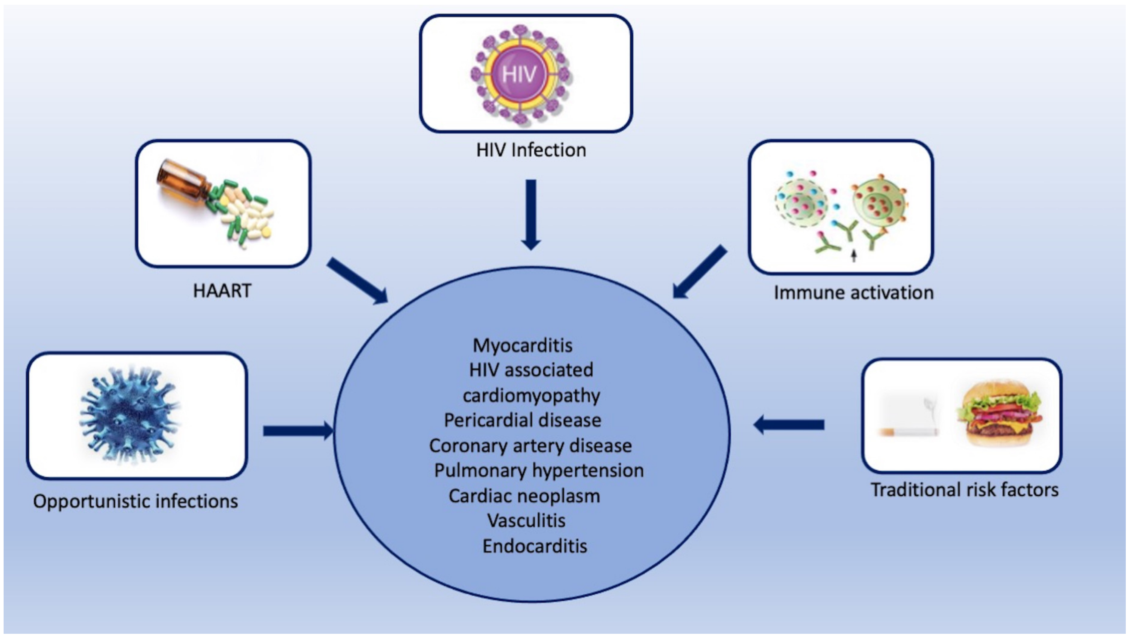
2. HIV-Associated Cardiovascular Complications

The cardiovascular complications of HIV include a broad range of cardiac, coronary, valvular, and vascular manifestations. The pathogenesis and imaging findings of these complications are summarized in the following sections.

2.1. Myocarditis

Myocarditis is seen in approximately one-third of HIV-infected patients in histopathology, without any specific cause in more than 80% of the cases [9]. The proposed etiologies for myocarditis include direct invasion by the HIV virus, opportunistic infections, and HIV-mediated indirect pathways of myocardial inflammation [10][11][12][13][14]. HIV also catalyzes a series of indirect pathways that induce myocardial inflammation and myocardial damage [15][16][17]. Myocardial damage resulting from these indirect inflammatory pathways leads to left ventricular systolic dysfunction and hemodynamic compromise [18].
TTE is a useful tool in the diagnostic assessment of suspected myocarditis. TTE investigates the wall thickness, chamber volume, and systolic and diastolic dysfunctions, and thus does not provide any direct evidence of myocarditis [19]. CMR is a promising tool for assessing myocardial inflammation. Comprehensive CMR imaging not only yields anatomic and functional cardiac information, but also provides information on myocardial tissue composition, including the degree of oedema, inflammation, and fibrosis (Figure 2). Several cardiac imaging studies have provided deep insight into the structural and functional alterations associated with HIV-related myocarditis. CMR parameters indicating myocardial inflammation are elevated in HIV-infected patients, even with subclinical diseases [20][21]. Myocardial functional alteration (systolic or diastolic dysfunctions) may be a result of this higher inflammatory burden [20].
Figure 2. Cardiac magnetic resonance imaging in a 43-year-old man with HIV-related perimyocarditis. (A) Mid-ventricular short-axis and (B) 3-chamber long-axis images showing linear mid-wall enhancement in the basal inferior and inferolateral lateral walls (white arrows). The overlying pericardium also shows mild thickening and enhancement. (C) T1 map and (D) T2 map at the mid-LV show a markedly elevated myocardial T1 time of 1214 ms (normal 1104 ms at 3.0 Tesla) and T2 time of 44 ms (normal 37.4 ms at 3.0 Tesla) consistent with diffuse myocardial fibrosis.
The presence of diastolic dysfunction in asymptomatic patients has also been observed in various TTE-based studies [22][23]. A study by Hsue et al. demonstrated a high prevalence of diastolic dysfunction in asymptomatic HIV-infected individuals. After adjustment for age and various traditional risk factors, HIV-infected individuals had 2.4-times greater odds of having diastolic dysfunction compared to controls [22]. CMR is also helpful in monitoring treatment response in HIV-related myocarditis. 

2.2. HIV Associated Cardiomyopathy

HIV infection is recognized as an important cause of dilated cardiomyopathy [24][25][26]. It is usually seen in the late stages of diseases and is associated with a low CD4 count [24]. The proposed etiological factors for HIV-associated cardiomyopathy include myocarditis, opportunistic infections, micronutrient deficiency, cardiac autoimmunity, and antiretroviral toxicity [27]. Patients with HIV-related cardiomyopathy show poor prognosis, with a reported median survival of 101 days, compared to a median survival of 472 days in patients with similar severity of HIV infection but normal hearts [28]. The gross pathological examination may show eccentric hypertrophy with dilated chambers and increased wall thickness. Conversely, there may be ventricular wall thinning. Other common features include endocardial fibroelastosis, apical mural thrombi, infective endocarditis, and apical mural thrombi [29].
TTE is the primary modality for assessing cardiomyopathy. In the pre-ART era, HIV-associated cardiomyopathy was recognized as systolic dysfunction with LV dilatation on TTE. However, after the onset of ART, HIV-associated cardiomyopathy is largely recognized as subclinical diastolic dysfunction [30]. Right ventricular (RV) dysfunction is common in HIV-infected patients, especially with dilated cardiomyopathy or HIV-associated pulmonary hypertension. Isolated RV dysfunction has been reported in both the pre-ART and ART eras [31].

2.3. Pericardial Diseases

The various HIV-related pericardial diseases include pericardial effusion, cardiac tamponade, pericarditis, and constrictive pericarditis [26][32][33][34][35][36][37][38][39]. Pericardial effusion was one of the common cardiac manifestations of HIV in the pre-HAART (highly active anti-retroviral therapy) era between 1985 and 1995, with a reported prevalence ranging from 5% to 45% in three major trials [26][32][33]. The etiology of pericardial effusion is unknown in asymptomatic patients, as they rarely need pericardiocentesis. Among symptomatic cases, infection and malignancy constitute approximately two-thirds of the cases. Other rare causes include chronic kidney diseases, hypothyroidism, and connective tissue diseases [34]. In developing countries, the most common cause of infective pericardial effusion is tuberculosis [35][36], while in developed countries, the common infective agents include streptococcus pneumoniae, staphylococcus aureus, chlamydia, listeria, and cryptococcus [37][38].

2.4. Coronary Artery Diseases

HIV-infected patients are at higher risk of coronary artery disease (CAD), with an estimated 1.5- to-2-fold higher risk compared to individuals without HIV [3][40]. The exact pathophysiology of the increased risk of CAD is not well understood. Previously, a higher prevalence of traditional risk factors such as smoking, substance abuse, and lipid abnormalities drew attention in HIV-infected patients [41][42]. Initial studies also focused on the positive association between lipid dystrophies and some ART medications [43]. However, recent studies show that HIV-positive patients are at significantly higher risk of non-calcific plaque compared to non-infected age-matched controls, even after adjustment for traditional risk factors [7].
Non-invasive coronary imaging allows early detection of atherosclerosis in patients with HIV by utilizing advances in both CT and MRI. The coronary artery calcium (CAC) score is a strong predictor of CAD and provides incremental risk stratification for major adverse cardiovascular events over traditional risk factors in the general population [44]. The utility of CAC has also been explored in HIV-infected individuals. In a prospective observational study of 843 HIV-infected patients with a median follow-up of 2.8 years, it was seen that a CAC score of 100 was associated with 3.3-times higher odds of myocardial infarction, independent of gender and age [45]. A potential limitation of CAC screening is its inability to identify non-calcific plaque [46]. CCTA is an established tool for characterizing coronary artery plaque. Plaque with low attenuation, spotty calcification, and positive remodeling is vulnerable and is associated with an increased risk of acute coronary syndromes [47]

2.5. Pulmonary Hypertension

HIV is an established risk factor for pulmonary hypertension (PH), with a significantly higher prevalence of PH in HIV-infected individuals compared with the general population (0.5 vs. 0.0015%) [48]. It has been hypothesized that HIV-protein-related factors cause persistent activation of metabolic and proliferation signaling pathways, leading to pulmonary vascular remodeling. The resultant medial hypertrophy, perivascular cuffing, and thrombosis lead to PH. Other factors contributing to PH include illicit drug abuse and concomitant secondary pulmonary infections [49]. The most commonly observed symptoms include progressive shortness of breath, pedal oedema, and cough.
The imaging features of HIV-related PH are similar to primary hypertension. The common echocardiographic findings of PH include dilation of the right heart chambers (98%), tricuspid regurgitation (64%), and paradoxical septal motion (40%). Other echocardiographic findings include RV hypertrophy, abnormal septal motion, and pulmonary valve insufficiency [50][51]. CT angiography plays a crucial role in the diagnostic workup of pulmonary hypertension.
The diagnostic workup of PH also includes right heart catheterization (RHC), which is the gold standard for diagnosing PH. The European Society of Cardiology guideline recommends the assessment of patients with signs of RV dysfunction using TTE as a screening tool, with a confirmed diagnosis dependent on results obtained from RHC. The hemodynamic parameters measured by RHC include cardiac output, mean pulmonary artery pressure, pulmonary artery wedge pressure, right atrial pressure, right ventricular pressure, pulmonary vascular resistance, and cardiac index [52].

2.6. Cardiac Neoplasm

HIV-infected patients show an increased frequency of various malignancies. Commonly prevalent malignancies associated with HIV include Kaposi sarcoma (KS), non-Hodgkin lymphoma (NHL), Hodgkin disease, squamous cell carcinoma, and leiomyosarcoma [53][54]. Common cardiac malignancies include KS and NHL.
KS was the first cardiac tumor seen in HIV-infected patients. It was first described in 1983 by Autran et al. in a young Haitian woman with AIDS [55]. The retrospective autopsy findings show a varying incidence of KS ranging from 12% to 28% [56][57]. In contrast to the classic KS seen in elderly Mediterranean men and endemic sub-Saharan African populations, HIV-associated Kaposi sarcoma is characterized by widespread dissemination and an aggressive nature [54]. HIV-associated KS usually involves the serous pericardium or subepicardial fat. There is a predilection for the subepicardial fat adjacent to the major coronary arteries, with or without the involvement of the adventitial layer of the pulmonary artery and ascending aorta [58].
Cardiac lymphoma is the second most common tumor involving the heart in HIV patients, with an estimated relative risk of 72.8 [53][54]. In the majority of cases, such lesions are high-grade B-cell tumors, including large-cell immunoblastic lymphoma or small-cell Burkitt or Burkitt-like lymphoma. The right atrium is most commonly involved, followed by the right ventricle, left ventricle, left atrium, and interatrial and interventricular septae [29]. The pericardial extension is commonly seen. Patients present with pericardial effusion, cardiac tamponade, congestive heart failure, and cardiac arrhythmias [59][60][61]. On chest radiographs, cardiac lymphoma shows cardiomegaly, pericardial effusion, and signs of heart failure [62]. Echocardiography shows a hypoechoic, polypoidal cardiac mass with or without pericardial effusion [62]. On CT, cardiac lymphoma appears isodense to the myocardium and shows heterogenous contrast enhancement [63]. On CMR, cardiac lymphoma appears iso-hypointense on T1 and iso-hyperintense on T2, and depicts heterogeneous contrast enhancement. Central necrosis is not seen, unlike in other tumors such as angiosarcoma [63].

2.7. Vasculitis

HIV-infected patients show a range of inflammatory vascular conditions caused by both infective and non-infective etiologies. There is scarce literature on infective vasculitis. Large vessels such as the aorta can be involved, producing mycotic aneurysms, especially in intravenous drug abusers. The common causative agents include staphylococcus aureus, salmonella, and mycobacterium tuberculosis [64][65][66]. The non-infective type can involve both large and small vessels. Large vessel disease may be aneurysmal or occlusive. The aneurysmal type is more common and may involve multiple arteries such as the aorta and common iliac, femoral, popliteal, and common carotid arteries [67]. The occlusive type is less common and has been reported in a few case reports [67][68].

2.8. Endocarditis

HIV-infected patients may develop infective or non-infective endocarditis. Infective endocarditis is usually seen in intravenous drug abusers, with a reported prevalence varying from 6.3% to 34% [24][69]. The clinical manifestations and survival rate of infective endocarditis in HIV-infected patients are similar to those in patients without HIV; however, late-stage HIV disease and infective endocarditis have a 30% higher mortality rate than asymptomatic HIV-positive patients [10][70]. The most common causative agent is staphylococcus aureus, followed by streptococcus viridians [70]. The clinical symptoms include fever, sweating, weight loss, and septic emboli. Possible complications include embolism; the left-sided endocarditis (mitral valve and aortic valve endocarditis) shows cerebral and myocardium embolism, while the right-sided endocarditis (tricuspid and pulmonary) shows pulmonary embolism. Another deleterious complication is the perforation of valvular leaflets or rupture of chordae tendinea, leading to acute valvular insufficiency and heart failure [71].
The transesophageal echocardiogram (TEE) is another valuable tool for assessing endocarditis. Compared to TTE, TEE has shown higher diagnostic accuracy in detecting small vegetations of endocarditis [72]. A recent study has reported that TTE performs better than CT for detecting valvular infective endocarditis-related lesions (vegetations, erosion) and is similar to CT for detecting paravalvular IE-related lesions (abscess, pseudoaneurysm). Furthermore, the relatively lower cost of TEE compared to CMR and the absence of radiation exposure compared to CT also contribute to its wide-scale use in clinical practice [73]. The CT imaging findings of septic pulmonary embolism include bilateral lung nodular lesions ranging from 0.5 to 3.5cm, often with a basal predominance [74][75]. These nodules may be well-delineated or poorly defined and may show cavitation in up to 50% of cases. 

3. Conclusions

Cardiovascular complications are commonly seen in HIV-infected patients. The spectrum of these complications is broad and includes myocarditis, dilated cardiomyopathy, pericardial effusion, coronary artery disease, endocarditis, vasculitis, and cardiac tumors. Imaging plays an important role in the early diagnosis of these diseases. The selection of imaging modalities should be tailored to the clinical presentation in order to investigate the underlying cause and facilitate evidence-based treatment.

References

  1. Mocroft, A.; Ledergerber, B.; Katlama, C.; Kirk, O.; Reiss, P.; d’Arminio Monforte, A.; Knysz, B.; Dietrich, M.; Phillips, A.; Lundgren, J. Decline in the AIDS and Death Rates in the EuroSIDA Study: An Observational Study. Lancet 2003, 362, 22–29.
  2. Hemkens, L.G.; Bucher, H.C. HIV infection and cardiovascular disease. Eur. Heart J. 2014, 35, 1373–1381.
  3. Triant, V.A.; Lee, H.; Hadigan, C.; Grinspoon, S.K. Increased acute myocardial infarction rates and cardiovascular risk factors among patients with human immunodeficiency virus disease. J. Clin. Endocrinol. Metab. 2007, 92, 2506–2512.
  4. Duprez, D.; Neuhaus, J.; Kuller, L.; Tracy, R.; Belloso, W.; De Wit, S. Inflammation, Coagulation and Cardiovascular Disease in HIV-Infected Individuals. PLoS ONE 2012, 7, e44454.
  5. Remick, J.; Georgiopoulou, V.; Marti, C.; Ofotokun, I.; Kalogeropoulos, A.; Lewis, W.; Butler, J. Heart Failure in Patients with Human Immunodeficiency Virus Infection: Epidemiology, Pathophysiology, Treatment, and Future Research. Circulation 2014, 129, 1781–1789.
  6. Erqou, S.; Lodebo, B.T.; Masri, A.; Altibi, A.M.; Echouffo-Tcheugui, J.B.; Dzudie, A.; Ataklte, F.; Choudhary, G.; Bloomfield, G.S.; Wu, W.-C.; et al. Cardiac Dysfunction among People Living with HIV. JACC Heart Fail. 2019, 7, 98–108.
  7. Post, W.S.; Budoff, M.; Kingsley, L.; Palella, F.J., Jr.; Witt, M.D.; Li, X.; George, R.T.; Brown, T.T.; Jacobson, L.P. Associations between HIV infection and subclinical coronary atherosclerosis. Ann. Intern. Med. 2014, 160, 458–467.
  8. Sood, V.; Jermy, S.; Saad, H.; Samuels, P.; Moosa, S.; Ntusi, N. Review of cardiovascular magnetic resonance in human immunodeficiency virus-associated cardiovascular disease. SA J. Radiol. 2017, 21, 1248.
  9. Herskowitz, A.; Wu, T.-C.; Willoughby, S.B.; Vlahov, D.; Ansari, A.A.; Beschorner, W.E.; Baughman, K.L. Mycarditis and Cardiotropic Viral Infection Associated with Severe Left Ventricular Dysfunction in Late-Stage Infection with Human Immunodeficiency Virus. J. Am. Coll. Cardiol. 1994, 24, 1025–1032.
  10. Barbaro, G.; Di Lorenzo, G.; Grisorio, B.; Barbarini, G.; the Gruppo Italiano per lo Studio Cardiologico dei Pazienti Affetti da AIDS Investigators. Cardiac involvement in the acquired immunodeficiency syndrome: A multicenter clinical-pathological study. AIDS Res. Hum. Retroviruses 1998, 14, 1071–1077.
  11. Barbaro, G. Cardiovascular manifestations of HIV infection. J. R. Soc. Med. 2001, 94, 384–390.
  12. Adair, O.V.; Randive, N.; Krasnow, N. Isolated toxoplasma myocarditis in acquired immune deficiency syndrome. Am. Heart J. 1989, 118, 856–857.
  13. Kinney, E.; Monsuez, J.; Kitzis, M.; Vittecoq, D. Treatment of AIDS-associated heart disease. Angiology 1989, 40, 970–976.
  14. Hofman, P.; Drici, M.D.; Gibelin, P.; Michiels, J.F.; Thyss, A. Prevalence of toxoplasma myocarditis in patients with the acquired immunodeficiency syndrome. Br. Heart J. 1993, 70, 376–381.
  15. Twu, C.; Liu, N.Q.; Popik, W.; Bukrinsky, M.; Sayre, J.; Roberts, J.; Rania, S.; Bramhandam, V.; Roos, K.P.; MacLellan, W.R.; et al. Cardiomyocytes Undergo Apoptosis in Human Immunodeficiency Virus Cardiomyopathy through Mitochondrion- and Death Receptor-Controlled Pathways. Proc. Natl. Acad. Sci. USA 2002, 99, 14386–14391.
  16. Fiala, M.; Popik, W.; Qiao, J.; Lossinsky, A.; Alce, T.; Tran, K. HIV-1 induces cardiomyopathyby cardiomyocyte invasion and gp120, tat, and cytokine apoptotic signaling. Cardiovasc. Toxicol. 2004, 4, 097–108.
  17. Barbaro, G. HIV-associated cardiomyopathy. Herz. Kardiovask. Erkrank. 2005, 30, 486–492.
  18. Duan, M.; Yao, H.; Hu, G.; Chen, X.; Lund, A.K.; Buch, S. HIV Tat induces expression of ICAM-1 in HUVECs: Implications for miR-221/-222 in HIV-associated cardiomyopathy. PLoS ONE 2013, 8, e60170.
  19. Olusegun-Joseph, D.A.; Ajuluchukwu, J.N.; Okany, C.C.; Mbakwem, A.C.; Oke, D.A.; Okubadejo, N.U. Echocardiographic patterns in treatment-naïve HIV-positive patients in Lagos, south-west Nigeria. Cardiovasc. J. Afr. 2012, 23, e1–e6.
  20. Luetkens, J.A.; Doerner, J.; Schwarze-Zander, C.; Wasmuth, J.-C.; Boesecke, C.; Sprinkart, A.M.; Schmeel, F.C.; Homsi, R.; Gieseke, J.; Schild, H.H.; et al. Cardiac Magnetic Resonance Reveals Signs of Subclinical Myocardial Inflammation in Asymptomatic HIV-Infected Patients. Circ. Cardiovasc. Imaging 2016, 9, e004091.
  21. Ntusi, N.A.; O’Dwyer, E.; Dorrell, L.; Piechnik, S.K.; Ferreira, V.M.; Karamitsos, T.D.; Sam, E.; Clarke, K.; Neubauer, S.; Holloway, C. HIV-1-Related Cardiovascular Disease Is Associated with Chronic Inflammation, Frequent Pericardial Effusions and Increased Myocardial Oedema. J. Cardiovasc. Magn. Resonan. 2016, 18, O104.
  22. Hsue, P.Y.; Hunt, P.W.; Ho, J.E.; Farah, H.H.; Schnell, A.; Hoh, R.; Martin, J.N.; Deeks, S.G.; Bolger, A.F. Impact of HIV Infection on Diastolic Function and Left Ventricular Mass. Circ. Heart Fail. 2010, 3, 132–139.
  23. Mendes, L.; Silva, D.; Miranda, C.; Sá, J.; Duque, L.; Duarte, N.; Brito, P.; Bernardino, L.; Poças, J. Impact of HIV Infection on Cardiac Deformation. Rev. Port. Cardiol. 2014, 33, 501–509.
  24. Barbaro, G.; Di Lorenzo, G.; Grisorio, B.; Barbarini, G. Incidence of dilated cardiomyopathy and detection of HIV in myocardial cells of HIV-positive patients. N. Engl. J. Med. 1998, 339, 1093–1099.
  25. Levy, W.S.; Simon, G.L.; Rios, J.C.; Ross, A.M. Prevalence of cardiac abnormalities in human immunodeficiency virus infection. Am. J. Cardiol. 1989, 63, 86–89.
  26. Himelman, R.B.; Chung, W.S.; Chernoff, D.N.; Schiller, N.B.; Hollander, H. Cardiac manifestations of human immunodeficiency virus infection: A two-dimensional echocardiographic study. J. Am. Coll. Cardiol. 1989, 13, 1030–1036.
  27. Lumsden, R.H.; Bloomfield, G.S. The Causes of HIV-Associated Cardiomyopathy: A Tale of Two Worlds. BioMed Res. Int. 2016, 2016, 1–9.
  28. Currie, P.F.; Jacob, A.J.; Foreman, A.R.; Elton, R.A.; Brettle, R.P.; Boon, N.A. Heart muscle disease related to HIV infection: Prognostic implications. BMJ 1994, 309, 1605–1607.
  29. D’Amati, G.; di Gioia, C.R.; Gallo, P. Pathological findings of HIV-associated cardiovascular disease. Ann. N. Y. Acad. Sci. 2001, 946, 23–45.
  30. Sliwa, K.; Carrington, M.J.; Becker, A.; Thienemann, F.; Ntsekhe, M.; Stewart, S. Contribution of the human immunodeficiency virus/acquired immunodeficiency syndrome epidemic to de novo presentations of heart disease in the Heart of Soweto Study cohort. Eur. Heart J. 2012, 33, 866–874.
  31. Simon, M.A.; Lacomis, C.D.; George, M.P.; Kessinger, C.; Weinman, R.; McMahon, D.; Gladwin, M.T.; Champion, H.C.; Morris, A. Isolated Right Ventricular Dysfunction in Patients with Human Immunodeficiency Virus Short Title: Right Ventricular Dysfunction in HIV. J. Card. Fail. 2014, 20, 414–421.
  32. Hecht, S.R.; Berger, M.; Van Tosh, A.; Croxson, S. Unsuspected cardiac abnormalities in the acquired immune deficiency syndrome. Echocardiogr. Study Chest 1989, 96, 805–808.
  33. Akhras, F.; Dubrey, S.; Gazzard, B.; Noble, M.I. Emerging patterns of heart disease in HIV infected homosexual subjects with and without opportunistic infections; a prospective colour flow doppler echocardiographic study. Eur. Heart J. 1994, 15, 68–75.
  34. Madhyastha, P.S.; Reddy, C.; Shetty, G.; Shastry, B.; Doddamani, A. A study of pericardial effusion in HIV positive patients and its correlation with the CD4 count. Gulhane Med. J. 2021, 63, 20–24.
  35. Ntsekhe, M.; Mayosi, B.M. Tuberculous pericarditis with and without HIV. Heart Fail. Rev. 2013, 18, 367–373.
  36. Reuter, H.; Burgess, L.J.; Doubell, A.F. Epidemiology of pericardial effusions at a large academic hospital in South Africa. Epidemiol. Infect. 2005, 133, 393–399.
  37. Gowda, R.M.; Khan, I.A.; Mehta, N.J.; Gowda, M.R.; Sacchi, T.J.; Vasavada, B.C. Cardiac tamponade in patients with human immunodeficiency virus disease. Angiology 2003, 54, 469–474.
  38. Boulanger, E.; Gérard, L.; Gabarre, J.; Molina, J.-M.; Rapp, C.; Abino, J.-F.; Cadranel, J.; Chevret, S.; Oksenhendler, E. Prognostic Factors and Outcome of Human Herpesvirus 8–Associated Primary Effusion Lymphoma in Patients with AIDS. J. Clin. Oncol. 2005, 23, 4372–4380.
  39. Wilkes, J.D.; Fidias, P.; Vaickus, L.; Perez, R.P. Malignancy related pericardial effusion. 127 cases from the Roswell Park Cancer Institute. Cancer 1995, 76, 1377–1387.
  40. Paisible, A.-L.; Chang, C.-C.H.; So-Armah, K.A.; Butt, A.A.; Leaf, D.A.; Budoff, M.; Rimland, D.; Bedimo, R.; Goetz, M.B.; Rodriguez-Barradas, M.C.; et al. HIV Infection, Cardiovascular Disease Risk Factor Profile, and Risk for Acute Myocardial Infarction. JAIDS J. Acquir. Immune Defic. Syndr. 2015, 68, 209–216.
  41. Krishnaswamy, G.; Chi, D.S.; Kelley, J.L.; Sarubbi, F.; Smith, J.K.; Peiris, A. The cardiovascular and metabolic complications of HIV infection. Cardiol. Rev. 2000, 8, 260–268.
  42. Ssinabulya, I.; Kayima, J.; Longenecker, C.; Luwedde, M.; Semitala, F.; Kambugu, A.; Ameda, F.; Bugeza, S.; McComsey, G.; Freers, J.; et al. Subclinical Atherosclerosis among HIV-Infected Adults Attending HIV/AIDS Care at Two Large Ambulatory HIV Clinics in Uganda. PLoS ONE 2014, 9, e89537.
  43. Bavinger, C.; Bendavid, E.; Niehaus, K.; Olshen, R.A.; Olkin, I.; Sundaram, V.; Wein, N.; Holodniy, M.; Hou, N.; Owens, D.K.; et al. Risk of Cardiovascular Disease from Antiretroviral Therapy for HIV: A Systematic Review. PLoS ONE 2013, 8, e59551.
  44. Hecht, H.S. Coronary artery calcium scanning: Past, present, and future. JACC Cardiovasc. Imaging 2015, 8, 579–596.
  45. Raggi, P.; Zona, S.; Scaglioni, R.; Stentarelli, C.; Ligabue, G.; Besutti, G.; Menozzi, M.; Santoro, A.; Malagoli, A.; Bellasi, A.; et al. Epicardial Adipose Tissue and Coronary Artery Calcium Predict Incident Myocardial Infarction and Death in HIV-Infected Patients. J. Cardiovasc. Comput. Tomogr. 2015, 9, 553–558.
  46. Choi, E.-K.; Choi, S.I.; Rivera, J.J.; Nasir, K.; Chang, S.-A.; Chun, E.J.; Kim, H.-K.; Choi, D.-J.; Blumenthal, R.S.; Chang, H.-J. Coronary Computed Tomography Angiography as a Screening Tool for the Detection of Occult Coronary Artery Disease in Asymptomatic Individuals. J. Am. Coll. Cardiol. 2008, 52, 357–365.
  47. Motoyama, S.; Sarai, M.; Harigaya, H.; Anno, H.; Inoue, K.; Hara, T.; Naruse, H.; Ishii, J.; Hishida, H.; Wong, N.D.; et al. Computed Tomographic Angiography Characteristics of Atherosclerotic Plaques Subsequently Resulting in Acute Coronary Syndrome. J. Am. Coll. Cardiol. 2009, 54, 49–57.
  48. Opravil, M.; Sereni, D. Natural history of HIV-associated pulmonary arterial hypertension: Trends in the HAART era. AIDS 2008, 22, S35–S40.
  49. Butrous, G. Human immunodeficiency virus-associated pulmonary arterial hypertension. Circulation 2015, 131, 1361–1370.
  50. Mesa, R.A.; Edell, E.S.; Dunn, W.F.; Edwards, W.D. Human immunodeficiency virus infection and pulmonary hypertension: Two new cases and a review of 86 reported cases. Mayo Clin. Proc. 1998, 73, 37–45.
  51. Mehta, N.J.; Khan, I.A.; Mehta, R.N.; Sepkowitz, D.A. HIV-related pulmonary hypertension: Analytic review of 131 cases. Chest 2000, 118, 1133–1141.
  52. Galiè, N.; Humbert, M.; Vachiery, J.-L.; Gibbs, S.; Lang, I.; Torbicki, A.; Simonneau, G.; Peacock, A.; Vonk Noordegraaf, A.; Beghetti, M.; et al. 2015 ESC/ERS Guidelines for the Diagnosis and Treatment of Pulmonary Hypertension: The Joint Task Force for the Diagnosis and Treatment of Pulmonary Hypertension of the European Society of Cardiology (ESC) and the European Respiratory Society (ERS): Endorsed By: Association for European Paediatric and Congenital Cardiology (AEPC), International Society for Heart and Lung Transplantation (ISHLT). Eur. Heart J. 2016, 37, 67–119.
  53. Mbulaiteye, S.M.; Parkin, D.M.; Rabkin, C.S. Epidemiology of AIDS-related malignancies: An international perspective. Hematol. Oncol. Clin. N. Am. 2003, 17, 673–696.
  54. O’Connor, P.G.; Scadden, D.T. AIDS oncology. Infect. Dis. Clin. N. Am. 2000, 14, 945–965.
  55. Autran, B. AIDS in a Haitian woman with cardiac Kaposi’s sarcoma and Whipple’s disease. Lancet 1983, 321, 767–768.
  56. Silver, M.A.; Macher, A.M.; Reichert, C.M.; Levens, D.L.; Parrillo, J.E.; Longo, D.L.; Roberts, W.C. Cardiac Involvement by Kaposi’s Sarcoma in Acquired Immune Deficiency Syndrome (AIDS). Am. J. Cardiol. 1984, 53, 983–985.
  57. Lewis, W. AIDS: Cardiac findings from 115 autopsies. Prog. Cardiovasc. Dis. 1989, 32, 207–215.
  58. Weatherald, J.; Boucly, A.; Chemla, D.; Savale, L.; Peng, M.; Jevnikar, M.; Jaïs, X.; Taniguchi, Y.; O’Connell, C.; Parent, F.; et al. Prognostic Value of Follow-up Hemodynamic Variables after Initial Management in Pulmonary Arterial Hypertension. Circulation 2018, 137, 693–704.
  59. Holladay, A.O.; Siegel, R.J.; Schwartz, D.A. Cardiac malignant lymphoma in acquired immune deficiency syndrome. Cancer 1992, 70, 2203–2207.
  60. Goldfarb, A.; King, C.L.; Rosenzweig, B.P.; Feit, F.; Kamat, B.R.; Rumancik, W.M.; Kronzon, I. Cardiac Lymphoma in the Acquired Immunodeficiency Syndrome. Am. Heart J. 1989, 118, 1340–1344.
  61. Constantino, A.; West, T.E.; Gupta, M.; Loghmanee, F. Primary cardiac lymphoma in a patient with acquired immune deficiency syndrome. Cancer 1987, 60, 2801–2805.
  62. Ciancarella, P.; Fusco, A.; Citraro, D.; Sperandio, M.; Floris, R. Multimodality imaging evaluation of a primary cardiac lymphoma. J. Saudi Heart Assoc. 2017, 29, 128–135.
  63. Dorsay, T.A.; Ho, V.B.; Rovira, M.J.; Armstrong, M.A.; Brissette, M.D. Primary cardiac lymphoma: CT and MR findings. J. Comput. Assist. Tomogr. 1993, 17, 978–981.
  64. Brasselet, C.; Maes, D.; Tassan, S.; Beguinot, I.; Jamet, B.; Nazeyrollas, P.; Metz, D.; Elaerts, J. Extensive mycotic coronary aneurysm detected by echocardiography: Apropos of a case. Arch. Mal. Coeur. Vaiss. 1999, 92, 1229–1233. (In French)
  65. Gouny, P.; Valverde, A.; Vincent, D.; Fadel, E.; Lenot, B.; Tricot, J.F.; Rozenbaum, W.; Nussaume, O. Human immunodeficiency virus and infected aneurysm of the abdominal aorta: Report of three cases. Ann. Vasc. Surg. 1992, 6, 239–243.
  66. Sellami, D.; Lucidarme, O.; Lebleu, L.; Grenier, P. Infected aneurysm of abdominal aorta: Early CT finding. J. Radiol. 2000, 81, 899–901. (In French)
  67. Chetty, R.; Batitang, S.; Nair, R. Large artery vasculopathy in HIV-positive patients: Another vasculitic enigma. Hum. Pathol. 2000, 31, 374–379.
  68. Marks, C.; Kuskov, S. Pattern of arterial aneurysms in acquired immunodeficiency disease. World J. Surg. 1995, 19, 127–132.
  69. Guillamon Toran, L.; Romeu Fontanillas, J.; Forcada Sainz, J.M.; Curos Abadal, A.; Larrousse Perez, E.; Valle Tudela, V. Heart pathology of extracardiac origin. I. Cardiac involvement in AIDS. Rev. Esp. Cardiol. 1997, 50, 721–728.
  70. Nahass, R.G.; Weinstein, M.P.; Bartels, J.; Gocke, D.J. Infective endocarditis in intravenous drug users: A comparison of human immunodeficiency virus type 1-negative and -positive patients. J. Infect. Dis. 1990, 162, 967–970.
  71. Erba, P.A.; Pizzi, M.N.; Roque, A.; Salaun, E.; Lancellotti, P.; Tornos, P.; Habib, G. Multimodality Imaging in Infective Endocarditis. Circulation 2019, 140, 1753–1765.
  72. Bai, A.D.; Steinberg, M.; Showler, A.; Burry, L.; Bhatia, R.S.; Tomlinson, G.L.; Bell, C.M.; Morris, A.M. Diagnostic accuracy of transthoracic echocardiography for infective endocarditis findings using transesophageal echocardiography as the reference standard: A meta-analysis. J. Am. Soc. Echocardiogr. 2017, 30, 639–646.e8.
  73. Sifaoui, I.; Oliver, L.; Tacher, V.; Fiore, A.; Lepeule, R.; Moussafeur, A.; Huguet, R.; Teiger, E.; Audureau, E.; Derbel, H.; et al. Diagnostic Performance of Transesophageal Echocardiography and Cardiac Computed Tomography in Infective Endocarditis. J. Am. Soc. Echocardiogr. 2020, 33, 1442–1453.
  74. Huang, R.M.; Naidich, D.P.; Lubat, E.; Schinella, R.; Garay, S.M.; McCauley, D.I. Septic pulmonary emboli: CT-radiographic correlation. AJR Am. J. Roentgenol. 1989, 153, 41–45.
  75. Kuhlman, J.E.; Fishman, E.K.; Teigen, C. Pulmonary septic emboli: Diagnosis with CT. Radiology 1990, 174, 211–213.
More
Information
Contributors MDPI registered users' name will be linked to their SciProfiles pages. To register with us, please refer to https://encyclopedia.pub/register : , , , , , ,
View Times: 375
Revisions: 2 times (View History)
Update Date: 25 May 2023
1000/1000
Video Production Service