Submitted Successfully!
To reward your contribution, here is a gift for you: A free trial for our video production service.
Thank you for your contribution! You can also upload a video entry or images related to this topic.
Version Summary Created by Modification Content Size Created at Operation
1 -- 3504 2023-05-25 03:54:39 |
2 format corrected. Meta information modification 3504 2023-05-25 05:08:55 |

Video Upload Options

Do you have a full video?

Confirm

Are you sure to Delete?
Cite
If you have any further questions, please contact Encyclopedia Editorial Office.
Ratan, Y.; Rajput, A.; Maleysm, S.; Pareek, A.; Jain, V.; Pareek, A.; Kaur, R.; Singh, G. Molecular Mechanism of Alzheimer’s Disease. Encyclopedia. Available online: https://encyclopedia.pub/entry/44803 (accessed on 26 April 2024).
Ratan Y, Rajput A, Maleysm S, Pareek A, Jain V, Pareek A, et al. Molecular Mechanism of Alzheimer’s Disease. Encyclopedia. Available at: https://encyclopedia.pub/entry/44803. Accessed April 26, 2024.
Ratan, Yashumati, Aishwarya Rajput, Sushmita Maleysm, Aaushi Pareek, Vivek Jain, Ashutosh Pareek, Ranjeet Kaur, Gurjit Singh. "Molecular Mechanism of Alzheimer’s Disease" Encyclopedia, https://encyclopedia.pub/entry/44803 (accessed April 26, 2024).
Ratan, Y., Rajput, A., Maleysm, S., Pareek, A., Jain, V., Pareek, A., Kaur, R., & Singh, G. (2023, May 25). Molecular Mechanism of Alzheimer’s Disease. In Encyclopedia. https://encyclopedia.pub/entry/44803
Ratan, Yashumati, et al. "Molecular Mechanism of Alzheimer’s Disease." Encyclopedia. Web. 25 May, 2023.
Molecular Mechanism of Alzheimer’s Disease
Edit

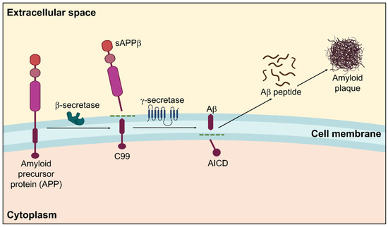
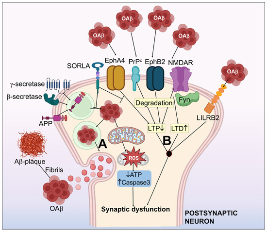
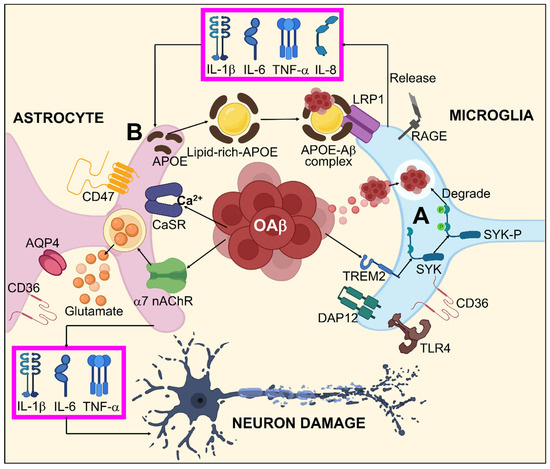
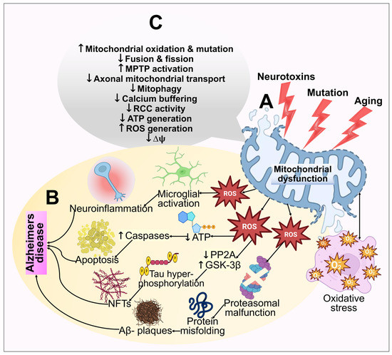
Alzheimer’s disease (AD) is the most prominent neurodegenerative disorder in the aging population. It is characterized by cognitive decline, gradual neurodegeneration, and the development of amyloid-β (Aβ)-plaques and neurofibrillary tangles, which constitute hyperphosphorylated tau. The early stages of neurodegeneration in AD include the loss of neurons, followed by synaptic impairment. Since the discovery of AD, substantial factual research has surfaced that outlines the disease’s causes, molecular mechanisms, and prospective therapeutics, but a successful cure for the disease has not yet been discovered. This may be attributed to the complicated pathogenesis of AD, the absence of a well-defined molecular mechanism, and the constrained diagnostic resources and treatment options. 

Alzheimer’s disease molecular mechanism risk factors

1. Amyloid β Hypothesis

For over three decades, the amyloid hypothesis, proposed by G. Higgins, J. Hardi [1], and D. Selkoe [2], has been the dominant and most widely accepted mechanistic theory of how AD develops. According to their theory, the accumulation of oligomeric Aβ (oAβ)-peptides are responsible for the pathophysiology causing downstream events such as neuroinflammation, the formation of neurofibrillary tangles (NFT), and vascular injury, encouraging dementia and cognitive deficits [3]. Their original theory primarily focused on the frequent occurrence of AD in Down’s syndrome patients due to the generation of significant amounts of Aβ-peptides since the amyloid precursor protein (APP) gene is positioned on the three 21 chromosomes [4].
Amyloidosis is a clinicopathological phenomenon where amyloid builds up in the body’s tissues and cells, generating amyloid plaques for various intricate reasons that eventually cause organ malfunction. It may run in the family or be acquired [5]. A systematic representation of amyloidosis in AD is displayed in Figure 1.
Figure 1. Amyloidogenic pathway in the pathogenesis of Alzheimer’s disease showing the formation of amyloid plaques. Within the membrane, β-secretase cleaves APP in the first instance, followed by γ-secretase. The extracellular amyloid-β that is released by the proteolytic breakdown of APP via the amyloidogenic pathway is susceptible to self-aggregation, resulting in the development of cytotoxic oligomers and insoluble Aβ fibrils/plaques.
Amyloidosis is categorized into two types based on where amyloid fibers are deposited: localized amyloidosis, which affects a particular tissue in a specific place, and systemic amyloidosis, which affects the entire body [6][7]. These amyloid proteins make up amyloid plaques [8]. The primary element that significantly contributes to the pathophysiology of AD and is often regarded as the principal reason for AD development is the amyloid β peptide [5][9].
APP is produced by blood arteries, blood cells, neurons, and astrocytes in confined numbers and is a more significant precursor molecule than Aβ [10]. Multiple physiological functions for APP have been postulated thus far. APP is crucial for brain growth, memory, and neuroplasticity [11]. In addition to being able to safeguard neurons, it also controls intercellular relations, managing neuronal development and neuroplasticity [12].
Extracellular domains of the APP control cellular adhesion to support neural circuits. APP homodimers allow Aβ to activate calcium channels, which further modulate neural signaling and neurotransmitter discharge [13][14]. More precisely, K+-Cl cotransporter 2 (KCC2) is stabilized on cellular membranes due to direct protein–protein interactions between APP and KCC2, which can modulate hippocampal γ-aminobutyric acid inhibition (GABAergic inhibition). APP reduction causes KCC2 to degrade more quickly through ubiquitination and tyrosine phosphorylation, which impairs γ-aminobutyric acid type A (GABAA) receptor-regulated inhibition and GABA reversal potential depolarization [10]. Soluble amyloid precursor protein (sAPP) cleavage molecules including sAPPα and sAPPβ are responsible for several facets of APP functionality, wherein the role of sAPPα has been thoroughly described. sAPPα has been demonstrated to be preventative against Aβ-induced toxicity and serves a significant role in neuroplasticity/survival [12].
Moreover, the central nervous system’s early embryonic processes and neuronal stem cell growth can be mediated by sAPPα [15]. In response to specific neuroprotective agents, it has been proposed that sAPPα could suppress cyclin-dependent kinase 5 (CDK5) activation induced by excitotoxicity and take part in diverse excitoprotective mechanisms [16]. Notably, in APP-deficient mice, sAPPα expression alone is sufficient to reverse defects, indicating that sAPPα might facilitate most APP functioning. It has been revealed that APP mutations accelerate the production of Aβ, which results in senile plaques and peripheral neuron degenerative alterations [17].
Depending on their cleavage products, APP processing can be classified as either amyloidogenic or non-amyloidogenic. In APP processing, the major proteolytic enzymes are α-, β-, and γ- secretase. The principal β-secretase in the brain is beta-site APP-cleaving enzyme 1 (BACE1) and γ-secretase. The full-length APP is broken down by α-secretase, which releases the sAPPα ectodomain beyond the cellular membrane while leaving a C-terminal APP fraction of 83 amino acids inside the plasma membrane. This process is known as the non-amyloidogenic pathway [17]. The consecutive APP proteolytic cleavage via β- and γ-secretase complex constitutes the amyloidogenic pathway. When APP is broken down by γ-secretase, amyloid peptides with varying chain lengths such as Aβ-37/38/39/40/42/43 can be produced [18][19]. The two main Aβ species in the brain are Aβ42 and Aβ40. Aβ42 has a greater potency for aggregating due to the hydrophobicity of its two terminal residues, albeit soluble Aβ40 is significantly more abundant than Aβ42. Hence, Aβ42 is primarily responsible for constituting the majority of amyloid plaques that are neurotoxic [20]. Correspondingly, Aβ42 is considered a principal performer in commencing plaque building in the pathophysiology of AD [21]. Moreover, it has been established that AD may be distinguished from other dementias by employing the Aβ42/38 ratio and levels of Aβ38/42 in the cerebral spinal fluid (CSF) [22][23][24]. By boosting Aβ synthesis and decreasing the Aβ40/Aβ42 ratio, dysregulated APP function likely aids in the etiology of AD [25]. Aβ protein is a 40–42 amino acid short peptide of 4.2 kDa [17]. Misfolded proteins with a stable conformation are called amyloid proteins. Additionally, an abnormal build-up of Aβ causes neurotoxicity [5]. Monomeric Aβ segments are soluble molecules that coalesce to produce insoluble oligomers, which then develop into neurologic plaques. The transfer of Aβ by the receptor for density lipoprotein receptor-related protein-1 (LRP1) and the receptor for advanced glycation end products (RAGE) is among the strategies the body uses to remove Aβ from the brain [26][27][28]. Clinical research has demonstrated that an imbalance between Aβ production and elimination causes abnormal metabolism, which in turn, causes extracellular protein build-up and misfolding, resulting in the establishment of amyloid plaques [29][30].
Compared to other cell types, nerve cells generate more Aβ, which is crucial for intercellular signaling and other typical physiological processes of the CNS [31]. Individuals with traumatic brain injury and PD accumulate Aβ, indicating a link between amyloid and neurodegenerative disorders [32]. Chronic stress causes the body to respond by ramping up the production of neural proteins, which results in the build-up of by-products such as phosphate. The phosphorylation of APP can be facilitated by high phosphate concentrations in the protein production area. Additionally, β-secretase engages in the subsequent phosphorylation of APP processing, which causes Aβ deposition. However, there are several circumstances where the bodily function that regulates the concentration of Aβ can become uncontrolled. For instance, natural Aβ can stimulate the production of extra APP, which is then phosphorylated and processed to become amyloid, increasing the concentration of Aβ. In peripheral neurons, Aβ elevated concentrations might also stimulate the synthesis of APP and amyloidosis. In the brain, a portion of Aβ misfolds and accumulates, generating hydrophobic exogenous oligomers that acquire the shape of plaques and fibers that harm synapses and neurons [33]. Substantial evidence such as the existence of APP mutations in familial AD patients points toward Aβ as a principal factor in disease development.
In the initial course of AD, patients have a synaptic malfunction; as the disease advances, synapses are lost. Synaptic loss is a primary pathogenic characteristic of AD and a reliable predictor of cognitive deterioration [34]. The degree of senile plaque development in the sick brain, however, does not always correlate with the severity of dementia that people with AD suffer. One viable argument is that soluble Aβ may indirectly contribute to AD pathophysiology by encouraging the development of neurofibrillary tangles [35]. The model for Aβ hypothesis in AD is displayed in Figure 2.
Figure 2. Systematic illustration of the amyloid-β hypothesis in Alzheimer’s disease. (A) APP processing to form Aβ, which simultaneously assembles as aggregates of the Aβ oligomer (oAβ) and form amyloid plaques. (B) Aβ-associated synaptic dysfunction by the impairment of LTP and LTD. Aβ receptors including NMDAR, PrPc, EphA4, EphB2 & LiLRB2 have been shown to induce synaptotoxicity by interaction with Aβ. EphA4-associated synaptic and cognitive malfunction may be inhibited by SORLA. Fyn kinase functions as an essential control mechanism for NMDAR related oAβ neurotoxicity. oAβ halts the normal mitochondrial function, which results in activated capsase-3, upregulated ROS, and decrease in ATP. This further worsens the synaptic dysfunction.

2. Tau Pathology toward Neurofibrillary Tangles

Tau is a cytosol protein mostly available in axons and is a neuronal microtubule-associated protein. The microtubule-associated protein tau (MAPT) gene possessing 16 exons is localized on chromosome 17, which encodes human tau [36][37][38]. Tau helps microtubules and related proteins assemble and remain stable [39]. By engaging on microtubules using its extensively conserved microtubule-binding repeat domains, tau also aids in regulating microtubule processes such as axon transport and neurite growth [40][41]. Microtubules oscillate between a stable phase and dynamical instability; efficient neural transmission and survival depend on optimal balancing among these two states [42]. This system relies on tau phosphorylation, which reduces tau’s capability for microtubules while maintaining their dynamic character to support the optimal neuron activity [43][44]. However, aberrant or excessive Tau phosphorylation reduces the integrity of microtubules, resulting in an elevation in neurite branching, a deduction in axonal transit, and synapse retraction, as shown in Figure 3 [42][45]. Neurodegenerative conditions like AD, ALS, and PD are featured by hyped phosphorylation of tau and the consequent micro tubular instability [46][47]. In the AD brain, hyperphosphorylated Tau could develop into oligomers, filaments of paired helical, and eventually neurofibrillary tangles [41][48][49][50]. Tau is more challenging for phosphatases to dephosphorylate once it has aggregated [51]. Oligomeric Tau could take effect as a “seed” and encourage additional Tau proteases in neighboring neurons to condense into fibrils [50][52][53]. It has been discovered that tau oligomers are the primary cause of axonal transport deficiencies in neurons, which can result in neural death [53][54]. Immunohistochemical staining was developed by researchers E. Braak and H. Braak to stage neuro-pathological Tau aggregation in the brain, and it has since been improved to make it easier for pathologists to determine the level of Tau deposition and whether AD needs to be identified in a post-mortem of the patient [55][56].
Figure 3. Systematic illustration for Aβ-mediated glial response in AD. (A) oAβ might activate microglia by binding to different Aβ receptors including TLR4, RAGE, LRP1, CD36, and specifically to TREM2, which stimulates the SYK pathway via DAP12 inducing Aβ degeneration. (B) Aβ dependent astrocyte dysfunction by enhanced interactions between Aβ/APOPE and LRP1 results in astrocyte activation by releasing TNF-α, IL-1β, IL-6, and IL-8. Furthermore, oAβ is also capable of direct astrocyte activation by AQP4, CD36, α7-nAchR, CD47, and CaSR. This astrocyte activation leads to neuronal damage through TNF-α, IL-1β, IL-6, and excitotoxication/irregulated homeostasis of glutamate.
The presence of Aβ, neural inflammation, enzymes, and oxidative stress that modulate phosphatases and kinases can all impact the phosphorylated tau-protein conformation [57][58]. Microtubule affinity-regulating kinase (MARK), CDK5, and glycogen synthase kinase 3 (GSK3) are the three enzymes that likely have a major impact [59][60][61]. The formation of Tau into neurofibrillary tangles is in close alliance with the neurodegeneration (i.e., neural demise) and brain atrophy seen in AD [55][62][63]. The hippocampus and entorhinal cortex are the first areas of the AD brain to be impacted, accompanied by areas of the temporal lobe and neocortex. During this period, patients may experience moderate cognitive impairment (MCI) [64]. The degeneration then progresses to the frontal portions of the cortex and occipital lobe, resulting in delayed personality alterations and trouble accomplishing daily tasks [65][66]. These frontal portions shrink while the ventricles are expanded. The primary pathogenic driver of ventricular enlargement and cortical atrophy is believed to be neuronal loss [67].

3. Mitochondrial Dysfunction and Reactive Oxygen Species (ROS) Generation

According to multiple evidence-based studies, the pathophysiology of AD may be influenced by mitochondrial dysfunction [68]. As a result of Aβ aggregation in the mitochondria of AD brains, disrupted mitochondrial conformation, reduced adenosine triphosphate (ATP) release and respiratory function, increased mitochondria-mediated oxidative stress, and poor mitochondria dynamics occur. The brain mitochondria of the patients suffering from AD and mouse models have both been reported to possess Aβ, which is responsible for neurodegeneration [69]. Irregularities in mitochondrial structure and functioning are associated with elevated mitochondrial Aβ. For example, reduced energy consumption related to mitochondria was noted in brain areas connected to amyloid plaques. Aβ also causes anomalies in mitochondrial function; due to decreased energy generation, abnormal alterations are also observed in the mitochondrial dynamics. Additionally, proteins linked to enhanced mitochondria fission and reduced fusion of mitochondria are amplified by Aβ exposure [70]. Unfortunately, it is still uncertain how mitochondrial dysfunction contributes to AD.
The oxygen consumption and metabolic rate of neurons are exceedingly high. As a result, to generate energy by oxidative phosphorylation, neurons depend on the numerous mitochondria in brain regions. ROS are primarily generated in mitochondria as by-products of oxidative phosphorylation, and routine homeostatic action in mitochondria frequently blocks excessive ROS formation. Furthermore, there are indications that oxidative assaults are critical in AD pathophysiology [71]. The idea that oxidative stress might be what causes AD pathogenesis triggered by Aβ is supported by the finding that oxidative stress emerges earlier in AD [72]. Aβ-peptides can elicit mitochondrial ROS generation, which releases cytochrome c and an apoptosis-inducing factor, causing malfunction of mitochondria, apoptosis, and the death of neurons [70][73]. In AD, appoptosin overexpression can cause the intrinsic caspase pathway to be activated. Prominently, decreased appoptosin expression can guard against Aβ’s neurotoxic effects [74]. Amyloid-binding cyclophilin D alcohol dehydrogenase are the few other mitochondrial proteins that have depicted a role toward mitochondrial dysfunction [75][76][77]. Mitochondrial dysfunction and oxidative stress in the pathogenesis of AD are illustrated in Figure 4.
Figure 4. An illustration of the mitochondrial dysfunction and oxidative stress in the pathogenesis of AD. (A) Multiple age-related processes, mutations, and toxic fluctuations such as metal exposure can all adversely affect mitochondria. Mitochondrial dysfunction further results in bioenergetic deficits, calcium imbalance, and free radical production. This causes oxidative stress, which exacerbates mitochondrial impairment, synaptic malfunction, cognitive decline, and memory loss. (B) The cellular redox equilibrium is disrupted by ROS generation or a compromised antioxidant arrangement, which leads to an oxidative imbalance and excessive ROS output. By adversely influencing mitochondrial energy reserves, disrupting energy metabolic processes, and impairing dynamics and mitophagy, elevated ROS reduces mitochondrial ΔΨm and ATP production. Caspase activity also rises as a result of ROS, which additionally starts the apoptotic process. However, excessive ROS generation inhibits phosphatase 2A (PP2A), which leads to glycogen synthase kinase 3 (GSK3) activation. This results in tau hyperphosphorylation and NFT buildup. (C) The functions of the mitochondria that are extensively hampered in AD have been highlighted.

4. Nitrosative Stress

Nitrosative stress arises when various defensive mechanisms fail to balance the formation of reactive nitrogen species (RNS), which harms intracellular constituents. The main component of RNS is nitric oxide (NO), which acts as a signaling molecule to control synaptic plasticity, neurotransmission, and brain growth. Significant cognitive impairment aligned to synapse malfunction and glial activation has been linked to nitrosative stress [78][79]. Due to S-nitrosylation, nitric oxide released as a consequence of Aβ in AD has been identified to trigger fission in mitochondria, resulting in synaptic dysfunction and neuronal death [80]. Since higher S-nitrosothiol (SNO)-CDK5 amounts were found in post-mortem samples of the AD brain and not in healthy samples, it has been determined that enhanced SNO-CDK5 activity possesses a part in the progression of AD [81]. Assessing the role of S-nitrosylation in nitrosative stress-initiated AD pathogenesis is made more accessible by the massive neuronal atrophy in the AD brain, accompanied by elevated S-nitrosylation of the peptides and a considerable proportion of altered sites of cysteine [82].

5. Protein Oxidation and Lipid Peroxidation

Multiple evidence-based findings imply that ROS might be crucial in the emergence of neurodegeneration in AD. ROS and RNS build up over time, which results in protein oxidation and lipid peroxidation. The brain also possesses an elevated proportion of unsaturated lipids, a high metal ion concentration, an elevated oxygen usage rate, and a poor antioxidant system. Consequently, both protein and lipid oxidation are particularly dangerous for the brain.

6. DNA Damage

DNA is nucleic acid found in the mitochondria (mtDNA) and nuclear (nDNA) material of living cells. Multiple lines of research have indicated that ROS generated as an oxidative phosphorylation by-product and environmental subjection to chemicals and radiation target nuclear and mitochondrial DNA for the genotoxic attack. Furthermore, investigations have demonstrated that because mtDNA is situated near the oxidative phosphorylation cascade, it is more vulnerable to genotoxic attack than nDNA [83].
The Tau protein, in conjunction with its function in microtubule dynamics, is essential for protecting the genomic DNA of neurons from oxidative stress and ROS. The modification in the tau protein may impair nucleic acid protection mechanisms and make hippocampus neurons more vulnerable to ROS-induced oxidative stress to their nuclear RNA and genomic DNA in AD patients. It has been established that ROS can damage DNA strands and play a role in subsequent AD disease-causing processes [84]. Mullaart et al. [85] found a two-fold rise in DNA destruction in neurons of the AD brain. They theorized that this might be one of the early identifiable pathogenic events in the transition from the normal to the AD brain. 8-Hydroxyguanine (8-OHG) is the most protruding DNA marker in most biological samples including blood cells, urine, and brain tissues [86]. 8-Oxoguanine-DNA glycosylase (OGG1) is a bifunctional enzyme that has apurinic/apyrimidinic lyase and DNA glycosylase properties. Evidence from research has shown two single-nucleotide polymorphisms in OGG1 caused by the substituted amino acid A53T and A288V. These polymorphic OGG1 proteins with the A53T and A288V mutations were found in 2007 in late-stage brain tissue AD patients but not in the controls [87].

7. Glial Cells in AD

Another characteristic of AD is neuroinflammation, which appears as gliosis and is marked by the activation and proliferation of the two main glial cell types in the brain, astrocytes and microglia. Numerous recently discovered AD risk genes such as triggering receptors expressed on myeloid cells-2 (TREM2) are only expressed in glial cells or are greatly concentrated in them. As a result, current research has placed a lot of emphasis on the probable impact that the glia may serve in the pathogenesis of AD. Pathogenic tau and Aβ species can bring neuroinflammation and gliosis. Glial cells and inflammation can control the development of Aβ and tau in a reciprocal manner. In general, it is thought that inappropriate microglial and astrocyte activation is a harmful event during the initiation of AD, and that blocking the formation of pro-inflammatory cytokines and the malignant glial responses to pathogenic Aβ and tau may prevent AD pathogenesis.

8. Proteasomal Dysfunction

By eliminating proteins that are inappropriately folded or clumped together, the ubiquitin-proteasome pathway (UPP) helps to maintain cellular integrity. When this system for removing undesirable protein complexes is disrupted, toxic and improperly folded proteins gather in brain cells, which is thought to be a pathogenic characteristic of AD. This route is crucial for the efficient elimination of aberrant protein garbage, which is essential for the viability and stability of neurons [88]. In order to continually remove defective proteins from neurons and block the aggregation of inappropriate proteins, the optimal functioning of UPP is of utmost importance [89].
Recent research has exhibited that the intracellular deposit of phosphorylated tau and Aβ protein clumps in AD patients directly impairs UPP. Brain tissue from patients with early AD had much less proteosome activity. The proteasome activity is reduced by 56% in AD patients due to the intraneural accumulation of paired helical filaments, which inhibits the proteasome. As a result, in the AD brain, the failure of UPP to remove phosphorylated tau and paired helical filament ultimately causes neuronal death [90]. Current findings have shown a strong correlation between ubiquitinated synaptic tau and hyperphosphorylation. This stable oligomerization of ubiquitinated synaptic tau results in elevated proteasome elements, proposing that a failure of the ubiquitin-proteasome system causes AD [91][92]. Although extant research has asserted a strong correlation between the build-up of hyperphosphorylated tau and malfunctioning UPP, none of these studies has explicitly stated whether the hyperphosphorylated tau is to blame for the UPP machinery’s impairment or the other way around [93]. As a result, further research employing cellular models are required to pinpoint the pathogenic event that causes aberrant neuron activity in AD patients.

9. Neuroinflammation

Infection, trauma, or toxic materials can cause a complicated series of inflammatory reactions in the brain system known as neuroinflammation. Microglia and astrocytes are important cells that indulge in inflammatory processes in the CNS and neuronal cells. By showing the existence of reactive microglia in the substantia nigra portion of post-mortem brain tissue from PD patients, Mc Geer et al. [94] made the initial discovery. The Aβ and tau tangles are surrounded by persistent microglial activation, which causes the loss of the homeostatic role of glial cells, developing a proinflammatory trait and exacerbating neurotoxicity. In the case of neuroinflammation, serum and brain specimens from AD patients include inflammatory mediators such TNF-α [95], IL-6, IL-β [96], and cyclooxygenase-2 (COX-2) [97].

References

  1. Hardy, J.; Duff, K.; Hardy, K.G.; Perez-Tur, J.; Hutton, M. Genetic Dissection of Alzheimer’s Disease and Related Dementias: Amyloid and Its Relationship to Tau. Nat. Neurosci. 1998, 1, 355–358.
  2. Selkoe, D.J.; Hardy, J. The Amyloid Hypothesis of Alzheimer’s Disease at 25 Years. EMBO Mol. Med. 2016, 8, 595–608.
  3. Hampel, H.; Hardy, J.; Blennow, K.; Chen, C.; Perry, G.; Kim, S.H.; Villemagne, V.L.; Aisen, P.; Vendruscolo, M.; Iwatsubo, T.; et al. The Amyloid- β Pathway in Alzheimer’s Disease. Mol. Pscychiatry 2021, 26, 5481–5503.
  4. Hardy, J.A.; Higgins, G.A. Alzheimer’s Disease: The Amyloid Cascade Hypothesis. Science 1992, 256, 184–185.
  5. Westermark, P.; Benson, M.D.; Buxbaum, J.N.; Cohen, A.S.; Frangione, B.; Ikeda, S.I.; Masters, C.L.; Merlini, G.; Saraiva, M.J.; Sipe, J.D. A Primer of Amyloid Nomenclature. Amyloid 2007, 14, 179–183.
  6. Glenner, G.G.; Caine, W. Wong Alzheimer’s Disease: Initial Report of the Purification and Characterization of a Novel Cerebrovascular Amyloid Protein. Biochem. Biophys. Res. Commun. 2012, 120, 885–890.
  7. Prasansuklab, A.; Tewin, T. Amyloidosis in Alzheimer’s Disease: The Toxicity of Amyloid Beta (Aβ), Mechanisms of Its Accumulation and Implications of Medicinal Plants for Therapy. Evid. Based Complement. Altern. Med. 2013, 2013, 413808.
  8. Merlini, G.; Bellotti, V. Molecular Mechanisms of Amyloidosis. N. Engl. J. Med. 2003, 349, 583–596.
  9. O’Brien, R.J.; Wong, P.C. Amyloid Precursor Protein Processing and Alzheimer’s Disease. Annu. Rev. Neurosci. 2011, 34, 185–204.
  10. Blennow, K.; de Leon, M.J.; Zetterberg, H. Alzheimer’s Disease. Lancet 2006, 368, 387–403.
  11. Nalivaeva, N.N.; Turner, A.J. The Amyloid Precursor Protein: A Biochemical Enigma in Brain Development, Function and Disease. FEBS Lett. 2013, 587, 2046–2054.
  12. Sadleir, K.R.; Kandalepas, P.C.; Buggia-Prévot, V.; Nicholson, D.A.; Thinakaran, G.; Vassar, R. Presynaptic Dystrophic Neurites Surrounding Amyloid Plaques Are Sites of Microtubule Disruption, BACE1 Elevation, and Increased Aβ Generation in Alzheimer’s Disease. Acta Neuropathol. 2016, 132, 235–256.
  13. Rice, H.C.; De Malmazet, D.; Schreurs, A.; Frere, S.; Van Molle, I.; Volkov, A.N.; Creemers, E.; Vertkin, I.; Nys, J.; Ranaivoson, F.M.; et al. Secreted Amyloid-b Precursor Protein Functions as a GABA B R1a Ligand to Modulate Synaptic Transmission. Science 2019, 363, eaao4827.
  14. Thinakaran, G.; Koo, E.H. Amyloid Precursor Protein Trafficking, Processing, and Function. J. Biol. Chem. 2008, 283, 29615–29619.
  15. Storey, E.; Cappai, R. The Amyloid Precursor Protein of Alzheimer’s Disease and the Aβ Peptide. Neuropathol. Appl. Neurobiol. 1999, 25, 81–97.
  16. Rogaev, E.I. Genetic Factors and a Polygenic Model of Alzheimer’s Disease. Genetika 1999, 35, 1558–1571.
  17. Devkota, S.; Williams, T.D.; Wolfe, M.S. Familial Alzheimer s Disease Mutations in Amyloid Protein Precursor Alter Proteolysis by γ-Secretase to Increase Amyloid β-Peptides of ≥45 Residues. J. Biol. Chem. 2021, 296, 100281.
  18. Bibl, M.; Mollenhauer, B.; Esselmann, H.; Lewczuk, P.; Klafki, H.W.; Sparbier, K.; Smirnov, A.; Cepek, L.; Trenkwalder, C.; Rüther, E.; et al. CSF Amyloid-β-Peptides in Alzheimer’s Disease, Dementia with Lewy Bodies and Parkinson’s Disease Dementia. Brain 2006, 129, 1177–1187.
  19. Bibl, M.; Mollenhauer, B.; Lewczuk, P.; Esselmann, H.; Wolf, S.; Trenkwalder, C.; Otto, M.; Stiens, G.; Rüther, E.; Kornhuber, J.; et al. Validation of Amyloid-β Peptides in CSF Diagnosis of Neurodegenerative Dementias. Mol. Psychiatry 2007, 12, 671–680.
  20. Iwatsubo, T.; Odaka, A.; Suzuki, N.; Mizusawa, H.; Nukina, N.; Ihara, Y. Visualization of Aβ42(43) and Aβ40 in Senile Plaques with End-Specific Aβ Monoclonals: Evidence That an Initially Deposited Species Is Aβ42(43). Neuron 1994, 13, 45–53.
  21. Jan, A.; Gokce, O.; Luthi-Carter, R.; Lashuel, H.A. The Ratio of Monomeric to Aggregated Forms of Aβ40 and Aβ42 Is an Important Determinant of Amyloid-β Aggregation, Fibrillogenesis, and Toxicity. J. Biol. Chem. 2008, 283, 28176–28189.
  22. Welge, V.; Fiege, O.; Lewczuk, P.; Mollenhauer, B.; Esselmann, H.; Klafki, H.W.; Wolf, S.; Trenkwalder, C.; Otto, M.; Kornhuber, J.; et al. Combined CSF Tau, p-Tau181 and Amyloid-β 38/40/42 for Diagnosing Alzheimer’s Disease. J. Neural Transm. 2009, 116, 203–212.
  23. Mulugeta, E.; Londos, E.; Ballard, C.; Alves, G.; Zetterberg, H.; Blennow, K.; Skogseth, R.; Minthon, L.; Aarsland, D. CSF Amyloid Β38 as a Novel Diagnostic Marker for Dementia with Lewy Bodies. J. Neurol. Neurosurg. Psychiatry 2011, 82, 160–164.
  24. Tang, W.; Huang, Q.; Wang, Y.; Wang, Z.Y.; Yao, Y.Y. Assessment of CSF Aβ42 as an Aid to Discriminating Alzheimer’s Disease from Other Dementias and Mild Cognitive Impairment: A Meta-Analysis of 50 Studies. J. Neurol. Sci. 2014, 345, 26–36.
  25. Jonsson, T.; Atwal, J.K.; Steinberg, S.; Snaedal, J.; Jonsson, P.V.; Bjornsson, S.; Stefansson, H.; Sulem, P.; Gudbjartsson, D.; Maloney, J.; et al. A Mutation in APP Protects against Alzheimer‘s Disease and Age-Related Cognitive Decline. Nature 2012, 488, 96–99.
  26. Selkoe, D.J. Clearing the Brain’s Amyloid Cobwebs. Neuron 2001, 32, 177–180.
  27. Deane, R.; Yan, S.D.; Submamaryan, R.K.; LaRue, B.; Jovanovic, S.; Hogg, E.; Welch, D.; Manness, L.; Lin, C.; Yu, J.; et al. RAGE Mediates Amyloid-β Peptide Transport across the Blood-Brain Barrier and Accumulation in Brain. Nat. Med. 2003, 9, 907–913.
  28. Deane, R.; Bell, R.D.; Sagare, A.; Zlokovic, B.V. Clearance of Amyloid-? Peptide Across the Blood-Brain Barrier: Implication for Therapies in Alzheimer’s Disease. CNS Neurlog. Disord. 2009, 8, 16–30.
  29. Jack, C.R.; Bennett, D.A.; Blennow, K.; Carrillo, M.C.; Dunn, B.; Haeberlein, S.B.; Holtzman, D.M.; Jagust, W.; Jessen, F.; Karlawish, J.; et al. NIA-AA Research Framework: Toward a Biological Definition of Alzheimer’s Disease. Alzheimer’s Dement. 2018, 14, 535–562.
  30. Levin, O.S.; Vasenina, E.E. Twenty-Five Years of the Amyloid Hypothesis of Alzheimer’s Disease: Advances, Failures and New Perspectives. Zhurnal Nevrol. i Psihiatr. Im. S.S. Korsakova 2016, 116, 3–9.
  31. Fukumoto, H.; Tomita, T.; Matsunaga, H.; Ishibashi, Y.; Saido, T.; Iwatsubo, T. Primary Cultures of Neuronal and Non-Neuronal Rat Brain Cells Secrete Similar Proportions of Amyloid β Peptides Ending at Aβ40 and Aβ42: Neuroreport. Neuroreport 1999, 10, 2965–2969.
  32. Tsitsopoulos, P.P.; Marklund, N. Amyloid-ß Peptides and Tau Protein as Biomarkers in Cerebrospinal and Interstitial Fluid Following Traumatic Brain Injury: A Review of Experimental and Clinical Studies. Front. Neurol. 2013, 4, 1–17.
  33. Maltsev, A.V.; Santockyte, R.; Bystryak, S.; Galzitskaya, O.V. Activation of Neuronal Defense Mechanisms in Response to Pathogenic Factors Triggering Induction of Amyloidosis in Alzheimer’s Disease. J. Alzheimer’s Dis. 2014, 40, 19–32.
  34. Palop, J.J.; Mucke, L. Amyloid-Β-Induced Neuronal Dysfunction in Alzheimer’s Disease: From Synapses toward Neural Networks. Nat. Neurosci. 2010, 13, 812–818.
  35. Greenberg, D.A.; Aminoff, M.J.; Roger, P.S. Clinical Neurology, 5th ed.; McGraw Hill: New York, NY, USA, 2002; Volume 139, pp. 1–236.
  36. Neve, R.L.; Harris, P.; Kosik, K.S.; Kurnit, D.M.; Donlon, T.A. Identification of CDNA Clones for the Human Microtubule-Associated Protein Tau and Chromosomal Localization of the Genes for Tau and Microtubule-Associated Protein 2. Mol. Brain Res. 1986, 1, 271–280.
  37. Goedert, M.; Spillantini, M.G.; Jakes, R.; Rutherford, D.; Crowther, R.A. Multiple Isoforms of Human Microtubule-Associated Protein Tau: Sequences and Localization in Neurofibrillary Tangles of Alzheimer’s Disease. Neuron 1989, 3, 519–526.
  38. Goedert, M.; Spillantini, M.G.; Potier, M.C.; Ulrich, J.; Crowther, R.A. Cloning and Sequencing of the CDNA Encoding an Isoform of Microtubule-Associated Protein Tau Containing Four Tandem Repeats: Differential Expression of Tau Protein MRNAs in Human Brain. EMBO J. 1989, 8, 393–399.
  39. Barbier, P.; Zejneli, O.; Martinho, M.; Lasorsa, A.; Belle, V.; Smet-Nocca, C.; Tsvetkov, P.O.; Devred, F.; Landrieu, I. Role of Tau as a Microtubule-Associated Protein: Structural and Functional Aspects. Front. Aging Neurosci. 2019, 11, 1–14.
  40. Johnson, G.V.W.; Stoothoff, W.H. Tau Phosphorylation in Neuronal Cell Function and Dysfunction. J. Cell. Sci. 2004, 117, 5721–5729.
  41. Goedert, M.; Wischik, C.M.; Crowther, R.A.; Walker, J.E.; Klug, A. Cloning and Sequencing of the CDNA Encoding a Core Protein of the Paired Helical Filament of Alzheimer Disease: Identification as the Microtubule-Associated Protein Tau. Proc. Nati. Acad. Sci. USA 1988, 85, 4051–4055.
  42. Dubey, J.; Ratnakaran, N.; Koushika, S.P. Neurodegeneration and Microtubule Dynamics: Death by a Thousand Cuts. Front. Cell. Neurosci. 2015, 9, 343.
  43. Lindwall, G.; Cole, R.D. Phosphorylation Affects the Ability of Tau Protein to Promote Microtubule Assembly. J. Biol. Chem. 1984, 259, 5301–5305.
  44. Mandelkow, E.M.; Biernat, J.; Drewes, G.; Gustke, N.; Trinczek, B.; Mandelkow, E. Tau Domains, Phosphorylation, and Interactions with Microtubules. Neurobiol. Aging 1995, 16, 355–362.
  45. Jameson, L.; Frey, T.; Zeeberg, B.; Dalldorf, F.; Caplow, M. Inhibition of Microtubule Assembly by Phosphorylation of Microtubule-Associated Proteins. Biochemistry 1980, 19, 2472–2479.
  46. Iqbal, K.; Zaidi, T.; Wen, G.Y.; Grundke-Iqbal, I.; Merz, P.A.; Shaikh, S.S.; Wisniewski, H.M.; Alafuzoff, I.; Winblad, B. Defective Brain Microtubule Assembly in Alzheimer’S Disease. Lancet 1986, 328, 421–426.
  47. Alonso, A.C.; Grundke-Iqbal, I.; Iqbal, K. Alzheimer’s Disease Hyperphosphorylated Tau Sequesters Normal Tau into Tangles of Filaments and Disassembles Microtubules. Nat. Med. 1996, 2, 783–787.
  48. Bancher, C.; Brunner, C.; Lassmann, H.; Budka, H.; Jellinger, K.; Wiche, G.; Seitelberger, F.; Grundke-Iqbal, I.; Iqbal, K.; Wisniewski, H.M. Accumulation of Abnormally Phosphorylated τ Precedes the Formation of Neurofibrillary Tangles in Alzheimer’s Disease. Brain Res. 1989, 477, 90–99.
  49. Jouanne, M.; Rault, S.; Voisin-Chiret, A.S. Tau Protein Aggregation in Alzheimer’s Disease: An Attractive Target for the Development of Novel Therapeutic Agents. Eur. J. Med. Chem. 2017, 139, 153–167.
  50. Hill, E.; Wall, M.J.; Moffat, K.G.; Karikari, T.K. Understanding the Pathophysiological Actions of Tau Oligomers: A Critical Review of Current Electrophysiological Approaches. Front. Mol. Neurosci. 2020, 13, 155.
  51. Miao, J.; Shi, R.; Li, L.; Chen, F.; Zhou, Y.; Tung, Y.C.; Hu, W.; Gong, C.X.; Iqbal, K.; Liu, F. Pathological Tau From Alzheimer’s Brain Induces Site-Specific Hyperphosphorylation and SDS- and Reducing Agent-Resistant Aggregation of Tau in Vivo. Front. Aging Neurosci. 2019, 11, 34.
  52. Medina, M.; Avila, J. The Role of Extracellular Tau in the Spreading of Neurofibrillary Pathology. Front. Cell. Neurosci. 2014, 8, 113.
  53. Shafiei, S.S.; Guerrero-Muñoz, M.J.; Castillo-Carranza, D.L. Tau Oligomers: Cytotoxicity, Propagation, and Mitochondrial Damage. Front. Aging Neurosci. 2017, 9, 83.
  54. Ward, S.M.; Himmelstein, D.S.; Lancia, J.K.; Binder, L.I. Tau Oligomers and Tau Toxicity in Neurodegenerative Disease. Biochem. Soc. Trans. 2012, 40, 667–671.
  55. Braak, H.; Braak, E. Neuropathological Stageing of Alzheimer-Related Changes. Acta Neuropathol. 1991, 82, 239–259.
  56. Braak, H.; Alafuzoff, I.; Arzberger, T.; Kretzschmar, H.; Tredici, K. Staging of Alzheimer Disease-Associated Neurofibrillary Pathology Using Paraffin Sections and Immunocytochemistry. Acta Neuropathol. 2006, 112, 389–404.
  57. Molinuevo, J.L.; Ayton, S.; Batrla, R.; Bednar, M.M.; Bittner, T.; Cummings, J.; Fagan, A.M.; Hampel, H.; Mielke, M.M.; Mikulskis, A.; et al. Current State of Alzheimer’s Fluid Biomarkers. Acta Neuropathol. 2018, 136, 821–853.
  58. Lewczuk, P.; Lelental, N.; Lachmann, I.; Holzer, M.; Flach, K.; Brandner, S.; Engelborghs, S.; Teunissen, C.E.; Zetterberg, H.; Molinuevo, J.L.; et al. Non-Phosphorylated Tau as a Potential Biomarker of Alzheimer’s Disease: Analytical and Diagnostic Characterization. J. Alzheimer’s Dis. 2017, 55, 159–170.
  59. Hugon, J.; Mouton-Liger, F.; Cognat, E.; Dumurgier, J.; Paquet, C. Blood-Based Kinase Assessments in Alzheimer’s Disease. Front. Aging Neurosci. 2018, 10, 338.
  60. Castro-Alvarez, J.F.; Alejandro Uribe-Arias, S.; Kosik, K.S.; Cardona-Gómez, G.P. Long- and Short-Term CDK5 Knockdown Prevents Spatial Memory Dysfunction and Tau Pathology of Triple Transgenic Alzheimer’s Mice. Front. Aging Neurosci. 2014, 6, 243.
  61. Kimura, T.; Tsutsumi, K.; Taoka, M.; Saito, T.; Masuda-Suzukake, M.; Ishiguro, K.; Plattner, F.; Uchida, T.; Isobe, T.; Hasegawa, M.; et al. Isomerase Pin1 Stimulates Dephosphorylation of Tau Protein at Cyclin-Dependent Kinase (Cdk5)-Dependent Alzheimer Phosphorylation Sites. J. Biol. Chem. 2013, 288, 7968–7977.
  62. Gómez-Isla, T.; Hollister, R.; West, H.; Mui, S.; Growdon, J.H.; Petersen, R.C.; Parisi, J.E.; Hyman, B.T. Neuronal Loss Correlates with but Exceeds Newofibriiary Tangles in Alzheimer’s Disease. Ann. Neurol. 1997, 41, 17–24.
  63. Smith, A.D. Imaging the Progression of Alzheimer Pathology through the Brain. Proc. Natl. Acad. Sci. USA 2002, 99, 4135–4137.
  64. Scahill, R.I.; Schott, J.M.; Stevens, J.M.; Rossor, M.N.; Fox, N.C. Mapping the Evolution of Regional Atrophy in Alzheimer’s Disease: Unbiased Analysis of Fluid-Registered Serial MRI. Proc. Natl. Acad. Sci. USA 2002, 99, 4703–4707.
  65. Nestor, S.M.; Rupsingh, R.; Borrie, M.; Smith, M.; Accomazzi, V.; Wells, J.L.; Fogarty, J.; Bartha, R. Ventricular Enlargement as a Possible Measure of Alzheimer’s Disease Progression Validated Using the Alzheimer’s Disease Neuroimaging Initiative Database. Brain 2008, 131, 2443–2454.
  66. Apostolova, L.G.; Green, A.E.; Babakchanian, S.; Hwang, K.S.; Chou, Y.Y.; Toga, A.W.; Thompson, P.M. Hippocampal Atrophy and Ventricular Enlargement in Normal Aging, Mild Cognitive Impairment (MCI), and Alzheimer Disease. Alzheimer Dis. Assoc. Disord. 2012, 26, 17–27.
  67. Serrano-Pozo, A.; Frosch, M.P.; Masliah, E.; Hyman, B.T. Neuropathological Alterations in Alzheimer Disease. Cold Spring Harb. Perspect. Med. 2011, 1, a006189.
  68. Abolhassani, N.; Leon, J.; Sheng, Z.; Oka, S.; Hamasaki, H.; Iwaki, T.; Nakabeppu, Y. Molecular Pathophysiology of Impaired Glucose Metabolism, Mitochondrial Dysfunction, and Oxidative DNA Damage in Alzheimer’s Disease Brain. Mech. Ageing Dev. 2017, 161, 95–104.
  69. Wang, X.; Su, B.; Lee, H.G.; Li, X.; Perry, G.; Smith, M.A.; Zhu, X. Impaired Balance of Mitochondrial Fission and Fusion in Alzheimer’s Disease. J. Neurosci. 2009, 29, 9090–9103.
  70. Ebenezer, P.J.; Weidner, A.M.; Levine, H.; Markesbery, W.R.; Murphy, M.P.; Zhang, L.; Dasuri, K.; Fernandez-Kim, S.O.K.; Bruce-Keller, A.J.; Gavilán, E.; et al. Neuron Specific Toxicity of Oligomeric Amyloid-β: Role for JUN-Kinase and Oxidative Stress. J. Alzheimer’s Dis. 2010, 22, 839–848.
  71. Butterfield, D.A.; Bader Lange, M.L.; Sultana, R. Involvements of the Lipid Peroxidation Product, HNE, in the Pathogenesis and Progression of Alzheimer’s Disease. Biochim. Biophys. Acta 2010, 1801, 924–929.
  72. Wanga, X.; Wanga, W.; Lia, L.; Perryb, G.; Leea, H.; Zhu, X. Oxidative Stress and Mitochondrial Dysfunction in Alzheimer’s Disease. Biochim. Biophys. Acta 2014, 1842, 1240–1247.
  73. Moreira, P.I.; Carvalho, C.; Zhu, X.; Smith, M.A.; Perry, G. Mitochondrial Dysfunction Is a Trigger of Alzheimer’s Disease Pathophysiology. Biochim. Biophys. Acta 2009, 1802, 1–38.
  74. Zhang, H.; Zhang, Y.W.; Chen, Y.; Huang, X.; Zhou, F.; Wang, W.; Xian, B.; Zhang, X.; Masliah, E.; Chen, Q.; et al. Appoptosin Is a Novel Pro-Apoptotic Protein and Mediates Cell Death in Neurodegeneration. J. Neurosci. 2012, 32, 15565–15576.
  75. Lustbader, J.W.; Cirilli, M.; Lin, C.; Xu, H.W.; Takuma, K.; Wang, N.; Caspersen, C.; Chen, X.; Pollak, S.; Chaney, M.; et al. ABAD Directly Links Aβ to Mitochondrial Toxicity in Alzheimer’s Disease. Science 2004, 304, 448–452.
  76. Du, H.; Guo, L.; Fang, F.; Chen, D.; Sosunov, A.A.; McKhann, G.M.; Yan, Y.; Wang, C.; Zhang, H.; Molkentin, J.D.; et al. Cyclophilin D Deficiency Attenuates Mitochondrial and Neuronal Perturbation and Ameliorates Learning and Memory in Alzheimer’s Disease. Nat. Med. 2008, 14, 1097–1105.
  77. Du, H.; Guo, L.; Zhang, W.; Rydzewska, M.; Yan, S. Cyclophilin D Deficiency Improves Mitochondrial Function and Learning/Memory in Aging Alzheimer Disease Mouse Model. Neurobiol. Aging 2011, 32, 398–406.
  78. Okamoto, S.; Nakamura, T.; Cieplak, P.; Chan, S.F.; Liao, L.; Saleem, S.; Han, X.; Clemente, A.; Sances, S.; Brechtel, C.; et al. S-Nitrosylation—Mediated Redox Transcriptional Switch Modulates Neurogenesis and Neuronal Cell Death. Cell. Rep. 2014, 8, 217–228.
  79. Medeiros, R.; Prediger, R.D.S.; Passos, G.F.; Pandolfo, P.; Duarte, F.S.; Franco, J.L.; Dafre, A.L.; Di Giunta, G.; Figueiredo, C.P.; Takahashi, R.N.; et al. Connecting TNF-α Signaling Pathways to INOS Expression in a Mouse Model of Alzheimer’s Disease: Relevance for the Behavioral and Synaptic Deficits Induced by Amyloid β Protein. J. Neurosci. 2007, 27, 5394–5404.
  80. Cho, D.-H.; Nakamura, T.; Fang, J.; Cieplak, P.; Godzik, A.; Gu, Z.; Lipton, S.A. S-Nitrosylation of Drp1 Mediates β-Amyloid-Related Mitochondrial Fission and Neuronal Injury. Science 2009, 324, 102–105.
  81. Qu, J.; Nakamura, T.; Cao, G.; Holland, E.A.; McKercher, S.R.; Lipton, S.A. S-Nitrosylation Activates Cdk5 and Contributes to Synaptic Spine Loss Induced by β-Amyloid Peptide. Proc. Natl. Acad. Sci. USA 2011, 108, 14330–14335.
  82. Zahid, S.; Khan, R.; Oellerich, M.; Ahmed, N.; Asif, A.R. Differential S-Nitrosylation of Proteins in Alzheimer’s Disease. Neuroscience 2014, 256, 126–136.
  83. Yakes, F.M.; Van Houten, B. Mitochondrial DNA Damage Is More Extensive and Persists Longer than Nuclear DNA Damage in Human Cells Following Oxidative Stress. Proc. Natl. Acad. Sci. USA 1997, 94, 514–519.
  84. Mecocci, P.; MacGarvey, U.; Beal, M.F. Oxidative Damage to Mitochondrial DNA Is Increased in Alzheimer’s Disease. Ann. Neurol. 1994, 36, 747–751.
  85. Mullaart, E.; Boerrigter, M.E.T.I.; Ravid, R.; Swaab, D.F.; Vijg, J. Increased Levels of DNA Breaks in Cerebral Cortex of Alzheimer’s Disease Patients. Neurobiol. Aging 1990, 11, 169–173.
  86. Jacoba, K.D.; Hootena, N.N.; Tadokorob, T.; Lohania, A.; Barnesa, J.; Evans, M.K. Alzheimer’s Disease Associated Polymorphisms in Human OGG1 Alter Catalytic Activity and Sensitize Cells to DNA Damage. Free Radic. Biol. Med. 2013, 63, 115–125.
  87. Mao, G.; Pan, X.; Zhu, B.B.; Zhang, Y.; Yuan, F.; Huang, J.; Lovell, M.A.; Lee, M.P.; Markesbery, W.R.; Li, G.M.; et al. Identification and Characterization of OGG1 Mutations in Patients with Alzheimer’s Disease. Nucleic Acids Res. 2007, 35, 2759–2766.
  88. Gadhave, K.; Bolshette, N.; Ahire, A.; Pardeshi, R.; Thakur, K.; Trandafir, C.; Istrate, A.; Ahmed, S.; Lahkar, M.; Muresanu, D.F.; et al. The Ubiquitin Proteasomal System: A Potential Target for the Management of Alzheimer’s Disease. J. Cell. Mol. Med. 2016, 20, 1392–1407.
  89. Keck, S.; Nitsch, R.; Grune, T.; Ullrich, O. Proteasome Inhibition by Paired Helical Filament-tau in Brains of Patients with Alzheimer’s Disease. J. Neurochem. 2003, 85, 115–122.
  90. Salon, M.L.; Morelli, L.; Castanño, E.M.; Soto, E.F.; Pasquini, J.M. Defective Ubiquitination of Cerebral Proteins in Alzheimer’s Disease. J. Neurosci. Res. 2000, 62, 302–310.
  91. Tai, H.C.; Serrano-Pozo, A.; Hashimoto, T.; Frosch, M.P.; Spires-Jones, T.L.; Hyman, B.T. The Synaptic Accumulation of Hyperphosphorylated Tau Oligomers in Alzheimer Disease Is Associated with Dysfunction of the Ubiquitin-Proteasome System. Am. J. Pathol. 2012, 181, 1426–1435.
  92. Aloisi, F. Immune Function of Microglia. Glia 2001, 36, 165–179.
  93. Lam, Y.A.; Pickart, C.M.; Alban, A.; Landon, M.; Jamieson, C.; Ramage, R.; Mayer, R.J.; Layfield, R. Inhibition of the Ubiquitin-Proteasome System in Alzheimer’s Disease. Proc. Natl. Acad. Sci. USA 2000, 97, 9902–9906.
  94. McGeer, P.L.; Itagaki, S.; Boyes, B.E.; McGeer, E.G. Reactive Microglia Are Positive for HLA-DR in the: Substantia Nigra of Parkinson’s and Alzheimer’s Disease Brains. Neurology 1988, 38, 1285–1291.
  95. Holmes, C.; Cunningham, C.; Zotova, E.; Woolford, J.; Dean, C.; Kerr, S.; Culliford, D.; Perry, V.H. Systemic Inflammation and Disease Progression in Alzheimer Disease. Neurology 2009, 73, 768–774.
  96. Blasko, I.; Veerhuis, R.; Stampfer-Kountchev, M.; Saurwein-Teissl, M.; Eikelenboom, P.; Grubeck-Loebenstein, B. Costimulatory Effects of Interferon-β and Interleukin-1β or Tumor Necrosis Factor α on the Synthesis of Aβ1-40 and Aβ1-42 by Human Astrocytes. Neurobiol. Dis. 2000, 7, 682–689.
  97. Caggiano, A.O.; Kraig, R.P. Prostaglandin E Receptor Subtypes in Cultured Rat Microglia and Their Role in Reducing Lipopolysaccharide-Induced Interleukin-1β Production. J. Neurochem. 1999, 72, 565–575.
More
Information
Subjects: Neurosciences
Contributors MDPI registered users' name will be linked to their SciProfiles pages. To register with us, please refer to https://encyclopedia.pub/register : , , , , , , ,
View Times: 496
Entry Collection: Neurodegeneration
Revisions: 2 times (View History)
Update Date: 25 May 2023
1000/1000