Submitted Successfully!
To reward your contribution, here is a gift for you: A free trial for our video production service.
Thank you for your contribution! You can also upload a video entry or images related to this topic.
Version Summary Created by Modification Content Size Created at Operation
1 -- 2153 2023-05-16 08:51:53 |
2 Format correct Meta information modification 2153 2023-05-17 16:14:29 |

Video Upload Options

We provide professional Video Production Services to translate complex research into visually appealing presentations. Would you like to try it?

Confirm

Are you sure to Delete?
Cite
If you have any further questions, please contact Encyclopedia Editorial Office.
Haastrup, M.O.; Vikramdeo, K.S.; Singh, S.; Singh, A.P.; Dasgupta, S. Protein Sorting at the Outer Mitochondrial Membrane. Encyclopedia. Available online: https://encyclopedia.pub/entry/44347 (accessed on 14 December 2024).
Haastrup MO, Vikramdeo KS, Singh S, Singh AP, Dasgupta S. Protein Sorting at the Outer Mitochondrial Membrane. Encyclopedia. Available at: https://encyclopedia.pub/entry/44347. Accessed December 14, 2024.
Haastrup, Mary Oluwadamilola, Kunwar Somesh Vikramdeo, Seema Singh, Ajay Pratap Singh, Santanu Dasgupta. "Protein Sorting at the Outer Mitochondrial Membrane" Encyclopedia, https://encyclopedia.pub/entry/44347 (accessed December 14, 2024).
Haastrup, M.O., Vikramdeo, K.S., Singh, S., Singh, A.P., & Dasgupta, S. (2023, May 16). Protein Sorting at the Outer Mitochondrial Membrane. In Encyclopedia. https://encyclopedia.pub/entry/44347
Haastrup, Mary Oluwadamilola, et al. "Protein Sorting at the Outer Mitochondrial Membrane." Encyclopedia. Web. 16 May, 2023.
Protein Sorting at the Outer Mitochondrial Membrane
Edit

Mitochondria are double membrane-bound organelles consisting of an outer membrane, intermembrane space (IMS), inner membrane, and matrix that play critical functions in cells including metabolism, energy production, regulation of intrinsic apoptosis, and maintenance of calcium homeostasis. Mitochondria are fascinatingly equipped with their own genome and machinery for transcribing and translating 13 essential proteins of the oxidative phosphorylation system (OXPHOS). The rest of the proteins (99%) that function in mitochondria in the various pathways described above are nuclear-transcribed and synthesized as precursors in the cytosol. These proteins are imported into the mitochondria by the unique mitochondrial protein import system that consists of seven machineries. Proper functioning of the mitochondrial protein import system is crucial for optimal mitochondrial deliverables, as well as mitochondrial and cellular homeostasis.

mitochondria outer membrane proteins

1. Introduction

Mitochondria are organelles present in almost all eukaryotic cells, and their number per cell depends on their energy demand. Organs with high metabolic activity, for example, heart muscles, kidneys, and the brain, contain the largest number of mitochondria [1][2]. Mitochondria are believed to be the descendants of an ancient prokaryote that underwent an endosymbiotic event with early eukaryotes [3]. Apart from their role in energy production, mitochondria are involved in numerous metabolic processes, including the biosynthesis of amino acids, lipids, heme, and Fe-S clusters [4]. In addition, they also play crucial functions in programmed cell death and maintenance of calcium homeostasis [4][5]. A mitochondrion is a double membrane-bound structure consisting of an outer membrane, intermembrane space (IMS), inner membrane, and matrix [6]. The outer membrane is permeable to solutes up to approximately 5 kDa and characterized by the presence of various enzymes and channels, such as carnitine palmitoyltransferase I, acyl-CoA synthetase, voltage-dependent anion channel (VDAC), and mitochondrial apoptosis-induced channel (MAC) [7]. On the other hand, the inner membrane is impermeable except through specific transporters and contains the enzyme complexes responsible for oxidative phosphorylation [6][8]. The IMS consists of enzymes including caspases, adenylyl kinase, and cytochrome c. Similarly, the mitochondrial matrix consists of several enzymes that take part in metabolic processes such as the tricarboxylic acid cycle and β oxidation of fatty acids.

Each mitochondrion has its own genome, which is a 16.5 kb double-stranded, closed circular DNA present in the mitochondrial matrix. The mtDNA is strictly maternally inherited and packaged into nucleoids, which is done to ensure its proper distribution and propagation [9]. The mitochondrial genome consists of 37 genes that encode approximately 1% of the total mitochondrial proteins (13 OXPHOS proteins), 2 ribosomal RNAs (12S and 16S rRNA), and 22 transfer RNAs [6]. The remaining 99% of mitochondrial proteins (~1500) are encoded by the nuclear genome, synthesized in the cytosol, and imported into the mitochondria by the mitochondrial protein import system, which consists of seven machineries (Figure 1). The translocase of the outer mitochondrial membrane (TOMM) machinery appears to be the most important of these machineries, as it is the first to come in contact with majority of the nuclear-encoded mitochondrial proteins to allow their entry into the intermembrane space. Subsequently, these proteins make use of any of the other machineries to get to their final destination. The choice of the next machinery depends on the targeting signal and the destination of the protein.


Figure 1. The mitochondrial protein import system. The mitochondrial protein import system consists of seven machineries, including the translocase of the outer mitochondrial membrane (TOMM) machinery, mitochondrial import machinery (MIM), sorting and assembly machinery (SAM), mitochondrial intermembrane space import and assembly machinery (MIA), translocase of the inner mitochondrial membrane 23 (TIMM 23) machinery, translocase of the inner mitochondrial membrane 22 (TIMM 22) machinery, and a presequence-associated motor (PAM). The figure was created with Biorender.com.

The mitochondrial outer membrane possesses two types of integral membrane proteins, including β-barrel proteins that are integrated into the OMM by multiple transmembrane β strands, and α-helical proteins, which are anchored in the OMM by one or more hydrophobic α-helical segments [4,10].

2. Sorting of β-Barrel Proteins into the Outer Mitochondrial Membrane
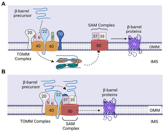
The presence of β-barrel proteins in the OMM is a key feature of the outer membrane of Gram-negative bacteria, reflecting the origin of mitochondria from prokaryotes [10]. Examples of these proteins are voltage-dependent anion channel (VDAC), TOMM40, and SAM50 [4]. The first machinery involved in the import of these β-barrel precursors is the TOMM machinery, which is composed of four receptors—TOMM20, TOMM70, and two molecules of TOMM22, as well as two molecules of the transmembrane channel—TOMM40, and three small subunits—TOMM5, TOMM6, and TOMM7—which are essential for complex stability and assembly [4][11]. However, it is unknown whether both TOMM40 channels take part in the translocation of incoming precursors across the OMM. In addition, how the two TOMM22 receptors cooperate during the recognition and binding of the incoming precursors remains to be elucidated.
After synthesis in the cytosol, the β-barrel precursors are recognized by the TOMM receptors and guided through the TOMM40 channel, through which they enter the IMS [10]. The identification of these β-barrel precursors by the TOMM receptors is directed by a targeting signal that consists of a β-hairpin element containing two adjacent β-strands, which are the two most C-terminal β strands of the precursor, and the connecting loop [4][12]. Although the exact sequence of the recognition of the targeting signal by the TOMM receptors is not completely understood, TOMM20, 70, and 22 have been shown to be crucial for the import of β barrel precursors [13][14]. Upon translocation through the TOMM40 channel, the precursors are bound to the small TIMM chaperones of the IMS, which exist as heterohexameric complexes, including the TIMM9–TIMM10 and TIMM8–TIMM13 complexes [4][10]. Of these, the TIMM9–TIMM10 complexes have been identified to be the main form involved in transfer of many hydrophobic proteins through the IMS [4][15]. These chaperones protect the β-barrel precursors from aggregation in the aqueous IMS and deliver the β-barrel precursors to the SAM complex (Figure 2A) [4][10], which consists of a membrane-integrated protein, SAM50, and two peripheral membrane proteins exposed to the cytosol, SAM35 and SAM37 [4]. The exact mechanisms governing the insertion of β-barrel proteins into the OMM are still not completely understood; however, it is believed that the SAM complex is responsible for the membrane insertion of these proteins (Figure 2A).
Figure 2. Import of β-barrel precursors into the outer mitochondrial membrane. (A) Upon translocation through the TOMM40 channel, the precursors bind to TIMM9–TIMM10 complex, which protect the β-barrel precursors from aggregation in the aqueous IMS and deliver the β-barrel precursors to the SAM complex. Subsequently, the precursors are folded in the SAM complex and laterally released into the lipid phase of the outer membrane. (B) SAM37 interacts with the cytosolic receptor domain of TOMM22, thereby linking the two complexes and leading to the formation of a TOMM–SAM supercomplex, which enables the binding of SAM35 to the β signal of the precursor, thereby allowing the direct transfer of the β-barrel precursors from TOMM to the SAM complex. Subsequently, the β-barrel precursors are inserted into the SAM50 channel, after which they are folded in the SAM complex and released laterally into the lipid phase of the outer membrane. IMS, intermembrane space. The figure was created with Biorender.com.
In contrast to the understanding that the β-barrel proteins are imported first into the IMS before being transferred to the SAM complex, Kutik et al. proposed that translocation of β-barrel precursors into the SAM complex is initiated by the binding of the last β-strand (β signal) to SAM35, a SAM subunit located on the cytosolic surface of mitochondria [16][17]. This signal binding then induces a conformational change that leads to opening of the SAM50 channel; thus, several β strands can be inserted into a hydrophilic, proteinaceous membrane environment [16]. Subsequently, the precursors are folded in the SAM complex and laterally released into the lipid phase of the outer membrane [16][17]. The binding of this β signal to a SAM subunit not integrated into the lipid phase of the outer membrane but embedded into a proteinaceous membrane environment by its close association with SAM50 molecules [16] suggests that the β-barrel precursors are not imported into the IMS but transferred directly from the TOMM complex to the SAM complex (Figure 2B).
In addition to the SAM components—SAM50, SAM35, and SAM37—which are required for β-barrel formation, TOMM22 has been shown to be required for β-barrel folding, as the oxidation of TOMM40, a β-barrel protein, was observed to be impaired in mitochondria lacking TOMM22 [13]. The promotion of β-barrel folding by TOMM22 could be due to its interaction with a fraction of SAM subunits, connecting the TOMM and SAM complexes and resulting in the generation of a TOMM–SAM supercomplex [13], suggesting that the formation of the supercomplex is essential for the folding of the β barrel. In another study by Wenz et al., it was shown that SAM37 is the sole SAM subunit responsible for the formation of the TOMM–SAM supercomplex, as deletion of SAM37 blocked the copurification of other SAM subunits with TOMM22His (His-tagged TOMM22) [18]. In addition, overexpression of SAM35 did not restore the interaction of TOMM and SAM complexes in SAM37Δ mitochondria [18]. Furthermore, the authors showed that SAM37 interacts with the cytosolic receptor domain of TOMM22, linking the two complexes and leading to the formation of a TOMM–SAM supercomplex (Figure 2B) [18]. The identification of this supercomplex supports the possibility of β-barrel precursors being transferred directly from TOMM to the SAM complex, as the two complexes are brought close to each other through the formation of the supercomplex, thereby enabling the binding of SAM35 to the β signal and the subsequent insertion of the β barrel precursors in the SAM50 channel. Therefore, further mechanistic studies are needed to determine whether this direct transfer is attainable, as well as to uncover novel insights about the step-by-step mechanisms involved in the import of nuclear-encoded β-barrel mitochondrial proteins.

3. Sorting of α-Helical Proteins into the Outer Mitochondrial Membrane
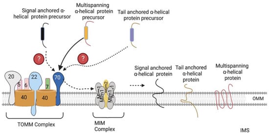
The mechanisms involved in the import of α-helical proteins is only partly understood. Three classes of α-helical proteins have been identified, including signal-anchored proteins, tail-anchored proteins, and polytopic (multispanning) outer-membrane proteins [4]. Signal-anchored and tail-anchored proteins contain an α-helical transmembrane segment at the N terminus and C terminus, respectively, which function as both membrane anchors and targeting signals, in addition to flanking positively charged amino acid residues [4]. In contrast, polytopic proteins possess multiple transmembrane segments that may contain targeting information; however, their exact targeting signals are unknown.
The MIM complex consisting of multiple copies of Mim1 and one or two copies of Mim2 has been identified as the machinery promoting the insertion of signal-anchored and polytopic α-helical proteins into the OMM (Figure 3) [4]. However, the molecular mechanisms through which it inserts these proteins into the OMM has not been elucidated yet [4]. No TOMM receptor has been identified to date that is required for the import of signal-anchored proteins [19][20]. In contrast, TOMM70 has been observed to recognize the precursors of polytopic proteins, after which, it binds them and transfers them to the MIM complex, which inserts them into the OMM (Figure 3) [21][22]. Previously, no proteinaceous machinery had been identified for the import of tail-anchored α-helical proteins; however, a recent study by Doan et al. showed that the import of the radiolabeled precursor of Fis1, a tail-anchored α-helical protein, into isolated mim1Δ and mim1-23 mitochondria was impaired [23]. In addition, re-expression of Mim1 in mim1Δ yeast mitochondria promoted the import of Fis1 [23]. Thus, the MIM complex is essential for the import of all three α-helical proteins. Besides the MIM complex, the low ergosterol content of the outer membrane favors the insertion of tail-anchored proteins into the OMM [24]. Likewise, the precursor of Ugo1, a polytopic protein, has been shown to be inserted into protein-free liposomes that mimic the phospholipid composition of the OMM [25], suggesting that the lipid composition of the mitochondria play a role in the optimal insertion of α-helical proteins.
 
Figure 3. Import of α-helical precursors into the outer mitochondrial membrane. Polytopic proteins are recognized by the TOMM70 receptor, after which TOMM70 binds to them and transfers them to the MIM complex, which inserts them into the OMM. Signal- and tail-anchored α-helical precursors are also inserted into the OMM by the MIM complex. The exact TOMM receptors recognizing these precursors have not been identified yet. OMM, outer mitochondrial membrane. The figure was created with Biorender.com.
Some OMM proteins are inserted into the outer membrane via routes distinct from those mentioned above. For example, Mcp3, an OMM protein, contains an N-terminal presequence and a stop-transfer signal, which allows it to be imported by the TOMM and TIMM23 machineries, after which it is released laterally into the inner membrane, where it is processed by the inner-membrane peptidase (IMP). Subsequently, it is released into the IMS and exported into the outer membrane by the MIM complex [26]. This suggests that other import machineries, apart from the import machineries located on the OMM, are involved in the import of OMM proteins. Therefore, more studies are needed to identify additional machineries involved in the import of OMM proteins and the proteins imported by these machineries. Furthermore, because the import of α-helical proteins is only understood in part, more studies with the aim of achieving an improved understanding of the mechanisms underlying the import of these proteins are needed.

References

  1. Wang, Z.; Ying, Z.; Bosy-Westphal, A.; Zhang, J.; Schautz, B.; Later, W.; Heymsfield, S.B.; Müller, M.J. Specific metabolic rates of major organs and tissues across adulthood: Evaluation by mechanistic model of resting energy expenditure. Am. J. Clin. Nutr. 2010, 92, 1369–1377.
  2. Pagliarini, D.J.; Calvo, S.E.; Chang, B.; Sheth, S.A.; Vafai, S.B.; Ong, S.-E.; Walford, G.A.; Sugiana, C.; Boneh, A.; Chen, W.K.; et al. A mitochondrial protein compendium elucidates complex I disease biology. Cell 2008, 134, 112–123.
  3. Archibald, J.M. Endosymbiosis and Eukaryotic Cell Evolution. Curr. Biol. 2015, 25, R911–R921.
  4. Wiedemann, N.; Pfanner, N. Mitochondrial Machineries for Protein Import and Assembly. Annu. Rev. Biochem. 2017, 86, 685–714.
  5. Srinivasan, S.; Guha, M.; Kashina, A.; Avadhani, N.G. Mitochondrial dysfunction and mitochondrial dynamics-The cancer connection. Biochim. Biophys. Acta Bioenerg. 2017, 1858, 602–614.
  6. Adebayo, M.; Singh, S.; Singh, A.P.; Dasgupta, S. Mitochondrial fusion and fission: The fine-tune balance for cellular homeostasis. FASEB J. 2021, 35, e21620.
  7. O’Rourke, B. Mitochondrial ion channels. Annu. Rev. Physiol. 2007, 69, 19–49.
  8. Lemasters, J.J. Modulation of mitochondrial membrane permeability in pathogenesis, autophagy and control of metabolism. J. Gastroenterol. Hepatol. 2007, 22, S31–S37.
  9. Gilkerson, R.W. Mitochondrial DNA nucleoids determine mitochondrial genetics and dysfunction. Int. J. Biochem. Cell Biol. 2009, 41, 1899–1906.
  10. Dudek, J.; Rehling, P.; van der Laan, M. Mitochondrial protein import: Common principles and physiological networks. Biochim. Biophys. Acta 2013, 1833, 274–285.
  11. Wang, W.; Chen, X.; Zhang, L.; Yi, J.; Ma, Q.; Yin, J.; Zhuo, W.; Gu, J.; Yang, M. Atomic structure of human TOM core complex. Cell Discov. 2020, 6, 67.
  12. Jores, T.; Klinger, A.; Groß, L.E.; Kawano, S.; Flinner, N.; Duchardt-Ferner, E.; Wöhnert, J.; Kalbacher, H.; Endo, T.; Schleiff, E.; et al. Characterization of the targeting signal in mitochondrial β-barrel proteins. Nat. Commun. 2016, 7, 12036.
  13. Qiu, J.; Wenz, L.-S.; Zerbes, R.M.; Oeljeklaus, S.; Bohnert, M.; Stroud, D.A.; Wirth, C.; Ellenrieder, L.; Thornton, N.; Kutik, S.; et al. Coupling of mitochondrial import and export translocases by receptor-mediated supercomplex formation. Cell 2013, 154, 596–608.
  14. Krimmer, T.; Rapaport, D.; Ryan, M.; Meisinger, C.; Kassenbrock, C.K.; Blachly-Dyson, E.; Forte, M.; Douglas, M.G.; Neupert, W.; Nargang, F.E.; et al. Biogenesis of porin of the outer mitochondrial membrane involves an import pathway via receptors and the general import pore of the TOM complex. J. Cell Biol. 2001, 152, 289–300.
  15. Wiedemann, N.; Truscott, K.N.; Pfannschmidt, S.; Guiard, B.; Meisinger, C.; Pfanner, N. Biogenesis of the protein import channel Tom40 of the mitochondrial outer membrane: Intermembrane space components are involved in an early stage of the assembly pathway. J. Biol. Chem. 2004, 279, 18188–18194.
  16. Kutik, S.; Stojanovski, D.; Becker, L.; Becker, T.; Meinecke, M.; Krüger, V.; Prinz, C.; Meisinger, C.; Guiard, B.; Wagner, R.; et al. Dissecting membrane insertion of mitochondrial beta-barrel proteins. Cell 2008, 132, 1011–1024.
  17. Diederichs, K.A.; Ni, X.; Rollauer, S.E.; Botos, I.; Tan, X.; King, M.S.; Kunji, E.R.S.; Jiang, J.; Buchanan, S.K. Structural insight into mitochondrial β-barrel outer membrane protein biogenesis. Nat. Commun. 2020, 11, 3290.
  18. Wenz, L.S.; Ellenrieder, L.; Qiu, J.; Bohnert, M.; Zufall, N.; van der Laan, M.; Pfanner, N.; Wiedemann, N.; Becker, T. Sam37 is crucial for formation of the mitochondrial TOM-SAM supercomplex, thereby promoting β-barrel biogenesis. J. Cell Biol. 2015, 210, 1047–1054.
  19. Ahting, U.; Waizenegger, T.; Neupert, W.; Rapaport, D. Signal-anchored proteins follow a unique insertion pathway into the outer membrane of mitochondria. J. Biol. Chem. 2005, 280, 48–53.
  20. Meineke, B.; Engl, G.; Kemper, C.; Vasiljev-Neumeyer, A.; Paulitschke, H.; Rapaport, D. The outer membrane form of the mitochondrial protein Mcr1 follows a TOM-independent membrane insertion pathway. FEBS Lett. 2008, 582, 855–860.
  21. Becker, T.; Wenz, L.-S.; Krüger, V.; Lehmann, W.; Müller, J.M.; Goroncy, L.; Zufall, N.; Lithgow, T.; Guiard, B.; Chacinska, A.; et al. The mitochondrial import protein Mim1 promotes biogenesis of multispanning outer membrane proteins. J. Cell Biol. 2011, 194, 387–395.
  22. Papic, D.; Krumpe, K.; Dukanovic, J.; Dimmer, K.S.; Rapaport, D. Multispan mitochondrial outer membrane protein Ugo1 follows a unique Mim1-dependent import pathway. J. Cell Biol. 2011, 194, 397–405.
  23. Doan, K.N.; Grevel, A.; Mårtensson, C.U.; Ellenrieder, L.; Thornton, N.; Wenz, L.-S.; Opaliński, Ł.; Guiard, B.; Pfanner, N.; Becker, T. The Mitochondrial Import Complex MIM Functions as Main Translocase for α-Helical Outer Membrane Proteins. Cell Rep. 2020, 31, 107567.
  24. Kemper, C.; Habib, S.; Engl, G.; Heckmeyer, P.; Dimmer, K.S.; Rapaport, D. Integration of tail-anchored proteins into the mitochondrial outer membrane does not require any known import components. J. Cell Sci. 2008, 121, 1990–1998.
  25. Vögtle, F.-N.; Keller, M.; Taskin, A.A.; Horvath, S.E.; Guan, X.L.; Prinz, C.; Opalińska, M.; Zorzin, C.; van der Laan, M.; Wenk, M.R.; et al. The fusogenic lipid phosphatidic acid promotes the biogenesis of mitochondrial outer membrane protein Ugo1. J. Cell Biol. 2015, 210, 951–960.
  26. Sinzel, M.; Tan, T.; Wendling, P.; Kalbacher, H.; Özbalci, C.; Chelius, X.; Westermann, B.; Brügger, B.; Rapaport, D.; Dimmer, K.S. Mcp3 is a novel mitochondrial outer membrane protein that follows a unique IMP-dependent biogenesis pathway. EMBO Rep. 2016, 17, 965–981.
More
Information
Contributors MDPI registered users' name will be linked to their SciProfiles pages. To register with us, please refer to https://encyclopedia.pub/register : , , , ,
View Times: 570
Revisions: 2 times (View History)
Update Date: 17 May 2023
1000/1000
Video Production Service