Submitted Successfully!
To reward your contribution, here is a gift for you: A free trial for our video production service.
Thank you for your contribution! You can also upload a video entry or images related to this topic.
Version Summary Created by Modification Content Size Created at Operation
1 -- 2041 2023-05-04 18:07:14 |
2 format change Meta information modification 2041 2023-05-05 10:42:09 |

Video Upload Options

Do you have a full video?

Confirm

Are you sure to Delete?
Cite
If you have any further questions, please contact Encyclopedia Editorial Office.
De Bardet, J.C.; Cardentey, C.R.; González, B.L.; Patrone, D.; Mulet, I.L.; Siniscalco, D.; Robinson-Agramonte, M.D.L.A. Cell Immortalization. Encyclopedia. Available online: https://encyclopedia.pub/entry/43791 (accessed on 03 July 2024).
De Bardet JC, Cardentey CR, González BL, Patrone D, Mulet IL, Siniscalco D, et al. Cell Immortalization. Encyclopedia. Available at: https://encyclopedia.pub/entry/43791. Accessed July 03, 2024.
De Bardet, Javier Curi, Celeste Ramírez Cardentey, Belkis López González, Deanira Patrone, Idania Lores Mulet, Dario Siniscalco, María De Los Angeles Robinson-Agramonte. "Cell Immortalization" Encyclopedia, https://encyclopedia.pub/entry/43791 (accessed July 03, 2024).
De Bardet, J.C., Cardentey, C.R., González, B.L., Patrone, D., Mulet, I.L., Siniscalco, D., & Robinson-Agramonte, M.D.L.A. (2023, May 04). Cell Immortalization. In Encyclopedia. https://encyclopedia.pub/entry/43791
De Bardet, Javier Curi, et al. "Cell Immortalization." Encyclopedia. Web. 04 May, 2023.
Cell Immortalization
Edit

Somatic human cells can divide a finite number of times, a phenomenon known as the Hayflick limit. It is based on the progressive erosion of the telomeric ends each time the cell completes a replicative cycle. Given this problem, researchers need cell lines that do not enter the senescence phase after a certain number of divisions. In this way, more lasting studies can be carried out over time and avoid the tedious work involved in performing cell passes to fresh media. However, some cells have a high replicative potential, such as embryonic stem cells and cancer cells. To accomplish this, these cells express the enzyme telomerase or activate the mechanisms of alternative telomere elongation, which favors the maintenance of the length of their stable telomeres. Researchers have been able to develop cell immortalization technology by studying the cellular and molecular bases of both mechanisms and the genes involved in the control of the cell cycle. Through it, cells with infinite replicative capacity are obtained. To obtain them, viral oncogenes/oncoproteins, myc genes, ectopic expression of telomerase, and the manipulation of genes that regulate the cell cycle, such as p53 and Rb, have been used.

Hayflick limit telomeres telomerase alternative telomere elongation immortalization

1. Introduction

Leonard Hayflick and Paul Moorhead observed in an in vitro study that human somatic cells can carry out a finite number of cell divisions, from which cell cycle arrest is triggered [1]. This study suggested that cells had a mechanism for counting the number of cell divisions, which was not causally related to time but to the number of replicative cycles [2]. However, it was not until 1965 that Hayflick postulated this theory, now known as Hayflick’s limit [3].
The molecular basis on which this phenomenon is based is the progressive shortening of telomeric DNA. Each time the cell completes a replicative cycle, telomeres shorten due to the terminal replication issue. In this way, when the telomere reaches a limited length, the mechanisms of cellular apoptosis and replicative senescence are triggered [4]. However, embryonic stem cells (ESCs) and cancer cells can keep the length of their telomeres stable, evading senescence and apoptosis and acquiring cellular immortality. Two molecular mechanisms associated with the maintenance of telomeric length have been elucidated in early studies: (1) transcriptional activation of the telomerase enzyme and (2) alternative lengthening of telomeres (ALT). The former is found in almost 90% of cancer cells, while the latter is the remaining percent [5][6].

1.1. Telomeres

Telomeres are nucleus-protein complexes located at the ends of the chromosomes of eukaryotic cells [7]. This structure prevents the linear ends of chromosomes from being recognized as double-strand DNA breaks(DSBs) [8]; their shortening constitutes a mechanism of tumor suppression by activating cellular senescence signals [9]. In humans, telomeric DNA is composed of tandem repeats of the hexanucleotide 5′-TTAGGG-3′sequence. This sequence extends from 3 to 12 kb in length, giving rise to a single chain rich in guanine residues (G strand) (Figure 1a). The protruding G chain can bend and invade the telomere double chain region and generate a loop structure, the T-loop, thathides the 3′end, as a primitive mechanism for telomere protection [10].
Figure 1. Structure of telomeres in humans. (a) Schematic representation of telomeric DNA in humans, composed of tandem repeats of the hexanucleotide sequence TTAGGG (G chain) and its complementary sequence in chain C. The G chain can extend 3–12kb in length (G-strand overhang). (b) Adjacent to the telomeres are the sub telomeric regions, also rich in repetitive DNA. Schematic representation of the shelterin or telosome complex. The TRF1 and TRF2 proteins have a binding domain for double-stranded DNA, whereas POT1 can only bind to single-stranded DNA. RAP1 exerts its function on the telomere through a TRF2-binding domain. TIN2 has binding domains for both TRF1 and TRF2, and through another domain, it binds to the TPP1-POT1 complex.
Associated with these tandem repeats is a six-protein complex called shelterins, including TRF1, TRF2, TIN2, RAP1, TPP1, and POT1 (Figure 1b). TRF1 and TRF2 contain a binding domain for dsDNA, while POT1 interacts with telomeric DNA. The other protein complex components do not have binding domains for telomeric DNA and exert their function through interaction with the TRF1 and TRF2 proteins. Shelterins prevent the DNA damage response mechanisms that DSBs activate from being triggered and in this way, prevent the occurrence of homology-directed repair (HDR) or binding processes non-homologous end joining (NHEJ). Likewise, TRF2 protein plays a key role in telomere protection since it makes the construction of the T-loop possible, giving telomeric DNA stability and inaccessibility at its ends. Therefore, telomeres devoid of shelterins lead to the fusion of the ends of chromosomes recognized as DSB that need to be repaired [11][12]. Even though telomeric shortening occurs after each cell cycle, other factors, such as oxidative stress, can influence the speed of telomeric shortening [13][14].

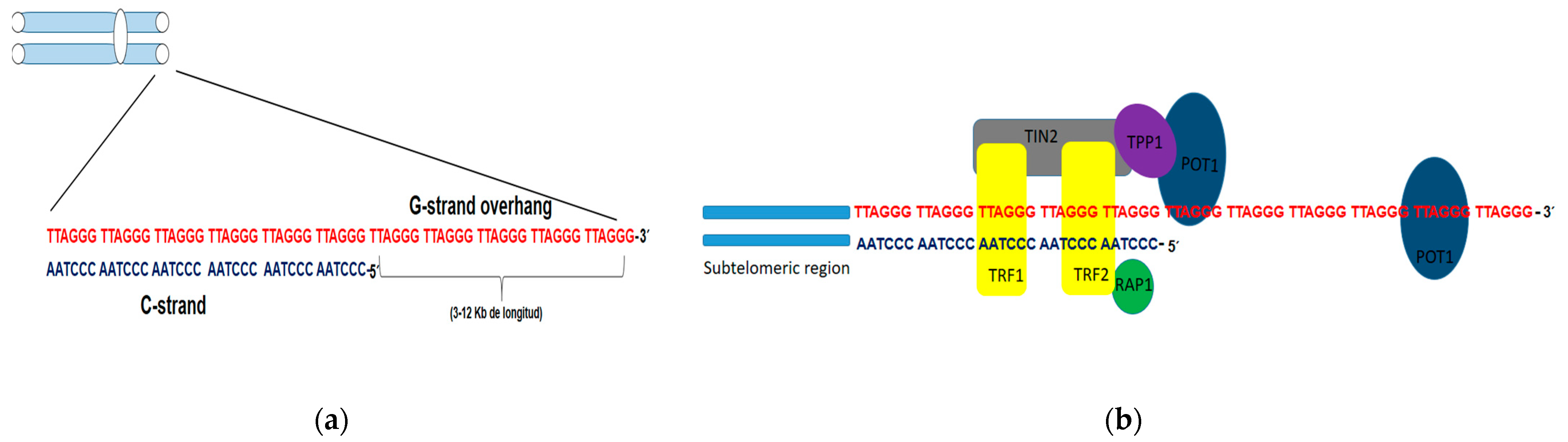
1.2. Mechanism Involved in Lengthening of Telomeres
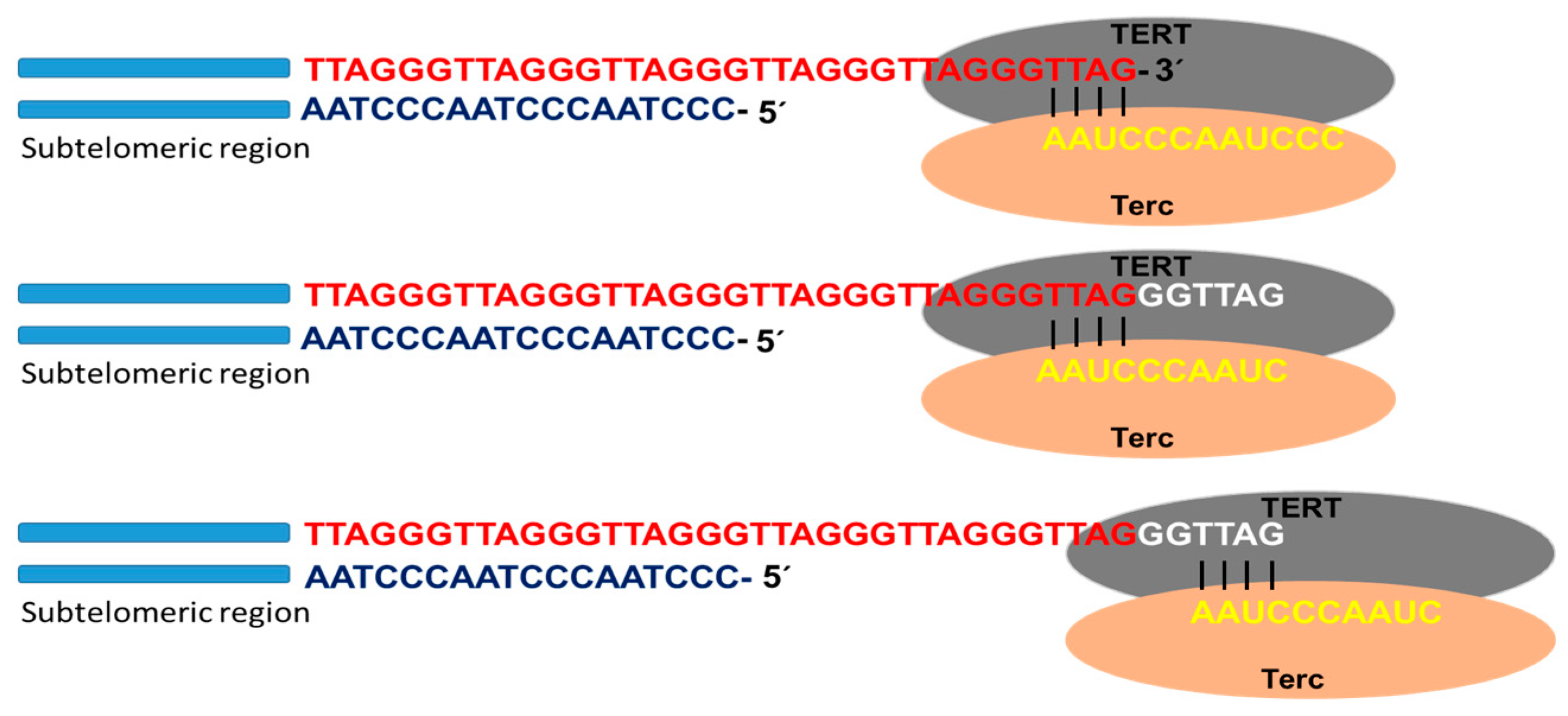
Telomerase

Telomerase is the specialized DNA polymerase responsible for the maintenance and elongation of telomeres. It consists of a ribonucleoprotein with a catalytic subunit with reverse transcriptase action (TERT) and an RNA fragment of sequence complementary to telomeric DNA (Terc: telomerase RNA component). Associated with the complex is dyskerin, a protein that contributes to its formation and stability. The catalytic site of telomerase adds deoxyribonucleotides using the RNA molecule as a template that hybridizes with the telomeric DNA strand (3′-OH end of the G strand). Through this mechanism, the enzyme is able to lengthen the telomere using a translocation movement each time it incorporates the complete hexanucleotide sequence (Figure 2). Once the elongation of the G strand has concluded, the replication of the C strand occurs by the conventional system of polymerases. In this way, the telomeric shortening product of the terminal replication problem is solved [15][16][17].
Figure 2. Telomerase activity. Schematic representation of telomere replication by telomerase, as well as the catalytic subunit with reverse transcriptase action (TERT) and the subunit containing the RNA template for telomere replication (Terc). Once the template RNA hybridizes with the telomeric DNA sequence of the G chain, at the 3′-OH end, the telomere polymerization process begins. Once the hexanucleotide sequence is added, the enzyme performs a translocation movement, and telomere elongation continues.
TERT is highly expressed in cells with high replication and self-renewal capacity, such as ESCs, germline cells, and most cancer cells. However, in adult stem cells (ASCs) and in activated lymphocytes, the activity of this enzyme was also demonstrated, although with much lower expression levels than in embryonic cells [5][18]. Except ASCs and activated lymphocytes, in the other somatic cells, telomerase activity decreases over time after birth, and subsequently, telomeres are shortened with cell divisions [19]. Therefore, telomeric shortening limits the capacity for replicative expansion in telomerase-negative cells, constituting a barrier to achieving cellular immortality and functioning as a biological clock [20]. On the other hand, there is evidence that even in somatic stem cells, telomerase levels are not satisfactory in avoiding telomeric shortening in the course of cellular aging [21].
After 50 or 60 cell cycles, telomeric shortening leads to replicative senescence due to genomic instability associated with fusion events and chromosomal breakage. However, to avoid genetic chaos, some cells can overcome this phase by acquiring mutations in p53 and Rb genes and other genes, encoding for proteins linked to cell cycle control mechanisms and telomerase activation. A small part of the cell population acquires immortality through this pathway and proceeds to carcinogenesis [20][22].
Studies in mice highlighted that the defects in telomeres, and following telomerase re-activation, they are able to trigger the induction of malignant tumors [23]. On the other hand, Barthel and colleagues observed the expression of TERT was present in almost 75% of the analyzed tumor samples, with 31% of samples exhibiting alterations in the TERT promoter and 53% showing methylation patterns [24]. Aberrant expression of TERT is due to mutually exclusive mutations in the TERT promoter (TPM) (−57 A > C; −124 C > T; −138/−139 CC > TT; −146 C > T) that produce transcription factor binding sites of the ETS (E26 transformation-specific) family, such as GA binding protein (GABP) [24][25]. These mutations are mostly heterozygous and induce the allele-specific re-expression of TERT; recruiting GABP promotes an epigenetic change at the chromatin level, passing to its active form [26].
TPMs are the most common non-coding driver of cancer mutations [27]. Indeed, approximately 85% of skin tumors carry them [28]. Likewise, they are coupled with a high expression of TERT and with poorer survival rates in several tumors, such as glioblastoma [29] and meningioma [30]. Furthermore, tumors that carry TPM shave a high risk of recurrence [31]. Similarly, it is worth noting the existence of other pathways able to promote TERT expression during tumor, such as amplifying the TERT gene, hypermethylation of the promoter, and chromosomal rearrangement [24][32].
Moreover, the induction of the molecular mechanisms of DNA repair is also an important factor determining the cellular sequela in response to telomere changes. In this condition, NHEJ activation triggers chromosomal fusion, while the induction of the HDR mechanism could mediate the telomere lengthening through the ALT pathway [20]. Consequently, activation of the telomere repair mechanisms could impact the progression of genomic instability and cancer induction by triggering the end’s union or obtaining cellular immortality through the ALT pathway. Because chromosome fusions and breakage of fused chromosomes could participate in cancer induction, further studies on DNA repair mechanisms that induce chromosomal fusions are required to better understand the role of telomeric shortening in cancer initiation [20][22].

2. Alternative Telomere Lengthening (ALT)

Even when telomerase is essential for obtaining cellular immortality, 10–15% of cancer cells can maintain stable telomere length through the ALT pathway [6]. The ALT mechanism is common in sarcomas and tumors of the central and peripheral nervous system but rare in common cancers such as breast cancer, colon cancer, and lung cancer and absent in lymphoma and thymoma [33]. Among cancers that arise in mesenchymal tissues, it was shown that 47% of osteosarcomas and 35% of soft tissue sarcomas (STS) use this mechanism of telomeric lengthening [34]. Furthermore, several differences between and within STS have been described [35]. In liposarcomas (LPS), ALT-positive tumors frequencies from 0% for well-differentiated LPS (characterized by amplification of the 12q13-15) to 80% for pleomorphic LPS, which show elevated levels of complex genomic alterations [36].
Some evidence suggests that the ALT mechanism could be present in normal somatic cells. This was seen early in studies in mice [37] and furthermore in primary human cells where the mechanism based on homologous recombination that resembles ALT seemed to be activated, transiently, in response to oxidative stress [38] or X-ray damage [39]. Similarly, intra-tumor heterogeneity was found in terms of telomere maintenance mechanisms (TMM) in neuroblastomas [40] and osteosarcomas [41], where several telomerase-positive cells have been detected, together with ALT-phenotyped cells.
In pluripotent stem cells (PSCs), telomere maintenance is carried out through telomerase expression, although the ALT pathway was found to play a fundamental role [42][43]. On the other hand, telomeres and the regulation of their length show marked differences between cancer cells and PSCs that use this mechanism. For example, tumor genomes employing the ALT pathway are unstable, exhibiting heterogeneous and dysfunctional telomeres, whereas PSCs possess long telomeres with a stable genome. The underlying mechanisms are not yet clear, but the ALT pathway is activated in PSCs by changes in epigenetic reprogramming [43].

3. Coexistence of Telomerase and the ALT Pathway

The fusion of cell lines that express one of the two mechanisms allowed for obtaining hybrids where once the genome was stabilized, only one of the mechanisms remained active. Therefore, hybridized cells show only one telomere maintenance mechanism (TMM) by the expression of telomerase or by the activation of the ALT pathway [44][45][46]. However, the ectopic overexpression of telomerase in ALT cells results in obtaining cell lines where both mechanisms coexist. In these cells, telomerase lengthens the shorter telomeres, while APBs and T-SCE can be detected with possible involvement in the maintenance of another subset of telomeres [44][47]. Therefore, the co-existence of both TMMs is only observed in the same cell when telomerase is over-expressed. Meanwhile, it has been shown that they can co-exist within the same tumor in different cells [18]. The coexistence of ALT-positive cells and telomerase has been reported in several types of tumors, which supports the concept of intra-tumor heterogeneity. Indeed, TMM activation is not necessary for initial tumorigenesis. Still, it may occur during tumor expansion, involving the activation of both ALT and telomerase in different types of cells from the same tumor. Conversely, all early-phase tumor cells could have activated one of the two TMMs, with the possibility of changing this choice later [48].

References

  1. Hayflick, L.; Moorhead, P.S. The Serial Cultivation of Human Diploid Cell Strains. Exp. Cell Res. 1961, 1, 585–621.
  2. Hayflick, L. How and why we age. Exp. Gerontol. 1998, 33, 377.
  3. Hayflick, L. The limited in vitro lifetime of human diploid cell strains. Exp. Cell Res. 1965, 37, 614–636.
  4. Blackburn, E.H. Telomeres—No end in sight. Cell 1994, 77, 621–623.
  5. Shay, J.W.; Bacchetti, S. A survey of telomerase activity in human cancer. Eur. J. Cancer 1997, 33, 787–791.
  6. Heaphy, C.M.; Subhawong, A.P.; Hong, S.M.; Goggins, M.G.; Montgomery, E.A.; Gabrielson, E.; Heaphy, C.M.; Subhawong, A.P.; Hong, S.M.; Goggins, M.G.; et al. Prevalence of the alternative lengthening of telomeres telomere maintenance mechanism in human cancer subtypes. Am. J. Pathol. 2011, 179, 1608–1615.
  7. Arnoult, N.; Karlseder, J. Complex interactions between the DNA-damage response and mammalian telomeres. Nat. Struct. Mol. Biol. 2015, 22, 859–866.
  8. Denchi, E.L.; de Lange, T. Protection of telomeres through independent control of ATM and ATR by TRF2 and POT1. Nature 2007, 448, 1068–1071.
  9. Harley, C.B.; Futcher, A.B.; Greider, C.W. Telomeres shorten during ageing of human fibroblasts. Nature 1990, 345, 458–460.
  10. Doksani, Y.; Wu, J.Y.; de Lange, T.; Zhuang, X. Super-resolution fluorescence imaging of telomeres reveals TRF2-dependent T-loop formation. Cell 2013, 155, 345–356.
  11. Rai, R.; Zheng, H.; He, H.; Luo, Y.; Multani, A.; Carpenter, P.B.; Chang, S. The function of classical and alternative non-homologous end-joining pathways in the fusion of dysfunctional telomeres. EMBO J. 2010, 29, 2598–2610.
  12. Okamoto, K.; Bartocci, C.; Ouzounov, I.; Diedrich, J.K.; Yates, J.R.; Denchi, E.L. A two-step mechanism for TRF2- mediated chromosome-end protection. Nature 2013, 494, 502–505.
  13. Maciejowski, J.; de Lange, T. Telomeres in Cancer: Tumour suppression and genome instability. Nat. Rev. 2017, 18, 175–186.
  14. Shay, J.W. Role of telomeres and telomerase in aging and cancer. Cancer Discov. 2016, 6, 584–593.
  15. Cohen, S.B.; Graham, M.E.; Lovrecz, G.O.; Bache, N.; Robinson, P.J.; Reddel, R.R. Protein composition of catalytically active human telomerase from immortal cells. Science 2007, 315, 1850–1853.
  16. Blackburn, E.H. Switching and signaling at the telomere. Cell 2001, 106, 661–673.
  17. Greider, C.W.; Blackburn, E.H. Identification of a specific telomere terminal transferase activity in Tetrahymena extracts. Cell 1985, 43, 405–413.
  18. De Vitis, M.; Berardinelli, F.; Sgura, A. Telomere lenght maintenence in Cancer: AT the Crossroad between Telomerase and Alternative Lenthening of Telomeres (ALT). Int. J. Mol. Sci. 2018, 19, 606.
  19. Harrington, L. Does the reservoir for self-renewal stem from the ends? Oncogene 2004, 23, 7283–7289.
  20. Gunes, C.; Lenhard, R.K. The role of the telomeres in stem cells and cancer. Cell 2013, 152, 390–393.
  21. Sperka, T.; Wang, J.; Rudolph, K.L. DNA damage checkpoints in stem cells, ageing and cancer. Nat. Rev. Mol. Cell Biol. 2012, 13, 579–590.
  22. Okamoto, K.; Seimiya, H. Revisiting telomeres shortening in Cancer. Cells 2019, 8, 107.
  23. Begus-Nahrmann, Y.; Hartmman, D.; Kraus, J.; Eshragi, P.; Scheffold, A.; Grieb, M.; Rasche, V.; Schirmacher, P.; Lee, H.W.; Kestler, H.A.; et al. Transient telomere dysfunction induces chromosomal instability and promotes carcinogenesis. J. Clin. Investig. 2012, 122, 2283–2288.
  24. Barthel, F.P.; Wei, W.; Tang, M.; Martinez-Ledesma, E.; Hu, X.; Amin, S.B.; Akdemir, K.C.; Seth, S.; Song, X.; Wang, Q.; et al. Systematic analysis of telomere length and somatic alterations in 31 cancer types. Nat. Genet. 2017, 49, 349–357.
  25. Bell, R.J.; Rube, H.T.; Kreig, A.; Mancini, A.; Fouse, S.D.; Nagarajan, R.P.; Choi, S.; Hong, C.; He, D.; Pekmezci, M.; et al. Cancer. The transcription factor GABP selectively binds and activates the mutant TERT promoter in cancer. Science 2015, 348, 1036–1039.
  26. Stern, J.L.; Theodorescu, D.; Vogelstein, B.; Papadopoulos, N.; Cech, T.R. Mutation of the TERT promoter, switch to active chromatin and monoallelic TERT expression in multiple cancers. Genes Dev. 2015, 29, 2219–2224.
  27. Weinhold, N.; Jacobsen, A.; Schultz, N.; Sander, C.; Lee, W. Genome-wide analysis of noncoding regulatory mutations in cancer. Nat. Genet. 2014, 46, 1160–1165.
  28. Hayward, N.K.; Wilmott, J.S.; Waddell, N.; Johansson, P.A.; Field, M.A.; Nones, K.; Patch, A.M.; Kakavand, H.; Alexandrov, L.B.; Burke, H.; et al. Whole-genome landscapes of major melanoma subtypes. Nature 2017, 545, 175–180.
  29. Labussière, M.; Boisselier, B.; Mokhtari, K.; DiStefano, A.L.; Rahimian, A.; Rossetto, M.; Ciccarino, P.; Saulnier, O.; Paterra, R.; Marie, Y.; et al. Combined analysis of TERT, EGFR, and IDH status defines distinct prognostic glioblastoma classes. Neurology 2014, 83, 1200–1206.
  30. Spiegl-Kreinecker, S.; Lötsch, D.; Neumayer, K.; Kastler, L.; Gojo, J.; Pirker, C.; Pichler, J.; Weis, S.; Kumar, R.; Webersinke, G.; et al. TERT promoter mutations are associated with poor prognosis and cell immortalization in meningioma. Neuro Oncol. 2018, 20, 1584–1593.
  31. Sahm, F.; Schrimpf, D.; Olar, A.; Koelsche, C.; Reuss, D.; Bissel, J.; Kratz, A.; Capper, D.; Schefzyk, S.; Hielscher, T.; et al. TERT promoter mutations and risk of recurrence in meningioma. J. Natl. Cancer Inst. 2016, 108, djv370.
  32. Lee, D.D.; Leão, R.; Komosa, M.; Gallo, M.; Zhang, C.H.; Lipman, T.; Remke, M.; Heidari, A.; Nunes, N.M.; Apolónio, J.; et al. DNA hypermethylation within TERT promoter upregulates TERT expression in cancer. J. Clin. Investig. 2019, 129, 223–229.
  33. Dilley, R.L.; Greenberg, R.A. Alternative telomere maintenance and cancer. Trends Cancer 2015, 1, 145–156.
  34. Henson, J.D.; Hannay, J.A.; McCarthy, S.W.; Royds, J.A.; Yeager, T.R.; Robinson, R.A.; Wharton, S.B.; Jellinek, D.A.; Arbuckle, S.M.; Yoo, J.; et al. A robust assay for alternative lengthening of telomeres in tumors shows the significance of alternative lengthening of telomeres in sarcomas and astrocytomas. Clin. Cancer Res. 2005, 11, 217–225.
  35. Liau, J.Y.; Lee, J.C.; Tsai, J.H.; Yang, C.Y.; Liu, T.L.; Ke, Z.L.; Hsu, H.H.; Jeng, Y.M. Comprehensive screening of alternative lengthening of telomeres phenotype and loss of telomeres phenotype and loss of ATRX expression in sarcomas. Mod. Pathol. 2015, 28, 1545–1554.
  36. Lee, J.C.; Jeng, Y.M.; Liau, J.Y.; Tsai, J.H.; Hsu, H.H.; Yang, C.Y. Alternative lengthening of telomeres and loss of ATRX are frequent events in pleomorphic and dedifferentiated liposarcomas. Mod. Pathol. 2015, 28, 1064–1073.
  37. Neumann, A.A.; Watson, C.M.; Noble, J.R.; Pickett, H.A.; Tam, P.P.; Reddel, R.R. Alternative lengthening of telomeres in normal mammalian somatic cells. Genes Dev. 2013, 27, 18–23.
  38. Coluzzi, E.; Buonsante, R.; Leone, S.; Asmar, A.J.; Miller, K.L.; Cimini, D.; Sgura, A. Transient ALT activation protects human primary cells from chromosome instability induced by low chronic oxidative stress. Sci. Rep. 2017, 7, 43309.
  39. De Vitis, M.; Berardinelli, F.; Coluzzi, E.; Marinaccio, J.; O’Sullivan, R.J.; Sgura, A.A. X-rays activate telomeric homologous recombination mediated repair in primary cells. Cells 2019, 8, 708.
  40. Pezzolo, A.; Pistorio, A.; Gambini, C.; Haupt, R.; Ferraro, M.; Erminio, G.; De Bernardi, B.; Garaventa, A.; Pistoia, V. Intratumoral diversity of telomere length in individual neuroblastoma tumors. Oncotarget 2015, 6, 7493–7503.
  41. Gocha, A.R.; Nuovo, G.; Iwenofu, O.H.; Groden, J. Human sarcomas are mosaic for telomerase-dependent and telomerase-independent telomere maintenance mechanisms: Implications for telomere-based therapies. Am. J. Pathol. 2013, 182, 41–48.
  42. Huang, Y.; Liang, P.; Liu, D.; Huang, J.; Songyang, Z. Telomere regulation in pluripotent stem cells. Protein Cell 2014, 5, 194–202.
  43. Liu, L. Linking Telomere Regulation to Stem Cell Pluripotency. Trends Genet. 2017, 33, 16–33.
  44. Perrem, K.; Colgin, L.M.; Neumann, A.A.; Yeager, T.R.; Reddel, R.R. Coexistence of alternative lengthening of telomeres and telomerase in hTERT- transfected GM847 cells. Mol. Cell Biol. 2001, 21, 3862–3875.
  45. Episkopou, H.; Draskovic, I.; VanBeneden, A.; Tilman, G.; Mattiussi, M.; Gobin, M.; Arnoult, N.; Londoño-Vallejo, A.; Decottignies, A. Decottignies A: Alternative lengthening of telomeres is characterized by reduced compaction of telomeric chromatin. Nucleic Acids Res. 2014, 42, 4391–4405.
  46. Episkopou, H.; Diman, A.; Claude, E.; Viceconte, N.; Decottignies, A. TSPYL5 depletion induces specific death of ALT cells through USP7-dependent proteasomal degradation of POT1. Mol. Cell 2019, 75, 469–482.
  47. Tilman, G.; Loriot, A.; VanBeneden, A.; Arnoult, N.; Londono-Vallejo, J.A.; DeSmet, C.; Decottignies, A. Subtelomeric DNA hypomethylation is not required for telomeric sister chromatid exchanges in ALT cells. Oncogene 2009, 28, 1682–1693.
  48. Bechter, O.E.; Zou, Y.; Walker, W.; Wright, W.E.; Shay, J.W. Telomeric recombination in mismatch repair deficient human colon cancer cells after telomerase inhibition. Cancer Res. 2004, 64, 3444–3451.
More
Information
Subjects: Cell Biology
Contributors MDPI registered users' name will be linked to their SciProfiles pages. To register with us, please refer to https://encyclopedia.pub/register : , , , , , ,
View Times: 253
Revisions: 2 times (View History)
Update Date: 05 May 2023
1000/1000
Video Production Service