You're using an outdated browser. Please upgrade to a modern browser for the best experience.
Submitted Successfully!
Thank you for your contribution! You can also upload a video entry or images related to this topic. For video creation, please contact our Academic Video Service.
Version Summary Created by Modification Content Size Created at Operation
1 Li Ren -- 2541 2023-05-04 07:49:13 |
2 update references and layout Rita Xu Meta information modification 2541 2023-05-04 08:04:25 |

Video Upload Options

We provide professional Academic Video Service to translate complex research into visually appealing presentations. Would you like to try it?

Confirm

Are you sure to Delete?
Yes No
Cite
If you have any further questions, please contact Encyclopedia Editorial Office.
Li, C.; Zhao, R.; Yang, H.; Ren, L. Hypoxic Microenvironment of Bone. Encyclopedia. Available online: https://encyclopedia.pub/entry/43735 (accessed on 07 December 2025).
Li C, Zhao R, Yang H, Ren L. Hypoxic Microenvironment of Bone. Encyclopedia. Available at: https://encyclopedia.pub/entry/43735. Accessed December 07, 2025.
Li, Chen, Rong Zhao, Hui Yang, Li Ren. "Hypoxic Microenvironment of Bone" Encyclopedia, https://encyclopedia.pub/entry/43735 (accessed December 07, 2025).
Li, C., Zhao, R., Yang, H., & Ren, L. (2023, May 04). Hypoxic Microenvironment of Bone. In Encyclopedia. https://encyclopedia.pub/entry/43735
Li, Chen, et al. "Hypoxic Microenvironment of Bone." Encyclopedia. Web. 04 May, 2023.
Hypoxic Microenvironment of Bone
Edit

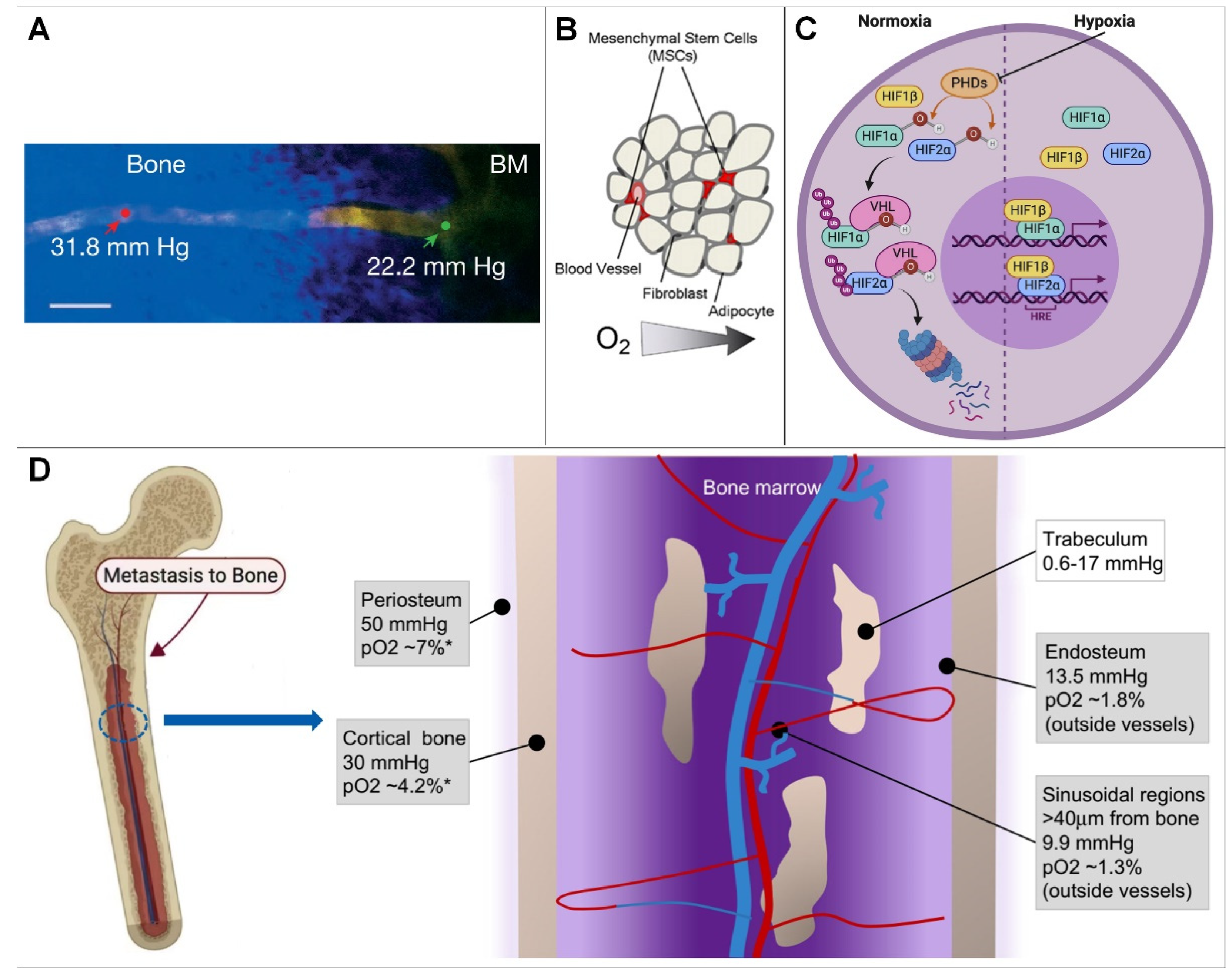
The normal physiological activities and functions of bone cells cannot be separated from the balance of the oxygenation level, and the physiological activities of bone cells are different under different oxygenation levels. At present, in vitro cell cultures are generally performed in a normoxic environment, and the partial pressure of oxygen of a conventional incubator is generally set at 141 mmHg (18.6%, close to the 20.1% oxygen in ambient air). This value is higher than the mean value of the oxygen partial pressure in human bone tissue. Additionally, the further away from the endosteal sinusoids, the lower the oxygen content. It follows that the construction of a hypoxic microenvironment is the key point of in vitro experimental investigation.

bone marrow hypoxic microenvironment cell culturing

1. Introduction

All organs of the human body are exposed to different oxygen tensions, and some organs are exposed to very low oxygen levels and different oxygen gradients. The changes of the bone marrow hypoxic microenvironment have a certain impact on human physiological conditions, and therefore oxygen tension and oxygen gradient need to be considered for cell cultures in vitro. Over the past few years, scientists have developed many tools and technologies to create various spatiotemporal gradients for cell research. However, the current methods of studying the physiological activities of cells under hypoxia cannot accurately control the level of oxygenation at the microscale.
The development of microfluidic platforms can overcome the inherent limitations of these methods. Microfluidic technology is one of the most promising technologies at present because it has strong controllability in both the space and time domains.

2. The Hypoxic Microenvironment of Bone

2.1. Overview of the Bone Hypoxic Microenvironment

2.1.1. Hypoxic Microenvironment Characteristics of Bone

Oxygen, an important source of energy for cellular metabolism and maintenance of the biological activity of the body, is transported to the whole body through the blood. To maintain oxygen homeostasis [1], hypoxic signaling pathways have matured to promote oxygen delivery and cellular adaptation to hypoxia [2]. Thus, hypoxic signaling plays an important role in development, tissue homeostasis, and pathological conditions [3][4].
Bone is a highly vascularized tissue that, however, harbors an extremely hypoxic microenvironment, with different regions of bone tissue characterized by different oxygen levels and oxygen gradients [5][6][7].
In the bone marrow compartment, quantification of bone marrow PO2 at several intravascular locations using two-photon light lifetime microscopy, as well as mathematical modeling, yielded values ranging from 11.7 to 31.7 mmHg, with a mean of 20.4 mmHg (2.7%) (Figure 1A), indicating that the bone marrow cavity is a hypoxic environment [8][9][10].
Figure 1. (A) Blood vessel projection image from bone to bone marrow. (B) In the hematopoietic stem cell niche, cells compete for scarce nutrients and oxygen. (C) HIF signaling pathway. (D) Hypoxic microenvironment in bone marrow. * = Estimated conversion to pO2 from mm Hg.
On cortical bone, the use of high-resolution imaging to study blood vessels and partial pressure of oxygen has been hampered by its thickness. Therefore, the partial pressure of oxygen to approximate cortical bone with a mathematical model is approximately 4.2% (30 mm Hg). Similar to the periosteum, oxygen levels in cortical bone are distributed as a result of presumably low cell density and low oxygen consumption [11].
On cancellous bone, the hypoxic microenvironment of the sinus region is similar to that of trabecular bone. When it is assumed that oxygen diffuses throughout the bone in a similar way to water (with the same diffusion coefficient), oxygen levels are expected to be even lower (0.08–2.4% or 0.6–17 mm Hg moving from the trabecular bone surface to the inner layer of osteocytes) [3][12][13][14]. Because this scenario has been predicted by mathematical models, the exact partial pressure of oxygen in trabecular bone remains unknown; however, models suggest that osteocytes embedded in the core of trabecular bone may experience a hypoxic environment [15][16].
At the endosteum, because the vasculature is different, smaller vessels are located closer to the endosteal surface by measuring PO2 at different locations within the BM. Vasculature with a diameter of 15 mm showed a higher partial pressure of oxygen (22.7 mmHg, 3.0%) than those with a diameter of 0.15 mm (19.5 mmHg, 2.6%; p < 0.03). Therefore, when analyzing PO2 values at different distances from the endosteum, researchers found that the lowest PO2 was located in a region of 0.40 mm [3][17]. These measurements revealed a moderate PO2 gradient with distance from the endosteum [18][19][20].

2.1.2. Causes of Bone Hypoxic Microenvironment Generation

Physiological hypoxia exists in many tissues of the human body, and a low-oxygen environment is necessary to maintain normal physiological functions, such as those of the liver and bone. The low-oxygen microenvironment of bone may be determined by cellular levels and oxygen consumption rates in specific regions of the skeleton, where hypoxic effects at regions of the sinusoidal vasculature may further determine regional oxygen gradients; in addition, blood in bone flows from the central arteries into the arterioles, capillaries, and finally into sinusoids, and is associated with a progressive decrease in blood velocity and oxygen delivery. It has been suggested that sinusoidal blood flow is approximately one tenth of arteriolar blood flow, so low oxygen tension in bone marrow may result from low oxygen levels in sinusoidal blood flow and high oxygen consumption by hematopoietic cells [1][4][21] because these hematopoietic stem cells in the bone marrow compete with each other for the scarce nutrition and oxygen supply in the capillaries close to them (Figure 1B) [22]. In summary, oxygen tension in bone is primarily controlled by cell density, oxygen consumption, and oxygenated blood supply [17][23].

2.2. Effect of Hypoxia on Bone Function

2.2.1. How Do Cells Sense Hypoxia?

How cells sense and adapt to oxygen levels in the microenvironment was previously unknown. Key to this process is the hypoxia-inducible factor (HIF) (Figure 1A). HIF is a binding protein present in humans and other mammals. It comprises four subunits, including O2-regulated α subunits (HIF-1α, HIF-2α, HIF-3α) and a β subunit (HIF-1β, or ARNT). Its main function is to induce bound proteins in response to low oxygen or anoxia. Among them, HIF-1β expression is generally unaffected by oxygen, but the expression of α-subunits is dependent on oxygen supply and has the shortest half-life [23][24].
In a normal oxygen environment, the oxygen-dependent domain (ODD) of α subunits is hydroxylated by proline hydroxylase domain enzymes (PHDs, including PHD 1, PHD 2, and PHD 3). Among these enzymes, PHD2 plays an important physiological function and PHD1 and PHD3 play a role in mitochondrial energy metabolism and innate immune responses, respectively (Figure 1C) [25][26]. HIF-α is ubiquitinated by the ubiquitin enzyme von Hippel Lindau tumor suppressor protein, and degraded, leading to a decline in intracellular HIF levels, which shapes the different sensing states of the cell in response to oxygen under different oxygen content environments:
(1)
In a hypoxic environment, HIF-α levels are elevated, resulting in the nuclear β dimerization of subunits. HIF-1 is an HIF-1 β and HIF-1 α of a heterodimeric protein complex that, upon binding to RNA polymerase II and interaction with major transcriptional coactivators such as P300, promotes the transcription of target genes. At the same time, it affects the expression levels of erythropoietin, glucose transporters, glycolytic enzymes, vascular endothelial growth factor, and other proteins, which ultimately help to increase oxygen delivery or promote metabolic adaptation to hypoxia, thereby regulating many important physiological behaviors, such as cell metabolism, erythropoiesis, and local angiogenesis [21][27].
(2)
Under normoxic conditions, inhibition of the asparagine hydroxylase factor hypoxia-1 transactivates the HIF-α of the conserved asparagine residues hydroxylated and interacts with P300 to ultimately repress HIF-induced transcription. HIF-3 α is thought to work by interacting with HIF-1 α competitive binding to inhibit HIF-1 α and HIF-2 β function [28]. Among these enzymes, PHD2 exerts major physiological functions. HIF-α is ubiquitinated and degraded by the ubiquitin protease tumor suppressor protein, resulting in reduced intracellular HIF levels, and, at the same time, a hypoxic environment can cause HIF-α levels to become elevated and associated with those in the nucleus β subunits undergoing dimerization [29][30][31]. Thereby, researchers can explore therapies for related diseases, such as ischemia, cancer, diabetes, stroke, infection, wound healing, and heart failure, by targeting the oxygen-sensing pathway.

2.2.2. Effect of Hypoxia on Bone Function

  • Hypoxia on cell differentiation
In the past few years, several authors have studied the effect of low oxygen concentrations on cell growth in different cell models, such as rat bone marrow-derived mesenchymal stem cells and mouse fibroblasts, respectively [32][33]. D’ippolito et al. [34] demonstrated that low oxygen concentrations (1%, 3%, 5%, and 10% O2) increase cell proliferation. At 3% O2, the cells have the most suitable growth environment. In addition, the low partial pressure of oxygen inhibits osteoblast differentiation, indicating that a low partial pressure environment is one of the essential conditions for cell culture, while illustrating that low partial pressure of oxygen is essential for maintaining mesenchymal stem cells in a poorly differentiated state. Moreover, Lennon et al. [33] reported that rat bone marrow stromal cells showed increased extracellular mineralization in cultures maintained at low PO2 compared to normoxic conditions. In addition, in several in vitro studies, low oxygen concentrations have been found to stimulate the differentiation process, inducing the cells to progress towards osteogenic, adipogenic, or chondrogenic lineages [35][36]. From this researchers can determine that cell differentiation and survival can be affected by different partial pressures of oxygen.
  • Hypoxia on skeleton development
The effects of hypoxic environments on bone metabolism and development are complex, mainly through HIFs and pathways related to osteoblasts and osteoclasts, and differ with time and oxygen concentration. Hypoxia can promote osteoblast generation and differentiation [37][38]. Short-term hypoxia may promote the early proliferation and differentiation of osteoblasts, while long-term hypoxia may inhibit their proliferation and differentiation. Under a hypoxic environment, HIF stimulates osteoclasts mainly through acidification, whereas it increases bone resorption capacity [13][24][39]. Hypoxia can lead to osteoclast formation and depends on the duration of hypoxia. Increased anaerobic metabolism and accumulation of acidic metabolites in hypoxia can cause mild acidification of the local microenvironment, and osteoclast activation is precisely dependent on extracellular acidification. Osteoclasts increase in number, volume, and bone resorption capacity, with the greatest effect occurring at 2% O2 [40][41].
Hypoxia simultaneously controls many activities of osteogenesis, including angiogenesis, bone repair, osteoblast function, metabolism, and the size and activity of osteoclasts. In particular, the HIF signaling pathway plays an important role in bone formation during fracture repair. Conditional deletion of HIF-1 or HIF-2 in osteoblasts results in a significant reduction in bone volume, whereas HIF-1 and HIF-2 are over-stabilized by the loss of VHL, leading to increased bone mass and thus changes in bone volume [42][43][44].
  • Bone remodeling
In addition to regulating organogenesis and development, hypoxia also plays an important role in maintaining tissue homeostasis. In mature bone tissue, the HIF signaling pathway is involved in the stabilization of the intraosseous environment. This pathway regulates osteoblast-to-osteoclast crosstalk and becomes an important mechanism in medical or pathological bone remodeling processes, such as odontoplasty and osteoporosis. In odontoplasty, driven by forces, mature bone undergoes coordinated tissue resorption and formation. HIF signaling is involved in the coordinated differentiation of osteoblasts and osteoclasts [45][46][47]. During orthodontic loading, pressure-induced local hypoxia contributes to the stabilization of HIF-1, up-regulates the expression of vascular endothelial growth factor and RANKL in osteoblasts, and promotes osteoclast differentiation [28][48][49]. RANKL from osteoblasts interacts with the osteoclast precursor RANK, promoting osteoclast activation, whereas OPG (osteoclast inhibitory factor or osteoprotegerin) inhibits osteoclast differentiation. HIF promotes osteoblast-mediated osteoclastogenesis in bone homeostasis by inducing OPG expression, which prevents the resorptive activity of osteoclasts [50][51][52].
In a hypoxic environment, the pathway involved in bone metabolism changes, affecting the maturation and differentiation of osteoblasts/osteoclasts. For osteoblasts, hypoxia mainly occurs at the early stage of their differentiation. Hypoxia promotes the early differentiation of osteoblasts and generates corresponding signals to stimulate the maturation and mineralization of the matrix [3][13][53][54]. Short-term hypoxia through up-regulation of HIF-1 α promotes matrix mineralization, promotes osteoblast differentiation and maturation, and accelerates osteogenesis. For osteoclasts, hypoxia can increase the production of osteoclasts, which has nothing to do with the differentiation stage of hypoxia, but the duration and severity of hypoxia may affect the differentiation of osteoclasts. During hypoxia, the anaerobic metabolism dominates and acid metabolites accumulate, leading to local mild acidosis. Existing studies have found that the severity and duration of tissue exposure to hypoxia and the stage of osteoblast differentiation in which hypoxia occurs may affect the growth and reconstruction of bone [5][55][56][57].
  • Osteoimmunology
The HIF signaling pathway enhances bone formation and bone immunity by regulating immune cell activity, important pathways, and important secretory molecules. Bone immunology includes the molecular and cellular interactions between osteoblasts, osteoclasts, lymphocytes, and hematopoietic cells [58][59][60].
In 2012, Dandajena et al. demonstrated that peripheral blood mononuclear cells co-cultured with osteoblasts can obtain functional osteoclasts by triggering HIF-mediated differentiation. In the pathogenesis of bone- or cartilage-erosive diseases, such as periodontitis and rheumatoid arthritis, HIF-1α plays an important role in synovitis and osteoclast formation of macrophages or monocytes. The formation of osteoclasts may be related to the stimulation of RANKL. Inhibition of HIF-1 α reduces osteoclast production by down-regulating the RANK/RANKL/OPG signaling pathway. The RANK/RANKL/OPG signaling pathway is one of the most classical signal pathways in bone immunology, and it plays an important role in immune organs and bone development [12][13][30][61].
The HIF-1 signaling pathway is closely related to the RANK/RANKL/OPG signaling pathway because it activates HIF-1 α and up-regulates the expression of RANK and RANKL. In addition, the involvement of the HIF-1 signaling pathway in bone immunology is believed to lead to B lymphocytes, IL-1 β, and S1P-S1PR1 signal pathway regulation [28][62][63].

2.2.3. Hypoxia and Bone-Related Diseases

  • Tumor bone metastasis
The reason for hypoxia caused by solid tumors is blood flow obstruction [10][28][64]. Increasing amounts of evidence show that hypoxia enhances the malignant phenotype of cancer cells by promoting angiogenesis, epithelial–mesenchymal transition (EMT), invasion, tumor stem-cell-like phenotype, tumor cell dormancy, and release of extracellular vesicles (EV). Most of them are HIF-dependent. HIF also plays a major regulatory role in angiogenesis, which is important in tumor metastasis [28][48][65].
  • Repair of bone defect
Bone defect refers to the damage to the integrity of bone structure caused by local bone deficiency due to congenital malformation, trauma, inflammation, tumor, and iatrogenic surgical treatment. Generally speaking, defects of limited scale can repair themselves by osteogenesis. HIFs play a key role in promoting acquired pathological ectopic osteogenesis; there may be similar oxygen sensing and action mechanisms that affect the final osteogenic effect by affecting the key cell activity and cytokine secretion.
Additionally, hypoxia is an indispensable factor in inducing angiogenesis. At the cellular level, hypoxia inhibits ATP consumption during metabolism, resulting in abnormal cell behavior. HIFs can regulate the expression of many important genes, which are essential for cell survival, angiogenesis, glycolysis, proliferation, and migration [30][66]. HIF is also a key factor to control the function of immune cells and plays an important role in inflammatory reaction.
  • Osteoporosis
Osteoporosis occurs when the balance between bone resorption and bone formation is disrupted during bone metabolism, resulting in bone loss and altered bone tissue architecture. In this process, osteoblasts and osteoclasts play a particularly important role. Under the condition of hypoxia, the growth and differentiation of osteoblasts is delayed and the mineralization of the bone matrix is inhibited, thus limiting the overall bone formation. Osteoblasts respond to the hypoxic environment, release adenosine triphosphate (ATP), inhibit bone formation, and stimulate osteoclasts, resulting in an increase in the number and activity of osteoclasts [21].
Hypoxia and hypoxic pathway proteins are influential for stromal cells in the bone/bone marrow environment. They directly regulate bone homeostasis, causing osteolytic damage, leading to the destruction of the normal structure of bone, resorption and loss of bone mass, and ultimately the occurrence of osteoporosis.

References

  1. Mohyeldin, A.; Garzon-Muvdi, T.; Quinones-Hinojosa, A. Oxygen in stem cell biology: A critical component of the stem cell niche. Cell Stem. Cell 2010, 7, 150–161.
  2. Spencer, J.A.; Ferraro, F.; Roussakis, E.; Klein, A.; Wu, J.; Runnels, J.M.; Zaher, W.; Mortensen, L.J.; Alt, C.; Turcotte, R.; et al. Direct measurement of local oxygen concentration in the bone marrow of live animals. Nature 2014, 508, 269–273.
  3. Chow, D.C.; Wenning, L.A.; Miller, W.M.; Papoutsakis, E.T. Modeling pO2 distributions in the bone marrow hematopoietic compartment. I. Krogh’s model. Biophys. J. 2001, 81, 675–684.
  4. Harrison, J.S.; Rameshwar, P.; Chang, V.; Bandari, P. Oxygen saturation in the bone marrow of healthy volunteers. Blood 2002, 99, 394.
  5. Johnson, R.W.; Sowder, M.E.; Giaccia, A.J. Hypoxia and Bone Metastatic Disease. Curr. Osteoporos. Rep. 2017, 15, 231–238.
  6. Bhaskar, A.; Tiwary, B.N. Hypoxia inducible factor-1 alpha and multiple myeloma. Int. J. Adv. Res. 2016, 4, 706–715.
  7. Toth, R.K.; Tran, J.D.; Muldong, M.T.; Nollet, E.A.; Schulz, V.V.; Jensen, C.C.; Hazlehurst, L.A.; Corey, E.; Durden, D.; Jamieson, C.; et al. Hypoxia-induced PIM kinase and laminin-activated integrin α6 mediate resistance to PI3K inhibitors in bone-metastatic CRPC. Am. J. Clin. Exp. Urol. 2019, 7, 297–312.
  8. Rankin, E.B.; Giaccia, A.J.; Schipani, E. A central role for hypoxic signaling in cartilage, bone, and hematopoiesis. Curr. Osteoporos. Rep. 2011, 9, 46–52.
  9. Alvarez-Martins, I.; Remedio, L.; Matias, I.; Diogo, L.N.; Monteiro, E.C.; Dias, S. The impact of chronic intermittent hypoxia on hematopoiesis and the bone marrow microenvironment. Pflug. Arch. 2016, 468, 919–932.
  10. Bendinelli, P.; Maroni, P.; Matteucci, E.; Desiderio, M.A. Cell and Signal Components of the Microenvironment of Bone Metastasis Are Affected by Hypoxia. Int. J. Mol. Sci. 2016, 17, 706.
  11. Gruneboom, A.; Hawwari, I.; Weidner, D.; Culemann, S.; Muller, S.; Henneberg, S.; Brenzel, A.; Merz, S.; Bornemann, L.; Zec, K.; et al. A network of trans-cortical capillaries as mainstay for blood circulation in long bones. Nat. Metab. 2019, 1, 236–250.
  12. Falank, C.; Fairfield, H.; Reagan, M.R. Signaling Interplay between Bone Marrow Adipose Tissue and Multiple Myeloma cells. Front. Endocrinol. 2016, 7, 67.
  13. Hiraga, T. Hypoxic Microenvironment and Metastatic Bone Disease. Int. J. Mol. Sci. 2018, 19, 3523.
  14. Hiraga, T. Bone metastasis: Interaction between cancer cells and bone microenvironment. J. Oral. Biosci. 2019, 61, 95–98.
  15. Chow, D.C.; Wenning, L.A.; Miller, W.M.; Papoutsakis, E.T. Modeling pO2 distributions in the bone marrow hematopoietic compartment. II. Modified Kroghian models. Biophys. J. 2001, 81, 685–696.
  16. Semenza, G.L. The hypoxic tumor microenvironment: A driving force for breast cancer progression. Biochim. Biophys. Acta 2016, 1863, 382–391.
  17. Zahm, A.M.; Bucaro, M.A.; Ayyaswamy, P.S.; Srinivas, V.; Shapiro, I.M.; Adams, C.S.; Mukundakrishnan, K. Numerical modeling of oxygen distributions in cortical and cancellous bone: Oxygen availability governs osteonal and trabecular dimensions. Am. J. Physiol. Cell Physiol. 2010, 299, C922–C929.
  18. Lee, J.S.; Kim, S.K.; Jung, B.J.; Choi, S.B.; Choi, E.Y.; Kim, C.S. Enhancing proliferation and optimizing the culture condition for human bone marrow stromal cells using hypoxia and fibroblast growth factor-2. Stem. Cell Res. 2018, 28, 87–95.
  19. Ohyashiki, J.H.; Umezu, T.; Ohyashiki, K. Exosomes promote bone marrow angiogenesis in hematologic neoplasia: The role of hypoxia. Curr. Opin. Hematol. 2016, 23, 268–273.
  20. Peck, S.H.; Bendigo, J.R.; Tobias, J.W.; Dodge, G.R.; Malhotra, N.R.; Mauck, R.L.; Smith, L.J. Hypoxic Preconditioning Enhances Bone Marrow-Derived Mesenchymal Stem Cell Survival in a Low Oxygen and Nutrient-Limited 3D Microenvironment. Cartilage 2021, 12, 512–525.
  21. De Bels, D.; Corazza, F.; Balestra, C. Oxygen sensing, homeostasis, and disease. N. Engl. J. Med. 2011, 365, 1845.
  22. Eliasson, P.; Jonsson, J.I. The hematopoietic stem cell niche: Low in oxygen but a nice place to be. J. Cell. Physiol. 2010, 222, 17–22.
  23. Taheem, D.K.; Jell, G.; Gentleman, E. Hypoxia Inducible Factor-1alpha in Osteochondral Tissue Engineering. Tissue Eng. Part B. Rev. 2020, 26, 105–115.
  24. Stegen, S.; Carmeliet, G. Hypoxia, hypoxia-inducible transcription factors and oxygen-sensing prolyl hydroxylases in bone development and homeostasis. Curr. Opin. Nephrol. Hypertens. 2019, 28, 328–335.
  25. Todd, V.M.; Johnson, R.W. Hypoxia in bone metastasis and osteolysis. Cancer Lett. 2020, 489, 144–154.
  26. Teh, S.W.; Koh, A.E.; Tong, J.B.; Wu, X.; Samrot, A.V.; Rampal, S.; Mok, P.L.; Subbiah, S.K. Hypoxia in Bone and Oxygen Releasing Biomaterials in Fracture Treatments Using Mesenchymal Stem Cell Therapy: A Review. Front. Cell Dev. Biol. 2021, 9, 634131.
  27. Semenza, G.L. Hypoxia-inducible factors in physiology and medicine. Cell 2012, 148, 399–408.
  28. Kim, E.J.; Yoo, Y.G.; Yang, W.K.; Lim, Y.S.; Na, T.Y.; Lee, I.K.; Lee, M.O. Transcriptional activation of HIF-1 by RORalpha and its role in hypoxia signaling. Arter. Thromb. Vasc. Biol. 2008, 28, 1796–1802.
  29. Xu, C.; Liu, X.; Zha, H.; Fan, S.; Zhang, D.; Li, S.; Xiao, W. A pathogen-derived effector modulates host glucose metabolism by arginine GlcNAcylation of HIF-1alpha protein. PLoS Pathog. 2018, 14, e1007259.
  30. Wan, C.; Shao, J.; Gilbert, S.R.; Riddle, R.C.; Long, F.; Johnson, R.S.; Schipani, E.; Clemens, T.L. Role of HIF-1alpha in skeletal development. Ann. N. Y. Acad. Sci. 2010, 1192, 322–326.
  31. Camacho-Cardenosa, M.; Camacho-Cardenosa, A.; Timon, R.; Olcina, G.; Tomas-Carus, P.; Brazo-Sayavera, J. Can Hypoxic Conditioning Improve Bone Metabolism? A Systematic Review. Int. J. Environ. Res. Public. Health 2019, 16, 1799.
  32. Parrinello, S.; Samper, E.; Krtolica, A.; Goldstein, J.; Melov, S.; Campisi, J. Oxygen sensitivity severely limits the replicative lifespan of murine fibroblasts. Nat. Cell Biol. 2003, 5, 741–747.
  33. Lennon, D.P.; Edmison, J.M.; Caplan, A.I. Cultivation of rat marrow-derived mesenchymal stem cells in reduced oxygen tension: Effects on in vitro and in vivo osteochondrogenesis. J. Cell. Physiol. 2001, 187, 345–355.
  34. D’Ippolito, G.; Diabira, S.; Howard, G.A.; Roos, B.A.; Schiller, P.C. Low oxygen tension inhibits osteogenic differentiation and enhances stemness of human MIAMI cells. Bone 2006, 39, 513–522.
  35. Fink, T.; Abildtrup, L.; Fogd, K.; Abdallah, B.M.; Kassem, M.; Ebbesen, P.; Zachar, V. Induction of adipocyte-like phenotype in human mesenchymal stem cells by hypoxia. Stem Cells 2004, 22, 1346–1355.
  36. Ren, H.; Cao, Y.; Zhao, Q.; Li, J.; Zhou, C.; Liao, L.; Jia, M.; Zhao, Q.; Cai, H.; Han, Z.C.; et al. Proliferation and differentiation of bone marrow stromal cells under hypoxic conditions. Biochem. Biophys. Res. Commun. 2006, 347, 12–21.
  37. Usategui-Martin, R.; Rigual, R.; Ruiz-Mambrilla, M.; Fernandez-Gomez, J.M.; Duenas, A.; Perez-Castrillon, J.L. Molecular Mechanisms Involved in Hypoxia-Induced Alterations in Bone Remodeling. Int. J. Mol. Sci. 2022, 23, 3233.
  38. Gorissen, B.; de Bruin, A.; Miranda-Bedate, A.; Korthagen, N.; Wolschrijn, C.; de Vries, T.J.; van Weeren, R.; Tryfonidou, M.A. Hypoxia negatively affects senescence in osteoclasts and delays osteoclastogenesis. J. Cell. Physiol. 2018, 234, 414–426.
  39. Yang, C.; Tian, Y.; Zhao, F.; Chen, Z.; Su, P.; Li, Y.; Qian, A. Bone Microenvironment and Osteosarcoma Metastasis. Int. J. Mol. Sci. 2020, 21, 6985.
  40. Gala, D.N.; Fabian, Z. To Breathe or Not to Breathe: The Role of Oxygen in Bone Marrow-Derived Mesenchymal Stromal Cell Senescence. Stem. Cells Int. 2021, 2021, 8899756.
  41. Ikeda, S.; Tagawa, H. Impact of hypoxia on the pathogenesis and therapy resistance in multiple myeloma. Cancer Sci. 2021, 112, 3995–4004.
  42. Li, L.; Li, A.; Zhu, L.; Gan, L.; Zuo, L. Roxadustat promotes osteoblast differentiation and prevents estrogen deficiency-induced bone loss by stabilizing HIF-1alpha and activating the Wnt/beta-catenin signaling pathway. J. Orthop. Surg. Res. 2022, 17, 286.
  43. Owen-Woods, C.; Kusumbe, A. Fundamentals of bone vasculature: Specialization, interactions and functions. Semin. Cell Dev. Biol. 2022, 123, 36–47.
  44. Gu, H.; Ru, Y.; Wang, W.; Cai, G.; Gu, L.; Ye, J.; Zhang, W.B.; Wang, L. Orexin-A Reverse Bone Mass Loss Induced by Chronic Intermittent Hypoxia Through OX1R-Nrf2/HIF-1alpha Pathway. Drug. Des. Devel Ther. 2022, 16, 2145–2160.
  45. Shang, T.; Li, S.; Zhang, Y.; Lu, L.; Cui, L.; Guo, F.F. Hypoxia promotes differentiation of adipose-derived stem cells into endothelial cells through demethylation of ephrinB2. Stem Cell Res. Ther. 2019, 10, 133.
  46. Sheng, L.; Mao, X.; Yu, Q.; Yu, D. Effect of the PI3K/AKT signaling pathway on hypoxia-induced proliferation and differentiation of bone marrow-derived mesenchymal stem cells. Exp. Ther. Med. 2017, 13, 55–62.
  47. Zhang, Y.; Hao, Z.; Wang, P.; Xia, Y.; Wu, J.; Xia, D.; Fang, S.; Xu, S. Exosomes from human umbilical cord mesenchymal stem cells enhance fracture healing through HIF-1alpha-mediated promotion of angiogenesis in a rat model of stabilized fracture. Cell Prolif. 2019, 52, e12570.
  48. Bai, H.; Wang, Y.; Zhao, Y.; Chen, X.; Xiao, Y.; Bao, C. HIF signaling: A new propellant in bone regeneration. Biomater. Adv. 2022, 138, 212874.
  49. Chen, K.; Zhao, J.; Qiu, M.; Zhang, L.; Yang, K.; Chang, L.; Jia, P.; Qi, J.; Deng, L.; Li, C. Osteocytic HIF-1alpha Pathway Manipulates Bone Micro-structure and Remodeling via Regulating Osteocyte Terminal Differentiation. Front. Cell Dev. Biol. 2021, 9, 721561.
  50. Ceradini, D.J.; Kulkarni, A.R.; Callaghan, M.J.; Tepper, O.M.; Bastidas, N.; Kleinman, M.E.; Capla, J.M.; Galiano, R.D.; Levine, J.P.; Gurtner, G.C. Progenitor cell trafficking is regulated by hypoxic gradients through HIF-1 induction of SDF-1. Nat. Med. 2004, 10, 858–864.
  51. Ma, Y.; Qiu, S.; Zhou, R. Osteoporosis in Patients With Respiratory Diseases. Front. Physiol. 2022, 13, 939253.
  52. Merceron, C.; Ranganathan, K.; Wang, E.; Tata, Z.; Makkapati, S.; Khan, M.P.; Mangiavini, L.; Yao, A.Q.; Castellini, L.; Levi, B.; et al. Hypoxia-inducible factor 2alpha is a negative regulator of osteoblastogenesis and bone mass accrual. Bone Res. 2019, 7, 7.
  53. Kimura, H.; Ota, H.; Kimura, Y.; Takasawa, S. Effects of Intermittent Hypoxia on Pulmonary Vascular and Systemic Diseases. Int. J. Environ. Res. Public. Health 2019, 16, 3101.
  54. Mylonis, I.; Simos, G.; Paraskeva, E. Hypoxia-Inducible Factors and the Regulation of Lipid Metabolism. Cells 2019, 8, 214.
  55. Ehrnrooth, E.; von der Maase, H.; Sørensen, B.S.; Poulsen, J.H.; Horsman, M.R. The ability of hypoxia to modify the gene expression of thymidylate synthase in tumour cells in vivo. Int. J. Radiat. Biol. 1999, 75, 885–891.
  56. Shu, S.; Wang, Y.; Zheng, M.; Liu, Z.; Cai, J.; Tang, C.; Dong, Z. Hypoxia and Hypoxia-Inducible Factors in Kidney Injury and Repair. Cells 2019, 8, 207.
  57. Wu, L.; Chen, Y.; Chen, Y.; Yang, W.; Han, Y.; Lu, L.; Yang, K.; Cao, J. Effect of HIF-1alpha/miR-10b-5p/PTEN on Hypoxia-Induced Cardiomyocyte Apoptosis. J. Am. Heart Assoc. 2019, 8, e011948.
  58. Norris, P.C.; Libreros, S.; Serhan, C.N. Resolution metabolomes activated by hypoxic environment. Sci. Adv. 2019, 5, eaax4895.
  59. Chicana, B.; Donham, C.; Millan, A.J.; Manilay, J.O. Wnt Antagonists in Hematopoietic and Immune Cell Fate: Implications for Osteoporosis Therapies. Curr. Osteoporos. Rep. 2019, 17, 49–58.
  60. Deynoux, M.; Sunter, N.; Herault, O.; Mazurier, F. Hypoxia and Hypoxia-Inducible Factors in Leukemias. Front. Oncol. 2016, 6, 41.
  61. Reagan, M.R.; Rosen, C.J. Navigating the bone marrow niche: Translational insights and cancer-driven dysfunction. Nat. Rev. Rheumatol. 2016, 12, 154–168.
  62. De Spiegelaere, W.; Cornillie, P.; Casteleyn, C.; Burvenich, C.; Van den Broeck, W. Detection of hypoxia inducible factors and angiogenic growth factors during foetal endochondral and intramembranous ossification. Anat. Histol. Embryol. 2010, 39, 376–384.
  63. Robins, J.C.; Akeno, N.; Mukherjee, A.; Dalal, R.R.; Aronow, B.J.; Koopman, P.; Clemens, T.L. Hypoxia induces chondrocyte-specific gene expression in mesenchymal cells in association with transcriptional activation of Sox9. Bone 2005, 37, 313–322.
  64. Byrne, N.M.; Summers, M.A.; McDonald, M.M. Tumor Cell Dormancy and Reactivation in Bone: Skeletal Biology and Therapeutic Opportunities. JBMR Plus 2019, 3, e10125.
  65. Zhuang, Y.; Zhao, Z.; Cheng, M.; Li, M.; Si, J.; Lin, K.; Yu, H. HIF-1alpha Regulates Osteogenesis of Periosteum-Derived Stem Cells Under Hypoxia Conditions via Modulating POSTN Expression. Front. Cell Dev. Biol. 2022, 10, 836285.
  66. Senel, K.; Baykal, T.; Seferoglu, B.; Altas, E.U.; Baygutalp, F.; Ugur, M.; Kiziltunc, A. Circulating vascular endothelial growth factor concentrations in patients with postmenopausal osteoporosis. Arch. Med. Sci. 2013, 9, 709–712.
More
Upload a video for this entry
Information
Subjects: Biology
Contributors MDPI registered users' name will be linked to their SciProfiles pages. To register with us, please refer to https://encyclopedia.pub/register : , , Hui Yang , Li Ren
View Times: 636
Revisions: 2 times (View History)
Update Date: 04 May 2023
1000/1000
Hot Most Recent
Notice
You are not a member of the advisory board for this topic. If you want to update advisory board member profile, please contact office@encyclopedia.pub.
OK
Confirm
Only members of the Encyclopedia advisory board for this topic are allowed to note entries. Would you like to become an advisory board member of the Encyclopedia?
Yes
No
Academic Video Service