Submitted Successfully!
To reward your contribution, here is a gift for you: A free trial for our video production service.
Thank you for your contribution! You can also upload a video entry or images related to this topic.
Version Summary Created by Modification Content Size Created at Operation
1 -- 1748 2023-04-27 17:35:54 |
2 format corrected. + 3 word(s) 1751 2023-05-04 03:43:07 |

Video Upload Options

Do you have a full video?

Confirm

Are you sure to Delete?
Cite
If you have any further questions, please contact Encyclopedia Editorial Office.
D’agnano, D.; Lo Cascio, S.; Correnti, E.; Raieli, V.; Sciruicchio, V. Visual Hallucinations in Migraine and Epilepsy. Encyclopedia. Available online: https://encyclopedia.pub/entry/43586 (accessed on 03 July 2024).
D’agnano D, Lo Cascio S, Correnti E, Raieli V, Sciruicchio V. Visual Hallucinations in Migraine and Epilepsy. Encyclopedia. Available at: https://encyclopedia.pub/entry/43586. Accessed July 03, 2024.
D’agnano, Daniela, Salvatore Lo Cascio, Edvige Correnti, Vincenzo Raieli, Vittorio Sciruicchio. "Visual Hallucinations in Migraine and Epilepsy" Encyclopedia, https://encyclopedia.pub/entry/43586 (accessed July 03, 2024).
D’agnano, D., Lo Cascio, S., Correnti, E., Raieli, V., & Sciruicchio, V. (2023, April 27). Visual Hallucinations in Migraine and Epilepsy. In Encyclopedia. https://encyclopedia.pub/entry/43586
D’agnano, Daniela, et al. "Visual Hallucinations in Migraine and Epilepsy." Encyclopedia. Web. 27 April, 2023.
Visual Hallucinations in Migraine and Epilepsy
Edit

Since the earliest descriptions of the simple visual hallucinations in migraine patients and in subjects suffering from occipital lobe epilepsy, several important issues have arisen in recognizing epileptic seizures of the occipital lobe, which often present with symptoms mimicking migraine. A detailed quantitative and qualitative clinical scrutiny of timing and characteristics of visual impairment can contribute to avoiding mistakes. Differential diagnosis, in children, might be challenging because of the partial clinical, therapeutic, and pathophysiological overlaps between the two diseases that often coexist. Ictal elementary visual hallucinations are defined by color, shape, size, location, movement, speed of appearance and duration, frequency, and associated symptoms and their progression. The evaluation of the distinctive clinical features of visual aura in migraine and visual hallucinations in occipital epilepsy could contribute to understanding the pathogenetic mechanisms of these two conditions. 

headache migraine visual hallucinations epilepsy

1. Visual Symptoms in Migraine

Among children and adolescents suffering from migraine (5% in pre-pubertal children, with percentages increasing throughout adolescence), approximately one-third have migraine with aura (MA) [1][2][3]. Typical aura without headache is rarely described and actual prevalence is still debated; some authors report from 1.8 to 2% [4][5], and others found that 44% of patients are affected by migraine with aura [6]. The most common type of aura is visual aura (87.1%), followed by sensory (38.5%) and speech and language auras (15.6%). A confusional state is present in 10.9% of patients [2], whereas olfactory hallucinations accompany approximately 3.9% of pediatric migraine aura cases [7]. Panayiotopoulos described well the clinical features of visual hallucination in migraine. It is now well-known that they differ from epileptic hallucinations in terms of color, shape, size, location and movement in the visual field, frequency, and associated symptoms. A peculiar and distinctive feature of the visual aura is the gradual onset and progression, in contrast to the sudden onset typical of epilepsy, cerebral ischemia, or hemorrhage [8]. Based on their characteristics, visual symptoms can be distinguished as “negative” (vision loss) or “positive” (with flashing, shimmering, or scintillating patterns) [9]; and simple (dots or other simple shapes) or complex (more prominent and more elaborate vision disturbances, such as the perception of fortification spectra and other illusions). Usually, elementary visual disturbances in migraine aura develop slowly within 4 min and usually last 15–20 min, and up to 60 min (IHS 2018); they have mainly uncolored or black and white patterns, although frequency of up to 40% of colorful aura is reported [10][11], and are represented by linear or zig-zag achromatic shapes [11][12], first appearing in the center of the visual field and then moving peripherally. In most cases, simple positive visual hallucinations are followed by a scotoma. Scintillating scotoma and blurry vision are the most frequent visual symptoms in children, followed by tunnel vision and zig-zag lines [13]. Moreover, most frequently than adults, children complain of color dysgnosia caused by unusual color brightness that prevents patients from recognizing color shades [13][14]; less frequently, adult patients complain of dimmed colors or achromatopsia, usually often associated with both prosopagnosia and loss of spatial orientation [15]. Theoretically, color perception changes could result from photophobia or a direct migraine effect on color processing, as Noseda et al. suggested, originating in the retina and thalamus rather than in cortical visual processing areas [16]. Visual aura rarely occurs with accompanying neurologic findings that are typical manifestations of epileptic seizures (i.e., tonic deviation of the eyes and alterations in consciousness). In our digression, it is essential to make a point: headaches are not always associated with auras and, in particular, auras do not always precede a headache. In this regard, the reseachers recall an uncommon diagnosis of typical aura without headache (TAH), which is neither accompanied nor followed by headache [17].

2. Visual Symptoms in Epilepsy

In most pediatric cases, visual disturbances are the typical manifestation of idiopathic childhood epilepsy with occipital paroxysms (Gastaut-type). This idiopathic epilepsy represents approximately 2 to 7% of benign childhood focal seizures [18], with an estimated prevalence of 0.3% in children and peak incidence between 8 and 9 years of age [18]. The hallmark of this childhood epilepsy syndrome is predominantly diurnal visual seizures [18] characterized by brief elementary visual hallucinations, deviation of the eyes, and blindness. Sometimes, autonomic symptoms follow the visual symptoms and evolve into a dyscognitive seizure. Usually, but not always, typical occipital paroxysms are detectable using EEG [19].
Elementary visual hallucinations are stereotyped, multi-colored, and circular. Frequently, they appear on the edge of a temporal hemifield, multiply in number and size, often move horizontally towards the other side, and may flash or be static. Usually, they develop within seconds and generally last up to 1 to 3 min. In a recent comparative analysis of visual symptoms, the median duration was reported as 56 s and 20 min in epileptic and adult patients affected by migraine, respectively [20]. Stereotypic lateralization of the visual phenomena was reported to be significantly more common in epilepsy than in migraine [20]. Visual hallucinations can occur in different diseases and might be caused by structural lesions in the posterior temporal–occipital regions, including cortical dysplasia, encephalomalacia, low-grade glial tumors, vascular malformations, and occipital calcifications [21].

3. Visual Symptoms in Both Conditions: Epilepsy and Migraine
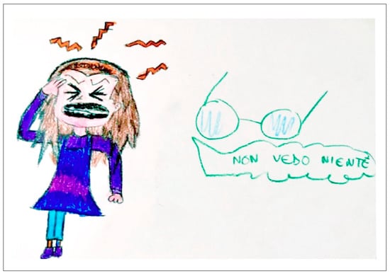
There is substantial variability in the characteristics of visual aura, with some commonalities across individuals. Recently, Viana et al. undertook a comprehensive review of all currently available data from clinical studies dealing with visual hallucinations in migraineur adults [22] and showed how different, and often complex, visual disturbances could be. Despite the smaller amount of evidence compared to adult patients, unusual visual aura has also been reported in children and adolescents. Polychromatic figures and formed shapes (e.g., dots, circles, triangles, squares, stars) [23] can mimic occipital seizures but duration, localization, and movement pattern, as previously mentioned, may provide guidance for the correct diagnosis (Figure 1).
Figure 1. Graphical representation of phosphenes. The researchers report two explanatory drawings, drawn by two children who arrived at the Department of Neuropsychiatry of Di Cristina Hospital in Palermo. (A) Graphic representation of phosphenes in a child with occipital epilepsy with seizure without loss of consciousness. (B) Graphic representation of phosphenes in a child with migraine with aura. The high similarity between the two symptoms complicates the differential diagnosis. (Courtesy of Dr. V. Raieli).
Unfortunately, visual disturbances such as visual loss (scotoma, amaurosis, and blindness), palinopsia, blurring vision, and macro or micropsia (as in the Alice in Wonderland syndrome) are more challenging and may mislead clinicians. In those cases, a detailed clinical history may reveal the event’s true nature and further direct investigations [24].

3.1. Visual Loss

Ictal amaurosis, blindness, or severe blurring of vision, limited to one hemifield, quadrant, or involving the entire visual field are common ictal phenomenon in idiopathic childhood occipital epilepsy (Gastaut-type), occurring in-two thirds of such patients and usually following the visual hallucinations, and last up to 5 min [25]. However, they may occasionally constitute the first symptom or the only ictal manifestation with abrupt onset [26][27][28]. In 70% of patients [29], it is associated with horizontal deviation of the eyes and progresses to varying associated focal convulsions. In the absence of any other seizure phenomenon, it may not be possible to differentiate migraine-induced transient cortical blindness from that induced by a seizure disorder. Migraine-induced transient cortical blindness can result from the scotoma resulting from the visual aura or can underlie a basilar or retinal migraine (Figure 2).
Figure 2. Graphical representation of blindness. The researchers report explanatory drawing of blindness, drawn by a child with migraine who visited the Department of Neuropsychiatry of Di Cristina Hospital in Palermo. Translation of the written portion: “I don’t see anything”. (Courtesy of Dr. V. Raieli).
Retinal migraine is sporadic among pediatric migraine patients, accounting for <2%; in this disorder, monocular scotoma or vision loss is reported, rather than hemifield deficits typical in the visual aura.

3.2. Palinopsia and Polioplia

Palinopsia is derived from the Greek words palin (again) and opsis (vision) and describes the perseveration of visual images. Palinopsia is a cardinal symptom of ictal clinical manifestations of occipital lobe seizures [28]. Belcastro et al. 2011 [30] described palinopsia events that lasted for seconds in 10% of 200 adults affected by migraine with and without aura. Polyopia is an uncommon visual phenomenon and an infrequent symptom of central nervous system disease, mainly occipital or temporal lesions [31], often associated with palinopsia, defined as the perception of many copies of objects or faces [32]. An interesting case report with a child affected by polioplia associated with the hemianopsia during migraine attack has been described in children affected by migraine [33][34]

3.3. Visual Snow

Visual snow is described as the electronic “snow” on a television set when the primary video signal is either reduced or absent, as well as pixelated fuzz and bubbles. It can be due to different etiologies and it has variable duration. When this symptom is associated with palinopsia, enhanced entoptic imagery, “nyctalopia”, and photophobia/photosensitivity, visual snow syndrome (VSS) can be diagnosed. In the anecdotal cases of migrainous patients reported in the literature [35], visual snow is usually short and without associated symptoms. In contrast, VSS was reported by Polster et al. [36] in two patients with suspected occipital epilepsy.

3.4. Other Complex Positive Symptoms

In migraineur children, visual hallucinations including landscapes, animals, human bodies, or faces, or the perception of mysterious objects that may aggregate to form complex scenes, are limited to anecdotical reports [37][38]. As epileptic symptoms, Panayiotopoulos described only three patients who reported “large objects, probably people, which I cannot identify”. Clear perception of people is reported in adults affected by different disease (hemorrhage, stroke, malformations, neoplasia) and are usually associated with medial temporolimbic seizure [39], and less commonly with lateral temporal seizure and rarely with occipital seizure [40]. In contrast, perceptual distortions of the patient’s body or objects in the environment, as described in Alice in Wonderland syndrome (AWS), are frequently reported in children [4][41]. AWS can present as micropsia (objects appear too small), macropsia (objects appear too large), metamorphopsia (objects appear too fat, thin, short, tall, etc.), teleopsia (objects appear further away than they are), and pelopsia (objects appear closer than they are). The symptom duration is widely variable, differs in each person, and resolves without sequelae. The cause of these symptoms is debated and remains unknown in almost 20% of cases [41]. In 2016, Blom et al. [41] showed that out of 166 published cases of AIWS, migraine was the most common cause (27.1%), followed by infections (22.9%), principally EBV (15.7%), brain lesions (7.8%), medication (6%) and drugs (6%), psychiatric disorders (3.6%), temporal or focal epilepsy (3%), disease of the peripheral nervous system (1.2%), and others (3%). In nearly 50% of patients [42], a family history of migraine or Alice in Wonderland syndrome can be detected. When occurring in children without other conditions, it usually predicts a higher chance of developing migraines in the future [9]. When AWS is associated with migraine, the symptoms can occur before, during, or after a migraine headache; micropsia and teleopsia have been more frequently reported [43].

References

  1. Victor, T.; Hu, X.; Campbell, J.; Buse, D.; Lipton, R. Migraine Prevalence by Age and Sex in the United States: A Life-Span Study. Cephalalgia 2010, 30, 1065–1072.
  2. Tarasco, V.; Grasso, G.; Versace, A.; Castagno, E.; Ricceri, F.; Urbino, A.F.; Pagliero, R. Epidemiological and Clinical Features of Migraine in the Pediatric Population of Northern Italy. Cephalalgia 2016, 36, 510–517.
  3. Genizi, J.; Khourieh Matar, A.; Zelnik, N.; Schertz, M.; Srugo, I. Frequency of Pediatric Migraine with Aura in a Clinic-Based Sample. Headache 2016, 56, 113–117.
  4. Shevell, M.I. Acephalgic Migraines of Childhood. Pediatr. Neurol. 1996, 14, 211–215.
  5. Pacheva, I.H.; Ivanov, I.S. Migraine Variants--Occurrence in Pediatric Neurology Practice. Clin. Neurol. Neurosurg. 2013, 115, 1775–1783.
  6. Ziegler, D.K.; Hassanein, R.S. Specific Headache Phenomena: Their Frequency and Coincidence. Headache 1990, 30, 152–156.
  7. Agessi, L.M.; Villa, T.R. Vestibular Migraine with Visual Aura and Olfactory Hallucination in Children: Two Case Reports. Neuropediatrics 2018, 49, 414–416.
  8. Raieli, V.; Capizzi, M.; Marino, A.; Di Nardo, G.; Raucci, U.; Parisi, P. Study on “Atypical” Migraine Auras in the Pediatric Age: The Role of Cortical Spreading Depression and the Physiopathogenetic Hypothesis Arising from Our Clinical Cases. Life 2022, 12, 450.
  9. Rastogi, R.G.; VanderPluym, J.; Lewis, K.S. Migrainous Aura, Visual Snow, and “Alice in Wonderland” Syndrome in Childhood. Semin. Pediatr. Neurol. 2016, 23, 14–17.
  10. Queiroz, L.P.; Friedman, D.I.; Rapoport, A.M.; Purdy, R.A. Characteristics of Migraine Visual Aura in Southern Brazil and Northern USA. Cephalalgia 2011, 31, 1652–1658.
  11. Russel, M.B.; Olesen, J. A Nosographic Analysis of the Migraine Aura in a General Population. Brain 1996, 119 Pt 2, 355–361.
  12. Schott, G.D. Exploring the Visual Hallucinations of Migraine Aura: The Tacit Contribution of Illustration. Brain 2007, 130, 1690–1703.
  13. Petrusic, I.; Pavlovski, V.; Vucinic, D.; Jancic, J. Features of Migraine Aura in Teenagers. J. Headache Pain 2014, 15, 87.
  14. Hadjikhani, N.; Vincent, M. Visual Perception in Migraine: A Narrative Review. Vision 2021, 5, 20.
  15. Lawden, M.C.; Cleland, P.G. Achromatopsia in the Aura of Migraine. J. Neurol. Neurosurg. Psychiatry 1993, 56, 708–709.
  16. Noseda, R.; Bernstein, C.A.; Nir, R.R.; Lee, A.J.; Fulton, A.B.; Bertisch, S.M.; Hovaguimian, A.; Cestari, D.M.; Saavedra-Walker, R.; Borsook, D.; et al. Migraine Photophobia Originating in Cone-Driven Retinal Pathways. Brain 2016, 139, 1971–1986.
  17. Olesen, J.; Bes, A.; Kunkel, R.; Lance, J.W.; Nappi, G.; Pfaffenrath, V.; Rose, F.C.; Schoenberg, B.S.; Soyka, D.; Tfelt-Hansen, P.; et al. The International Classification of Headache Disorders, 3rd Edition (Beta Version). Cephalalgia 2013, 33, 629–808.
  18. Caraballo, R.; Koutroumanidis, M.; Panayiotopoulos, C.P.; Fejerman, N. Idiopathic Childhood Occipital Epilepsy of Gastaut: A Review and Differentiation from Migraine and Other Epilepsies. J. Child Neurol. 2009, 24, 1536–1542.
  19. Gastaut, H. Benign Epilepsy of Childhood with Occipital Spike and Wave Complexes; Andermann, F., Lugaresi, E., Eds.; Butterworths: Boston, MA, USA, 1987.
  20. Hartl, E.; Angel, J.; Rémi, J.; Schankin, C.J.; Noachtar, S. Visual Auras in Epilepsy and Migraine–An Analysis of Clinical Characteristics. Headache 2017, 57, 908–916.
  21. Jobst, B.C. Focal Seizures with Auditory Hallucinations. In Atlas of Epilepsies; Springer: London, UK, 2010; pp. 447–450.
  22. Viana, M.; Tronvik, E.A.; Do, T.P.; Zecca, C.; Hougaard, A. Clinical Features of Visual Migraine Aura: A Systematic Review. J. Headache Pain 2019, 20, 64.
  23. Shapiro, H.F.J.; Lebel, A. Pediatric Episodic Migraine with Aura: A Unique Entity? Children 2021, 8, 228.
  24. Flanagan, D.; Ghose, K. Co-Morbidity of Migraine and Epilepsy: A Review of Clinical Features. J. Headache Pain 2000, 1, 137.
  25. Guerrini, R.; Genton, P. Epileptic Syndromes and Visually Induced Seizures. Epilepsia 2004, 45 (Suppl. S1), 14–18.
  26. Panayiotopoulos, C.P. Benign Childhood Partial Seizures and Related Epileptic Syndromes; John Libbey & Company Ltd.: London, UK, 1999.
  27. Panayiotopoulos, C. Benign Childhood Focal Seizures and Related Epileptic Syndromes. In The Epilepsies: Seizures, Syndromes and Management; Bladon Medical Pubblishing: Chipping Norton, UK, 2005.
  28. Adcock, J.E.; Panayiotopoulos, C.P. Occipital Lobe Seizures and Epilepsies. J. Clin. Neurophysiol. 2012, 29, 397–407.
  29. Rajapakse, T.; Buchhalter, J. The Borderland of Migraine and Epilepsy in Children. Headache 2016, 56, 1071–1080.
  30. Belcastro, V.; Maria Cupini, L.; Corbelli, I.; Pieroni, A.; D’Amore, C.; Caproni, S.; Gorgone, G.; Ferlazzo, E.; Di Palma, F.; Sarchielli, P.; et al. Palinopsia in Patients with Migraine: A Case-Control Study. Cephalalgia 2011, 31, 999–1004.
  31. Kataoka, H.; Ueno, S. Cerebral Polyopia and Palinopsia in a Patient with Occipital Lobe Epilepsy. Epilepsy Behav. 2009, 14, 684–686.
  32. Bender, M.B.; Rudolph, S.H.; Stacy, C. The Neurology of the Visual and Oculomotor Systems; Baker, A.B., Baker, J.R., Eds.; Harper-Row: Philadelphia, PA, USA, 1986; Volume 43.
  33. Raieli, V.; Eliseo, G.L.; La Franca, G.; La Vecchia, M.; Puma, D.; Eliseo, M. Cerebral Polyopia in Migraine: A Clinical Case. J. Headache Pain 2000, 1, 127–129.
  34. Raieli, V.; Monastero, R.; Santangelo, G.; Eliseo, G.L.; Eliseo, M.; Camarda, R. Red Ear Syndrome and Migraine: Report of Eight Cases. Headache 2002, 42, 147–151.
  35. Simpson, J.C.; Goadsby, P.J.; Prabhakar, P. Positive Persistent Visual Symptoms (Visual Snow) Presenting as a Migraine Variant in a 12-Year-Old Girl. Pediatr. Neurol. 2013, 49, 361–363.
  36. Polster, T.; Schaefer, S.; Panzer, A. Visual Snow–Two Adolescents with a New Syndrome Different from Migraine or Epilepsy. Neuropediatrics 2019, 50, GNP-FV20.
  37. Smith, R.A.; Wright, B.; Bennett, S. Hallucinations and Illusions in Migraine in Children and the Alice in Wonderland Syndrome. Arch. Dis. Child. 2015, 100, 296–298.
  38. Akiyama, O.; Kondo, A.; Akiyama, I. Paediatric Migraine with Visual Hallucination Auras Appearing in the Form of a Human Body. BMJ Case Rep. 2019, 12, e232358.
  39. Gloor, P.; Olivier, A.; Quesney, L.F.; Andermann, F.; Horowitz, S. The Role of the Limbic System in Experiential Phenomena of Temporal Lobe Epilepsy. Ann. Neurol. 1982, 12, 129–144.
  40. Sakamoto, Y.; Suzuki, R.; Ohara, T.; Miyagi, T.; Osaki, M.; Nishimura, K.; Toyoda, K. Complex Visual Hallucinations as the Sole Manifestation of Symptomatic Temporo-Occipital Lobe Epilepsy Due to Old Intracerebral Hemorrhage. Seizure 2014, 23, 244–246.
  41. Blom, J.D. Alice in Wonderland Syndrome: A Systematic Review. Neurol. Clin. Pract. 2016, 6, 259–270.
  42. Liu, A.M.; Liu, J.G.; Liu, G.W.; Liu, G.T. “Alice in Wonderland” Syndrome: Presenting and Follow-up Characteristics. Pediatr. Neurol. 2014, 51, 317–320.
  43. Swartwood, S.; Pham, K.; Candee, M.S. Pediatric Migraine Phenomena and Variants: Don’t Let Them Go Over Your Head. Curr. Pain. Headache Rep. 2020, 24, 47.
More
Information
Subjects: Neurosciences
Contributors MDPI registered users' name will be linked to their SciProfiles pages. To register with us, please refer to https://encyclopedia.pub/register : , , , ,
View Times: 490
Revisions: 2 times (View History)
Update Date: 04 May 2023
1000/1000
Video Production Service