Your browser does not fully support modern features. Please upgrade for a smoother experience.
Submitted Successfully!
 +1 credit
Thank you for your contribution! You can also upload a video entry or images related to this topic.
For video creation, please contact our Academic Video Service.
| Version | Summary | Created by | Modification | Content Size | Created at | Operation |
|---|---|---|---|---|---|---|
| 1 | Kamil Nelke | -- | 2400 | 2023-04-27 12:03:22 | | | |
| 2 | Rita Xu | Meta information modification | 2400 | 2023-04-28 03:18:31 | | |
Video Upload Options
We provide professional Academic Video Service to translate complex research into visually appealing presentations. Would you like to try it?
Cite
If you have any further questions, please contact Encyclopedia Editorial Office.
Nelke, K.; Łuczak, K.; Pawlak, W.; Janeczek, M.; Pasicka, E.; Morawska-Kochman, M.; Błaszczyk, B.; Błaszczyk, T.; Dobrzyński, M. Unilateral Condylar Hyperplasia. Encyclopedia. Available online: https://encyclopedia.pub/entry/43569 (accessed on 26 July 2026).
Nelke K, Łuczak K, Pawlak W, Janeczek M, Pasicka E, Morawska-Kochman M, et al. Unilateral Condylar Hyperplasia. Encyclopedia. Available at: https://encyclopedia.pub/entry/43569. Accessed July 26, 2026.
Nelke, Kamil, Klaudiusz Łuczak, Wojciech Pawlak, Maciej Janeczek, Edyta Pasicka, Monika Morawska-Kochman, Bartłomiej Błaszczyk, Tomasz Błaszczyk, Maciej Dobrzyński. "Unilateral Condylar Hyperplasia" Encyclopedia, https://encyclopedia.pub/entry/43569 (accessed July 26, 2026).
Nelke, K., Łuczak, K., Pawlak, W., Janeczek, M., Pasicka, E., Morawska-Kochman, M., Błaszczyk, B., Błaszczyk, T., & Dobrzyński, M. (2023, April 27). Unilateral Condylar Hyperplasia. In Encyclopedia. https://encyclopedia.pub/entry/43569
Nelke, Kamil, et al. "Unilateral Condylar Hyperplasia." Encyclopedia. Web. 27 April, 2023.
Copy Citation
Unilateral condylar hyperplasia (UCH) is a pathology most commonly present in one side of the mandible, characterized by an abnormal condylar process of progressive overgrowth in time, causing visible changes in the anatomy, shape, and size of condylar head and neck elongation, along with visible facial asymmetry features.
condylar hyperplasia
unilateral condylar hyperplasia
condylectomy
1. Introduction
Each mandibular asymmetry (MA) case should be carefully evaluated. UCH remains a challenge for surgeons since each case might be different. This situation is related to the volume of overgrowth, the duration of growth and its intensity, the scope of symptoms, and the formation of both dentoalveolar and skeletal disturbances, and many possible algorithms that could be used to treat it are known [1][2][3][4][5].
Clinically, the visible central incisor dental midline and/or upper/lower lips’ frenulum deviate from the central midline of the body, along with disproportions in the symmetry of the cupid’s bow, the lips’ angle exposure, and lip position. Skeletal, dental, and soft/hard tissue disproportions might occur, which are related to maxillary bone tilting and the scope of asymmetry, mainly impacted by time and the intensity of pathological growth [6][7]. Present bone and teeth discrepancies influence facial contour and balance, which influence facial appearance aspects, such as lip contour and symmetry, the angle of the lips, chin position, subnasal and sublabial balance, zygomaticomalar area prominence, and others [1][2][3][4][5][8][9][10].
The presented pathology might start early, at 9–12 years of age (±2 years), and can have various forms of intensity and growth activity, and even have a natural cessation period and self-limiting over time [1][2][3][4][5][9]. In most cases, mandibular asymmetry should be carefully examined and established, especially in young, growing patients. Growing children and young adults are most affected by this condition, and a higher female predominance (F:M ratio) is noted and estimated at 2:1 or 3:1 [9][10][11][12][13]. On the other hand, some authors emphasize no sex-related occurrence of this pathological condition, while others underline a higher occurrence rate in the left condyle area [9][10]. Raijmakers et al.’s meta-analysis study in 2012 explored 10 studies with 275 patients with UCH [10]. The presented results indicated a female predominance, along with a higher occurrence rate on the left side of the mandible, which has also been confirmed in other studies [11]. A study on two Indian families by Mahajan suggests a possible Y-linked or autosomal dominant etiology of UCH [12]. Both mother- and son-related genetics need further genetic evaluation. A similar rare genetic link was reported by Yang et al. in two siblings with mirror image UCH [13]. The role of potential etiological factors is still not fully known; however, some endocrine and hormone-related factors are also taken into consideration [9][10][11][12][13][14]. Possible etiological factors may be various since no evidence-based relationship has so far been noted in the literature. Some literature reviews suggest the following factors: trauma-related, metabolic hyperactivity, endocrinal distortions (growth factors, insulin growth factors (IGF), hormonal estrogen–progesterone related, and others), arthrosis, genetic factors, hereditary or acquired factors, or others [13][14][15][16][17].
2. Mandibular Asymmetries Differentiation
An important topic is the differentiation between UCH and any other forms of mandibular asymmetries (MA), which can be related to different scopes of etiological factors, including: trauma, inflammation, TMJ abnormalities, condyle/head anatomical variations, benign/malignant TMJ tumors, cases with syndromic/non-syndromic genetic syndromes, or others. Mandibular asymmetries can be related to various factors. Anatomically, asymmetry can be described as the under- or over-development of one side of the mandible (ramus, angle, body, symphysis, or condyles) from various factors. Chia et al. underline some key factors that might divide MA into developmental (hemimandibular hyperplasia (HH), hemimandibular elongation (HE), hemifacial microsomia, Parry-Romberg), pathological (cysts, tumors, idiopathic condylar resorption, infections), traumatic (condylar fracture), and functional once (mandibular displacements, luxations, and others) [18]. Other developmental or genetic-related factors co-existing with mandibular asymmetry might include changes in the eye socket, external/internal ear area, temporomandibular area joint structural morphology, zygomatic and maxillary bones, or facial nerve functioning [1][2][3][4][5][8][15][16][17][19].
Other classifications of MA with recognition of dental, skeletal, and occlusal asymmetries features include Bishara et al.’s studies [2]. The mentioned asymmetry has been divided into dental, skeletal, muscular, functional, and a combination of all. On the other hand, a study by Reyeneke et al. divided MA into congenital, developmental, post-traumatic, and pathology-related categories [3]. Other classifications are also known [4][5].
Concerning the temporomandibular joint (TMJ), one of its components is the mandibular condyle (MC), along with its head and neck. Its position in the glenoid fossa of the temporal bone is limited anteriorly by the articular eminence. During growth and maturation, appropriate condyle growth ensures its adequate shape, size, and position. When some growth disturbances or abnormalities occur, various forms of condylar disfigurement might be present. Growth disturbances in the first trimester might result in condyle aplasia, hypoplasia, or related disorders. An understanding of the anatomy of the MC within the glenoid fossa can help in any surgery planning in UCH cases [13][14][15][16][17][20].
3. Classifications
Some types of mandibular asymmetry (MA), named mandibular condyle hyperplasia (CH) or unilateral condyle hyperplasia (UCH), are related to one-sided pathological growth in one of the affected condyles of an unknown origin. The presence of MA might start early in puberty and last even until the patient is fully grown, or the pathological growth might become self-limiting over time, in either way causing visible mandible asymmetry and lack of facial balance with the presence of various dentoalveolar discrepancies [7]. In 1986, Obwegeser and Makek used the first classification to distinguish hemimandibular hyperplasia (HH), an abnormal self-limiting condylar growth, resulting in pathologic one-sided bone overgrowth of the entire mandible on the affected side (Figure 1), as well as other forms such as hemimandibular elongation (HE) and a mixed, rare form HH-HE [6]. Currently, a different classification presented by Wolford et al., which includes some temporomandibular joint manifestations, is also known [1]. Types IA (the most common) and IB have excessive growth in the horizontal vector, while IIA and IIB have excessive growth in the vertical vector. Type III is caused by benign tumors (e.g., chondroma, osteoma), while type IV (the rarest) is caused by malignant tumors (e.g., chondrosarcoma, osteosarcoma) in the TMJ area [1].
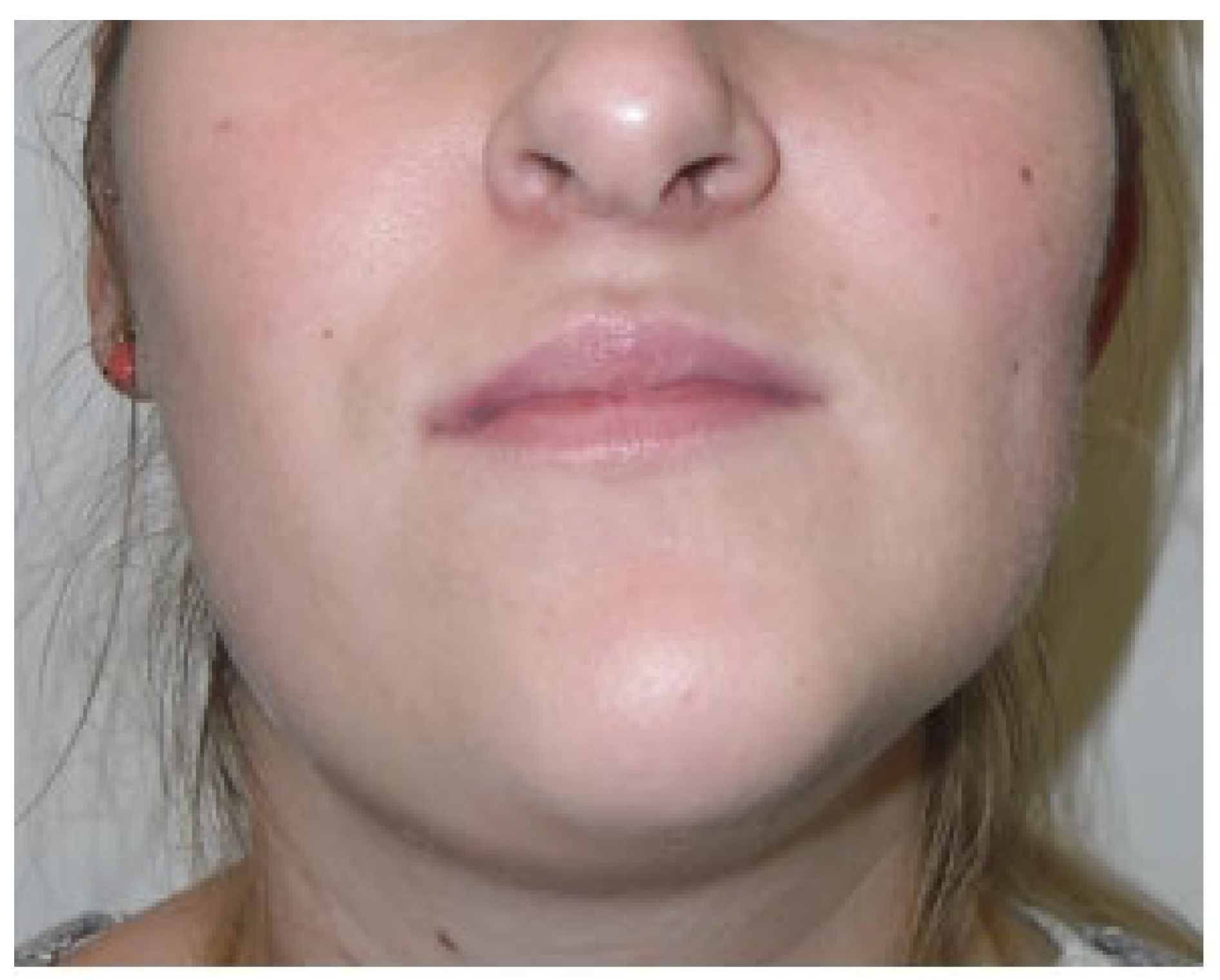
Figure 1. Typical facial appearance of UCH. A notable elongation and overgrowth of the right side of the face with chin asymmetry is visible. Changes in proportions of mouth angles are clearly visible along with the different positions of mandibular gonial diameters.
After the MA etiology and scope of changes within the mandibular body are evaluated, it is quite easy to identify UCH and schedule the necessary proceedings (Table 1).
Table 1. Authors’ proposal on the scope of approaches. Each of the presented cases should be scheduled for orthodontic treatment because of various scopes of dentoalveolar discrepancies. (1) Case A: asymmetry (MA) without pathological growth and no UCH symptoms: scheduled for orthodontic or surgical camouflage or solely bone remodeling if no dentoalveolar discrepancies are present. (2) Case B–C: rapid or slowly progressive in time FA with MA asymmetry and pathological growth still present in the affected condyle confirmed in SPECT and during clinical examination: mandatory condylectomy followed by orthodontic treatment. After 3–6 months the scope of asymmetry, degree of maxillary tilting, and patient’s willingness to undergo total symmetry correction decide the final approaches. If the scope of mandibular overgrowth is extensive some surgical camouflage or bone remodeling procedures can be chosen. (3) Case D: low growth in SPECT with a tendency to decrease in time: early orthodontics followed by patient monitoring. If the orthodontics is sufficient, then the treatment is finalized. On the other hand, when growth has caused asymmetrical bone discrepancies and orthodontics alone is not sufficient a decision on either BSSO/Lefort I or surgical camouflage could be used. (4) Case E: growth became limited over time: orthodontics is sufficient to prevent MA and FA. In some difficult cases, either BIMAX, BSSO, Lefort I, or another approach can be scheduled based on each individual case. Abbreviations: BIMAX—includes BSSO and Lefort I procedures performed simultanously; ORT—orthodontic treatement.
| 1. | Growth Activity | SPECT Monitoring | Condylectomy | Maxillary Tilting | Presence of Overgrowth | |
|---|---|---|---|---|---|---|
| No growth present | Are orthodontics sufficient? | Surgical camo | ||||
| A | Not present | One study | NO | ORT or surgical camo | Genioplasty - Wing-osteotomy - Marginectomy - Bone drilling and chiseling - Corrective ostectomies - Bone remodeling procedures - Facial contouring - Facial implants |
|
| Some forms of growth: | YES | NO | ||||
| B | Active growth: rapidly progressive in time | SPECT: at least two studies | YES | ± BIMAX BSSO/Lefort I ± ORT ± ORT camo ± Surg camo |
± BSSO ± ORT ± ORT camo ± Surg camo |
|
| C | Active growth: slowly progressive in time | SPECT: at least two studies | YES | ± BIMAX BSSO/Lefort I ± ORT ± ORT camo ± Surg camo |
± BSSO ± ORT ± ORT camo ± Surg camo |
|
| D | Growth self-limiting in time | Each one per year till absence of growth | NO | ± Surg camo BSSO/Lefort I ± ORT ± ORT camo |
± BSSO ± ORT ± ORT camo ± Surg camo |
|
| E | Growth cessation | One study | NO | ± Lefort I ± BSSO ± ORT ± ORT camo |
± ORT ± ORT camo ± Surg camo |
|
4. Signs and Symptoms
A close, detailed patient evaluation might be made according to clinical and radiological signs and symptoms. Facial and/or mandibular asymmetry can cause various co-existing problems related to facial appearance, chewing, bite, occlusion, TMJ function, and in some cases even breathing and an unbalanced proper posture [20][21][22][23][24].
To begin with, the scope of mandibular asymmetry should be closely evaluated since its intensity and occurrence and changes in patients’ skeletal appearance are greatly correlated with the excess of growth pathology [1][2][3][4][5][6][7][8][18][19]. Some typical signs and symptoms can be addressed according to the Obwegeser, Wolford, or Kaban classifications of asymmetries, in detailed relation to the type of mandibular and skeletal asymmetry [1][2][3][4][5][6][7][8][18][19][20][21]. First of all, a detailed evaluation of any pain-related TMJ joint symptoms should be performed since, in some cases, patients’ TMJ pain in UCH might be present and result in condyle volume enlargement and its possible dysfunction. An enlarged condyle head might have a different shape and size with exophytic growth present on its surface or even some osteosclerosis or increased bone density [17][20][21][22][23][24]. The shape and size of the affected side are also age- and growth-related. It is worth noting that growth finishes earlier in females than males, at approximately 18 ± 1 year of age in females and up until 21 years of age in males [2][13][22]. Because of different pathological growth factors and the timing of its occurrence in UCH, the scope of mandibular overgrowth might greatly impact not only the shape, size, and position of the mandible. Secondary bone asymmetry, deviation, or lack of balance might also be present in the maxillary bone and might influence the maxillary sinus volume, along with the nasal septum. The degree of dentoalveolar discrepancy has an impact on proper chewing, bite, and occlusion, along with disrupted facial balance [3][10][18].
In the dental aspect, an open bite with the presence of various dentoalveolar discrepancies is found. Often, an open bite on the affected side is present, followed by a crossbite on the contralateral side. Dental balance is always disturbed. Along with mandibular asymmetrical growth, teeth position might result in various forms of open bites and crossbites on the affected side, along with others. The incisor midline may be deviated and shifted towards the healthy side with one of the possible variances in the volume of the increased anteroposterior and mediolateral dimensions of the right side upper and lower dental arches. Incisor tilting towards the affected side is common. Both dental, skeletal, and even labial frenulum symmetry lines can deviate. The types of open bite and molar contact are mostly dependable on the value of UCH condyle growth. Growth in vertical or horizontal aspects might lead to features of a skeletal class III or skeletal open bite (class II is very rarely seen). In slow-growing cases, the maxillary molars are capable of maintaining some contact with the mandibular molars. An open bite with a posterior crossbite might occur. In HH/HE cases, similar features of mandibular prognathism with malocclusion class III according to angle are quite common [2][3][13][18][22].
Skeletal manifestations with visible mandibular asymmetry consisted in most cases of the mandibular ramus’s vertical height being enlarged, followed by a typical antegonial notch, which can be less or more visible on CT/panx [8]. Secondly, the chin position might deviate towards the healthy side, be slightly protruded, or be a combination of the two. The body and mandibular ramus on the affected side may be elongated with an increased anteroposterior thickness of the mandibular body, and the characteristic shape of the gonial angle with a low-settled mandibular canal towards the inferior mandibular border is most commonly found [23][24][25][26][27]. The lateral aspect of the mandibular base can also be enlarged and curved. The chin position might be protruded and deviated in a skeletal class III-like matter. The aspect of increased cortical thickness in the overgrowth bone might be more visible in the affected condyle area. In some situations, the increased volume of the head might decrease mouth opening and some mandibular movements and cause a more visible open bite over time or have a continuous small growth causing simultaneous down growth of the maxillary bone on the affected side.
Some atypical bone features might include asymmetrical growth of the maxillary bones, changes in the volume and size of maxillary sinuses, an increased horizontal volume of the maxillary dental process on the affected side, or other case-specified situations. Some authors also conclude incidence of changes in the shape of the anterior nares, deviation in the nasal septum, and palatal cant. These could also be related to disrupted nasal breathing because of the scope of skeletal malocclusion [14][27][28][29][30].
Soft tissues are characteristics for chin asymmetry, sloping rima oris, lips asymmetry, deviation of the mandibular angle, and convex profile on the affected side, which, in some cases, is also related to alleged eye asymmetry, which is related to head lining for patients’ subconscious adjustment for asymmetry (Figure 2). A study by Fariña et al. with detailed measurements on 20 patients’ vertically increased mandibular ramus concluded that the mandibular lingula is the lowest and most stable reference point for any measurements, while the sigmoidal notch is neither a suitable nor a stable reference point [29][30]. On the other hand, a study by Olate et al., which measured various reference lines between the condylar head and midline, suggests that UCH always influences facial transversal asymmetry [30].
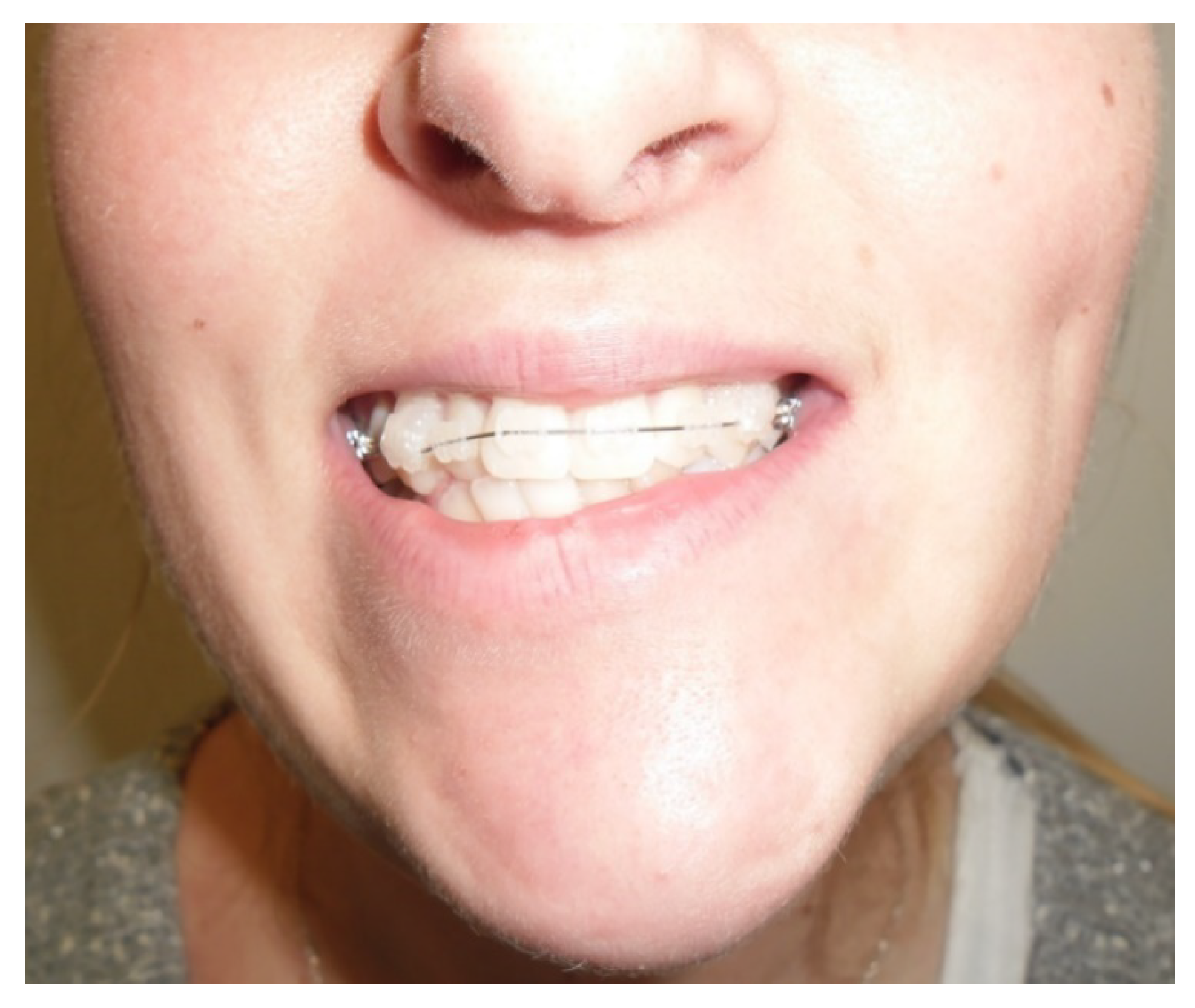
Figure 2. Visible lack of balance in lip contour with deviated maxillary bite plane and open bite present on the affected right side. The degree of maxillary deviation is related to the degree and volume of abnormal growth in the affected condyle over time. Open bite suggests an increased volume of pathological growth and low compensatory maxillary downward rotation. In the following cases, orthodontic treatment has its limitations, even after a condylectomy.
References
- Wolford, L.M.; Movahed, R.; Perez, D.E. A Classification System for Conditions Causing Condylar Hyperplasia. J. Oral Maxillofac. Surg. 2014, 72, 567–595.
- Bishara, S.E.; Burkey, P.S.; Kharouf, J.G. Dental and Facial Asymmetries: A Review. Angle Orthod. 1994, 64, 89–98.
- Reyneke, J.P. Orthognathic Essentials of Surgery: Second Edition; Quintessence Books: Chandler Drive, IL, USA, 2010.
- Iyer, J.; Hariharan, A.; Cao, U.M.N.; Tran, S.D. Acquired Facial, Maxillofacial, and Oral Asymmetries—A Review Highlighting Diagnosis and Management. Symmetry 2021, 13, 1661.
- Andrade, N.N.; Mathai, P.; Aggarwal, N. Facial Asymmetry. In Oral and Maxillofacial Surgery for the Clinician; Bonanthaya, K., Panneerselvam, E., Manuel, S., Kumar, V.V., Rai, A., Eds.; Springer Nature: Singapore, 2021; pp. 1549–1576. ISBN 9789811513466.
- Obwegeser, H.L.; Makek, M.S. Hemimandibular Hyperplasia—Hemimandibular Elongation. J. Maxillofac. Surg. 1986, 14, 183–208.
- Rodrigues, D.B.; Castro, V. Condylar Hyperplasia of the Temporomandibular Joint: Types, Treatment, and Surgical Implications. Oral Maxillofac. Surg. Clin. N. Am. 2015, 27, 155–167.
- Wink, J.D.; Goldstein, J.A.; Paliga, J.T.; Taylor, J.A.; Bartlett, S.P. The Mandibular Deformity in Hemifacial Microsomia: A Reassessment of the Pruzansky and Kaban Classification. Plast. Reconstr. Surg. 2014, 133, 174e–181e.
- Almeida, L.E.; Zacharias, J.; Pierce, S. Condylar Hyperplasia: An Updated Review of the Literature. Korean J. Orthod. 2015, 45, 333–340.
- Raijmakers, P.G.; Karssemakers, L.H.E.; Tuinzing, D.B. Female Predominance and Effect of Gender on Unilateral Condylar Hyperplasia: A Review and Meta-Analysis. J. Oral Maxillofac. Surg. 2012, 70, e72–e76.
- Nelke, K.H.; Pawlak, W.; Morawska-Kochman, M.; Łuczak, K. Ten Years of Observations and Demographics of Hemimandibular Hyperplasia and Elongation. J. Craniomaxillofac. Surg. 2018, 46, 979–986.
- Mahajan, M. Unilateral Condylar Hyperplasia—A Genetic Link? Case Reports. Natl. J. Maxillofac. Surg. 2017, 8, 58–63.
- Yang, J.; Lignelli, J.L.; Ruprecht, A. Mirror Image Condylar Hyperplasia in Two Siblings. Oral Surg. Oral Med. Oral Pathol. Oral Radiol. Endod. 2004, 97, 281–285.
- Portelli, M.; Gatto, E.; Matarese, G.; Militi, A.; Catalfamo, L.; Gherlone, E.; Lucchese, A. Unilateral Condylar Hyperplasia: Diagnosis, Clinical Aspects and Operative Treatment. A Case Report. Eur. J. Paediatr. Dent. 2015, 16, 99–102.
- Mehrotra, D.; Dhasmana, S.; Kamboj, M.; Gambhir, G. Condylar Hyperplasia and Facial Asymmetry: Report of Five Cases. J. Maxillofac. Oral Surg. 2011, 10, 50–56.
- Singh, V.; Verma, A.; Attresh, G.; Batra, J. Ortho-Surgical Management of Condylar Hyperplasia: Rare Case Reports. Natl. J. Maxillofac. Surg. 2014, 5, 54–59.
- Ma, X.; Wang, H.; Zhang, X. . Hua Xi Kou Qiang Yi Xue Za Zhi 2014, 32, 150–152.
- Chia, M.S.; Naini, F.B.; Gill, D.S. The Aetiology, Diagnosis and Management of Mandibular Asymmetry. Orthod. Updat. 2008, 1, 44–52.
- Madrid, J.R.P.; Montealegre, G.; Gomez, V. A New Classification Based on the Kaban’s Modification for Surgical Management of Craniofacial Microsomia. Craniomaxillofac. Trauma. Reconstr. 2010, 3, 1–7.
- Fisch, A.W.; Espinosa, C.I.; Quezada, S.R. Facial Asymmetry Secondary to Mandibular Condylar Hyperplasia. A Case Report. Rev. Odontol. Mex. 2011, 15, 251–256.
- Brionne, C.; Cadre, B.; Laroche, Y.; Lhotellier, J.; Maze, M.; Raffre, A.; Sorel, O. The Diagnosis of Mandibular Assymmetries. J. Dentofac. Anom. Orthod. 2013, 16.
- Alyamani, A.; Abuzinada, S. Management of Patients with Condylar Hyperplasia: A Diverse Experience with 18 Patients. Ann. Maxillofac. Surg. 2012, 2, 17–23.
- Arora, K.S.; Bansal, R.; Mohapatra, S.; Pareek, S. Review and Classification Update: Unilateral Condylar Hyperplasia. BMJ Case Rep. 2019, 12, e227569.
- Nitzan, D.W.; Katsnelson, A.; Bermanis, I.; Brin, I.; Casap, N. The Clinical Characteristics of Condylar Hyperplasia: Experience with 61 Patients. J. Oral Maxillofac. Surg. 2008, 66, 312–318.
- Khawaja, S.N.; Crow, H.; Gonzalez, Y. Goldenhar Syndrome and Pain-Related Temporomandibular Disorders. A Case Report. N. Y. State Dent. J. 2016, 82, 21–24.
- GN, S.; Sharma, M.L.; JK, D.R.; Goel, S.; Srivastava, S. Facial Asymmetry in Young Adults with Condylar Hyperplasia-Unusual Changes in the Facial Bones. J. Clin. Diagn. Res. 2015, 9, ZD21–ZD23.
- Kawamoto, H.K.; Kim, S.S.; Jarrahy, R.; Bradley, J.P. Differential Diagnosis of the Idiopathic Laterally Deviated Mandible. Plast. Reconstr. Surg. 2009, 124, 1599–1609.
- De Stefano, A.A.; Di Chicco, A.; Alessandra, I.; Emanuela, S.; Guercio-Mónaco, E.; Galluccio, G. Unilateral Condylar Hyperplasia: A Thee-Dimensional CBCT Morphometric and Volumetric Evaluation of Mandibular Condyle by Open-Source Softwares. Int. J. Morphol. 2021, 39.
- Fariña, R.; Bravo, R.; Villanueva, R.; Valladares, S.; Hinojosa, A.; Martinez, B. Measuring the Condylar Unit in Condylar Hyperplasia: From the Sigmoid Notch or from the Mandibular Lingula? Int. J. Oral Maxillofac. Surg. 2017, 46, 857–860.
- Olate, S.; Cantín, M.; Alister, J.P.; Uribe, F.; Navarro, P.; Olate, G.; de Moraes, M. Relationship Between Condylar Size and Transverse Facial Asymmetry in Subject with Condylar Hyperplasia. Int. J. Morphol. 2013, 31, 937–941.
More
Information
Subjects:
Dentistry, Oral Surgery & Medicine
Contributors
MDPI registered users' name will be linked to their SciProfiles pages. To register with us, please refer to https://encyclopedia.pub/register
:
View Times:
977
Revisions:
2 times
(View History)
Update Date:
28 Apr 2023
Table of Contents


Notice
You are not a member of the advisory board for this topic. If you want to update advisory board member profile, please contact office@encyclopedia.pub.
OK
Confirm
Only members of the Encyclopedia advisory board for this topic are allowed to note entries. Would you like to become an advisory board member of the Encyclopedia?
Yes
No
${ textCharacter }/${ maxCharacter }
Submit
Cancel
Back
Comments
${ item }
|
${ item.createdUser.fullName }
${ item.createdAt }
${ item.vote }
${ item.reply }
Delete
${ reply.createdUser.fullName }
${ reply.createdAt }
${ reply.vote }
Delete
There is no reply to this comment~
${ item.replyTextCharacter }/${ item.replyMaxCharacter }
Submit
Cancel
More
No more~
There is no comment~
${ textCharacter }/${ maxCharacter }
Submit
Cancel
${ selectedItem.replyTextCharacter }/${ selectedItem.replyMaxCharacter }
Submit
Cancel
Confirm
Are you sure to Delete?
Yes
No