Submitted Successfully!
To reward your contribution, here is a gift for you: A free trial for our video production service.
Thank you for your contribution! You can also upload a video entry or images related to this topic.
Version Summary Created by Modification Content Size Created at Operation
1 -- 3330 2023-04-20 02:28:57 |
2 add 4. Conclusions Meta information modification 3330 2023-04-20 02:36:24 | |
3 update references and layout -1316 word(s) 2014 2023-04-20 05:51:07 |

Video Upload Options

Do you have a full video?

Confirm

Are you sure to Delete?
Cite
If you have any further questions, please contact Encyclopedia Editorial Office.
Wang, J.; Zhou, X.; Elazab, S.T.; Park, S.; Hsu, W.H. Pharmacokinetics of Macrolide Antibiotics in Respiratory Infection. Encyclopedia. Available online: https://encyclopedia.pub/entry/43270 (accessed on 20 August 2024).
Wang J, Zhou X, Elazab ST, Park S, Hsu WH. Pharmacokinetics of Macrolide Antibiotics in Respiratory Infection. Encyclopedia. Available at: https://encyclopedia.pub/entry/43270. Accessed August 20, 2024.
Wang, Jianzhong, Xueying Zhou, Sara T. Elazab, Seung-Chun Park, Walter H. Hsu. "Pharmacokinetics of Macrolide Antibiotics in Respiratory Infection" Encyclopedia, https://encyclopedia.pub/entry/43270 (accessed August 20, 2024).
Wang, J., Zhou, X., Elazab, S.T., Park, S., & Hsu, W.H. (2023, April 20). Pharmacokinetics of Macrolide Antibiotics in Respiratory Infection. In Encyclopedia. https://encyclopedia.pub/entry/43270
Wang, Jianzhong, et al. "Pharmacokinetics of Macrolide Antibiotics in Respiratory Infection." Encyclopedia. Web. 20 April, 2023.
Pharmacokinetics of Macrolide Antibiotics in Respiratory Infection
Edit

Macrolide antibiotics are important drugs to combat infections. The pharmacokinetics (PK) of these drugs are essential for the determination of their optimal dose regimens, which affect antimicrobial pharmacodynamics and treatment success. For most drugs, the measurement of their concentrations in plasma/serum is the surrogate for drug concentrations in target tissues for therapy.

serum/plasma concentrations pharmacokinetics macrolide antibiotics

1. Introduction

Macrolide antibiotics are a family of compounds featured by the existence of a macrocyclic lactone ring of ≥12 members [1][2]. The macrolide molecule is hydrophobic and is distributed in the extracellular fluid. In consideration of their satisfactory bioavailability via oral administration, superior tissue penetration and broad efficacy against many pulmonary pathogens, macrolides are extensively used as first-line antibiotics for the treatment of respiratory bacterial infections [3].
Pharmacokinetics (PK) describe the chronological movement of drugs within the body, i.e., the time course of the drug concentrations of serum/plasma or tissue fluid. Understanding the PK plays an important role in monitoring the antibiotic exposure in a patient. PK are most frequently evaluated by measuring the drug concentrations of serum/plasma. In addition to PK, the optimal dosing of an antibiotic also relies on the pharmacodynamics (PD) of the drug. The PD of a drug describe the relationship between drug concentration and pharmacological activity. Usually, the measurement of drug exposure is based on serum/plasma concentration–time course data [4]. Traditional PD are based on serum/plasma concentrations of a drug which achieve equilibrium with tissues. The selection of antibiotic concentrations aims to obtain an ideal exposure that should maximize the antibacterial activity and minimize antibiotic resistance [4][5][6][7]. Thus, most investigators have focused on the drug concentrations of serum/plasma for the PK/PD studies. However, the interstitial tissue is a site invaded by most bacteria [8]. The concentration of an antibiotic in the interstitial fluid (ISF) of the target tissue is essential for assessing the antibacterial effects [8]. Therefore, researchers realize that only the free (unbound) antibiotic concentration in the ISF of the target site is in charge of the antibacterial activity and is more applicable in determining clinical efficacy than serum/plasma concentration [9][10][11][12]. However, for most antibiotics, serum/plasma concentrations have been used to calculate PK/PD for the determination of the optimal dose regimens [11]. Meanwhile, the traditional PD parameters based on the serum/plasma concentration of a macrolide antibiotic are not suitable for the management of respiratory infections due to their much higher concentrations in the respiratory tract than serum/plasma [4]. Plasma/serum concentrations of macrolide antibiotics (e.g., clarithromycin, tildipirosin, gamithromycin, tilmicosin, telithromycin, and tulathromycin) in animals following the administration of recommended doses are, overall, substantially lower than their minimum inhibitory concentrations (MICs) [13]. The aforementioned studies indicated that the length of time when the drug concentration remains higher than MIC of the pathogen at the infection site, which provides more therapeutically relevant data than depending on serum/plasma concentrations [14].

2. ISF Concentrations of Macrolide Antibiotics in the Lower Respiratory Tract

For most drugs in general, the measurement of their concentrations in the serum/plasma is the surrogate for drug concentrations in target tissues for therapy [4]. However, most infections appear in tissues instead of the blood [15]. There is increasing interest in the relationship between the PK and PD of antimicrobial agents [11][16][17], such as MIC, Cmax/MIC, and AUC24/MIC, which rely on the serum/plasma concentration as the PK input value and MIC as the PD input value [9]. Further understanding of PK and PD is attainable via meticulous PK investigations, the use of the free drug concentration of serum/plasma in confirming PK values, and the analysis of drug concentrations of tissues and ISF [18]. The precise assessment of the drug concentration at the infection site is necessary for the optimal therapy in patients. Measurements in special compartments (such as epithelial lining fluid) or confirming concentrations in ISF contributes to the understanding of macrolide concentrations at the infection site, thereby leading to the high therapeutic efficacy of macrolides. Following a macrolide administration, different tissues may contain different macrolide concentrations. In a mouse model, it was found that after clarithromycin administration to Streptococcus pneumoniae-inoculated mice, the incubation of lung and thigh tissues yielded very different bacteria counts: a drastic reduction in the lung and an increase in the thigh [19]. Thus, consistent bacterial killing was observed in the lung model of infection whereas no drug effect was seen in the thigh model. These results implied that following the macrolide administration, its concentration is tissue-dependent. The lungs may have a much higher macrolide concentration than the thigh. Others also reported that lung tissues contain higher macrolide concentrations than those of muscle [20], fat [21], and skin [22]. In addition, higher clarithromycin concentrations were found in pulmonary epithelial lining fluid (PELF) than the serum of mice [19]. In another mouse study, ~19-fold higher clarithromycin concentrations were found in lungs than plasma following a 100 mg/kg clarithromycin dose [23]. The unique characteristic of macrolide antibiotics with a higher concentration in lungs and PELF than serum/plasma is an exciting phenomenon.

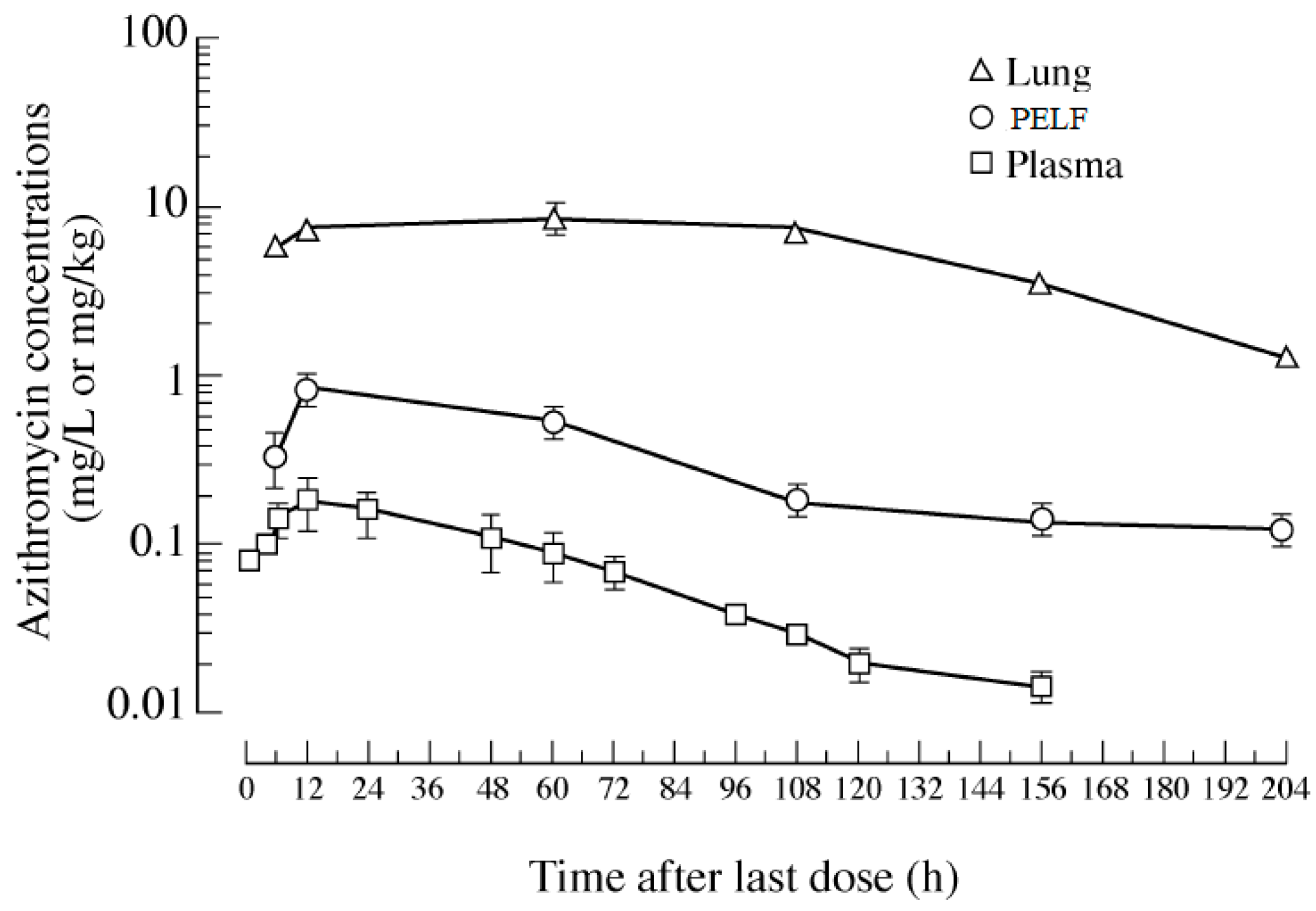
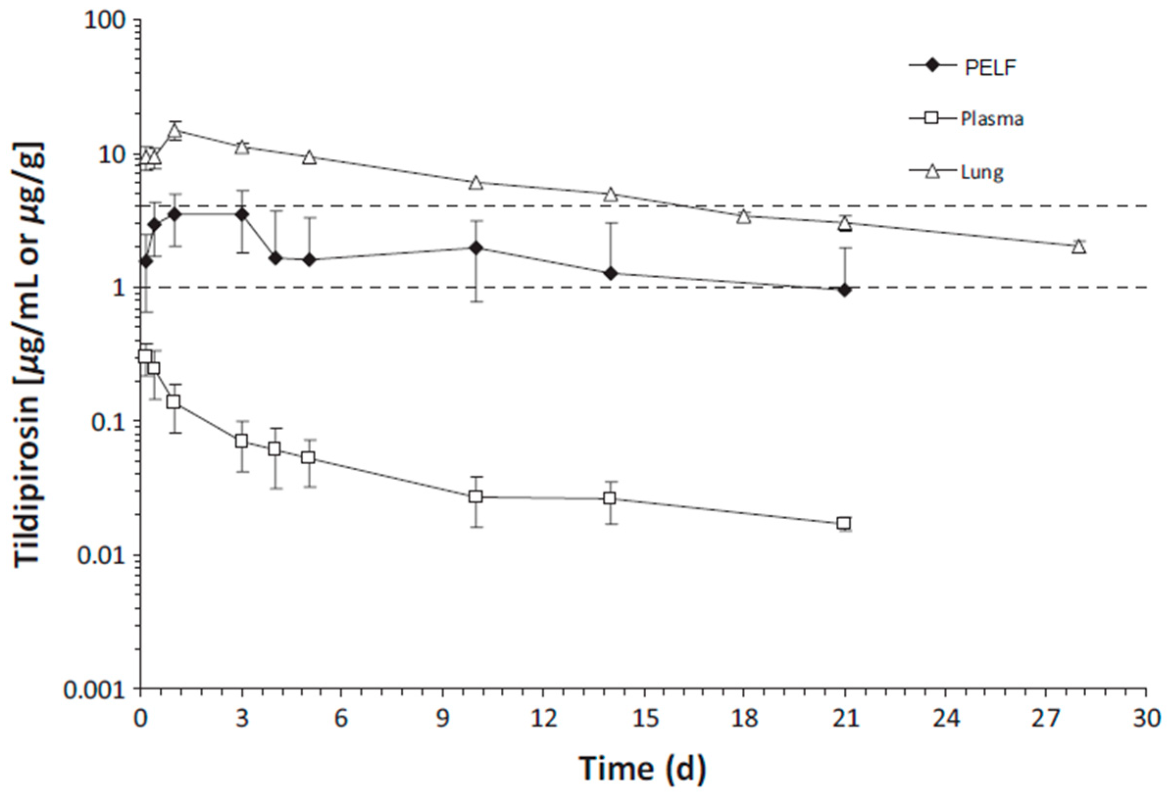
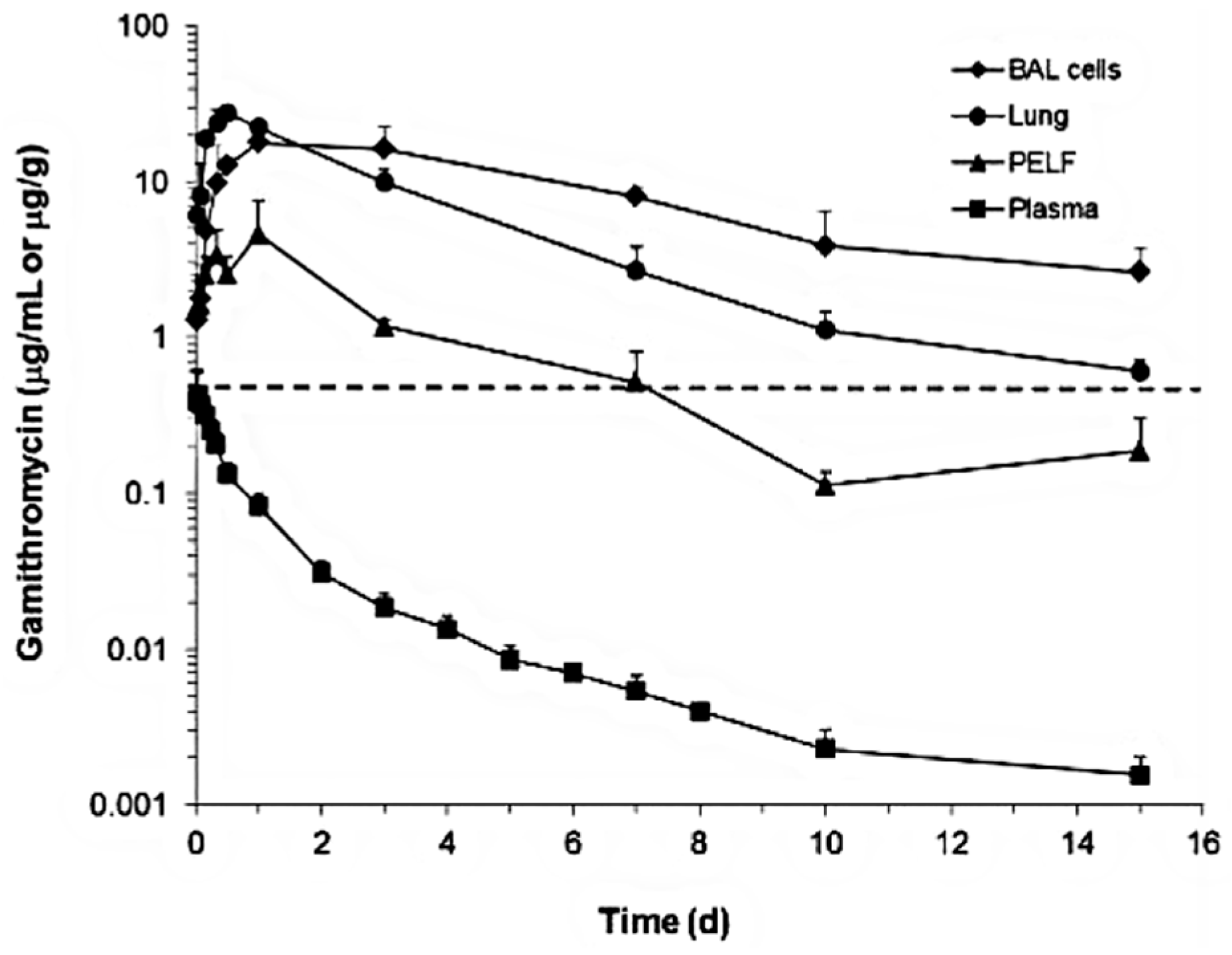
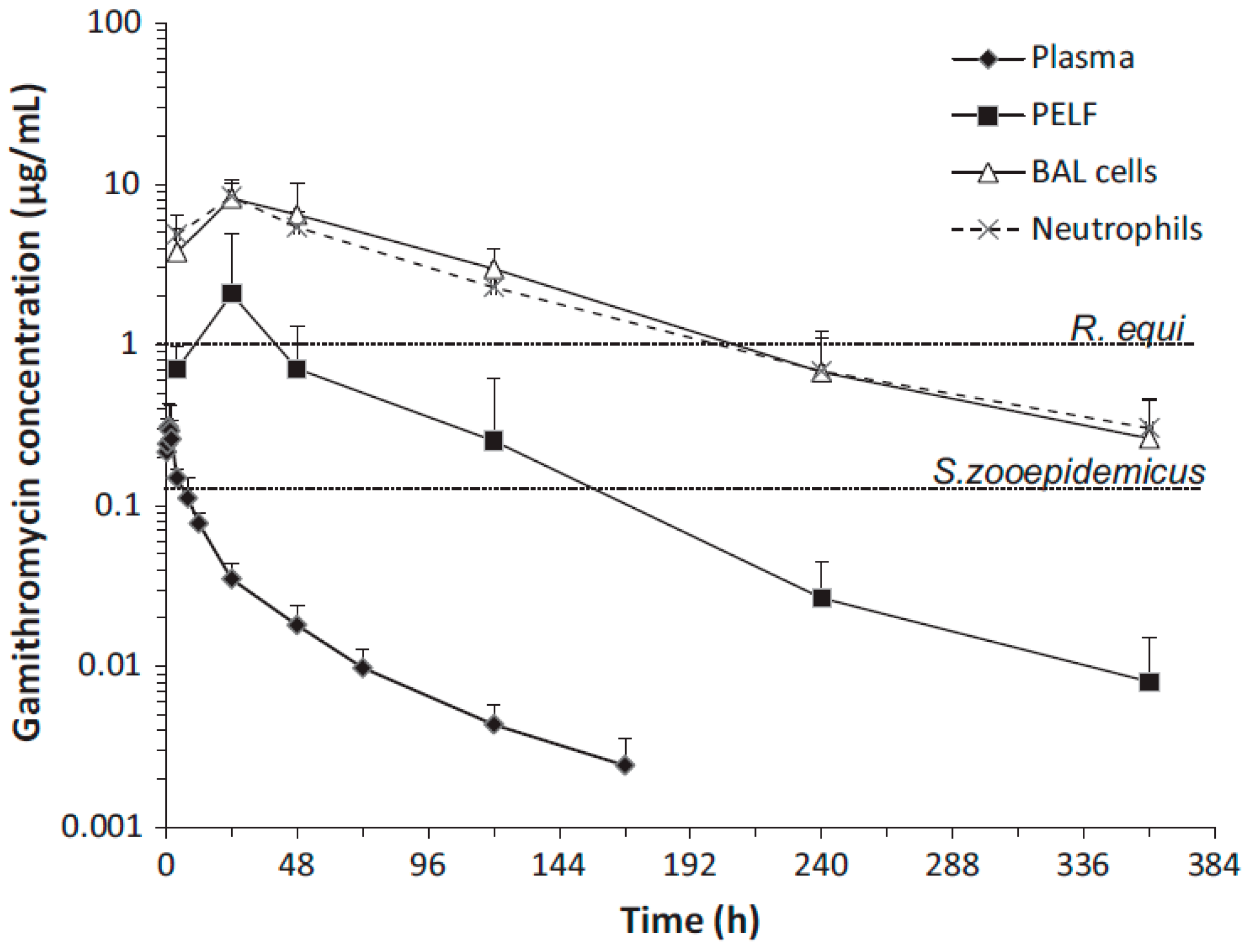
3. Concentrations of Macrolide Antibiotics in Plasma/Serum, Airway Fluid, and Tissues

Since the respiratory tract tissue and its epithelial lining fluid contain higher macrolide concentrations than plasma/serum, tremendous research interest has been focused on the area of macrolide concentrations in the airway tissues and associated ISF [12][24]. However, the PK of macrolide antibiotics (e.g., erythromycin, tylosin, azithromycin, clarithromycin, tildipirosin, gamithromycin, tilmicosin, and tulathromycin) in plasma/serum and target tissues, particularly those of the respiratory tract, have not been fully elucidated. Macrolide concentrations are ~10-fold higher in the airway epithelial fluid than in plasma [25]. In a human study, the researchers examined the PK of azithromycin in plasma, lung tissue and PELF in patients after oral administration of 500 mg azithromycin for 3 d. This study examined the pharmacokinetics of azithromycin in plasma, lung tissue, and PELF in patients after the oral administration of 500 mg azithromycin for 3 d. It was found that the plasma azithromycin concentrations were only ~10% and ~1% those of bronchial fluid (BF) and lung, respectively (Figure 1) [26]. The results showed that after azithromycin administration, the drug concentrations were: lung > PELF > plasma. In another two human studies, upon the administration of a single 500 mg oral dose of azithromycin, the tissue concentrations exceeded the minimum inhibitory concentration that inhibited 90% of likely pathogens (MIC90), and phagocytic concentrations reached >200 times serum concentrations [27][28]. In another human study, concentrations of clarithromycin and azithromycin in PELF exceeded serum concentrations by 20-fold 24 h after the last dose of drug administration [29]. Telithromycin also has excellent penetration into bronchopulmonary tissues. In humans, concentrations of telithromycin in PEFL exceeded serum concentrations by 12-fold 24 h after the last dose of drug administration [29][30].
Figure 1. Concentrations vs. time semilogarithmic plot of azithromycin 500 mg per human patient once daily for 3 days in lung tissue, PELF and plasma.
This phenomenon has been documented in cattle after the administration of tildipirosin as well [13][31][32]. The findings in cattle by Menge et al. [31] showed that the tildipirosin concentrations in lungs collected postmortem (4–240 h after dosing) exceeded those in BF (the ratio of lung/BF concentrations was ~2.5:1) and the ratio of lung/plasma was 27.6:1–214.5:1 (Figure 2). The results showed that after tildipirosin administration, the drug concentrations were: lung > PELF > plasma. The ratio of tildipirosin concentrations of BF/plasma determined from the same animals increased from 5.2:1 (4 h) to 72.3:1 (240 h or 10 days after administration) and then declined to 56.0:1 at the last collection time point of BF and plasma (504 h or 21 days) [31]. The plasma concentrations of tildipirosin were below the MIC90 of all three pathogens throughout the study. In contrast, the lung had drug concentrations higher than the MIC90 of H. somni for 18 d and the MIC90 of 2 other pathogens for >28 d. PELF had drug concentrations higher than the MIC90 of H. somni for 3 d and the MIC90 of 2 other pathogens for 21 d [31].
Figure 2. Tildipirosin concentration vs. time (d) in bovine plasma (μg/mL), lung tissue, and PELF (μg/g) from cattle following a single SC administration at 4 mg/kg body weight. The dotted lines represent the MIC90 for Mannheimia haemolytica and Pasteurella multocida (1 µg/mL) and for Histophilus somni (4 µg/mL).
Additionally, the concentrations of gamithromycin (Figure 3 and Figure 4) [33][34] and tulathromycin (Figure 5) in bovine lung tissues were also much higher than those of plasma [35]. The results from Giguere, S. et al. (2011) showed that after gamithromycin administration, the drug concentrations were: BAL cells > lung > PELF > plasma [33]. Gamithromycin concentrations in BAL cells and blood neutrophils were 26–732 and 33–563 times higher than concurrent plasma concentrations, respectively. The ratios ranged 4.7:1–127:1 for PELF/plasma, 16:1–650:1 for lung tissue/plasma, and 3.2:1–2135:1 for BAL/plasma [33]. Both BAL cells and lung had drug concentrations above MIC90 for >14 d; PELF had drug concentrations higher than MIC90 for >6 d; and plasma had drug concentrations below MIC90 throughout the study [33]. The results from Berghaus, L.J. et al. (2012) showed that after gamithromycin administration, the drug concentrations were: BAL cells = neutrophils > PELF > plasma [34]. Both BAL cells and neutrophils had drug concentrations above the MIC90 of both pathogens for >336 h (14 d); PELF had drug concentrations higher than the MIC90 of R. equi for 48 h (2 d) and higher than the MIC90 of S. zooepidemicus for >144 h (6 d). Plasma had drug concentrations below the MIC90 of both pathogens throughout the study [34]. The plasma concentrations of tulathromycin in cattle were substantially lower than the lung tissue concentrations, with AUC0-360 (area under the plasma concentration–time curve) values for PELF, PELF cells, and lung homogenate of cattle (Figure 5) [35]. The results showed that after tulathromycin administration, the drug concentrations were: PELF cells > lungs > PELF > plasma. Likewise, tulathromycin concentrations in PELF were ~10 times higher than plasma concentrations with the IV/IM route in horses [36]. The AUC0–17 days in BF and PELF were 223 and 90 times, respectively, the corresponding values of plasma. The PELF and BF concentration profiles of tulathromycin revealed that there was disparity in the local PK of tulathromycin at various anatomical structures of the lung. Therefore, drug concentrations in PELF may not be a precise alternative of drug concentrations for different pulmonary compartments [37]. The equilibrium between plasma–BF and plasma–PELF does not occur instantaneously, and the concentration–time profiles in both compartments are different [38][39]. The PELF and BF drug concentrations are mainly affected by the alveolar area and bronchial section, respectively. The larger blood flow and surface area of the airway compartment compared with the alveolar and bronchial areas may account for, at least partially, the differences in tulathromycin profiles at those two lung compartments [37].
Figure 3. Gamithromycin concentrations in plasma, bronchoalveolar lavage (BAL) cells and pulmonary epithelial lining fluid (PELF) (µg/mL) and lung tissue (µg/g) of healthy Angus calves following gamithromycin administration (6 mg/kg, SC). The dotted line represents the MIC90 for Mannheimia haemolytica (0.5 µg/mL) [33].
Figure 4. Gamithromycin concentrations in plasma, BAL cells, PELF, and neutrophils of 6 foals following a single IM dose of gamithromycin (6 mg/kg). The dotted horizontal lines represent the MIC90 of Rhodococcus equi (1 µg/mL) and Streptococcus zooepidemicus (0.125 µg/mL) isolates [34].
Figure 5. Tulathromycin concentrations for 360 h following a single 2.5 mg/kg IM dose to cattle.
Similarly, the Cmax and AUC of tilmicosin in foals were higher for PELF than for serum (Figure 6) [40]. The results showed that after tilmicosin administration, the drug concentrations were: lung > BAL cells > PELF = neutrophils > serum. Following intragastric administration, the ratios of PELF/plasma telithromycin concentrations in foals were 5.89:1 and 5.64:1 at 4 and 24 h, respectively [41].
Figure 6. Tilmicosin concentrations in serum, bronchoalveolar lavage (BAL) cells, pulmonary epithelial lining fluid (PELF) (µg/mL), and lung tissue (µg/g) of 7 foals following a single IM dose of tilmicosin (10 mg/kg).

4. Conclusions

Although the change in plasma/serum concentration-time and the change in airway ISF-concentration-time of macrolide antibiotics are proportional, macrolide antibiotics in plasma/serum do not reflect the antibacterial activity of the airway ISF. Thus, if the ISF concentrations of a macrolide antibiotic is efficiently and precisely collected by the least invasive method, e.g., microdialysis, practical PK/PD parameters can be obtained for such a study. Nevertheless, the PK data of airway ISF, the site of bacterial infection, are more important in setting the optimal dose regimen of a macrolide than the PK data of plasma/serum.

References

  1. Spagnolo, P.; Fabbri, L.M.; Bush, A. Long-term macrolide treatment for chronic respiratory disease. Eur. Respir. J. 2013, 42, 239–251.
  2. Mazzei, T.; Mini, E.; Novelli, A.; Periti, P. Chemistry and mode of action of macrolides. J. Antimicrob. Chemother. 1993, 31 (Suppl. C), 1–9.
  3. Bearden, D.T.; Rodvold, K.A. Penetration of macrolides into pulmonary sites of infection. Infect. Med. 1999, 16, 480A–484A.
  4. Drusano, G.L. Infection site concentrations: Their therapeutic importance and the macrolide and macrolide-like lass of antibiotics. Pharmacotherapy 2005, 25, 150S–158S.
  5. Barza, M. Anatomical barriers for antimicrobial agents. Eur. J. Clin. Microbiol. Infect. Dis. 1993, 12 (Suppl. S1), S31–S35.
  6. Barza, M. Pharmacokinetics of antibiotics in shallow and deep compartments. J. Antimicrob. Chemother. 1993, 31 (Suppl. D), 17–27.
  7. Landersdorfer, C.B.; Nation, R.L. Limitations of antibiotic MIC-based PK-PD metrics: Looking back to move forward. Front. Pharmacol. 2021, 12, 3024.
  8. Matzneller, P.; Krasniqi, S.; Kinzig, M.; Sörgel, F.; Hüttner, S.; Lackner, E.; Müller, M.; Zeitlinger, M. Blood, Tissue, and intracellular concentrations of azithromycin during and after end of therapy. Int. J. Antimicrob. Agents. 2013, 57, 1736–1742.
  9. Liu, P.; Müller, M.; Derendorf, H. Rational dosing of antibiotics: The use of plasma concentrations versus tissue concentrations. Int. J. Antimicrob. Agents 2002, 19, 285–290.
  10. Toutain, P.L.; Bousquet-Melou, A. Free drug fraction vs. free drug concentration: A matter of frequent confusion. J. Vet. Pharmacol. Ther. 2002, 25, 460–463.
  11. Toutain, P.L.; del Castillo, J.R.E.; Bousquet-Mélou, A. The pharmacokinetic–pharmacodynamic approach to a rational regimen for antibiotics. Res. Vet. Sci. 2002, 73, 105–114.
  12. Gonzalez, D.; Schmidt, S.; Derendorf, H. Importance of relating efficacy measures to unbound drug concentrations for anti-infective agents. Clin. Microbiol. Rev. 2013, 26, 274–288.
  13. Rose, M.; Menge, M.; Bohland, C.; Zschiesche, E.; Wilhelm, C.; Kilp, S.; Metz, W.; Allan, M.; Ropke, R.; Nurnberger, M. Pharmacokinetics of tildipirosin in porcine plasma, lung tissue, and bronchial fluid and effects of test conditions on in vitro activity against reference strains and field isolates of Actinobacillus pleuropneumoniae. J. Vet. Pharmacol. Ther. 2013, 36, 140–153.
  14. Huang, R.A.; Letendre, L.T.; Banav, N.; Fischer, J.; Somerville, B. Pharmacokinetics of gamithromycin in cattle with comparison of plasma and lung tissue concentrations and plasma antibacterial activity. J. Vet. Pharmacol. Ther. 2010, 33, 227–237.
  15. Giguere, S.; Tessman, R.K. Rational dosing of antimicrobial agents for bovine respiratory disease: The use of plasma versus tissue concentrations in predicting efficacy. Int. J. Appl. Res. Vet. M 2009, 9, 342–355.
  16. Ball, P.; Baquero, F.; Cars, O.; File, T.; Garau, J.; Klugman, K.; Low, D.E.; Rubinstein, E.; Wise, R. Antibiotic therapy of community respiratory tract infections: Strategies for optimal outcomes and minimized resistance emergence. J. Antimicrob. Chemother. 2002, 49, 31–40.
  17. Togami, K.; Chono, S.; Morimoto, K. Distribution characteristics of clarithromycin and azithromycin, macrolide antimicrobial agents used for treatment of respiratory infections, in lung epithelial lining fluid and alveolar macrophages. Biopharm. Drug Dispos. 2011, 32, 389–397.
  18. Di, L.; Kerns, E.H. Chapter 37—Pharmacokinetic Methods. In Drug-Like Properties, 2nd ed.; Di, L., Kerns, E.H., Eds.; Academic Press: Boston, MA, USA, 2016; pp. 455–461.
  19. Maglio, D.; Capitano, B.; Banevicius, M.A.; Geng, Q.; Nightingale, C.H.; Nicolau, D.P. Differential efficacy of clarithromycin in lung versus thigh infection models. Chemotherapy 2004, 50, 63–66.
  20. Bachtold, K.A.; Alcorn, J.M.; Boison, J.O.; Matus, J.L.; Woodbury, M.R. Pharmacokinetics and lung and muscle concentrations of tulathromycin following subcutaneous administration in white-tailed deer (Odocoileus virginianus). J. Vet. Pharmacol. Ther. 2016, 39, 292–298.
  21. Romanet, J.; Smith, G.W.; Leavens, T.L.; Baynes, R.E.; Wetzlich, S.E.; Riviere, J.E.; Tell, L.A. Pharmacokinetics and tissue elimination of tulathromycin following subcutaneous administration in meat goats. Am. J. Vet. Res. 2012, 73, 1634–1640.
  22. Kobuchi, S.; Kabata, T.; Maeda, K.; Ito, Y.; Sakaeda, T. Pharmacokinetics of macrolide antibiotics and transport into the interstitial fluid: Comparison among erythromycin, clarithromycin, and azithromycin. Antibiotics 2020, 9, 199.
  23. Okamoto, H.; Miyazaki, S.; Tateda, K.; Ishii, Y.; Yamaguchi, K. In vivo efficacy of telithromycin (HMR3647) against Streptococcus pneumoniae and Haemophilus influenzae. Antimicrob. Agents Chemother. 2001, 45, 3250–3252.
  24. Ambrose, P.G.; Bhavnani, S.M.; Ellis-Grosse, E.J.; Drusano, G.L. Pharmacokinetic-pharmacodynamic considerations in the design of hospital-acquired or ventilator-associated bacterial pneumonia studies: Look before you leap! Clin. Infect. Dis. 2010, 51, S103–S110.
  25. Kanoh, S.; Rubin, B.K. Mechanisms of Action and Clinical Application of Macrolides as Immunomodulatory Medications. Clin. Microbiol. Rev. 2010, 23, 590–615.
  26. Danesi, R.; Lupetti, A.; Barbara, C.; Ghelardi, E.; Chella, A.; Malizia, T.; Senesi, S.; Alberto Angeletti, C.; Del Tacca, M.; Campa, M. Comparative distribution of azithromycin in lung tissue of patients given oral daily doses of 500 and 1000 mg. J. Antimicrob. Chemother. 2003, 51, 939–945.
  27. Firth, A.; Prathapan, P. Azithromycin: The First Broad-spectrum Therapeutic. Eur. J. Med. Chem. 2020, 207, 112739.
  28. Kong, F.Y.; Rupasinghe, T.W.; Simpson, J.A.; Vodstrcil, L.A.; Fairley, C.K.; McConville, M.J.; Hocking, J.S. Pharmacokinetics of a single 1g dose of azithromycin in rectal tissue in men. PLoS ONE 2017, 12, e0174372.
  29. Zuckerman, J.M. Macrolides and ketolides: Azithromycin, clarithromycin, telithromycin. Infect. Dis. Clin. N. Am. 2004, 18, 621–649.
  30. Muller-Serieys, C.; Soler, P.; Cantalloube, C.; Lemaitre, F.; Gia, H.P.; Brunner, F.; Andremont, A. Bronchopulmonary disposition of the ketolide telithromycin (HMR 3647). Antimicrob. Agents Chemother. 2001, 45, 3104–3108.
  31. Menge, M.; Rose, M.; Bohland, C.; Zschiesche, E.; Kilp, S.; Metz, W.; Allan, M.; Ropke, R.; Nurnberger, M. Pharmacokinetics of tildipirosin in bovine plasma, lung tissue, and bronchial fluid (from live, nonanesthetized cattle). J. Vet. Pharmacol. Ther. 2012, 35, 550–559.
  32. Torres, F.; Santamaria, R.; Jimenez, M.; Menjón, R.; Ibanez, A.; Collell, M.; Azlor, O.; Fraile, L. Pharmacokinetics of tildipirosin in pig tonsils. J. Vet. Pharmacol. Ther. 2016, 39, 199–201.
  33. Giguere, S.; Huang, R.; Malinski, T.J.; Dorr, P.M.; Tessman, R.K.; Somerville, B.A. Disposition of gamithromycin in plasma, pulmonary epithelial lining fluid, bronchoalveolar cells, and lung tissue in cattle. Am. J. Vet. Res. 2011, 72, 326–330.
  34. Berghaus, L.J.; Giguère, S.; Sturgill, T.L.; Bade, D.; Malinski, T.J.; Huang, R. Plasma pharmacokinetics, pulmonary distribution, and in vitro activity of gamithromycin in foals. J. Vet. Pharmacol. Ther. 2012, 35, 59–66.
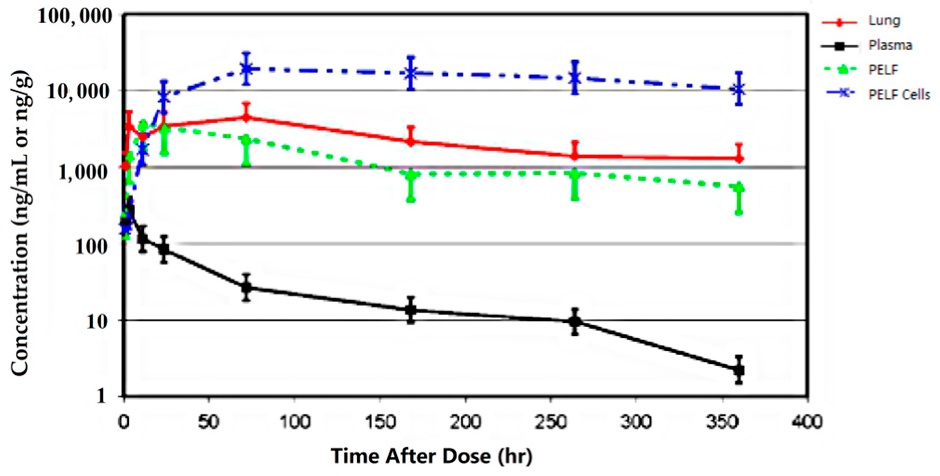
  35. Cox, S.R.; McLaughlin, C.; Fielder, A.E.; Yancey, M.; Bowersock, T.; Garcia-Tapia, D.; Bryson, L.; Lucas, M.J.; Robinson, J.A.; Nanjiani, I.; et al. Rapid and prolonged distribution of tulathromycin into lung homogenate and pulmonary epithelial lining fluid of holstein calves following a single subcutaneous administration of 2.5 mg/kg body weight. Int. J. Appl. Res. Vet. Med. 2010, 8, 129–137.
  36. Leventhal, H.R.; McKenzie, H.C.; Estell, K.; Council-Troche, M.; Davis, J.L. Pharmacokinetics and pulmonary distribution of Draxxin® (tulathromycin) in healthy adult horses. J. Vet. Pharmacol. Ther. 2021, 44, 714–723.
  37. Villarino, N.; Brown, S.A.; Martín-Jiménez, T. Understanding the pharmacokinetics of tulathromycin: A pulmonary perspective. J. Vet. Pharmacol. Ther. 2014, 37, 211–221.
  38. Villarino, N.; Lesman, S.; Fielder, A.; García-Tapia, D.; Cox, S.; Lucas, M.; Robinson, J.; Brown, S.A.; Martín-Jiménez, T. Pulmonary pharmacokinetics of tulathromycin in swine. Part 2: Intra-airways compartments. J. Vet. Pharmacol. Ther. 2013, 36, 340–349.
  39. Villarino, N.; Lesman, S.; Fielder, A.; García-Tapia, D.; Cox, S.; Lucas, M.; Robinson, J.; Brown, S.A.; Martín-Jiménez, T. Pulmonary pharmacokinetics of tulathromycin in swine. Part I: Lung homogenate in healthy pigs and pigs challenged intratracheally with lipopolysaccharide of Escherichia coli. J. Vet. Pharmacol. Ther. 2013, 36, 329–339.
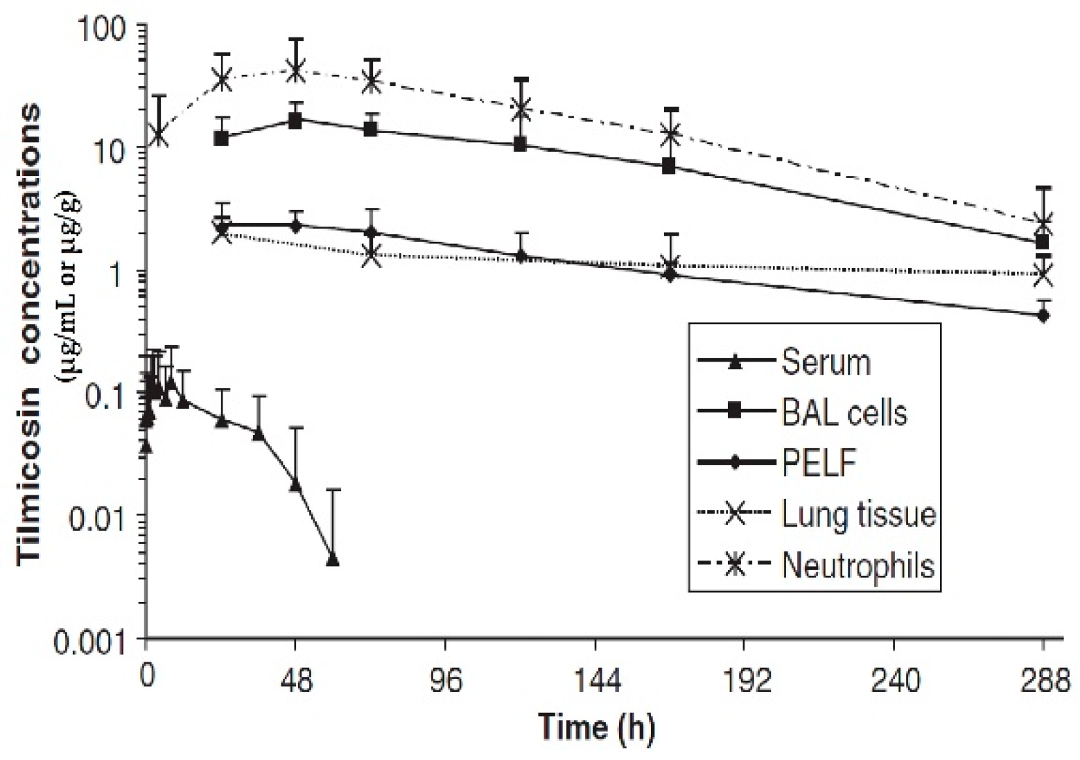
  40. Womble, A.; Giguère, S.; Murthy, Y.V.S.N.; Cox, C.; Obare, E. Pulmonary disposition of tilmicosin in foals and in vitro activity against Rhodococcus equi and other common equine bacterial pathogens. J. Vet. Pharmacol. Ther. 2006, 29, 561–568.
  41. Javsicas, L.; Giguère, S.; Womble, A.Y. Disposition of oral telithromycin in foals and in vitro activity of the drug against macrolide-susceptible and macrolide-resistant Rhodococcus equi isolates. J. Vet. Pharmacol. Ther. 2010, 33, 383–388.
More
Information
Subjects: Allergy
Contributors MDPI registered users' name will be linked to their SciProfiles pages. To register with us, please refer to https://encyclopedia.pub/register : , , , ,
View Times: 402
Revisions: 3 times (View History)
Update Date: 20 Apr 2023
1000/1000
ScholarVision Creations