Submitted Successfully!
To reward your contribution, here is a gift for you: A free trial for our video production service.
Thank you for your contribution! You can also upload a video entry or images related to this topic.
Version Summary Created by Modification Content Size Created at Operation
1 -- 4280 2023-04-19 16:55:45 |
2 update references and layout -73 word(s) 4207 2023-04-20 05:31:39 |

Video Upload Options

Do you have a full video?

Confirm

Are you sure to Delete?
Cite
If you have any further questions, please contact Encyclopedia Editorial Office.
Aziz, R.; Falanga, M.; Purenovic, J.; Mancini, S.; Lamberti, P.; Guida, M. Natural Biodegradable Nano Polymers in Cardiac Tissue Engineering. Encyclopedia. Available online: https://encyclopedia.pub/entry/43249 (accessed on 04 September 2024).
Aziz R, Falanga M, Purenovic J, Mancini S, Lamberti P, Guida M. Natural Biodegradable Nano Polymers in Cardiac Tissue Engineering. Encyclopedia. Available at: https://encyclopedia.pub/entry/43249. Accessed September 04, 2024.
Aziz, Rabia, Mariarosaria Falanga, Jelena Purenovic, Simona Mancini, Patrizia Lamberti, Michele Guida. "Natural Biodegradable Nano Polymers in Cardiac Tissue Engineering" Encyclopedia, https://encyclopedia.pub/entry/43249 (accessed September 04, 2024).
Aziz, R., Falanga, M., Purenovic, J., Mancini, S., Lamberti, P., & Guida, M. (2023, April 19). Natural Biodegradable Nano Polymers in Cardiac Tissue Engineering. In Encyclopedia. https://encyclopedia.pub/entry/43249
Aziz, Rabia, et al. "Natural Biodegradable Nano Polymers in Cardiac Tissue Engineering." Encyclopedia. Web. 19 April, 2023.
Natural Biodegradable Nano Polymers in Cardiac Tissue Engineering
Edit

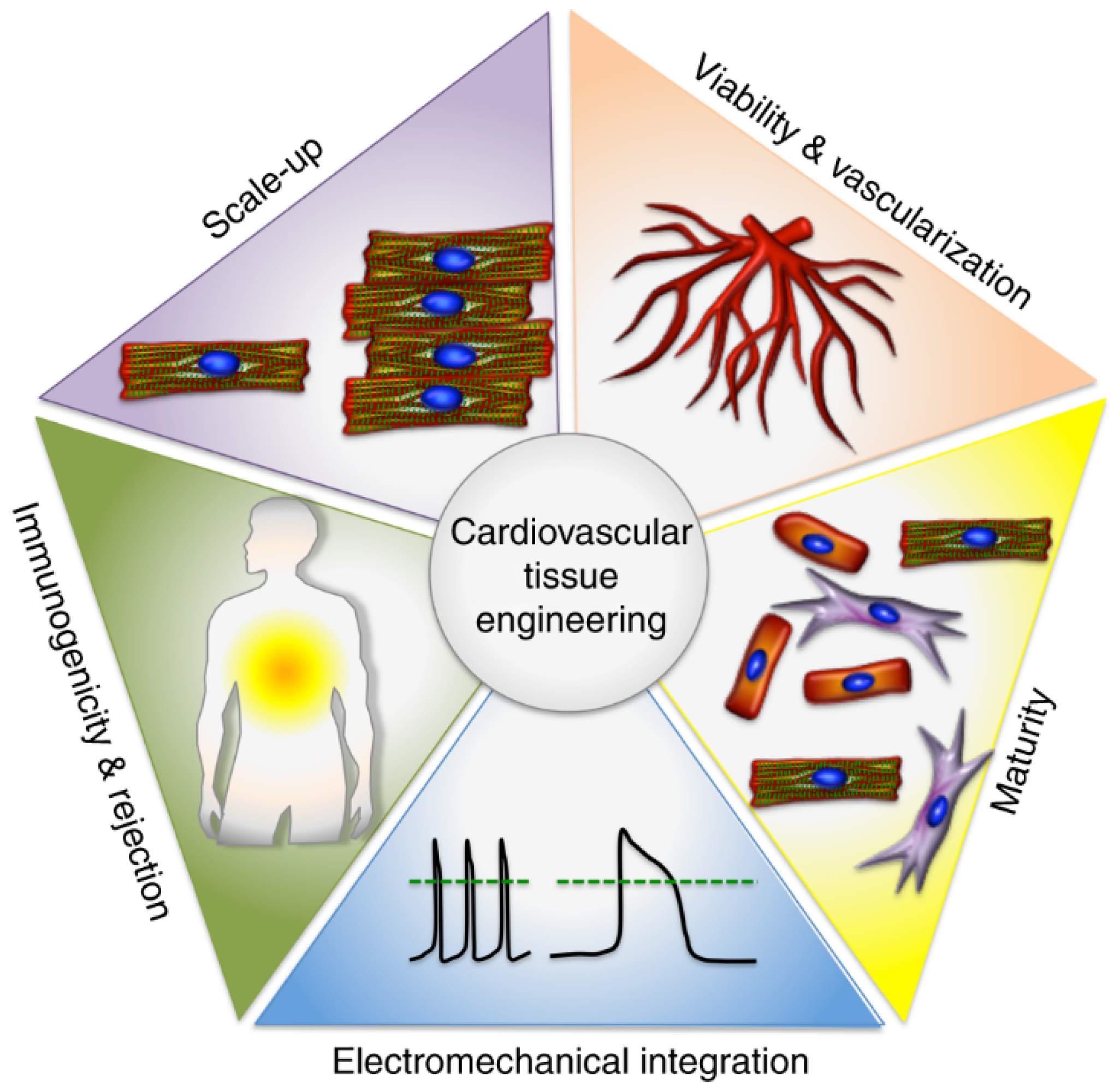
As cardiac diseases, which mostly result in heart failure, are increasing rapidly worldwide, heart transplantation seems the only solution for saving lives. This practice is not always possible due to several reasons, such as scarcity of donors, rejection of organs from recipient bodies, or costly medical procedures. In the framework of nanotechnology, nanomaterials greatly contribute to the development of these cardiovascular scaffolds as they provide an easy regeneration of the tissues. Functional nanofibers can be used in the production of stem cells and in the regeneration of cells and tissues. The small size of nanomaterials, leads to changes in their chemical and physical characteristics that could alter their interaction and exposure to stem cells with cells and tissues.

nanotechnology functional nanofibers cardiovascular scaffolds cardiac tissue engineering biodegradable nano polymers

1. Introduction

Cardio Vascular Diseases (CVD) are one of the most prominent causes of mortality in both genders worldwide; thus, they have been marked as a significant public health burden. According to the World Health Organization’s (WHO) estimation, in 2019, 8.9 million people died because of cardiovascular diseases, accounting for 32% of all deaths worldwide. CVDs are linked to heart and vascular problems, including the most frequent atherosclerosis, cerebrovascular disease, ischemic heart disease, and strokes. Clinically, these diseases occur in the mid or older age, after years of unhealthy diet habits, lack of physical activity, alcoholism, and smoking, while obesity, high levels of cholesterol, blood pressure, and diabetes are common risk factors [1].
The apparent global rise in heart failure prevalence is not always related to an increase in heart failure incidence, which, indeed, has been found to be constant or even declining in some studies [2]. The population’s aging, combined with improved heart failure survival (as a result of advancements in treatments and diagnostic technology), may account for the increase in prevalence, while the decrease in incidence (thanks to prevention programs) may be attributed to the treatment of acute coronary syndromes. Among them, cardiac transplantation remains the most effective and efficient treatment. However, the persistent shortage of donor organs and tissues is a serious obstacle to transplantation. Additionally, heart transplant recipients confront severe obstacles to their long-term survival in the form of acute immunosuppression and chronic immune refusal. In light of these observations, there is, therefore, a strong need to find new ways to improve heart failure care [3].
All the issues related to cardiac transplantation and the limited capacity of cardiomyocytes to repair the heart after an acute myocardial infarction (MI) are the primary reasons why much research in regenerative medicine has been developing.
Tissue engineering is a technique that integrates biological sciences and engineering to create functional tissue analogs, such as synthetic heart tissue, which may be used in place of an organ or as mechanical aid [1].
The successful production of contractile cardiomyocytes (CM) from human-induced pluripotent stem cells (hiPSCs) [4] is a significant development in cardiovascular tissue engineering [5]. This is because CMs are normally non-proliferative in the myocardium. Some studies have shown that hiPSC-CM-derived synthetic myocardial tissue is effective in treating heart failure in both small and large preclinical animal models. Some obstacles exist that prevent the widespread clinical use of hiPSC-CM-derived engineered cardiac tissue [6][7][8][9][10]. Scalable three-dimensional (3D) created tissues face challenges in several areas, including survival, electrical integration, immunological response, and maturity/function of hiPSC-CMs (Figure 1).
Figure 1. Cardiovascular tissue engineering bottlenecks.
Two primary technologies are helpful in the fields of tissue regeneration and tissue engineering: (1) biomaterial technology for the creation of three-dimensional porous scaffolds to facilitate direct tissue formation from dissociated cells; and (2) bioreactor cultivation of three-dimensional cell constructs during ex-vivo tissue engineering that aims to recreate the normal stresses as well as flows experienced by heart tissue [11]. Both of these technologies are currently in development. Scaffolds, which have a porous structure that allows for the diffusion of donor cells and growth factors to stimulate and direct the growth of new, healthy tissue; and hydrogels, which are water-insoluble, cross-linked polymer matrices with a high water content that are easily able to be infused into damaged heart tissue and fabricated into a broad range of tissue engineering constructs. Scaffolds and hydrogels are the two main classes of materials that have been traditionally used in cardiac tissue engineering [12].
The properties of biodegradable polymers, such as their optimal porosity, flexible degradation rates, biocompatibility, as well as elastomeric characteristics (which can mechanically favor tissue contraction, which is an inherent part of cardiac function), have attracted a lot of attention for the applications of cardiac tissue engineering. These characteristics include the capacity of scaffolds to maintain their mechanical properties during the process of tissue growth, their capacity to gradually degrade into biocompatible products, and their capacity to accept cells, growth factors, and other such components [13][14].
The two principal classes of biodegradable polymers are synthetic and natural polymers. Natural biodegradable polymers (NAbioPOLY) are polymers derived from the environment. Collagen, gelatin, fibrin, alginate, chitosan, matrigel, and silk are examples of natural polymers. Moreover, other natural nanofibers are also present, such as mussel-derived silk, spider-based nanofibers, sea-silk-based nanofibers, and snail-based nanofibers. These natural nanofibers, along with silk nanofibers created from silkworms, have been utilized in a variety of tissue engineering applications. Natural biodegradable polymers have a number of advantages, including abundant accessibility, biodegradability, and renewability, while their disadvantages include insufficient electrical conductivity, rapid degradation, weak mechanical properties, and immunoreactivity. Man-made polymers are referred to as synthetic polymers. These involve Poly(lactic acid), Poly(glycolic acid), polycaprolactone, Poly(glycolic acid), polycaprolactone, etc.
In addition to controlled structures, stable mechanical properties, flexibility, and the absence of immunological concerns, synthetic biodegradable polymers also exhibit lower biocompatibility and lack of cell attachment. Researchers have developed novel natural/synthetic composites in an effort to mitigate the drawbacks of both natural and synthetic polymers (a combination of both natural and synthetic polymers). In this manner, composite properties have been significantly enhanced. Included in this category are PLA/chitosan, TiO2-PEG/chitosan, and gelatin/PCL/graphene. Natural/synthetic composites exhibit superior biocompatibility, mechanical strength, electrical conductivity, and biological properties [15].

2. Natural Biodegradable Nanopolymers Used in Cardiac Tissue Engineering

Recent advances in the incorporation of nanoparticles into tissue engineering have been made [16]. Nanomaterials may be particularly useful in cardiovascular tissue engineering. The latter is concerned with regenerating damaged cardiac tissue, sometimes through the induction of cell proliferation with regenerative potential, such as mesenchymal stem, embryonic stem, as well as induced pluripotent stem cells [17]. Numerous efforts are being made to develop polymer scaffolds or patches that can be used to support tissue regeneration or repair. Cardiovascular tissue engineering has been tested by using biomaterials such as hydrogels, electrospun polymers, and 3D-printed cardiac patches [18]. Additionally, injectable hydrogels have been developed to facilitate clinical use by allowing for easy delivery to injured myocardium without the need for invasive approaches. Following modification with bioactive molecules such as microRNA, peptides, or growth factors, these materials may be repurposed. Although these biomolecules are susceptible to degradation in a physiological environment, nanoparticles can aid in their stabilization. Nanoparticles have been found to be beneficial in these efforts by stimulating neighboring cells and acting as platforms for bioactive molecule modification [15][19].
Synthetic polymers and natural polymers are the two main types of biodegradable polymers. Polymers that are naturally biodegradable are referred to as NAbioPOLY. Synthetic polymers are those that are created in a laboratory. This category includes substances such as poly(lactic acid), poly(glycolic acid), poly(glycolic acid), poly(caprolactone), etc. [15]. Natural polymers include things such as silk, gelatin, collagen, chitosan, alginate, and chitosan. There are pros and cons to using natural biodegradable polymers. Pros include their abundant availability, biodegradability, and renewability; cons include their low electrical conductivity, quick disintegration, weak mechanical capabilities, and immunogenicity (Figure 2).
Figure 2. Types of biomaterials used in cardiac tissue engineering.
Natural polymers are derived from natural sources, for example, plants and animals. These natural polymers are formed of nano-structured molecules and are, therefore, classified as nanomaterials [20]. Because of their biocompatibility, renewability, biodegradability, and widespread availability, these natural polymers have been employed in different cardiovascular tissue engineering applications [21]. NAbioPOLYs such as collagen [22], gelatin [23], fibrin gel [24], alginate [25], chitosan [26], matrigel [27] and silk [28] are frequently employed in cardiac tissue engineering. They will be discussed in the following subsections.
The primary feature of NAbioPOLYs that puts them at the frontline of cardiac tissue engineering is their biocompatibility. Additional important properties that have been studied and found to be acceptable for cardiac applications include mechanical properties and rate of degradation. Hydrogels are soft materials among these biomaterials, and they are ideally suited for cardiac repair because they are made from naturally occurring matrices such as collagen, gelatin, fibrinogen, alginate, matrigel, chitosan, and silk. To date, there have only been a handful of clinical trials using hydrogels made from NAbioPOLYs [29].
In order to more closely resemble normal tissue structure, including cardiac tissue, NAbioPOLYs have been transformed into other types of 3D structures with tailored porosity. These are advantageous because they can be processed using a wide variety of methods and boast a wide range of mechanical properties that can be fine-tuned to meet the needs of individual patients. The development of big perfusable vessels is still difficult, despite the fact that many approaches have been taken to increase vascularity inside the cardiac patches. To avoid the potentially fatal arrhythmic effects of cell injection in large quantities, more study is needed to determine how to enhance functional coupling between the graft and host cardiomyocytes. Intriguing advances in cardiac tissue engineering are on the horizon thanks to multi-material structures made possible by 3D printing techniques and featuring structures tailored to individual patients. These scaffolds aim to mimic the properties of natural cardiac tissue as closely as possible [30].

2.1. Collagen

Collagen is the most frequently employed natural polymer in tissue engineering since it is found in the ECM of nearly every human tissue. Its use has increased due to its good biocompatibility and weak antigenicity. Collagen has been utilized in a variety of applications over the years due to its pro-vascularization biocompatibility, high cellular activity, hyposensitivity, biodegradability, and low toxication [31]. Collagen provides several advantages for cardiac tissue engineering, including heat reversibility, biocompatibility and high cellular activity, hyposensitivity, biodegradability, and low toxication [32].
Collagen, comprising skin, bone, tendons, ligaments, and cartilage, is found in almost all human tissues. Collagen types I, II, III, and IV are frequently explored in tissue engineering [33]. Among them, thanks to its biocompatibility, type I collagen is mostly utilized in tissue engineering [34]. It comprises 75–85% [29] of the ECM in the heart and has the added benefit of being relatively nonimmunogenic. Although collagens can be subdivided into fibrillar and non-fibrillar components, type I collagen consists of two alpha-1 chains and one alpha-2 chain, forming long fibers whose characteristics are dependent on the density and spatial alignment [35]. These non-fibrillar parts can bind to membranes or create networks [36][37].
The human body, and especially the native myocardium, are primarily composed of collagen I. The latter, like all collagens, has a three-helical structure at its molecular level. These molecules, when left to their own devices, self-assemble into fibrils, which are then organized into collagen fibers of variable diameters in the body. As a result, the tensile mechanical contributions of these fibrils and fibers are maximized in tissues subjected to high mechanical stresses, such as tendons, ligaments, and muscles. Long channels surrounding cardiac muscle bundles and bestowing to the anisotropy of native myocardium are formed by collagen fibers, the primary component of the endomysium in the myocardium. It has been shown that resident cells can apply stresses to initially disordered collagen hydrogel scaffolds, causing them to become organized over time [38][39].
Collagen-based biomaterials have been the focus of recent studies for the treatment of illnesses such as myocardial infarction. Particles, like growth factors or peptides, can be transported by these materials to stimulate differentiation as well as patterning [40]. Based on these preliminary findings, researchers have intramyocardial injection methods, as they allow for targeted, direct administration to the heart muscle. However, surgery is required for this method, and there is also the risk of the substance leaking out into the surrounding tissue [41]. So-called “cardiac patches” are a substitute for this kind of administration. Cardiac patches contain certain qualities that are not unique to collagen but are nonetheless useful. These patches have the ability to infiltrate models and have high engraftment levels [42], and one feature is the possibility of cultivating cells ex vivo to encourage the proper invasion of the patch. Eventually, autologous bone marrow mononuclear cells seeded into a 3D collagen type I matrix (for the regeneration of ischemic myocardium) increased the thickness of the infarct scar with viable tissue, assisted normalized cardiac wall stress in wounded regions, limited ventricular remodeling, and improved diastolic function [43].
For cardiac tissue engineering, scaffolds of collagens, particularly nano-fibrous scaffolds, are being studied. Punnoose et al. (2015) describe one of the most straightforward new approaches for the development of nano-fibrous scaffolds from collagen by the use of an electric field among the solution of polymer and grounded collector. Fluoroalcohols (e.g., 1,1,1,3,3,3–hexafluoro-2-propanol (HFIP) and 2,2,2-trifluoroethanol (TFE)) to manufacture nano-fibrous scaffolds from collagen type I, which generally has been the favored for the manufacture of biomaterials based on collagen. Though fluoroalcohols are caustic and expensive, numerous investigations have been conducted with the goal of discovering a benign and economically viable solvent. For instance, Punnoose et al. (2015) stated the utilization of a simple benign binary solvent solution comprised of acetic acid and dimethylsulphoxide for electrospinning the nanofibers from type I collagen whose diameter ranges from 200–1100 nm in one study. This solvent was not just inexpensive but also preserved the inherent properties of electrospun collagen [44].

2.2. Gelatin

Gelatin is a natural polymer that is similar to collagen, from which it is derived. Once collagen has been separated out, gelatin can be extracted in one of two ways: alkaline hydrolysis or acid hydrolysis. The latter is how the IP (Isoelectric Point) of gelatin is calculated. Type A, with an IP value less than 5, is the designation given to gelatin after it has been acid hydrolyzed. Type B gelatin, with an IP of less than 9, is the result of extraction in an alkaline medium. Denaturalization gives gelatin its characteristic linear structure, made up of Gly-X-Y (mostly proline and hydroxyproline) sequences. The RGD (Arginylglycylaspartic acid) motif, a different set of amino acids in the structure, also aids in cell adhesion, proliferation, and differentiation [45][46][47][48].
In addition to promoting cell adhesion, differentiation, and proliferation without eliciting an immune response, its biocompatible, biodegradable, and low-toxic properties also make it easily digested by the body’s own enzymes (metalloproteinases) [49][50]. Because of its low price, it has been used in a variety of applications (microparticles for bone regeneration enhancement, wound dressing, hydrogels for the controlled release of chemotherapeutic agents in the treatment of cancer) and in a wide variety of tissues (bone, skeletal, neural) [51][52].
However, gelatin is renowned for its capacity to take in liquid. Porosity guarantees a diffusion of nutrients and oxygen for proper cell growth, making it a highly desirable property in tissue regeneration [53]. Nevertheless, some gelatin-based cell delivery systems have evidenced a poor cell survival rate [54], suggesting that porous structures do not always fulfill every requirement for facilitating the exchange of products for cell survival. Finding a way to use gelatin-based systems to ensure proper pore size, which may lead to an elevated rate of cell survival, is thus the current research focus [48].
Gelatin is a possible biomaterial for cardiovascular tissue regeneration since collagen is abundantly found in the extracellular matrix of numerous organs, including the heart. Gelatin hydrogels have several limitations, including a lack of durability, mechanical stability, and high-water content. Thus, gelatin-based biomaterials have been produced by cross-linking by enzymatic, chemical, or physical cross-linking. Synthesizing gelatin-based nanofibrous scaffolds is a technique to increase their strength in cardiovascular tissue applications. Elamparithi et al. (2016) [55] recently electrospun gelatin nanofibrous matrices and investigated them for primary cardiomyocyte development and function, using a benign binary solvent (acetic acid, dimethylsulfoxide (DMSO)). Channels and grooves bio-printed in three dimensions can have a significant effect on cell behavior, phenotypic, and morphology. Tijore et al. (2018) studied the development of a stem cell myocardial lineage using a gelatin scaffold. They discovered that by the 3D printing hydrogel of gelatin which cross-linked with the enzyme MTGase and reliable cell alignment, micro-channels could be created [56].

2.3. Fibrin Gel

Fibrin is a biopolymer that forms naturally during the coagulation process, making it a biomaterial with several potential applications [29][57]. Fibrin is an extensively employed natural polymer in cardiovascular tissue engineering, most notably for the encapsulation of cardiac cells [58]. The fibrin is formed during hemostatic coagulation by the prompt polymerization of fibrinogen monomers at room temperature with the help of the proteolytic enzyme thrombin as a cross-linking agent. The mechanical characteristics and the rates of gelation of fibrin have been shown to be directly connected with the variation of ratio for fibrinogen/thrombin. Due to its better biocompatibility, predictable rate of degradation, natural hydrogel properties, and absence of toxicity, fibrin remained widely promoted and utilized in cardiovascular tissue engineering. This has allowed for the creation of small-diameter vessels that are both resistant to systolic pressures in vivo and immunologically compatible with the patient, thereby reducing the likelihood of graft rejection. New technologies and the adaptability of fibrin (which can be used as glue or as engineered microbeads) have increased the biopolymer’s utility. Adipose, bone, cardiac, cartilage, liver, nervous, ocular, skin, tendons, and ligaments are just some of the tissues that have been successfully regrown using fibrin as a biological scaffold alone or in combination with other materials. Bioactive peptides, as well as growth factors delivered via a heparin-binding delivery system, can further enhance its efficacy. The geometry of its structure can be changed into appropriate and predictable forms using cutting-edge technologies such as inkjet printing and magnetically influenced self-assembly. Because of its versatility and adaptability to in vitro manipulation, fibrin provides unique biomaterial properties. In order to create a 3D bioengineered tissue that can be used as an in vitro model system, fibrin gel casting was employed to bioengineer functional vascular smooth muscle (VSM) strips from primary human aortic VSM cells. Microthreads made of fibrin, with a diameter of about 155–165 (μ
m), can be used to promote the growth and alignment of cells in a longitudinal direction using contact guidance. Compared to simple hydrogels, which also promote cell survival and engraftment, this type of scaffold appears to offer an advantage by more closely approximating the architecture of the target tissue and facilitating cell alignment. Large wounds can benefit greatly from the use of fibrin micro threads, as they eliminate the need for prevascularization [59].
However, it has several important disadvantages, including poor mechanical qualities, gel shrinkage, and the possibility of disease transmission; all of these factors prevent fibrin gel from being an excellent option for tissue engineering [60].

2.4. Alginate

Alginates are a class of naturally occurring polysaccharides that are considered biodegradable, biocompatible, non-toxic, as well as non-immunogenic [61]. Natural alginate is a polysaccharide that makes up 18–40% of the biomass of some brown algae (including Ascophyllum nodosum, Macrocystis pyrifera, Laminaria hyperborea, and others) [62]. Numerous approaches have been employed to modify the gelation and mechanical characteristics of alginate, including conjugation of several materials, cross-linking, and immobilization of particular ligands. Sondermeijer et al. (2017) synthesized a scaffold made of alginate with a covalently bonded synthetic cyclic RGDfK (ArgGlyAspDPheLys) peptide to enhance cell survival and angiogenesis in injured myocardial tissue. The modified version of scaffolds supplied through (Human Mesenchymal Precursor cells) hMPCs was investigated in rats with myocardial infarction, and increased vascularization at the infarct border zone was obtained with scaffolds seeded with 1106 hMPCs compared to scaffolds planted with 3106 hMPCs and scaffolds without cells (7 days). Additionally, one week after implantation, the epicardial scaffolds demonstrated no foreign body response to the scaffold material. As a result, a 3D alginate scaffold purified and enhanced with a cyclic RGDfK peptide was able to boost cell survival and angiogenesis, indicating its non-immunogenic and biocompatible qualities [63].
Bioengineered cardiac grafts, stem cell delivery, acellular injectable tissue support and reconstruction, and the description of multiple growth factors in a manner reminiscent of nature are just some of the applications of alginate hydrogels. Clinical trials of two injectable alginate implants have already begun, demonstrating the good prospect of alginate-based approaches to myocardial repair and regeneration [62].
Cardiac tissue engineering and regeneration are two areas where alginate has shown great potential and versatility. Biocompatibility, mild gelation conditions, and easy improvements to create alginate derivatives with unique properties are the most appealing aspects of alginate for these applications. Several applications have made use of alginate biomaterials, including stem cell delivery, 3D microenvironment design for functional cardiac tissue formation, and bio-inspired design of systems for controlled release and presentation of multiple combinations of bioactive molecules and regenerative factors [62].

2.5. Chitosan

Chitosan is a linear polysaccharide comprised of -(14)-linked D-glucosamine and N-acetyl-D-glucosamine. It is formed through the deacetylation of chitin. The chitin deacetylation process not only regulates the degree of deacetylation (DD) but also modifies the average molecular weight of chitosan [64]. Chitosan has low toxicity and good biocompatibility due to its structural resemblance to natural glycosaminoglycans. Chitosan biodegrades into harmless compounds during in vivo tissue applications via enzymatic hydrolysis. Chitosan and its derivatives are frequently used to coat or graft onto scaffold surfaces to enhance cell identification and cytocompatibility in tissue engineering applications [65]. Unfortunately, chitosan is insoluble at physiological pH. Hydrogels based on chitosan have recently been evaluated as a biodegradable material for applications in cardiovascular tissue engineering due to their controllable biodegradability and biocompatible degradation products. According to Xu et al. (2017), the temperature-responsive chitosan-based hydrogel was used to transport MSCs. Researchers found that a temperature-responsive chitosan hydrogel has improved cell retention and graft size in an ischemic heart, enhanced the effects of MSCs on the formation of neo vasculature, and promoted cardiomyocyte differentiation in the MSCs. Additionally, this hydrogel has improved the function of the heart and hemodynamics in rats with myocardial infarctions after the five weeks of transplantation in the infarcted area [66][67].

2.6. Matrigel

Engelbreth–Holm-Swarm (EHS) murine sarcoma cells produce matrigel, a gelatinous extracellular matrix (ECM) protein that is frequently utilized as a cell culture matrix. Although matrigel is often utilized as a coating for the substrate to promote cell adhesion, matrigel hydrogels are also used to repair heart tissue. Hydrogels composed of matrigel include important cytokines growth factors for the growth of cells and have been used as a cell-compatible gel in the past [68]. However, the fact that it is derived from animals makes it unfit for therapeutic use (derived from murine sarcoma cells) [26], Zhang et al. (2017) created the vascularized pacemaker tissues in matrigel using endothelial progenitor cells (EPCs) and cardiac progenitor cells (CPCs). Cardiac sinus node dysfunction was explored by implanting a tissue-engineered cardiac pacemaker (TECP) made from CPC-derived pace-making cells in culture for 21 days and then implanting it in vivo for four weeks. The vascularization of TECP (vTECP) was performed using a mix of CPCs and EPCs (implanted into rat hearts). Individual survival was increased with TECP implantation in sinus node injury animals, and the inserted TECP demonstrated electrical activity. Additionally, the optimal ratio of EPCs to CPC-derived pacemaker cells (1:1) resulted in the greatest vascularization in vitro for vTECP. Additionally, vTECP insertion boosted electrical activity in vivo. Nevertheless, the primary disadvantage was an inefficient vasculature in engineered tissues, which poses a significant barrier to their utilization in cardiac tissue engineering [69].

2.7. Silk

The glands of arthropods such as spiders, silkworms, flies, scorpions, mites, and bees are responsible for producing the silky fibers that are commonly referred to as “silks” [70]. Silks from different species have vastly different structures, compositions, and properties. Silkworms are the primary source of the most well-known and widely used silks. If a silkworm does not eat mulberry leaves, it is not considered a mulberry silkworm, and vice versa. Silk from the Saturniidae family, including the Antheraea assamensis and Antheraea mylitta species, is used to make things other than mulberry silk, which is spun by the Bombyx mori (B. mori) silkworm. Non-mulberry silkworms produce less silk than mulberry silkworms. Core silk fibrion (SF) and outer silk sericin (SS) make up the bulk of B. mori silk. In most cases, SF contributes between 60 and 80% of the overall cocoon weight, while SS contributes between 15 and 35%. The sericin layer also contains 1–5% nonsericin-like components, such as sugars, wax, and pigments. Textile production relies on silk. Recently, the field of biomedicine has become increasingly interested in silk as more is learned about its structure and properties [71].
Silk is an innovative biomaterial for tissue engineering that has been researched recently. Silk has been examined because it is similar to fibrin/fibronectin in design, mechanical characteristics, and degradation rates but does not produce pathological hypertrophy [72]. Silk-based scaffolds exhibit therapeutic effects and sustain cardiac lineage differentiation in animal models [73]. In cardiac tissue studies, orientation is essential for sarcomere maintenance and development, notably titin protein overexpression [28][74]. Yeshi et al. (2021) proposed a Polypyrrole (PPy) scaffold coated with silk fibroin. Here, PPy was mixed with a silk fibroin (SF) solution to electrospin PPy-encapsulated SF nanofibers, which is an alternative to the more conventional aqueous coating. Electrospinning has been identified as a promising technique for producing nanofibrous mats imitating the structure of extracellular matrix (ECM) protein fibers [75].
Apart from nanofibers of silk derived from silkworms, other natural nanofibers are also present, for example, mussel-derived silk, spider-based nanofibers, and sea-silk-based nanofibers, snail-based nanofibers and have been used in several applications in tissue engineering [76].

References

  1. Huang, N.F.; Serpooshan, V.; Morris, V.B.; Sayed, N.; Pardon, G.; Abilez, O.J.; Nakayama, K.H.; Pruitt, B.L.; Wu, S.M.; Yoon, Y.S.; et al. Big bottlenecks in cardiovascular tissue engineering. Commun. Biol. 2018, 1, 199.
  2. Woodruff, R.C.; Tong, X.; Jackson, S.L.; Loustalot, F.V.; Vaughan, A.S. Abstract 9853: Trends in National Death Rates from Heart Disease in the United States, 2010–2020. Circulation 2022, 146, A9853.
  3. Amezcua, R.; Shirolkar, A.; Fraze, C.; Stout, D.A. Nanomaterials for Cardiac Myocyte Tissue Engineering. Nanomaterials 2016, 6, 133.
  4. Takahashi, K.; Tanabe, K.; Ohnuki, M.; Narita, M.; Ichisaka, T.; Tomoda, K.; Yamanaka, S. Induction of pluripotent stem cells from adult human fibroblasts by defined factors. Cell 2007, 131, 861–872.
  5. Lian, X.; Zhang, J.; Azarin, S.M.; Zhu, K.; Hazeltine, L.B.; Bao, X.; Hsiao, C.; Kamp, T.J.; Palecek, S.P. Directed cardiomyocyte differentiation from human pluripotent stem cells by modulating Wnt/β-catenin signaling under fully defined conditions. Nat. Protoc. 2013, 8, 162–175.
  6. Yang, X.; Pabon, L.; Murry, C.E. Engineering adolescence: Maturation of human pluripotent stem cell–derived cardiomyocytes. Circ. Res. 2014, 114, 511–523.
  7. Ye, L.; Chang, Y.H.; Xiong, Q.; Zhang, P.; Zhang, L.; Somasundaram, P.; Lepley, M.; Swingen, C.; Su, L.; Wendel, J.S.; et al. Cardiac repair in a porcine model of acute myocardial infarction with human induced pluripotent stem cell-derived cardiovascular cells. Cell Stem Cell 2014, 15, 750–761.
  8. Miki, K.; Uenaka, H.; Saito, A.; Miyagawa, S.; Sakaguchi, T.; Higuchi, T.; Shimizu, T.; Okano, T.; Yamanaka, S.; Sawa, Y. Bioengineered myocardium derived from induced pluripotent stem cells improves cardiac function and attenuates cardiac remodeling following chronic myocardial infarction in rats. Stem Cells Transl. Med. 2012, 1, 430–437.
  9. Kawamura, M.; Miyagawa, S.; Miki, K.; Saito, A.; Fukushima, S.; Higuchi, T.; Kawamura, T.; Kuratani, T.; Daimon, T.; Shimizu, T.; et al. Feasibility, safety, and therapeutic efficacy of human induced pluripotent stem cell-derived cardiomyocyte sheets in a porcine ischemic cardiomyopathy model. Circulation 2012, 126, S29–S37.
  10. Nakane, T.; Masumoto, H.; Tinney, J.P.; Yuan, F.; Kowalski, W.J.; Ye, F.; LeBlanc, A.J.; Sakata, R.; Yamashita, J.K.; Keller, B.B. Impact of cell composition and geometry on human induced pluripotent stem cells-derived engineered cardiac tissue. Sci. Rep. 2017, 7, 45641.
  11. Shachar, M.; Cohen, S. Cardiac Tissue Engineering, Ex-Vivo: Design Principles in Biomaterials and Bioreactors. Heart Fail. Rev. 2003, 8, 271–276.
  12. Saludas, L.; Pascual-Gil, S.; Prósper, F.; Garbayo, E.; Blanco-Prieto, M. Hydrogel based approaches for cardiac tissue engineering. Int. J. Pharm. 2017, 523, 454–475.
  13. Rodrigues, I.C.P.; Kaasi, A.; Maciel Filho, R.; Jardini, A.L.; Gabriel, L.P. Cardiac tissue engineering: Current state-of-the-art materials, cells and tissue formation. Einstein 2018, 16, eRB4538.
  14. Kroeze, R.J.; Helder, M.N.; Govaert, L.E.; Smit, T.H. Biodegradable Polymers in Bone Tissue Engineering. Materials 2009, 2, 833–856.
  15. Aziz, R. Applications of Nanomaterials in Tissue Engineering and Regenerative Medicine. In Bio-Manufactured Nanomaterials: Perspectives and Promotion; Pal, K., Ed.; Springer International Publishing: Cham, Switzerland, 2021; pp. 187–202.
  16. Hasan, A.; Morshed, M.; Memic, A.; Hassan, S.; Webster, T.J.; Marei, H.E.S. Nanoparticles in tissue engineering: Applications, challenges and prospects. Int. J. Nanomed. 2018, 13, 5637–5655.
  17. Nguyen, A.H.; Marsh, P.; Schmiess-Heine, L.; Burke, P.J.; Lee, A.; Lee, J.; Cao, H. Cardiac tissue engineering: State-of-the-art methods and outlook. J. Biol. Eng. 2019, 13, 57.
  18. Fleischer, S.; Feiner, R.; Dvir, T. Cutting-edge platforms in cardiac tissue engineering. Curr. Opin. Biotechnol. 2017, 47, 23–29.
  19. R Amin, D.; Sink, E.; Narayan, S.P.; Abdel-Hafiz, M.; Mestroni, L.; Peña, B. Nanomaterials for Cardiac Tissue Engineering. Molecules 2020, 25, 5189.
  20. Garbayo, E.; Pascual-Gil, S.; Prosper, F.; Blanco-Prieto, M.J. 19—Bioresorbable Polymers for Next-Generation Cardiac Scaffolds; Woodhead Publishing: Sawston, UK, 2017; pp. 445–467.
  21. Doppalapudi, S.; Katiyar, S.; Domb, A.J.; Khan, W. Biodegradable Natural Polymers BT—Advanced Polymers in Medicine; Springer International Publishing: Cham, Switzerland, 2015; pp. 33–66.
  22. Adamcova, M.; Baka, T.; Dolezelova, E.; Aziriova, S.; Krajcirovicova, K.; Karesova, I.; Stanko, P.; Repova, K.; Simko, F. Relations between markers of cardiac remodelling and left ventricular collagen in an isoproterenol-induced heart damage model. J. Physiol. Pharmacol. 2019, 70, 8.
  23. Tijore, A.; Irvine, S.A.; Sarig, U.; Mhaisalkar, P.; Baisane, V.; Venkatraman, S. Contact guidance for cardiac tissue engineering using 3D bioprinted gelatin patterned hydrogel. Biofabrication 2018, 10, 25003.
  24. Ye, Q.; Zünd, G.; Benedikt, P.; Jockenhoevel, S.; Hoerstrup, S.P.; Sakyama, S.; Hubbell, J.A.; Turina, M. Fibrin gel as a three dimensional matrix in cardiovascular tissue engineering. Eur. J. Cardio-Thorac. Surg. 2000, 17, 587–591.
  25. Liberski, A.; Latif, N.; Raynaud, C.; Bollensdorff, C.; Yacoub, M. Alginate for cardiac regeneration: From seaweed to clinical trials. Glob. Cardiol. Sci. Pract. 2016, 2016, e201604.
  26. Mohammadi Nasr, S.; Rabiee, N.; Hajebi, S.; Ahmadi, S.; Fatahi, Y.; Hosseini, M.; Bagherzadeh, M.; Ghadiri, A.M.; Rabiee, M.; Jajarmi, V.; et al. Biodegradable Nanopolymers in Cardiac Tissue Engineering: From Concept Towards Nanomedicine. Int. J. Nanomed. 2020, 15, 4205–4224.
  27. Ou, L.; Li, W.; Zhang, Y.; Wang, W.; Liu, J.; Sorg, H.; Furlani, D.; Gäbel, R.; Mark, P.; Klopsch, C.; et al. Intracardiac injection of matrigel induces stem cell recruitment and improves cardiac functions in a rat myocardial infarction model. J. Cell. Mol. Med. 2011, 15, 1310–1318.
  28. Di Felice, V.; Serradifalco, C.; Rizzuto, L.; De Luca, A.; Rappa, F.; Barone, R.; Di Marco, P.; Cassata, G.; Puleio, R.; Verin, L.; et al. Silk fibroin scaffolds enhance cell commitment of adult rat cardiac progenitor cells. J. Tissue Eng. Regen. Med. 2015, 9, 51–64.
  29. Majid, Q.A.; Fricker, A.T.; Gregory, D.A.; Davidenko, N.; Hernandez Cruz, O.; Jabbour, R.J.; Owen, T.J.; Basnett, P.; Lukasiewicz, B.; Stevens, M.; et al. Natural Biomaterials for Cardiac Tissue Engineering: A Highly Biocompatible Solution. Front. Cardiovasc. Med. 2020, 7, 192.
  30. Yaacob, A.; Jamaludin, N.S. Biodegradable Polymers for Cardiac Tissue Engineering BT—Handbook of Biodegradable Materials; Springer International Publishing: Cham, Switzerland, 2023; pp. 979–1013.
  31. Copes, F.; Pien, N.; Van Vlierberghe, S.; Boccafoschi, F.; Mantovani, D. Collagen-based tissue engineering strategies for vascular medicine. Front. Bioeng. Biotechnol. 2019, 7, 166.
  32. Lin, Y.L.; Chen, C.P.; Lo, C.M.; Wang, H.S. Stiffness-controlled three-dimensional collagen scaffolds for differentiation of human Wharton’s jelly mesenchymal stem cells into cardiac progenitor cells. J. Biomed. Mater. Res. Part A 2016, 104, 2234–2242.
  33. Ricard-Blum, S. The collagen family. Cold Spring Harbor Perspect. Biol. 2011, 3, a004978.
  34. Mirsadraee, S.; Wilcox, H.E.; Watterson, K.G.; Kearney, J.N.; Hunt, J.; Fisher, J.; Ingham, E. Biocompatibility of acellular human pericardium. J. Surg. Res. 2007, 143, 407–414.
  35. Jang, Y.; Park, Y.; Kim, J. Engineering Biomaterials to Guide Heart Cells for Matured Cardiac Tissue. Coatings 2020, 10, 925.
  36. Fang, Y.; Zhang, T.; Song, Y.; Sun, W. Assessment of various crosslinking agents on collagen/chitosan scaffolds for myocardial tissue engineering. Biomed. Mater. 2020, 15, 45003.
  37. Gotenstein, J.R.; Koo, C.C.; Ho, T.W.; Chisholm, A.D. Genetic Suppression of Basement Membrane Defects in Caenorhabditis elegans by Gain of Function in Extracellular Matrix and Cell-Matrix Attachment Genes. Genetics 2018, 208, 1499–1512.
  38. Pang, Y.; Wang, X.; Lee, D.; Greisler, H.P. Dynamic quantitative visualization of single cell alignment and migration and matrix remodeling in 3-D collagen hydrogels under mechanical force. Biomaterials 2011, 32, 3776–3783.
  39. Kaiser, N.J.; Kant, R.J.; Minor, A.J.; Coulombe, K.L.K. Optimizing Blended Collagen-Fibrin Hydrogels for Cardiac Tissue Engineering with Human iPSC-derived Cardiomyocytes. ACS Biomater. Sci. Eng. 2019, 5, 887–899.
  40. Wu, W.Q.; Peng, S.; Song, Z.Y.; Lin, S. Collagen biomaterial for the treatment of myocardial infarction: An update on cardiac tissue engineering and myocardial regeneration. Drug Deliv. Transl. Res. 2019, 9, 920–934.
  41. Johnson, T.D.; Christman, K.L. Injectable hydrogel therapies and their delivery strategies for treating myocardial infarction. Expert Opin. Drug Deliv. 2013, 10, 59–72.
  42. Mohamed, M.A.; Islas, J.F.; Schwartz, R.J.; Birla, R.K. Electrical Stimulation of Artificial Heart Muscle: A Look into the Electrophysiologic and Genetic Implications. ASAIO J. 2017, 63, 333–341.
  43. Chachques, J.C.; Trainini, J.C.; Lago, N.; Masoli, O.H.; Barisani, J.L.; Cortes-Morichetti, M.; Schussler, O.; Carpentier, A. Myocardial assistance by grafting a new bioartificial upgraded myocardium (MAGNUM clinical trial): One year follow-up. Cell Transplant. 2007, 16, 927–934.
  44. Punnoose, A.M.; Elamparithi, A.; Kuruvilla, S. Electrospun Type 1 Collagen Matrices Using a Novel Benign Solvent for Cardiac Tissue Engineering. J. Cell. Physiol. 2015, 231, 744.
  45. C Echave, M.; S Burgo, L.; L Pedraz, J.; Orive, G. Gelatin as biomaterial for tissue engineering. Curr. Pharm. Des. 2017, 23, 3567–3584.
  46. Tabata, Y.; Ikada, Y. Protein release from gelatin matrices. Adv. Drug Deliv. Rev. 1998, 31, 287–301.
  47. Bello, A.B.; Kim, D.; Kim, D.; Park, H.; Lee, S.H. Engineering and functionalization of gelatin biomaterials: From cell culture to medical applications. Tissue Eng. Part B Rev. 2020, 26, 164–180.
  48. Lukin, I.; Erezuma, I.; Maeso, L.; Zarate, J.; Desimone, M.F.; Al-Tel, T.H.; Dolatshahi-Pirouz, A.; Orive, G. Progress in Gelatin as Biomaterial for Tissue Engineering. Pharmaceutics 2022, 14, 1177.
  49. Echave, M.C.; Sánchez, P.; Pedraz, J.L.; Orive, G. Progress of gelatin-based 3D approaches for bone regeneration. J. Drug Deliv. Sci. Technol. 2017, 42, 63–74.
  50. Echave, M.C.; Hernáez-Moya, R.; Iturriaga, L.; Pedraz, J.L.; Lakshminarayanan, R.; Dolatshahi-Pirouz, A.; Taebnia, N.; Orive, G. Recent advances in gelatin-based therapeutics. Expert Opin. Biol. Ther. 2019, 19, 773–779.
  51. Askari, E.; Naghib, S.M.; Zahedi, A.; Seyfoori, A.; Zare, Y.; Rhee, K.Y. Local delivery of chemotherapeutic agent in tissue engineering based on gelatin/graphene hydrogel. J. Mater. Res. Technol. 2021, 12, 412–422.
  52. Daikuara, L.Y.; Yue, Z.; Skropeta, D.; Wallace, G.G. In vitro characterisation of 3D printed platelet lysate-based bioink for potential application in skin tissue engineering. Acta Biomater. 2021, 123, 286–297.
  53. Zhang, Z.; Rong, Z.; Wu, G.; Wang, Y.; Tan, Z.; Zheng, J.; Jin, Y.; Liang, Z.; Liu, C.; Guo, J.; et al. Gelatin-CaO2/SAP/PLGA composite scaffold enhances the reparation of critical-sized cranial defects by promoting seed cell survival. Appl. Mater. Today 2021, 22, 100960.
  54. Seliktar, D. Designing cell-compatible hydrogels for biomedical applications. Science 2012, 336, 1124–1128.
  55. Elamparithi, A.; Ravi, M.; Balachandran, C.; Rao, S.; Paul, S.F. Biocompatibility Evaluation of Electrospun Collagen, Gelatin, Polycaprolactone and their Composite Matrices in Rattus Norvegicus. Indian Vet. J. 2016, 93, 49–51.
  56. Alonzo, M.; AnilKumar, S.; Roman, B.; Tasnim, N.; Joddar, B. 3D Bioprinting of cardiac tissue and cardiac stem cell therapy. Transl. Res. J. Lab. Clin. Med. 2019, 211, 64–83.
  57. de Melo, B.A.G.; Jodat, Y.A.; Cruz, E.M.; Benincasa, J.C.; Shin, S.R.; Porcionatto, M.A. Strategies to use fibrinogen as bioink for 3D bioprinting fibrin-based soft and hard tissues. Acta Biomater. 2020, 117, 60–76.
  58. Li, Y.; Meng, H.; Liu, Y.; Lee, B.P. Fibrin Gel as an Injectable Biodegradable Scaffold and Cell Carrier for Tissue Engineering. Sci. World J. 2015, 2015, 685690.
  59. Barsotti, M.C.; Felice, F.; Balbarini, A.; Di Stefano, R. Fibrin as a scaffold for cardiac tissue engineering. Biotechnol. Appl. Biochem. 2011, 58, 301–310.
  60. Shaikh, F.M.; Callanan, A.; Kavanagh, E.G.; Burke, P.E.; Grace, P.A.; McGloughlin, T.M. Fibrin: A natural biodegradable scaffold in vascular tissue engineering. Cells Tissues Organs 2008, 188, 333–346.
  61. Mi, F.L.; Sung, H.W.; Shyu, S.S. Drug release from chitosan–alginate complex beads reinforced by a naturally occurring cross-linking agent. Carbohydr. Polym. 2002, 48, 61–72.
  62. Ruvinov, E.; Cohen, S. Alginate biomaterial for the treatment of myocardial infarction: Progress, translational strategies, and clinical outlook: From ocean algae to patient bedside. Adv. Drug Deliv. Rev. 2016, 96, 54–76.
  63. Sondermeijer, H.P.; Witkowski, P.; Seki, T.; van der Laarse, A.; Itescu, S.; Hardy, M.A. The Use of Biocompatible Alginate Scaffolds Covalently Modified with Cyclic RGDfK Peptides to Improve Survival of Transplanted Cells and Angiogenesis in Damaged Myocardium. Tissue Eng. Part A 2017.
  64. Kim, C.H.; Park, S.J.; Yang, D.H.; Chun, H.J. Chitosan for Tissue Engineering. Adv. Exp. Med. Biol. 2018, 1077, 475–485.
  65. Deng, C.; Li, F.; Griffith, M.; Ruel, M.; Suuronen, E.J. Application of Chitosan-Based Biomaterials for Blood Vessel Regeneration. Macromol. Symp. 2010, 297, 138–146.
  66. Xu, B.; Li, Y.; Deng, B.; Liu, X.; Wang, L.; Zhu, Q.L. Chitosan hydrogel improves mesenchymal stem cell transplant survival and cardiac function following myocardial infarction in rats. Exp. Ther. Med. 2017, 13, 588–594.
  67. Sobczak-Kupiec, A.; Iqbal, M.J.; Qureshi, M.Z.; Mansoor, Q.; Nabavi, S.M.; Purenovic, J.; Yaylim, I.; Farooqi, A.A.; Ismail, M. Role of TRAIL and miR-34a as Therapeutic Agents in Prostate Cancer: Increasing the Armory of Micro-Musketeers. In Molecular Oncology: Underlying Mechanisms and Translational Advancements; Farooqi, A.A., Ismail, M., Eds.; Springer International Publishing: Cham, Switzerland, 2017; pp. 237–245.
  68. Chae, M.P.; Hunter-Smith, D.J.; Murphy, S.V.; Findlay, M.W. 15—3D bioprinting adipose tissue for breast reconstruction. In 3D Bioprinting for Reconstructive Surgery; Thomas, D.J., Jessop, Z.M., Whitaker, I.S., Eds.; Woodhead Publishing: Sawston, UK, 2018; pp. 305–353.
  69. Zhang, L.; Li, X.; Yu, X.; Li, Y.; Sun, A.; Huang, C.; Xu, F.; Guo, J.; Sun, Y.; Zhang, X.; et al. Construction of vascularized pacemaker tissues by seeding cardiac progenitor cells and endothelial progenitor cells into Matrigel. Life Sci. 2017, 179, 139–146.
  70. Altman, G.H.; Diaz, F.; Jakuba, C.; Calabro, T.; Horan, R.L.; Chen, J.; Lu, H.; Richmond, J.; Kaplan, D.L. Silk-based biomaterials. Biomaterials 2003, 24, 401–416.
  71. Cao, T.T.; Zhang, Y.Q. Processing and characterization of silk sericin from Bombyx mori and its application in biomaterials and biomedicines. Mater. Sci. Eng. C Mater. Biol. Appl. 2016, 61, 940–952.
  72. Stoppel, W.L.; Hu, D.; Domian, I.J.; Kaplan, D.L.; Black, L.D.R. Anisotropic silk biomaterials containing cardiac extracellular matrix for cardiac tissue engineering. Biomed. Mater. 2015, 10, 34105.
  73. Song, Y.; Wang, H.; Yue, F.; Lv, Q.; Cai, B.; Dong, N.; Wang, Z.; Wang, L. Silk-Based Biomaterials for Cardiac Tissue Engineering. Adv. Healthc. Mater. 2020, 9, e2000735.
  74. Roacho-Pérez, J.A.; Garza-Treviño, E.N.; Moncada-Saucedo, N.K.; Carriquiry-Chequer, P.A.; Valencia-Gómez, L.E.; Matthews, E.R.; Gómez-Flores, V.; Simental-Mendía, M.; Delgado-Gonzalez, P.; Delgado-Gallegos, J.L.; et al. Artificial Scaffolds in Cardiac Tissue Engineering. Life 2022, 12, 1117.
  75. Liang, Y.; Mitriashkin, A.; Lim, T.T.; Goh, J.C.H. Conductive polypyrrole-encapsulated silk fibroin fibers for cardiac tissue engineering. Biomaterials 2021, 276, 121008.
  76. Tian, D.; Yu, D.N.; Xu, Y.M.; Ding, X.Y.; Zhang, Z.Y.; Wan, C.L.; He, J.H. Electrospun Mussel-derived Silk Fibers. Recent Patents Nanotechnol. 2020, 14, 14–20.
More
Information
Contributors MDPI registered users' name will be linked to their SciProfiles pages. To register with us, please refer to https://encyclopedia.pub/register : , , , , ,
View Times: 249
Revisions: 2 times (View History)
Update Date: 20 Apr 2023
1000/1000
ScholarVision Creations