Submitted Successfully!
To reward your contribution, here is a gift for you: A free trial for our video production service.
Thank you for your contribution! You can also upload a video entry or images related to this topic.
Version Summary Created by Modification Content Size Created at Operation
1 -- 4419 2023-04-17 12:35:40 |
2 format correct Meta information modification 4419 2023-04-18 02:06:46 |

Video Upload Options

Do you have a full video?

Confirm

Are you sure to Delete?
Cite
If you have any further questions, please contact Encyclopedia Editorial Office.
Mir, R.H.; Mir, P.A.; Uppal, J.; Chawla, A.; Patel, M.; Bardakci, F.; Adnan, M.; Mohi-Ud-Din, R. Plant-Derived Proteasome Inhibitors in Developing Cancer Therapeutics. Encyclopedia. Available online: https://encyclopedia.pub/entry/43118 (accessed on 02 July 2024).
Mir RH, Mir PA, Uppal J, Chawla A, Patel M, Bardakci F, et al. Plant-Derived Proteasome Inhibitors in Developing Cancer Therapeutics. Encyclopedia. Available at: https://encyclopedia.pub/entry/43118. Accessed July 02, 2024.
Mir, Reyaz Hassan, Prince Ahad Mir, Jasreen Uppal, Apporva Chawla, Mitesh Patel, Fevzi Bardakci, Mohd Adnan, Roohi Mohi-Ud-Din. "Plant-Derived Proteasome Inhibitors in Developing Cancer Therapeutics" Encyclopedia, https://encyclopedia.pub/entry/43118 (accessed July 02, 2024).
Mir, R.H., Mir, P.A., Uppal, J., Chawla, A., Patel, M., Bardakci, F., Adnan, M., & Mohi-Ud-Din, R. (2023, April 17). Plant-Derived Proteasome Inhibitors in Developing Cancer Therapeutics. In Encyclopedia. https://encyclopedia.pub/entry/43118
Mir, Reyaz Hassan, et al. "Plant-Derived Proteasome Inhibitors in Developing Cancer Therapeutics." Encyclopedia. Web. 17 April, 2023.
Plant-Derived Proteasome Inhibitors in Developing Cancer Therapeutics
Edit

Homeostasis between protein synthesis and degradation is a critical biological function involving a lot of precise and intricate regulatory systems. The ubiquitin-proteasome pathway (UPP) is a large, multi-protease complex that degrades most intracellular proteins and accounts for about 80% of cellular protein degradation. The proteasome, a massive multi-catalytic proteinase complex that plays a substantial role in protein processing, has been shown to have a wide range of catalytic activity and is at the center of this eukaryotic protein breakdown mechanism. As cancer cells overexpress proteins that induce cell proliferation, while blocking cell death pathways, UPP inhibition has been used as an anticancer therapy to change the balance between protein production and degradation towards cell death. Natural products have unique chemical diversity, which results in diversity in their biological activities and drug-like properties. Physical chemistry has been able to recognize the high structural diversity of natural products. Their efficacy is related to the complexity of their well-organized three-dimensional chemical and steric properties, which offer many advantages in terms of efficiency and the selectivity of molecular targets.

proteasome cancer natural

1. Emodin

Aloe vera’s major active component, emodin (Figure 1), is a natural anthraquinone that exhibits anti-cancer and anti-inflammatory activity [1][2]. Emodin suppresses the cell cycle, stimulates the production of HIF-1α (hypoxia-inducible factor 1α), and prevents the production of the latter by addition reactions between carcinogenic agents and DNA [3]. Its resemblance to tricyclic anthracyclines, such as doxorubicin, appears to be an intriguing benchmark in the quest for the structural and pharmacological associations with anticancer activity. The biggest drawback of emodin for probable use as a treatment is its significant in-vivo toxicity and low bioavailability. In the gut and liver, this molecule experiences profound glucuronidation [4][5].
Figure 1. Plant-derived proteasome inhibitors: (1) emodin; (2) syringic acid; (3) curcumin; (4) celastrol; (5) pristimerin; (6) triptolide; (7) shikonin; (8) withaferin A; (9) gambogic acid; (10) resveratrol; (11) quercetin; (12) genistein; (13) kaempferol.
Emodin is likewise a strong blocker of the human 26S proteasome. It suppresses chymotrypsin-like activity (IC50 = 1.22 mM), caspase-related action (IC50 = 0.24 mM), and trypsin-related action (IC50 = 20.85 mM), and enhances endogenous proteins’ ubiquitination in the cell. Emodin also enhances nucleophilic assault in active positions of the proteasome. Docking studies suggest that emodin restricts the caspase-like proteasomal action by creating H-bonds and hydrophobic interactions, whereas the suppression of chymotrypsin-related action originates mostly from creating hydrophobic interactions [6]. HSP (heat shock protein) might be intricated in another mechanism that controls the UPS pathway reaction. Many cancers have high levels of HSPs, which help foster tumor growth. HSPs protect cancerous cells from programmed cell death and promote cancer growth by stabilizing the active products of a mutant gene [7].
Emodin derivatives influence the UPS system indirectly by blocking the production of a complex between signaling proteins and Hsp90 (a chaperone protein), which are often carcinogenic. Although model studies do not rule out the probability that the analog binds to both Hsp90 and Her2/neu, it has been discovered that an azide derivative of methyl anthraquinone impairs Hsp90 and Her2/neu binding. As a result, Her2/neu is degraded by proteasomes, resulting in cell death [8]. By activating the dissociation of ERa and HSP90, aloe-emodin (1,8-dihydroxy-3-hydroxymethylanthraquinone) inhibits the proliferation of estrogen-dependent breast cancer cells. Importantly, instead of translocating to the nucleus and acting as a transcriptional stimulator, the released ERa protein is ubiquitinated [9].

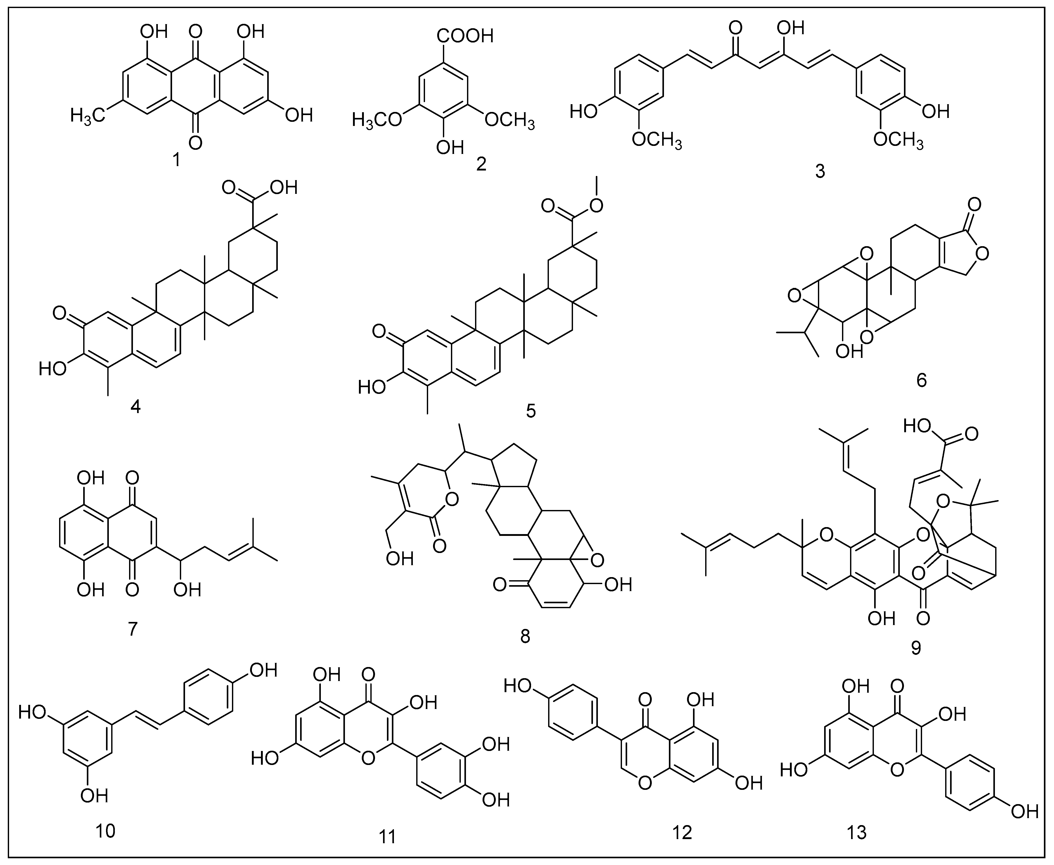
2. Syringic Acid Derivatives

Syringic acid is a phenolic compound present in olives, pumpkin, dates, honey, spices, grapes, acai palm, and other plants that inhibits the Nox/PTP-k/EGFR pathway and has a substantial anti-proliferative action on colon, breast, and skin cancer cells [10][11][12]. Its anti-mitogenic effect is linked to the concurrent inhibition of the binding of NFkB to DNA, proteasomal suppression (ChT-L, PGPH), angiogenesis suppression, and cell sensitization to basic anticancer drugs (vincristine (130-times), camptothecin (500-times), taxol (3134-times), 5FU (20,000-times), vinblastine (1000-times), and doxorubicin (210-times) compared to these drugs alone in vitro [10]. Semi-synthetic analogs of this acid have been synthesized, demonstrating substantial antagonistic efficacy against the binding sites of the 20S proteasome. The following syringic acid analogs exhibited significant mitogenic actions on human malignant melanoma cells with negligible toxicity to breast and colon cancer cells: methyl 4-hydroxy-3,5-dimethoxybenzoate and benzyl 4-hydroxy-3,5-dimethoxybenzoate. The compounds block the proteasome catalytic activity (chymotrypsin-related, trypsin-related, and PGPH), preventing cellular proliferation and promoting programmed cell death [13].

3. Curcumin

Curcumin, a chemical found in turmeric, is now a hot topic in cancer research. It has long been utilized in Asian food and medicine, and it is now used worldwide. Curcumin has recently sparked renewed attention, as its numerous therapeutic properties have been discovered in scientific investigations, and it is still extensively employed in India for medical purposes [14].
Curcumin is a well-recognized effective natural monomer found in the Zingiberaceae family of plants. It can maintain intracellular Nrf2 by blocking ubiquitin and proteasome. Under standard circumstances, Nrf2 is extracted from the cytoplasm by associating with Keap1, a bridging protein that promotes Nrf2 ubiquitination and consequent protein denaturation by acting as a bridging protein among Nrf2 and the Cullin3/Rbx1/E3 ligase complex. Under stress, the Cullin3/Rbx1/E3 ligase blocks the Nrf2, and Nrf2 aggregates ubiquitination in the nucleus, which leads to an increase in cellular protective genes’ transcription. The stimulation of Nrf2 produced by curcumin is reduced in cells treated with a cysteine 151 mutant Keap1 protein substituted by serine, revealing that serine is the primary target for curcumin-modified Keap1, enabling the secretion of Nrf2. As a result, the curcumin’s unsaturated carbonyl component may be critical for attaching to Keap1 and stabilizing Nrf2 by preventing ubiquitination and proteasomal destruction [15].
In glioblastoma (GBM), curcumin modulates the programmed cell death of glioblastoma cells through modifying the protein expression of connexin 43 (Cx43), and thus it can be utilized as an adjunctive treatment. Cx43 protein expression is suppressed by curcumin, although its gene remains unaffected. The cell death produced by TMZ is also exacerbated by curcumin therapy. Curcumin-mediated degradation of Cx43 is inhibited by MG132, a proteasome antagonist, showing that the destruction happens via the ubiquitin-proteasomal cascade. Curcumin is also an autophagy-inducing substance. Autophagy is a type of non-specific breakdown. Ubiquitin is involved in the specific autophagy of ubiquitinated proteins via autophagosomes [16].
Curcumin increases BK protein expression in A7r5 cells but does not impact its gene expression, suggesting that BK gene transcription is unaffected but maintains BK protein by lowering its deterioration. Curcumin also boosts BK channel expression and its half-life in HEK293 cells. The proteasome antagonist MG-132 can remove curcumin in both circumstances. Curcumin also promotes BK protein production by suppressing proteasome degradation and triggering the ERK signaling pathway, enhancing BK channels’ function [17].
Curcumin stimulates autophagy and blocks TGF and Smad signal transmission. Suppression of tetrapeptide repeat domain (TTC3) and mediation of ubiquitin of Smad specific E3 ubiquitin regulatory factor 2 (SMURF2) and proteasomal destruction may be involved in Smad 2 and Smad 3 polyubiquitin degradation, limiting the epithelial–mesenchymal transition of hepatocytes [18].
EBNA1 modulates Epstein–Barr virus’s (EBV) DNA replication, promoting cellular growth, survival, and cancer, and is a promising approach for EBV-associated ailments. Curcumin decreases the level of EBV nuclear antigen, and hence limits the growth of EBV-related human nasopharyngeal cancerous cells via the ubiquitin proteasomal mechanism [19]. Through NOX4 facilitating the formation of reactive oxygen species, imipramine, in conjunction with curcumin, stimulates proteasomal action and reduces the level of post-translational c-FLIP and MCL-1 in a proteasomal way, inducing programmed cell death by modulating PSMA5 levels [20].
In human cystic fibrosis (CF) bronchial epithelial cells, curcumin lowers the nuclear expression of transcriptional stimulatory protein 1 (SP1), which is among the critical reasons for the enhanced production of basic TLR2. A curcumin-induced SP1 decrease can be reduced using MG-132, a proteasome antagonist. Curcumin is thought to suppress TLR2’s gene abundance and function in CF bronchial epithelial cells by speeding up SP1 destruction through the ubiquitin-proteasome cascade [21]. Curcumin reduces the production of ALKB homolog 5 (ALKHB5), which can enhance the expression of mA-modified TNF receptor-associated factor 4 (TRAF4) mRNA, and so decreases HFD-mediated obesity. As an E3 ubiquitin ligase, TRAF4 increases the denaturation of the differentiating modulator. The PPAR of adipose cells suppress adipogenesis via the ubiquitin-proteasome cascade [22].
Curcumin, in conjugation with TNF-associated apoptotic cell death-mediating ligand, can target carcinogenesis synergistically, while causing no harm to conventional proximal renal tubular epithelial cells. The conjugated treatment causes caspase-based cell death, suppresses the proteasome, stimulates the JNK-CHOP cascade driven by ROS, and enables cancerous kidney cells susceptible to TNF-associated apoptotic cell death [23]. Curcumin causes ubiquitination of malignant SIRT1 and consequent proteasomal disintegration to suppress the expression of malignant SIRT1 protein, decreasing the malignancy of human colon cancer cells by irreversibly altering the cysteine 67 motif of SIRT1 [24].
It protects PC12 cells by stimulating the proteasome mechanism, whereas rotenone therapy causes significant protein degradation. Curcumin, in conjunction with carfilzomib, a pharmacological proteasome antagonist, triggers apoptotic cell death sensitized to multiple proteasomes, while being harmless to non-cancerous cells. Curcumin is even a potent inducer of the 26S proteasome, which primarily targets DYRK2 and the 26S proteasome, and a specific blocker of bispecific tyrosine regulated kinase 2 (DYRK2). Curcumin inhibits DYRK2-induced phosphorylation of the 26S proteasome. It can enhance the level of p53 and trigger cell death by activating mitochondrial caspases as proteasome inhibitors [25]. RSV (respiratory syncytial virus) is a myxoviridae virus with a negative-stranded RNA that causes bronchitis, asthma, and chronic lower respiratory tract ailments in newborns and young children. It is a potent proteasomal suppressant that reduces RSV replication in Vero cells by inhibiting the stimulation of nuclear factor-kappa B and suppresses degradation of IKB induced by the proteosome [26].
In a biphasic way, curcumin and its polyphenolic analog (didemethylcurcumin, CUIII) can influence proteasome functioning. At nanomolar doses, curcumin and CUIII promote proteasomal action, whereas at micromolar values, they decrease it. Curcumin has consistently outperformed CUIII in terms of activity [27]. Curcumin increases the expression of miR-142–3p, while decreasing PSMB5 protein levels, thereby reducing the chymotrypsin-related functionality of the 20S proteasome nucleus. Furthermore, p300, a histone acetyltransferase, can inhibit the level of miR-142-3p [28].
Curcumin monoacetate and diacetate were synthesized first, and their action on the proliferation of HCT-11 colon cancer cell lines was examined, along with that of curcumin. All compounds demonstrated the same effect on HCT-116 cells. However, curcumin monoacetate showed less efficiency in SW480 cells than the others. The acetates were substantially less powerful in the context of proteasome suppression when CT-like activity was evaluated. The ability of the various curcumin amino acid conjugates to constrain pure 20S proteasome was then evaluated, in addition to the curcumin. The amino acid conjugates were more effective suppressants of the 20S proteasome than unmodified curcumin. The conjugates were then tested in LNCaP prostate cancer cell lines to determine how they affected cellular proliferation. Overall, the findings imply that hydrophilic curcumin amino acid analogs could be a useful strategy to connect curcumin’s chemotherapeutic properties for cancer therapy [29][30].

4. Celastrol

Anti-cancer properties of celastrol, a component isolated from a Chinese plant called “Thunder of God Vine,” have been explored in a wide range of investigations. In one such investigation, celastrol was found to be a proteasome inhibitor [31][32][33][34][35]. Celastrol has been used for thousands of years to treat a number of inflammatory diseases, and this research found that it structurally resembles quercetin, a flavonoid that is included in many dietary products. Using computer docking studies, it was put to the test against oridonin, a chemical from a different Chinese plant that was thought to have very little proteasome-inhibiting power. Celastrol stopped the 20S proteasome’s CT-like activity at high concentrations, but oridonin did not have any effect, no matter how much it was concentrated. PC-3 prostate cancer cells that were still alive when celastrol was given, but not when oridonin was given, had a rise in proteins that were targeted by the proteasome.
Celastrol caused apoptosis, which was shown by a rise in caspase-3 levels and the cleavage of PARP. Celastrol was next examined in LNCaP prostate cancer cells that were positive for androgen receptor (AR) and shown to inhibit the proteasome and reduce the levels of AR [34][35]. This discovery was crucial because of AR’s significance in developing prostate cancer cells. Celastrol was also effective at reducing tumor growth in animals injected with PC-3 prostate cancer cells. Celastrol blocked 65–82 percent of the growth of breast cancer cells. There was a study that found that celastrol and pristimerin could help temozolomide work well on different types of melanomas cell lines [34][35][36][37]. Both combinations of treatments worked better than temozolomide alone at controlling cell growth in the SK-MEL-173 cell line. Celastrol and temozolomide worked better together, when they were used together in four other resistant melanoma cell lines. Each line had a lower IC50 for temozolomide. They also looked into whether celastrol could stop the proteasome in SK-MEL-173. A chemical called celastrol was put into the cells. Then, Western blot analysis was performed on the cells to look for ubiquitinated proteins. However, there were more IKB and ubiquitinated proteins, which meant that the proteasome was slowed right down because it took longer to break down. In the first step, IKB was phosphorylated, which means that NFKB can be activated. This makes genes in the nucleus more likely to be turned on and off. This research also looked into how NFKB and celastrol work together in the same way. Celastrol and temozolomide were both used to treat melanoma cells. When celastrol was used first, followed by temozolomide, the phosphorylation of IKB was blocked, which further blocked the activation of NFKB. There were no changes in the level of NFKB expression. A crucial stage in NFKB’s ability to influence gene expression was its inability to travel to the nucleus. If this is true, it could help us understand celastrol’s role in slowing cell growth in melanoma lines previously resistant to the drug [35][37].

5. Pristimerin

Another organic compound derived from Chinese plant Celastrus hypoleucus has been investigated for its ability to treat cancer. Pristimerin is the methyl ester of celastrol, and it has also been used in cancer treatment research. In docking investigations, pristimerin, which is also used to treat inflammation, was extremely potent. Prostate cancer cells such as PC-3, and numerous other cell lines with high AR levels, experience reduced AR levels in response to celastrol. Inhibition of the proteasome by pristimerin caused cell death via interfering with the AR signaling pathway [35][38].

6. Triptolide

Another component obtained from the Chinese “Thunder of God Vine” is triptolide, inhibiting the proteasome. Triptolide inhibited cell proliferation successfully in both MDA-MB-231 breast cancer and PC-3 prostate cancer cells. Breast cancer cells were marginally more amenable to therapy than PC-3 cells [35][39]. Triptolide was able to efficiently inhibit the proteasome’s CT-like activity while the PC-3 cells were still alive. This occurred in a dose- and time-dependent manner. Additionally, it destroyed both MDA-MB-231 and PC-3 cells but was more effective against breast cancer cells. Triptolide does not appear to inhibit the function of the purified proteasome, but it can inhibit the proteasome in living cells. To begin, the triptolide molecule may be unable to attach to the 20S proteasome’s β5 subunit due to certain linkages in it. Second, triptolide may inhibit the activity of other components of the 26S proteasome, such as the 20S subunit. The researchers propose that if triptolide is adjusted appropriately, it may be extremely effective at combating cancer [35][39].

7. Shikonin

Shikonin is a naphthoquinone chemical obtained from the root of “Lithospermum erythrorhizon”, a plant that has been used in Chinese medicine for centuries to heal disease-related cuts, sores, and burns. Today, it has been demonstrated that when cancer cell lines are treated with it, they undergo apoptosis. There is currently no understanding of how shikonin induces apoptosis. At various dosages, the researchers discovered that shikonin inhibited the 26S proteasome in both human PC3 prostate cancer cells and murine hepatocellular carcinoma H22 lines [35][40]. Shikonin also induced apoptosis in these cell lines. Additionally, it has been proven that the proteasome is inhibited before cell death. On mice, an in vivo experiment was conducted using P388 leukemia cells. Only one shikonin-treated animal survived the 28-day experiment, but all control mice died after 23 days [35][37][40].

8. Withaferin A

Withania somnifera, commonly referred to as Indian winter cherry, produces withaferin A, a steroidal lactone. For centuries, traditional medicine practitioners in Southeast Asia have used the plant to cure everything, from tumors and wounds to age-related illnesses. Additionally, withaferin A has been researched for its anti-cancer properties to understand its efficacy and mode of action better. According to a study by Yang, Shi, et al., in 2007, withaferin A suppressed the 20S proteasome effectively, with the β5 subunit as its principal target [35][41][42]. In computer docking experiments, withaferin A was discovered to attach to the 20S proteasome’s β5 subunit, inhibiting its CT-like activity. When examined in vitro, purified 20S proteasome was found to inhibit CT-like activity. When withaferin A was used to treat PC3 prostate cancer xenografts, the results were identical. In these cells, apoptosis was also detected, as demonstrated by Western blotting for indicators of apoptosis. Proteasome inhibition by withaferin A increased PARP and caspase-3, as along with morphological alterations in the cells.
Similarly, in LNCaP cells, ubiquitinated proteins were elevated, and androgen-receptor expression decreased over time. Finally, a mouse xenograft of a human PC-3 was generated. Withaferin A-treated mice had tumors that were significantly smaller than those of the control mice, and one of the treated mice was completely tumor-free. Proteasome-targeted proteins such as IκB-α, p27, and Bax were found to have enhanced ubiquitination in the tumor tissues examined after the experiment [35][42].

9. Gambogic Acid

It is the principal pigment of gamboge resin from various Garcinia species. It has been used for centuries in traditional Chinese medicine to cure human diseases, including cancer. Chinese regulators have approved gambogic acid (GA) in clinical trials to treat a range of cancers [43][44]. Numerous putative targets for GA have been revealed, but its molecular targets remain a mystery. GA is equally as effective as bortezomib at inhibiting tumor proteasome activity, but with a lower risk of side-effects. Additionally, intracellular CYP2E1 metabolizes GA to provide it with proteasome-inhibitory properties. There is a lot more CYP2E1 gene expression in tumor tissues than in many normal tissues, which leads to tissue-specific proteasome inhibition and toxicity for cancers. Various cancer cell types have shown these effects to be effective [45][46].

10. Resveratrol

It is a well-known phytoestrogen found in red wine and among other foods. Despite its various health-enhancing characteristics, its effect on HER2+/Erα breast cancer cells appear to be significantly unfavorable. The resveratrol-induced accumulation of ∆16HER2, the production of ∆16HER2/HER heterodimers, and the stimulation of the mTORC1/p70S6K/4EBP1 cascade all contribute to the rise and proliferation of cancer cells, in both in vivo and in vitro settings [47][48][49][50]. Many studies have shown that proteasome-inhibiting can have a beneficial or negative effect, depending on the disease entity (particularly the type of malignancy). There is evidence that resveratrol can reduce the activity of NF-kB and the TNF-α protein, which are both important players in inflammation in neurodegenerative and cardiovascular illnesses, and in many other inflammatory conditions [50][51][52]. Glioma stem cells (GSCs; the chemotherapy-resistant cells responsible for tumor growth and relapse) are less able to self-renew and initiate (form, multiply) tumors in glioblastoma cells when resveratrol is present. Activation of the p53/p21 pathway and reduction of NANOG through proteasomal degradation are both caused by resveratrol, and this suggests a treatment method for glioma that could be used [50][53].

11. Quercetin

The flavonoid quercetin can be found in onions, apples, citrus fruits, and green leafy plants. It has potent antitumor activity, which is multidirectional. Additionally, there is evidence that it possesses anti-inflammatory effects. Quercetin suppresses the activity of all three catalytic subunits of trypsin-like (T-L), proteasomal chymotrypsin-like (ChT-L), and caspase-like/peptidyl-glutamyl peptide-hydrolyzing-like (PGPH) enzymes, and the ChT-L subunit has the largest inhibitory impact [50][54][55]. Quercetin causes apoptosis in cancer epithelial cells because it stops the activity of the proteasome and makes more polyubiquitinated proteins. This flavonoid also causes autophagy by stopping mTOR from working. Quercetin is also more effective than kaempferol or myricetin at blocking the 26S and 20S proteasomes in Jurkat T cells. This is because quercetin is an antioxidant [50][56][57]. It was found that a drug’s capacity to inhibit the proteasome depended on the number of OH groups it contained. The more OH groups there are, the more effective it is. Additionally, the quantity of hydroxyl groups in flavonoids has a significant effect on how antioxidants and cells function. The presence of an unsaturated bond in the C-ring of flavonoids can also affect their ability to prevent toxins from entering the body. This is demonstrated by apigenin, which has an IC50 value 21 times that of naringenin, which lacks an unsaturated bond in the center. Unsaturation on carbon C4 makes the compound more active. The threonine β5 subunit’s N-terminal end can be attacked by nucleophiles, making it more active. In contrast, the presence of saturated ring C makes it less effective at blocking [50][57][58]. It may also be because quercetin and kaempferol have aromatic ketone groups that are easy for nucleophiles to attack. This could be why the proteasome’s ChT-L activity is slowed down [34][50].

12. Genistein

Genistein, an isoflavone present in soybeans, has been shown to stop the development of human cancer cells and greatly reduce the chance of developing hormone-dependent cancers. It has been demonstrated to cause apoptosis that is p53-dependent in previous studies [50][59][60][61]. The anti-chymotrypsin-like activity of proteasome has been demonstrated to be inhibited by this polyphenol both in vitro and in vivo. The in vitro activity of genistein is mediated via interaction with the proteasome subunit β5. IkB, Kip1, and the pro-apoptotic Bax protein become ubiquitinated as a result, and apoptosis is subsequently triggered. Healthy cells do not undergo proteasome inhibition or apoptosis; only malignant fibroblasts do. The mechanism through which genistein affects tumor cells is mainly by targeting the topoisomerases II (Top II) [50][62][63][64]. A drug called doxorubicin, for example, is used to fight cancer because it damages DNA by attacking Top II. According to a study, the proapoptotic effect of genistein on HeLa cells may be due to a decreased transcription level. However, there is considerable evidence that Top II inhibitors have a significant likelihood of producing translocations that result in secondary malignancies [65].

13. Kaempferol

Kaempferol, a flavonol present in various foods, viz., tomatoes, grapes, apples, green tea, and broccoli, is a popular compound. It has been shown to have a variety of qualities, including prooxidative and antioxidant effects, based on the quantity in the cell and the environment. It exhibited several properties, including prooxidative activity (greater production of ROS in glioblastoma cells), anticancer activity (suppression of, among others, RSK2, Src, p53 action, suppression of Akt activity, and suppression of ERK activity), and antiangiogenic action [50][66][67][68]. Kaempferol has been demonstrated to inhibit the degradation of MAGEA6, a ubiquitin ligand for AMPKα1. It is believed that a deficiency of AMPKα1 in the body could result in the development and destruction of human DNA.
Additionally, it makes human glioblastoma cells more susceptible to TRAIL-induced apoptosis, as survivin is degraded by the proteasome. When Akt is inactive, survivin becomes less stable. This occurs because the phosphorylated kinase stabilizes survivin, making it more lasting [50][69][70][71]. Another possible reason the genome does not stay stable is when there are changes to DNA methylation. DNMT methyltransferases are important to the process of DNA methylation. Qiu and his colleagues discovered that kaempferol promotes the proteasomal pathway in bladder cancer cells, activating the DNMT3B methyltransferase degradation process. PI3K and Akt signals regulate the proteasomal degradation of DNMT3B; and p-Akt inhibits the degradation of DNMT3B when it interacts with it. Several previous investigations have demonstrated that kaempferol inhibits the PI3K/Akt pathway, increasing the ubiquitination of DNMT3B and making it simpler to eliminate [50][72].

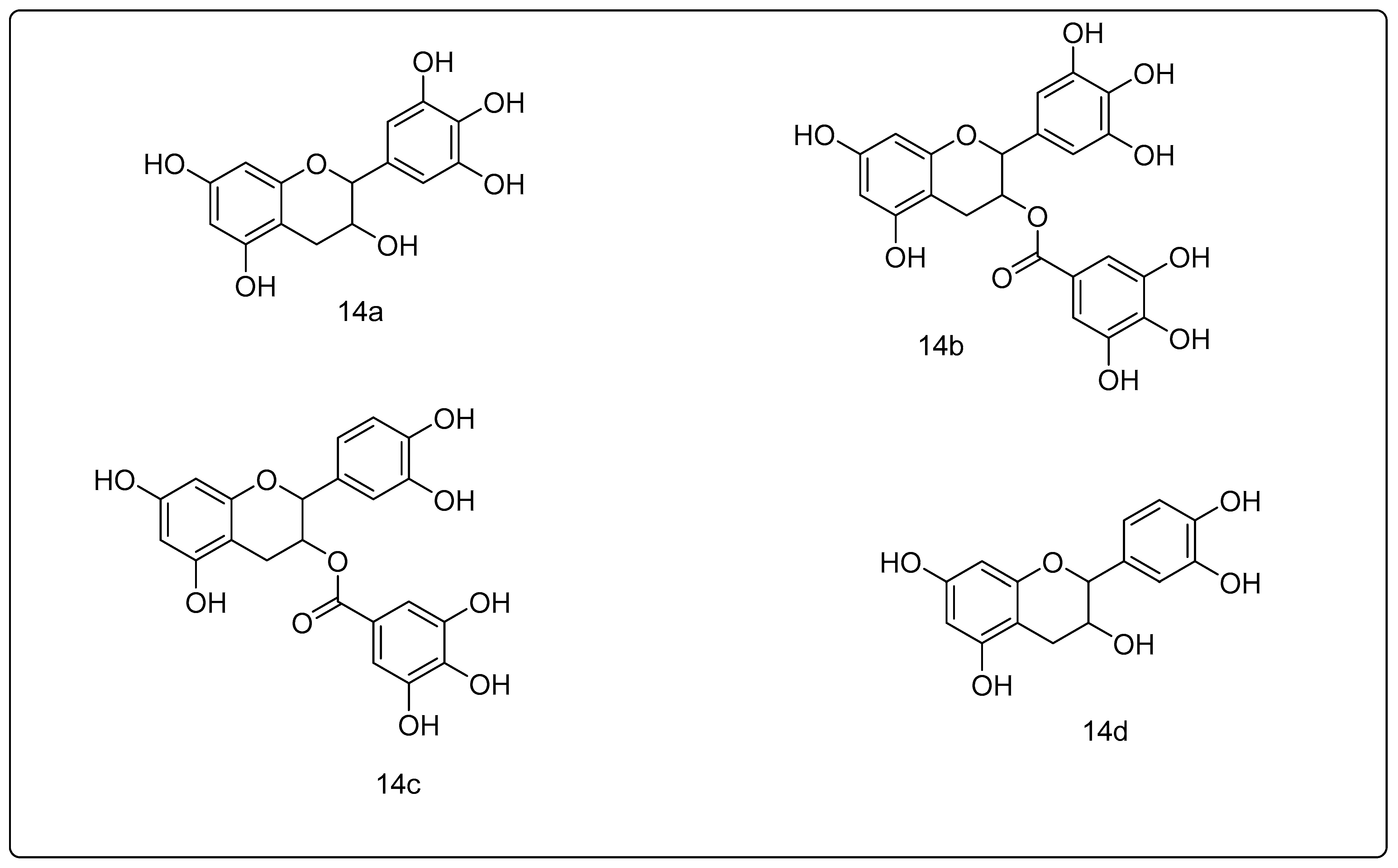
14. Green Tea Polyphenols

The main polyphenols in the tea plant are epigallocatechin (EGC), epigallocatechin-3-gallate (EGCG), epicatechin 3-gallate (ECG), and epicatechin (EC) (Figure 2), and their epimers. Both the 5 and 1 catalytic subunits of the proteasome were shown to be inhibited by EGCG. The identical chemical, on the other hand, is unable to suppress the two subunits [73][74][75][76][77][78][79][80][81]. At 80–200 nM, epigallocatechin-3-gallate (EGCG) inhibits proteasomal activity in cancer cells in vitro. The effective doses are necessary for proteasome suppression to rise to the 1–10 μM range in vivo, which corresponds to the range of concentrations reported in serum, following green tea ingestion [78]. EGCG has high selectivity for cancer cells, as it has a minor inhibitory effect on non-cancer cells’ proteolysis [82]. The capacity of laboratory synthesized EGCG amides and analogs utilizing changed ester linkages and rings to suppress the five subunits of the proteasome is similar to that of the natural substance [81]. The ester bonds present in EGCG, EGC, ECG, and EC appear to be decisive of their proteasomal antagonistic capacities, as the latter are not correlated with GCG (gallocatechin 3-gallate), GC (gallocatechin), CG (catechin-3-gallate), and C (catechin), or their relating epimers, which lack the aforementioned ester bonds. Lactacystin, the most extensively used proteasome antagonist, has an ester link in its active state (lactone) [78].
Figure 2. Green tea polyphenols: (14a) epigallocatechin; (14b) epigallocatechin gallate; (14c) epicatechin gallate; (14d) epicatechin.
Manufactured compounds of tea polyphenols have previously been patented for proteasome suppression and cancer therapy [78]. EGCG is unstable under physiological circumstances, irrespective of its effectiveness as a proteasome antagonist. It is suggested that in vivo biotransformation mechanisms such as methylation diminish its capacity to suppress the proteasome. As a result, attempts have been undertaken to improve its bio-absorption by developing prodrugs that demonstrated promising outcomes in vitro in cell-free and cellular mechanisms, exhibiting greater effects than the known natural molecule [77][83]. With the aid of a proteasome denaturation cascade, EGCG decreases the accumulation of pulmonary fibrosis-associated mutant surfactant protein-A2 (SP-A2), but the proteasomal antagonist MG-132 can counteract the EGCG-mediated aggregate diminution [84].
Cannabinoids cause the Rap1GAPII proteasome to be degraded by G (alpha) o/i, lowering its integrity and activating Rap1, resulting in neurite development. Proteasome antagonists can prevent Rap1 activation caused by this mechanism [85]. Gallic acid reduces the amount of EGFR via speeding up EGFR transformation, causing death in EGFR mutant non-small cell lung cancer cells (MSCLC), although proteasome antagonists can counteract this action [86].
Green tea is rich in polyphenols, which have anti-inflammatory and anti-cancer effects. Epicatechin (EC), epigallocatechin (EGC), epigallocatechin-3-gallate (ECG), and epigallocatechin-3-gallate (EGG) are all anticancer chemicals found in this famous drink. EGCG, which has the greatest antineoplastic effects among the green tea components, is a component of particular interest [87]. EGCG is anti-proliferative (it is believed that it is potent than 5-fluorouracil, which is employed to manage colon cancer), pro-apoptotic, anti-angiogenic [88], and anti-neoplastic [87].
The efficacy of EGCG in the management of malignancies of the gastrointestinal tract, ovarian, breast, prostate, pancreatic, and lung cancer has been proven by epidemiological studies [89].

References

  1. Pecere, T.; Gazzola, M.V.; Mucignat, C.; Parolin, C.; Dalla Vecchia, F.; Cavaggioni, A.; Basso, G.; Diaspro, A.; Salvato, B.; Carli, M.J.C.R. Aloe-emodin is a new type of anticancer agent with selective activity against neuroectodermal tumors. Cancer Res. 2000, 60, 2800–2804.
  2. Monisha, B.A.; Kumar, N.; Tiku, A. Emodin and its role in chronic diseases. Anti-Inflamm. Nutraceuticals Chronic Dis. 2016, 928, 47–73.
  3. Hsu, S.-C.; Chung, J.-G.J.B. Anticancer potential of emodin. BioMedicine 2012, 2, 108–116.
  4. Liu, W.; Feng, Q.; Li, Y.; Ye, L.; Hu, M.; Liu, Z.J.T. Coupling of UDP-glucuronosyltransferases and multidrug resistance-associated proteins is responsible for the intestinal disposition and poor bioavailability of emodin. Toxicol. Appl. Pharmacol. 2012, 265, 316–324.
  5. Xing, J.Y.; Song, G.P.; Deng, J.P.; Jiang, L.Z.; Xiong, P.; Yang, B.J.; Liu, S.S. Antitumor Effects and Mechanism of Novel Emodin Rhamnoside Derivatives against Human Cancer Cells. PLoS ONE 2015, 10, e0144781.
  6. He, Y.; Huang, J.; Wang, P.; Shen, X.; Li, S.; Yang, L.; Liu, W.; Suksamrarn, A.; Zhang, G.; Wang, F.J.O. Emodin potentiates the antiproliferative effect of interferon α/β by activation of JAK/STAT pathway signaling through inhibition of the 26S proteasome. Oncotarget 2016, 7, 4664.
  7. Calderwood, S.K.; Gong, J. Heat shock proteins promote cancer: It’s a protection racket. Trends Biochem. Sci. 2016, 41, 311–323.
  8. Yan, Y.-Y.; Zheng, L.-S.; Zhang, X.; Chen, L.-K.; Singh, S.; Wang, F.; Zhang, J.-Y.; Liang, Y.-J.; Dai, C.-L.; Gu, L.-Q. Blockade of Her2/neu binding to Hsp90 by emodin azide methyl anthraquinone derivative induces proteasomal degradation of Her2/neu. Mol. Pharm. 2011, 8, 1687–1697.
  9. Gililland, J.M.; Anderson, L.A.; Erickson, J.; Pelt, C.E.; Peters, C.L. Mean 5-year clinical and radiographic outcomes of cementless total hip arthroplasty in patients under the age of 30. BioMed Res. Int. 2013, 2013, 1–7.
  10. Abaza, M.-S.; Al-Attiyah, R.A.; Bhardwaj, R.; Abbadi, G.; Koyippally, M.; Afzal, M.J.P. Syringic acid from Tamarix aucheriana possesses antimitogenic and chemo-sensitizing activities in human colorectal cancer cells. Pharm. Biol. 2013, 51, 1110–1124.
  11. Kampa, M.; Alexaki, V.-I.; Notas, G.; Nifli, A.-P.; Nistikaki, A.; Hatzoglou, A.; Bakogeorgou, E.; Kouimtzoglou, E.; Blekas, G.; Boskou, D.J. Antiproliferative and apoptotic effects of selective phenolic acids on T47D human breast cancer cells: Potential mechanisms of action. Breast Cancer Res. 2004, 6, R63–R74.
  12. Ha, S.J.; Lee, J.; Park, J.; Kim, Y.H.; Lee, N.H.; Kim, Y.E.; Song, K.-M.; Chang, P.-S.; Jeong, C.-H.; Jung, S.K. Syringic acid prevents skin carcinogenesis via regulation of NoX and EGFR signaling. Biochem. Pharmacol. 2018, 154, 435–445.
  13. Carlsson, J.; Davidsson, S.; Helenius, G.; Karlsson, M.; Lubovac, Z.; Andrén, O.; Olsson, B.; Klinga-Levan, K.J. A miRNA expression signature that separates between normal and malignant prostate tissues. Cancer Cell Int. 2011, 11, 14.
  14. Mir, R.H.; Mir, P.A.; Shah, A.J.; Banday, N.; Sabreen, S.; Maqbool, M.; Jan, R.; Shafi, N.; Masoodi, M.H. Curcumin as a privileged scaffold molecule for various biological targets in drug development. Stud. Nat. Prod. Chem. 2022, 73, 405–434.
  15. Shin, J.W.; Chun, K.-S.; Kim, D.-H.; Kim, S.-J.; Kim, S.H.; Cho, N.-C.; Na, H.-K.; Surh, Y.-J. Curcumin induces stabilization of Nrf2 protein through Keap1 cysteine modification. Biochem. Pharmacol. 2020, 173, 113820.
  16. Huang, B.-R.; Tsai, C.-H.; Chen, C.-C.; Way, T.-D.; Kao, J.-Y.; Liu, Y.-S.; Lin, H.-Y.; Lai, S.-W.; Lu, D.-Y. Curcumin promotes connexin 43 degradation and temozolomide-induced apoptosis in glioblastoma cells. Am. J. Chin. Med. 2019, 47, 657–674.
  17. Chen, Q.; Tao, J.; Hei, H.; Li, F.; Wang, Y.; Peng, W.; Zhang, X.J. Up-regulatory effects of curcumin on large conductance Ca2+-activated K+ channels. PLoS ONE 2015, 10, e0144800.
  18. Kong, D.; Zhang, Z.; Chen, L.; Huang, W.; Zhang, F.; Wang, L.; Wang, Y.; Cao, P.; Zheng, S.J. Curcumin blunts epithelial-mesenchymal transition of hepatocytes to alleviate hepatic fibrosis through regulating oxidative stress and autophagy. Redox Biol. 2020, 36, 101600.
  19. Liu, L.; Yang, J.; Ji, W.; Wang, C.J.B. Curcumin Inhibits Proliferation of Epstein–Barr Virus-Associated Human Nasopharyngeal Carcinoma Cells by Inhibiting EBV Nuclear Antigen 1 Expression. BioMed Res. Int. 2019, 2019, 1–10.
  20. Seo, S.U.; Kim, T.H.; Kim, D.E.; Min, K.-J.; Kwon, T.K. NOX4-mediated ROS production induces apoptotic cell death via down-regulation of c-FLIP and Mcl-1 expression in combined treatment with thioridazine and curcumin. Redox Biol. 2017, 13, 608–622.
  21. Chaudhary, N.; Ueno-Shuto, K.; Ono, T.; Ohira, Y.; Watanabe, K.; Nasu, A.; Fujikawa, H.; Nakashima, R.; Takahashi, N.; Suico, M.A.J.B.; et al. Curcumin down-regulates toll-like receptor-2 gene expression and function in human cystic fibrosis bronchial epithelial cells. Biol. Pharm. Bull. 2019, 42, 489–495.
  22. Chen, Y.; Wu, R.; Chen, W.; Liu, Y.; Liao, X.; Zeng, B.; Guo, G.; Lou, F.; Xiang, Y.; Wang, Y.J. Curcumin prevents obesity by targeting TRAF4-induced ubiquitylation in m6A-dependent manner. EMBO Rep. 2021, 22, e52146.
  23. Obaidi, I.; Cassidy, H.; Ibanez Gaspar, V.; McCaul, J.; Higgins, M.; Halász, M.; Reynolds, A.L.; Kennedy, B.N.; McMorrow, T.J.B. Curcumin sensitizes kidney cancer cells to TRAIL-induced apoptosis via ROS mediated activation of JNK-CHOP pathway and upregulation of DR4. Biology 2020, 9, 92.
  24. Buratta, S.; Chiaradia, E.; Tognoloni, A.; Gambelunghe, A.; Meschini, C.; Palmieri, L.; Muzi, G.; Urbanelli, L.; Emiliani, C.; Tancini, B.J. Effect of Curcumin on Protein Damage Induced by Rotenone in Dopaminergic PC12 Cells. Int. J. Mol. Sci. 2020, 21, 2761.
  25. Banerjee, S.; Ji, C.; Mayfield, J.E.; Goel, A.; Xiao, J.; Dixon, J.E.; Guo, X.J. Ancient drug curcumin impedes 26S proteasome activity by direct inhibition of dual-specificity tyrosine-regulated kinase 2. Proc. Natl. Acad. Sci. USA 2018, 115, 8155–8160.
  26. Obata, K.; Kojima, T.; Masaki, T.; Okabayashi, T.; Yokota, S.; Hirakawa, S.; Nomura, K.; Takasawa, A.; Murata, M.; Tanaka, S.J. Curcumin prevents replication of respiratory syncytial virus and the epithelial responses to it in human nasal epithelial cells. PLoS ONE 2013, 8, e70225.
  27. Khan, T.K.; You, Y.; Nelson, T.J.; Kundu, S.; Pramanik, S.K.; Das, J. Modulation of proteasome activity by curcumin and didemethylcurcumin. J. Biomol. Struct. Dyn. 2021, 40, 8332–8339.
  28. Liu, L.; Fu, Y.; Zheng, Y.; Ma, M.; Wang, C. Curcumin inhibits proteasome activity in triple-negative breast cancer cells through regulating p300/miR-142–3p/PSMB5 axis. Phytomedicine 2020, 78, 153312.
  29. Wan, S.B.; Yang, H.; Zhou, Z.; Cui, Q.C.; Chen, D.; Kanwar, J.; Mohammad, I.; Dou, O.P.; Chan, T.H. Evaluation of curcumin acetates and amino acid conjugates as proteasome inhibitors. Int. J. Mol. Med. 2010, 26, 447–455.
  30. Yue, X.; Zuo, Y.; Ke, H.; Luo, J.; Lou, L.; Qin, W.; Wang, Y.; Liu, Z.; Chen, D.; Sun, H.J. Identification of 4-arylidene curcumin analogues as novel proteasome inhibitors for potential anticancer agents targeting 19S regulatory particle associated deubiquitinase. Biochem. Pharmacol. 2017, 137, 29–50.
  31. Dai, Y.; DeSano, J.; Tang, W.; Meng, X.; Meng, Y.; Burstein, E.; Lawrence, T.S.; Xu, L. Natural proteasome inhibitor celastrol suppresses androgen-independent prostate cancer progression by modulating apoptotic proteins and NF-kappaB. PLoS ONE 2010, 5, e14153.
  32. Pang, X.; Yi, Z.; Zhang, J.; Lu, B.; Sung, B.; Qu, W.; Aggarwal, B.B.; Liu, M. Celastrol suppresses angiogenesis-mediated tumor growth through inhibition of AKT/mammalian target of rapamycin pathway. Cancer Res. 2010, 70, 1951–1959.
  33. Raja, S.M.; Clubb, R.J.; Ortega-Cava, C.; Williams, S.H.; Bailey, T.A.; Duan, L.; Zhao, X.; Reddi, A.L.; Nyong, A.M.; Natarajan, A. Anticancer activity of Celastrol in combination with ErbB2-targeted therapeutics for treatment of ErbB2-overexpressing breast cancers. Cancer Biol. Ther. 2011, 11, 263–276.
  34. Yang, H.; Chen, D.; Cui, Q.C.; Yuan, X.; Dou, Q.P. Celastrol, a triterpene extracted from the Chinese “Thunder of God Vine,” is a potent proteasome inhibitor and suppresses human prostate cancer growth in nude mice. Cancer Res. 2006, 66, 4758–4765.
  35. Soave, C.L.; Guerin, T.; Liu, J.; Dou, Q.P. Targeting the ubiquitin-proteasome system for cancer treatment: Discovering novel inhibitors from nature and drug repurposing. Cancer Metastasis Rev. 2017, 36, 717–736.
  36. Mahajan, K.; Malla, P.; Lawrence, H.R.; Chen, Z.; Kumar-Sinha, C.; Malik, R.; Shukla, S.; Kim, J.; Coppola, D.; Lawrence, N.J. ACK1/TNK2 regulates histone H4 Tyr88-phosphorylation and AR gene expression in castration-resistant prostate cancer. Cancer Cell 2017, 31, 790–803.e8.
  37. Chen, M.; Rose, A.E.; Doudican, N.; Osman, I.; Orlow, S.J. Celastrol synergistically enhances temozolomide cytotoxicity in melanoma cells. Mol. Cancer Res. 2009, 7, 1946–1953.
  38. Yang, H.; Landis-Piwowar, K.R.; Lu, D.; Yuan, P.; Li, L.; Reddy, G.P.V.; Yuan, X.; Dou, Q.P. Pristimerin induces apoptosis by targeting the proteasome in prostate cancer cells. J. Cell. Biochem. 2008, 103, 234–244.
  39. Lu, L.; Kanwar, J.; Schmitt, S.; Cui, Q.C.; Zhang, C.; Zhao, C.; Dou, Q.P. Inhibition of tumor cellular proteasome activity by triptolide extracted from the Chinese medicinal plant ‘thunder god vine’. Anticancer Res. 2011, 31, 1–10.
  40. Yang, H.; Zhou, P.; Huang, H.; Chen, D.; Ma, N.; Cui, Q.C.; Shen, S.; Dong, W.; Zhang, X.; Lian, W. Shikonin exerts antitumor activity via proteasome inhibition and cell death induction in vitro and in vivo. Int. J. Cancer 2009, 124, 2450–2459.
  41. Mirjalili, M.H.; Moyano, E.; Bonfill, M.; Cusido, R.M.; Palazón, J. Steroidal lactones from Withania somnifera, an ancient plant for novel medicine. Molecules 2009, 14, 2373–2393.
  42. Yang, H.; Shi, G.; Dou, Q.P. The tumor proteasome is a primary target for the natural anticancer compound Withaferin A isolated from “Indian winter cherry”. Mol. Pharmacol. 2007, 71, 426–437.
  43. Kashyap, D.; Mondal, R.; Tuli, H.S.; Kumar, G.; Sharma, A.K. Molecular targets of gambogic acid in cancer: Recent trends and advancements. Tumor Biol. 2016, 37, 12915–12925.
  44. Zhou, Z.; Wan, J. Phase I human tolerability trial of gambogic acid. Chin. J. New Drugs 2007, 16, 679–682.
  45. Li, X.; Liu, S.; Huang, H.; Liu, N.; Zhao, C.; Liao, S.; Yang, C.; Liu, Y.; Zhao, C.; Li, S. Gambogic acid is a tissue-specific proteasome inhibitor in vitro and in vivo. Cell Rep. 2013, 3, 211–222.
  46. Shi, X.; Chen, X.; Li, X.; Lan, X.; Zhao, C.; Liu, S.; Huang, H.; Liu, N.; Liao, S.; Song, W. Gambogic acid induces apoptosis in imatinib-resistant chronic myeloid leukemia cells via inducing proteasome inhibition and caspase-dependent Bcr-Abl downregulation. Clin. Cancer Res. 2014, 20, 151–163.
  47. Andreani, C.; Bartolacci, C.; Wijnant, K.; Crinelli, R.; Bianchi, M.; Magnani, M.; Hysi, A.; Iezzi, M.; Amici, A.; Marchini, C. Resveratrol fuels HER2 and ERα-positive breast cancer behaving as proteasome inhibitor. Aging 2017, 9, 508.
  48. Mir, R.H.; Banday, N.; Sabreen, S.; Shah, A.J.; Jan, R.; Wani, T.U.; Farooq, S.; Bhat, Z.A. Resveratrol: A potential drug candidate with multispectrum therapeutic application. Stud. Nat. Prod. Chem. 2022, 73, 99–137.
  49. Kwon, K.J.; Kim, J.N.; Kim, M.K.; Lee, J.; Ignarro, L.J.; Kim, H.J.; Shin, C.Y.; Han, S.H. Melatonin synergistically increases resveratrol-induced heme oxygenase-1 expression through the inhibition of ubiquitin-dependent proteasome pathway: A possible role in neuroprotection. J. Pineal Res. 2011, 50, 110–123.
  50. Golonko, A.; Pienkowski, T.; Swislocka, R.; Lazny, R.; Roszko, M.; Lewandowski, W. Another look at phenolic compounds in cancer therapy the effect of polyphenols on ubiquitin-proteasome system. Eur. J. Med. Chem. 2019, 167, 291–311.
  51. Bradley, J. TNF-mediated inflammatory disease. J. Pathol. A J. Pathol. Soc. Great Br. Irel. 2008, 214, 149–160.
  52. Silswal, N.; Reddy, N.; Qureshi, N. Resveratrol Modulates Cytokine Expression in LPS-induced Human Monocytes: Role of Proteasome Subunits. FASEB J. 2016, 30, 597.6.
  53. Sato, A.; Okada, M.; Shibuya, K.; Watanabe, E.; Seino, S.; Suzuki, K.; Narita, Y.; Shibui, S.; Kayama, T.; Kitanaka, C. Resveratrol promotes proteasome-dependent degradation of Nanog via p53 activation and induces differentiation of glioma stem cells. Stem Cell Res. 2013, 11, 601–610.
  54. Hashemzaei, M.; Delarami Far, A.; Yari, A.; Heravi, R.E.; Tabrizian, K.; Taghdisi, S.M.; Sadegh, S.E.; Tsarouhas, K.; Kouretas, D.; Tzanakakis, G. Anticancer and apoptosis-inducing effects of quercetin in vitro and in vivo. Oncol. Rep. 2017, 38, 819–828.
  55. Dosenko, V.; Nagibin, V.; Tumanovskaya, L.; Zagorii, V.Y.; Moibenko, A. Effect of quercetin on the activity of purified 20S and 26S proteasomes and proteasomal activity in isolated cardiomyocytes. Biochem. (Mosc.) Suppl. Ser. B Biomed. Chem. 2007, 1, 40–44.
  56. Klappan, A.K.; Hones, S.; Mylonas, I.; Brüning, A. Proteasome inhibition by quercetin triggers macroautophagy and blocks mTOR activity. Histochem. Cell Biol. 2012, 137, 25–36.
  57. Chen, D.; Daniel, K.G.; Chen, M.S.; Kuhn, D.J.; Landis-Piwowar, K.R.; Dou, Q.P. Dietary flavonoids as proteasome inhibitors and apoptosis inducers in human leukemia cells. Biochem. Pharmacol. 2005, 69, 1421–1432.
  58. Chen, D.; Chen, M.S.; Cui, Q.C.; Yang, H.; Dou, Q.P. Structure-proteasome-inhibitory activity relationships of dietary flavonoids in human cancer cells. Front Biosci. 2007, 12, 1935–1945.
  59. Zhu, J.; Zhang, C.; Qing, Y.; Cheng, Y.; Jiang, X.; Li, M.; Yang, Z.; Wang, D. Genistein induces apoptosis by stabilizing intracellular p53 protein through an APE1-mediated pathway. Free Radic. Biol. Med. 2015, 86, 209–218.
  60. Zhang, Z.; Wang, C.-Z.; Du, G.-J.; Qi, L.-W.; Calway, T.; He, T.-C.; Du, W.; Yuan, C.-S. Genistein induces G2/M cell cycle arrest and apoptosis via ATM/p53-dependent pathway in human colon cancer cells. Int. J. Oncol. 2013, 43, 289–296.
  61. Wu, T.-C.; Lin, Y.-C.; Chen, H.-L.; Huang, P.-R.; Liu, S.-Y.; Yeh, S.-L. The enhancing effect of genistein on apoptosis induced by trichostatin A in lung cancer cells with wild type p53 genes is associated with upregulation of histone acetyltransferase. Toxicol. Appl. Pharmacol. 2016, 292, 94–102.
  62. Kazi, A.; Daniel, K.G.; Smith, D.M.; Kumar, N.B.; Dou, Q.P. Inhibition of the proteasome activity, a novel mechanism associated with the tumor cell apoptosis-inducing ability of genistein. Biochem. Pharmacol. 2003, 66, 965–976.
  63. Zhou, N.; Yan, Y.; Li, W.; Wang, Y.; Zheng, L.; Han, S.; Yan, Y.; Li, Y. Genistein inhibition of topoisomerase IIα expression participated by Sp1 and Sp3 in HeLa cell. Int. J. Mol. Sci. 2009, 10, 3255–3268.
  64. Azarova, A.M.; Lin, R.-K.; Tsai, Y.-C.; Liu, L.F.; Lin, C.-P.; Lyu, Y.L. Genistein induces topoisomerase IIbeta-and proteasome-mediated DNA sequence rearrangements: Implications in infant leukemia. Biochem. Biophys. Res. Commun. 2010, 399, 66–71.
  65. Nitiss, J.L. Targeting DNA topoisomerase II in cancer chemotherapy. Nat. Rev. Cancer 2009, 9, 338–350.
  66. Sharma, V.; Joseph, C.; Ghosh, S.; Agarwal, A.; Mishra, M.K.; Sen, E. Kaempferol induces apoptosis in glioblastoma cells through oxidative stress. Mol. Cancer Ther. 2007, 6, 2544–2553.
  67. Shields, M. Pharmacognosy: Fundamentals, Applications and Strategies; Elsevier: Atlanta, GA, USA, 2017.
  68. Kim, S.-H.; Choi, K.-C. Anti-cancer effect and underlying mechanism(s) of kaempferol, a phytoestrogen, on the regulation of apoptosis in diverse cancer cell models. Toxicol. Res. 2013, 29, 229–234.
  69. Han, B.; Yu, Y.-Q.; Yang, Q.-L.; Shen, C.-Y.; Wang, X.-J. Kaempferol induces autophagic cell death of hepatocellular carcinoma cells via activating AMPK signaling. Oncotarget 2017, 8, 86227.
  70. Xu, H.; Zhou, Y.; Coughlan, K.A.; Ding, Y.; Wang, S.; Wu, Y.; Song, P.; Zou, M.-H. AMPKα1 deficiency promotes cellular proliferation and DNA damage via p21 reduction in mouse embryonic fibroblasts. Biochim. Biophys. Acta (BBA) Mol. Cell Res. 2015, 1853, 65–73.
  71. Siegelin, M.D.; Reuss, D.E.; Habel, A.; Herold-Mende, C.; Von Deimling, A. The flavonoid kaempferol sensitizes human glioma cells to TRAIL-mediated apoptosis by proteasomal degradation of survivin. Mol. Cancer Ther. 2008, 7, 3566–3574.
  72. Qiu, W.; Lin, J.; Zhu, Y.; Zhang, J.; Zeng, L.; Su, M.; Tian, Y. Kaempferol modulates DNA methylation and downregulates DNMT3B in bladder cancer. Cell. Physiol. Biochem. 2017, 41, 1325–1335.
  73. Seely, D.; Mills, E.J.; Wu, P.; Verma, S.; Guyatt, G. The effects of green tea consumption on incidence of breast cancer and recurrence of breast cancer: A systematic review and meta-analysis. Integr. Cancer Ther. 2005, 4, 144–155.
  74. Arab, L.; Il’yasova, D.J. The epidemiology of tea consumption and colorectal cancer incidence. J. Nutr. 2003, 133, 3310S–3318S.
  75. Imai, K.; Suga, K.; Nakachi, K.J. Cancer-preventive effects of drinking green tea among a Japanese population. Prev. Med. 1997, 26, 769–775.
  76. Mir, R.H.; Masoodi, M.H. Anti-inflammatory plant polyphenolics and cellular action mechanisms. Curr. Bioact. Compd. 2020, 16, 809–817.
  77. Dou, Q.; Landis-Piwowar, K.; Chen, D.; Huo, C.; Wan, S.; Chan, T.J. Green tea polyphenols as a natural tumour cell proteasome inhibitor. Inflammopharmacology 2008, 16, 208–212.
  78. Nam, S.; Smith, D.M.; Dou, Q.P. Ester bond-containing tea polyphenols potently inhibit proteasome activity in vitro and in vivo. J. Biol. Chem. 2001, 276, 13322–13330.
  79. Thangapazham, R.L.; Singh, A.K.; Sharma, A.; Warren, J.; Gaddipati, J.P.; Maheshwari, R.K. Green tea polyphenols and its constituent epigallocatechin gallate inhibits proliferation of human breast cancer cells in vitro and in vivo. Cancer Lett. 2007, 245, 232–241.
  80. Smith, D.M.; Wang, Z.; Kazi, A.; Li, L.-H.; Chan, T.-H.; Dou, Q.P. Synthetic analogs of green tea polyphenols as proteasome inhibitors. Mol. Med. 2002, 8, 382–392.
  81. Kazi, A.; Wang, Z.; Kumar, N.; Falsetti, S.C.; Chan, T.-H.; Dou, Q.P. Structure-activity relationships of synthetic analogs of (-)-epigallocatechin-3-gallate as proteasome inhibitors. Anticancer Res. 2004, 24, 943–954.
  82. Chen, Z.P.; Schell, J.B.; Ho, C.-T.; Chen, K.Y. Green tea epigallocatechin gallate shows a pronounced growth inhibitory effect on cancerous cells but not on their normal counterparts. Cancer Lett. 1998, 129, 173–179.
  83. Kuhn, D.; Lam, W.H.; Kazi, A.; Daniel, K.G.; Song, S.; Chow, L.; Chan, T.H.; Dou, Q.P. Synthetic peracetate tea polyphenols as potent proteasome inhibitors and apoptosis inducers in human cancer cells. Front. Biosci. Landmark 2005, 10, 1010–1023.
  84. Quan, Y.; Li, L.; Dong, L.; Wang, S.; Jiang, X.; Zhang, T.; Jin, P.; Fan, J.; Mao, S.; Fan, X.; et al. Epigallocatechin-3-gallate (EGCG) inhibits aggregation of pulmonary fibrosis associated mutant surfactant protein A2 via a proteasomal degradation pathway. Int. J. Biochem. Cell Biol. 2019, 116, 105612.
  85. Jordan, J.D.; He, J.C.; Eungdamrong, N.J.; Gomes, I.; Ali, W.; Nguyen, T.; Bivona, T.G.; Philips, M.R.; Devi, L.A.; Iyengar, R.J. Cannabinoid receptor-induced neurite outgrowth is mediated by Rap1 activation through Gαo/i-triggered proteasomal degradation of Rap1GAPII. J. Biol. Chem. 2005, 280, 11413–11421.
  86. Nam, B.; Rho, J.K.; Shin, D.-M.; Son, J.J. Gallic acid induces apoptosis in EGFR-mutant non-small cell lung cancers by accelerating EGFR turnover. Bioorg. Med. Chem. Lett. 2016, 26, 4571–4575.
  87. Zhang, L.; Wei, Y.; Zhang, J.J. Novel mechanisms of anticancer activities of green tea component epigallocatechin-3-gallate. Anti-Cancer Agents Med. Chem. 2014, 14, 779–786.
  88. Xiang, L.-P.; Wang, A.; Ye, J.-H.; Zheng, X.-Q.; Polito, C.A.; Lu, J.-L.; Li, Q.-S.; Liang, Y.-R. Suppressive effects of tea catechins on breast cancer. Nutrients 2016, 8, 458.
  89. Ju, J.; Lu, G.; Lambert, J.D.; Yang, C.S. Inhibition of carcinogenesis by tea constituents. Semin. Cancer Biol. 2007, 17, 395–402.
More
Information
Contributors MDPI registered users' name will be linked to their SciProfiles pages. To register with us, please refer to https://encyclopedia.pub/register : , , , , , , ,
View Times: 215
Revisions: 2 times (View History)
Update Date: 18 Apr 2023
1000/1000
Video Production Service