Submitted Successfully!
To reward your contribution, here is a gift for you: A free trial for our video production service.
Thank you for your contribution! You can also upload a video entry or images related to this topic.
Version Summary Created by Modification Content Size Created at Operation
1 -- 1661 2023-04-12 16:42:16 |
2 format correct Meta information modification 1661 2023-04-13 02:44:51 | |
3 format correct Meta information modification 1661 2023-04-13 02:47:24 |

Video Upload Options

Do you have a full video?

Confirm

Are you sure to Delete?
Cite
If you have any further questions, please contact Encyclopedia Editorial Office.
Hossain, M.M.; Sultana, F.; Li, W.; Tran, L.P.; Mostofa, M.G. Sclerotinia sclerotiorum (Lib.) de Bary against Host Defense. Encyclopedia. Available online: https://encyclopedia.pub/entry/42996 (accessed on 08 May 2024).
Hossain MM, Sultana F, Li W, Tran LP, Mostofa MG. Sclerotinia sclerotiorum (Lib.) de Bary against Host Defense. Encyclopedia. Available at: https://encyclopedia.pub/entry/42996. Accessed May 08, 2024.
Hossain, Md. Motaher, Farjana Sultana, Weiqiang Li, Lam-Son Phan Tran, Mohammad Golam Mostofa. "Sclerotinia sclerotiorum (Lib.) de Bary against Host Defense" Encyclopedia, https://encyclopedia.pub/entry/42996 (accessed May 08, 2024).
Hossain, M.M., Sultana, F., Li, W., Tran, L.P., & Mostofa, M.G. (2023, April 12). Sclerotinia sclerotiorum (Lib.) de Bary against Host Defense. In Encyclopedia. https://encyclopedia.pub/entry/42996
Hossain, Md. Motaher, et al. "Sclerotinia sclerotiorum (Lib.) de Bary against Host Defense." Encyclopedia. Web. 12 April, 2023.
Sclerotinia sclerotiorum (Lib.) de Bary against Host Defense
Edit

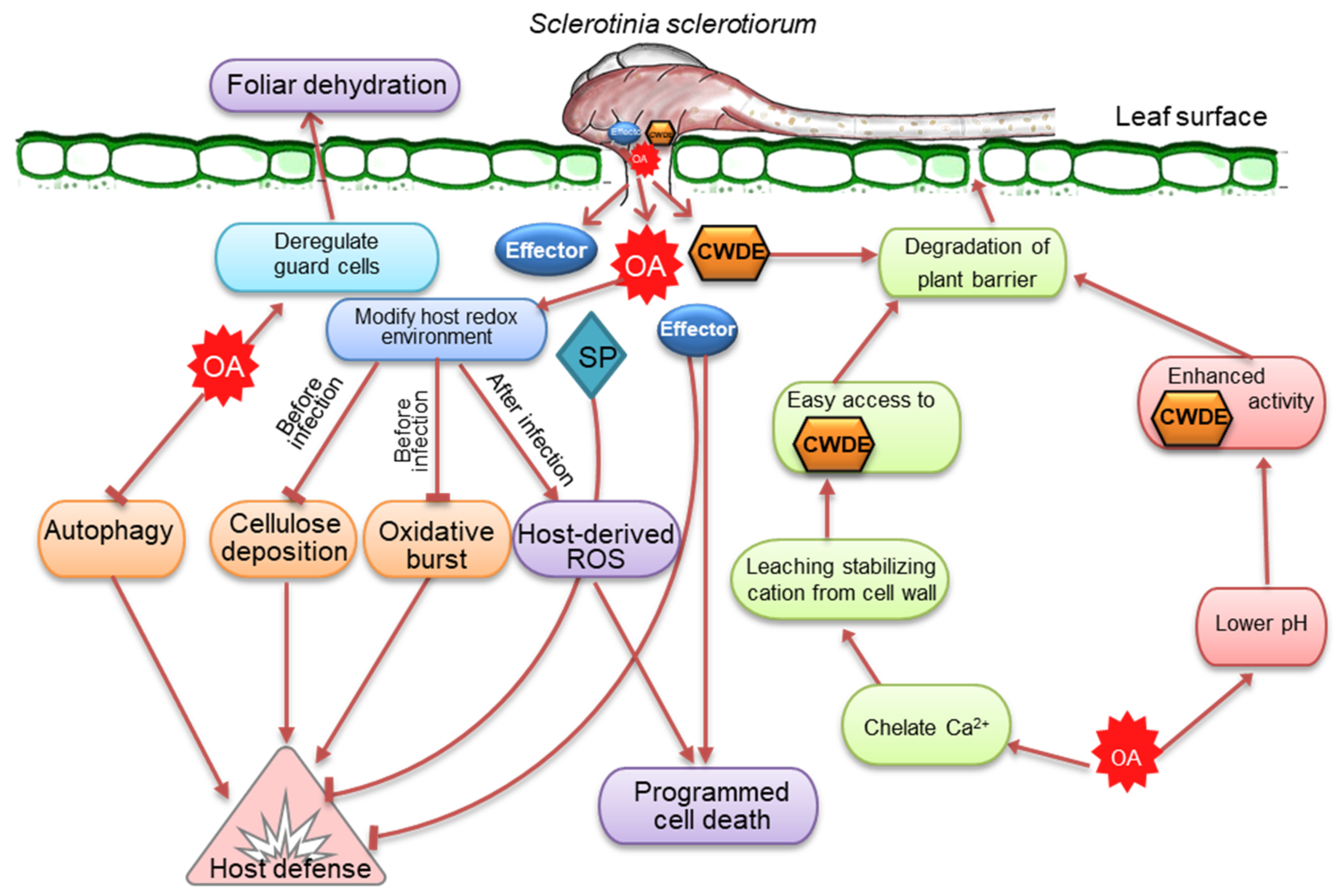
Sclerotinia sclerotiorum (Lib.) de Bary is a broad host-range fungus that infects an inclusive array of plant species and afflicts significant yield losses globally. S. sclerotiorum possesses an immense arsenal of disease weaponry to subsist and succeed under widespread environmental conditions. The massive pathogenic arsenal of S. sclerotiorum, including oxalic acid (OA), CWDEs, and small secretory proteins (effectors), has long been associated with virulence. A subtle interplay between these virulence factors serves various regulatory functions in host cells, allowing S. sclerotiorum to colonize the host, evade or inhibit the host defense system, and cause disease. Understanding and studying these mechanisms is crucial for detecting pathways of genetic interventions that could result in improved control of this disease.

cAMP cell wall degrading enzymes effectors life cycle

1. Oxalic Acid, a Versatile Toxin and Broad-Spectrum Pathogenicity Factor

Of the various virulence factors, extensive research has been conducted on the relationship between white mold disease and the production of OA by S. sclerotiorum [1][2]. This fungus is well-known for its ability to acidify the surroundings via OA secretion. The OA-deficient mutants of the fungus have been shown to be poorly pathogenic [3], suggesting that the pathogenesis of S. sclerotiorum requires accumulating high quantities of OA. While OA-minus mutants can still infect and colonize hosts to variable degrees, it is appropriate to view OA more as a colonization factor rather than a crucial compatibility element [4]. OA is not directly toxic to plant cells but plays a more sophisticated role as a signaling molecule that seizes control of host cells and creates an environment favorable for S. sclerotiorum growth. OA alters a number of physiological processes in host plants. OA promotes cell wall degradation by increasing the activity of polygalacturonases, inhibiting plant-protection enzymes, subduing oxidative burst, deregulating stomatal guard cell closure, mediating pH signaling, inducing apoptosis-like cell death, and altering cellular redox status in plants (Figure 1) [3][5][6][7][8][9][10]. The production of OA seems to suit the overall strategy of S. sclerotiorum to acidify its ambient environment to facilitate infection in several ways. Many of the fungal enzymes (such as polygalacturonase) secreted during the invasion of plant tissues have maximal activities at a low pH [7]. It is postulated that OA could increase Sclerotinia virulence by adjusting the apoplastic pH to a level conducive to the enzymatic breakdown of plant cell walls. OA can also weaken plant cell walls via acidity, thereby facilitating invasion [7]. Interestingly, no ROS production is detected at acidic pH [10]. It is evident that S. sclerotinia uses OA to elicit apoptosis-like programmed cell death, and the induction of this process is commenced by a reducing environment generated by the fungus in the host cell [6]. As a direct consequence of this redox manipulation, the fungus subverts host defense responses, inhibits oxidative burst, and prepares the infection court for disease establishment [6]. These findings imply that the effect of OA on the reduction of pH in the ambient environment is significantly greater than that of OA alone as an essential component of the infection process caused by S. sclerotiorum. Intriguingly, the regulation of host defense by OA appears to be biphasic. In the early phases of infection, OA suppresses host defenses by culminating ROS buildup and callose deposition in the host [6]. OA sequesters Ca2+ liberated during cell wall collapse, thereby protecting the development of S. sclerotiorum hyphae from hazardous Ca2+ concentrations [11]. In the later stages of infection, OA quashes antioxidant enzyme activities and stimulates ROS generation, concurrently damaging the membranes of host cells [12]. These findings suggest that OA is a cross-functional toxin crucial to the widespread pathogenic success of S. sclerotiorum.
The dicarboxylic acid OA can be produced through the oxidation of glyoxylate or glycolaldehyde or the hydrolysis of oxaloacetate [13]. However, current research indicates that OA biosynthesis entirely depends on the oxaloacetate acetylhydrolase (OAH) activity [14]. OAH mediates the C-C cleavage of oxaloacetate during the OA biosynthesis process [4]. Depending on the growth substrate, either the tricarboxylic acid (TCA) cycle or the glyoxylate cycle may produce oxaloacetate precursor [15]. Experimental evidence from S. sclerotiorum and B. cinerea gene-deletion mutants supports the hypothesis that the expressed enzyme activity is responsible for catalyzing the last step in the OA biosynthesis process [16]. Three types of OA-degrading enzymes are found throughout evolutionary lineages: OxDC (EC 4.1.1.2), OxOx (EC 1.2.3.4), and oxalylCoA decarboxylase (OXC, EC 4.1.1.8). S. sclerotiorum exhibits oxalate decarboxylase (OxDCs) activity but not OxOx [17], while only bacterial species have been identified to possess OXC activity. OxDC catalyzes the production of formate and CO2 from OA, and its activity is widespread among fungi and bacteria [18].
Figure 1. The frontline weapons or virulence factors of Sclerotinia sclerotiorum important for developing and regulating the infection process in plants. S. sclerotiorum releases oxalic acid (OA), which modulates reactive oxygen species (ROS) and the activity of cell wall-degrading enzymes (CWDEs) while dampening host defense responses and suppressing autophagy. At the early stage, OA suppresses plant defensive responses and modifies the host redox state by inhibiting callose deposition and host oxidative burst and establishing a reducing environment. At a later stage, OA-induced ROS causes apoptotic-like programmed cell death and disease. Because of its affinity for Ca2+, OA may degrade plant barriers by draining the stabilization of cations from the plant cell wall. OA accumulation also lowers the pH and activates CWDEs, which are necessary to destroy infection surfaces. Effectors and other secreted proteins (SP) have a similar role in cell death and host defensive responses.

2. Arsenal of Cell Wall-Degrading Enzymes Reflects Broad Host Preference

An essential component of the lifecycle of S. sclerotiorum is its capacity to break down a wide range of complex plant polysaccharides by CWDEs (Figure 1). Cellulose, hemicellulose, and pectin form a complex network of polysaccharides that constitute plant cell walls [19]. This network is the target of CAZymes, a group of proteins and enzymes that break down plant cell wall polysaccharides into simple monomers [20]. These monomers serve as carbon sources and allow access to internal plant tissues. At least 118 CAZymes are explicitly associated with plant cell wall degradation by S. sclerotiorum; however, many other CAZymes presumably contribute to this activity [21]. Several endo- and exo-polygalacturonases (PGs) are known to break down unesterified pectate polymers, middle lamella structural polysaccharides, and primary plant cell wall components. Endo-PGs catalyze the hydrolysis of homogalacturonan, whereas exo-PGs fragment the substrate and release potential nutrients by removing monomeric or dimeric glycosyl groups from pectic cell wall polysaccharides [22]. PGs are also recognized as potential virulence factors in several pathosystems [23][24][25]; however, convincing proof for their universal involvement in virulence is missing. The principal hemicellulose constituent of plant cell walls, xylan, is comprised of xylose and arabinose connected by β-(1–4)-linked xylopyranose. Endo β-1, 4-xylanase is the major enzyme that degrades xylan [26]. Studies have highlighted the critical functions of endo-β-1, 4-xylanase in the development and pathogenicity of S. sclerotiorum [27]. Other important groups of mannan- and cellulose-degrading enzymes are also highly upregulated [21]. These indicate that S. sclerotiorum utilizes a diverse array of enzymes that play significant roles in its infection strategy across many host species.

3. Secretory Effectors, Critical Components in the Necrotrophic Infection Strategy

Numerous small, secreted proteins of S. sclerotiorum have been reported as having effector-like features or effector-like activities, which are released either extracellularly or within the cytoplasm of plant cells to disarm host defense mechanisms and enhance fungal infection (Figure 1) [28][29]. Until now, a number of S. sclerotiorum effectors associated with virulence have been identified. During the infection of soybean seedlings, S. sclerotiorum produces a basic endopolygalacturonase (PG) isoform early in the process that elicits Ca2+-mediated signaling and programmed cell death [30]. A polygalacturonase (PG) enzyme, called Sspg1d, reacts with proteins that have a C2 domain in Brassica napus [31]. Necrosis and ethylene-inducing peptides encoded by two genes, SsNep1 and SsNep2, induce necrosis in tobacco (Nicotiana tabacum) leaves during heterologous expression [32]. A protein with a signal peptide and 302 amino acid residues in the integrin alpha N-terminal domain superfamily, SSITL, inhibits defense reactions in Arabidopsis thaliana [33]. A putative-secreted protein disrupting pathogen virulence has also been reported [34]. A cutinase encoded by SsCut induces cell death in an array of plants, including rice (Oryzae staiva), G. max, maize (Zea mays), B. napus, wheat (Triticum aestivum), and A. thaliana [35]. A small cysteine-rich protein containing a cyanoviron-N homology (CVNH) domain, SsSSVP1, is required for pathogen virulence [36]. This putative effector is believed to alter plant energy metabolism in order to promote S. sclerotiorum infection. A small virulence-related protein 1, without obvious functional domains, has been found explicitly in S. sclerotiorum and B. cinerea [37].
In addition, several other effectors, such as chorismate mutase (SsCM1) [38], compound appressorium formation-related protein 1 (SsCaf1) [39], BAX inhibitor-1 (SsBI1) [40], superoxide dismutase (SsSodI) [41], catalase (SsCat1) [42], a protein analogous to a Magnaporthe grisea protein elicitor (SsPemG1) [43], and ceratoplantanin effector (SsCP1) [44], have been documented in S. sclerotiorum. The vast majority of the putative proteinaceous effectors characterized in S. sclerotiorum cause cell death in host tissue. S. sclerotiorum genome analysis has recently identified a novel class of necrosis-inducing effector 2 (SsNE2) [45]. On the contrary, a few biotrophy-related candidate effectors have been spotted, such as salicylate hydroxylase and a lysin motif (LysM) domain protein [28]. These effector genes were found to be upregulated within the first 24 h after infection. In biotrophic and hemibiotrophic pathogens, recognizing the effectors often leads to a hypersensitive reaction (HR) to contain the pathogens (Jones and Dangl, 2006) [46]. Contrariwise, for necrotrophic infections, the elicitation of HR responses by effectors delivers substrates to stimulate host colonization [14][47]. Additionally, S. sclerotiorum exhibits a host-specific expression of candidate effectors, as Guyon et al. [48] showed that the expression patterns of 16 effector candidates varied significantly between the hosts S. lycospersicum, Nicotiana benthamiana, and susceptible and resistant accessions of A. thaliana. According to previous studies, the host-specific expression of putative effectors was more limited than anticipated. Only one of the effector candidate genes (sscle 08g064180) identified by Derbyshire et al. [49] and Amselem et al. [21] was expressed differently between B. napus and Lupinus angustifolius, where the expression was significantly higher in B. napus compared to L. angustifolius. This putative effector is anticipated to have a coiled-coil domain, although little is known about the role of such domain in mediating effectors’ functions.

References

  1. Xu, T.; Li, J.; Yu, B.; Liu, L.; Zhang, X.; Liu, J.; Pan, H.; Zhang, Y. Transcription Factor SsSte12 was involved in mycelium growth and development in Sclerotinia sclerotiorum. Front. Microbiol. 2018, 9, 2476.
  2. Derbyshire, M.C.; Newman, T.E.; Khentry, Y.; Owolabi, T.A. The evolutionary and molecular features of the broad-host-range plant pathogen Sclerotinia sclerotiorum. Mol. Plant Pathol. 2022, 23, 1075–1090.
  3. Cessna, S.G.; Sears, V.E.; Low, P.S.; Dickman, M. Oxalic acid, a pathogenicity factor for Sclerotinia sclerotiorum, suppresses the oxidative burst of the host plant. Plant Cell. 2000, 12, 2191–2200.
  4. Liang, X.; Liberti, D.; Li, M.; Kim, Y.T.; Hutchens, A.; Wilson, R.; Rollins, J.A. Oxaloacetate acetylhydrolase gene mutants of Sclerotinia sclerotiorum do not accumulate oxalic acid but do produce limited lesions on host plants. Mol. Plant Pathol. 2015, 16, 559–571.
  5. Rollins, J.A.; Dickman, M.B. pH signaling in Sclerotinia sclerotiorum: Identification of a pacC/RIMI homolog. Appl. Environ. Microbiol. 2001, 67, 75–81.
  6. Williams, B.; Kabbage, M.; Kim, H.J.; Britt, R.; Dickman, M.B. Tipping the balance: Sclerotinia sclerotiorum secreted oxalic acid suppresses host defenses by manipulating the host redox environment. PLoS Pathog. 2011, 7, e1002107.
  7. Bateman, D.F.; Beer, S.V. Simultaneous production and synergistic action of oxalic acid and polygalacturonase during pathogenesis by Sclerotium rolfsii. Phytopathology 1965, 55, 204–211.
  8. Favaron, F.; Sella, L.; D’Ovidio, R. Relationships among endo-polygalacturonase, oxalate, pH, and plant polygalacturonase-inhibiting protein (PGIP) in the interaction between Sclerotinia sclerotiorum and soybean. Mol. Plant Microbe Interact. 2004, 17, 1402–1409.
  9. Guimaraes, R.L.; Stotz, H.U. Oxalate production by Sclerotinia sclerotiorum deregulates guard cells during infection. Plant Physiol. 2004, 136, 3703–3711.
  10. Kim, K.S.; Min, J.Y.; Dickman, M.B. Oxalic acid is an elicitor of plant programmed cell death during Sclerotinia sclerotiorum disease development. Mol. Plant Microb. Interact. 2008, 21, 605–612.
  11. Heller, A.; Witt-Geiges, T. Oxalic acid has an additional, detoxifying function in Sclerotinia sclerotiorum pathogenesis. PLoS ONE 2013, 8, e72292.
  12. Fagundes-Nacarath, I.R.F.; Debona, D.; Oliveira, A.T.H.; Hawerroth, C.; Rodrigues, F.A. Biochemical responses of common bean to white mold potentiated by phosphites. Plant Physiol. Biochem. 2018, 132, 308–319.
  13. Dutton, M.V.; Evans, C.S. Oxalate production by fungi: Its role in pathogenicity and ecology in the soil environment. Can. J. Microbiol. 1996, 42, 881–895.
  14. Liang, X.F.; Rollins, J.A. Mechanisms of broad host range necrotrophic pathogenesis in Sclerotinia sclerotiorum. Phytopathology 2018, 108, 1128–1140.
  15. Liberti, D.; Rollins, J.A.; Dobinson, K.F. Peroxisomal carnitine acetyl transferase influences host colonization capacity in Sclerotinia sclerotiorum. Mol. Plant Microb. Interact. 2013, 26, 768–780.
  16. Rollins, J.; Cuomo, C.; Dickman, M.; Kohn, L. Genomics of Sclerotinia sclerotiorum. In Genomics of Plant-Associated Fungi and Oomycetes: Dicot Pathogens; Dean, R.A., Lichens-Park, A., Kole, C., Eds.; Springer: Berlin/Heidelberg, Germany, 2014; pp. 1–17. ISBN 978-3-662-44056-8.
  17. Magro, P.; Marciano, P.; Lenna, P. Enzymatic oxalate decarboxylation in isolates of Sclerotinia sclerotiorum. FEMS Microbiol. Lett. 1988, 49, 49–52.
  18. Mäkelä, M.R.; Hildén, K.; Lundell, T.K. Oxalate decarboxylase: Biotechnological update and prevalence of the enzyme in filamentous fungi. Appl. Microbiol. Biotechnol. 2010, 87, 801–814.
  19. Held, M.A.; Jiang, N.; Basu, D.; Showalter, A.M.; Faik, A. Plant Cell Wall Polysaccharides: Structure and Biosynthesis. In Polysaccharides; Ramawat, K., Mérillon, J.M., Eds.; Springer: Cham, Switzerland, 2015; ISBN 978-3-319-03751-6.
  20. Blanco-Ulate, B.; Morales-Cruz, A.; Amrine, K.C.H.; Labavitch, J.M.; Powell, A.L.T.; Cantu, D. Genome-wide transcriptional profiling of Botrytis cinerea genes targeting plant cell walls during infections of different hosts. Front. Plant Sci. 2014, 5, 435.
  21. Amselem, J.; Cuomo, J.C.A.; van Kan, J.A.L.; Viaud, M.; Benito, E.P.; Couloux, A.; Coutinho, P.M.; de Vries, R.P.; Dyer, P.S.; Fillinger, S.; et al. Genomic analysis of the necrotrophic fungal pathogens Sclerotinia sclerotiorum and Botrytis cinerea. PLoS Genet. 2011, 7, e1002230.
  22. Kars, I.; Van Kan, J.A. Extracellular enzymes and metabolites involved in pathogenesis of Botrytis. In Botrytis: Biology, Pathology and Control; Elad, Y., Williamson, B., Tudzynski, P., Delen, N., Eds.; Springer: Dordrecht, The Netherlands, 2004; pp. 99–118. ISBN 978-1-4020-6586-6.
  23. Wagner, F.; Kusserow, H.; Schafer, W. Cloning and targeted disruption of two polygalacturonase genes in Penicillium olsonii. FEMS Microbiol. Lett. 2000, 186, 293–299.
  24. Garcia-Maceira, F.I.; Di Pietro, A.; Huertas-Gonzalez, M.D.; Ruiz-Roldan, M.C.; Roncero, M.I.G. Molecular characterization of an endopolygalacturonase from Fusarium oxysporum expressed during early stages of infection. Appl. Environ. Microbiol. 2001, 67, 2191–2196.
  25. Kars, I.; Krooshof, G.H.; Wagemakers, L.; Joosten, R.; Benen, J.A.; Van Kan, J.A. Necrotizing activity of five Botrytis cinerea endopolygalacturonases produced in Pichia pastoris. Plant J. 2005, 43, 213–225.
  26. Collins, T.; Gerday, C.; Feller, G. Xylanases, xylanase families and extremophilic xylanases. FEMS Microbiol. Rev. 2005, 29, 3–23.
  27. Yu, Y.; Xiao, J.; Du, J.; Yang, Y.; Bi, C.; Qing, L. Disruption of the gene encoding endo-β-1, 4-xylanase affects the growth and virulence of Sclerotinia sclerotiorum. Front. Microbiol. 2016, 7, 1787.
  28. Seifbarghi, S.; Borhan, M.H.; Wei, Y.; Coutu, C.; Robinson, S.J.; Hegedus, D.D. Changes in the Sclerotinia sclerotiorum transcriptome during Infection of Brassica napus. BMC Genom. 2017, 18, 266.
  29. Heard, S.; Brown, N.A.; Hammond-Kosack, K. An interspecies comparative analysis of the predicted secretomes of the necrotrophic plant pathogens Sclerotinia sclerotiorum and Botrytis cinerea. PLoS ONE 2015, 10, e0130534.
  30. Zuppini, A.; Navazio, L.; Sella, L.; Castiglioni, C.; Favaron, F.; Mariani, P. An endopolygalacturonase from Sclerotinia sclerotiorum induces calcium-mediated signaling and programmed cell death in soybean cells. Mol. Plant Microb. Interact. 2005, 18, 849–855.
  31. Wang, J.X.; Ma, H.X.; Chen, Y. Sensitivity of Sclerotinia sclerotiorum to boscalid in Jiangsu Province of China. Crop Prot. 2009, 28, 882–886.
  32. Dallal Bashi, Z.; Hegedus, D.D.; Buchwaldt, L.; Rimmer, S.R.; Borhan, M.H. Expression and regulation of Sclerotinia sclerotiorum necrosis and ethylene-inducing peptides (NEPs). Mol. Plant Pathol. 2010, 11, 43–53.
  33. Zhu, W.; Wei, W.; Fu, Y.; Cheng, J.; Xie, J.; Li, G.; Yi, X.; Kang, Z.; Dickman, M.B.; Jiang, D. A secretory protein of necrotrophic fungus Sclerotinia sclerotiorum that suppresses host resistance. PLoS ONE 2013, 8, e53901.
  34. Liang, X.; Rollins, J.A. An oxalate decarboxylase gene functions in the early infection processes of Sclerotinia sclerotiorum. Phytopathology 2013, 103, 81.
  35. Zhang, H.; Wu, Q.; Cao, S.; Zhao, T.; Chen, L.; Zhuang, P.; Zhou, X.; Gao, Z. A novel protein elicitor (SsCut) from Sclerotinia sclerotiorum induces multiple defense responses in plants. Plant Mol. Biol. 2014, 86, 495–511.
  36. Lyu, X.; Shen, C.; Fu, Y.; Xie, J.; Jiang, D.; Li, G.; Cheng, J. A small secreted virulence-related protein is essential for the necrotrophic interactions of Sclerotinia sclerotiorum with its host plants. PLoS Pathog. 2016, 12, e1005435.
  37. Lyu, X.; Shen, C.; Fu, Y.; Xie, J.; Jiang, D.; Li, G.; Cheng, J. The microbial opsin homolog Sop1 is involved in Sclerotinia sclerotiorum development and environmental stress response. Front. Microbiol. 2016, 6, 1504.
  38. Kabbage, M.; Williams, B.; Dickman, M.B. Cell death control: The interplay of apoptosis and autophagy in the pathogenicity of Sclerotinia sclerotiorum. PLoS Pathog. 2013, 9, e1003287.
  39. Xiao, X.; Xie, J.; Cheng, J.; Li, G.; Yi, X.; Jiang, D.; Fu, Y. Novel secretory protein Ss-Caf1 of the plant-pathogenic fungus Sclerotinia sclerotiorum is required for host penetration and normal sclerotial development. Mol. Plant Microb. Interact. 2014, 27, 40–55.
  40. Yu, Y.; Xiao, J.; Yang, Y.; Bi, C.; Qing, L.; Tan, W. Ss-Bi1 encodes a putative BAX inhibitor-1 protein that is required for full virulence of Sclerotinia sclerotiorum. Physiol. Mol. Plant Pathol. 2015, 90, 115–122.
  41. Xu, L.; Chen, W. Random T-DNA mutagenesis identifies a Cu/Zn superoxide dismutase gene as a virulence factor of Sclerotinia sclerotiorum. Mol. Plant Microb. Interact. 2013, 26, 431–441.
  42. Yarden, O.; Veluchamy, S.; Dickman, M.B.; Kabbage, M. Sclerotinia sclerotiorum catalase SCAT1 affects oxidative stress tolerance, regulates ergosterol levels and controls pathogenic development. Physiol. Mol. Plant Pathol. 2014, 85, 34–41.
  43. Pan, Y.; Xu, Y.; Li, X.; Yao, C.; Gao, Z. SsPemG1 encodes an elicitor-homologous protein and regulates pathogenicity in Sclerotinia sclerotiorum. Physiol. Mol. Plant Pathol. 2015, 92, 70–78.
  44. Yang, G.; Tang, L.; Gong, Y.; Xie, J.; Fu, Y.; Jiang, D.; Li, G.; Collinge, D.B.; Chen, W.; Cheng, J. A cerato-platanin protein SsCP1 targets plant PR1 and contributes to virulence of Sclerotinia sclerotiorum. New Phytol. 2018, 217, 739–755.
  45. Seifbarghi, S.; Borhan, M.H.; Wei, Y.; Ma, L.; Coutu, C.; Bekkaoui, D.; Hegedus, D.D. Receptor-Like Kinases BAK1 and SOBIR1 are required for necrotizing activity of a novel group of Sclerotinia sclerotiorum necrosis-inducing effectors. Front. Plant Sci. 2020, 11, 1021.
  46. Jones, J.D.G.; Dangl, J.L. The plant immune system. Nature 2006, 444, 323.
  47. Wang, Y.; Wang, Y. Trick or treat: Microbial pathogens evolved apoplastic effectors modulating plant susceptibility to infection. Mol. Plant Microb. Interact. 2018, 31, 6–12.
  48. Guyon, K.; Balague, C.; Roby, D.; Raffaele, S. Secretome analysis reveals effector candidates associated with broad host range necrotrophy in the fungal plant pathogen Sclerotinia sclerotiorum. BMC Genom. 2014, 15, 336.
  49. Derbyshire, M.; Denton-Giles, M.; Hegedus, D.; Seifbarghy, S.; Rollins, J.; Van Kan, J.; Seidl, M.F.; Faino, L.; Mbengue, M.; Navaud, O.; et al. The complete genome sequence of the Phytopathogenic fungus Sclerotinia sclerotiorum reveals insights into the genome architecture of broad host range pathogens. Genome Biol. Evol. 2017, 9, 593–618.
More
Information
Contributors MDPI registered users' name will be linked to their SciProfiles pages. To register with us, please refer to https://encyclopedia.pub/register : , , , ,
View Times: 421
Revisions: 3 times (View History)
Update Date: 13 Apr 2023
1000/1000