Submitted Successfully!
To reward your contribution, here is a gift for you: A free trial for our video production service.
Thank you for your contribution! You can also upload a video entry or images related to this topic.
Version Summary Created by Modification Content Size Created at Operation
1 -- 4679 2023-04-11 11:54:31 |
2 Reference format revised. Meta information modification 4679 2023-04-13 08:03:19 |

Video Upload Options

Do you have a full video?

Confirm

Are you sure to Delete?
Cite
If you have any further questions, please contact Encyclopedia Editorial Office.
Werth, R. Causes of Dyslexia. Encyclopedia. Available online: https://encyclopedia.pub/entry/42932 (accessed on 07 October 2024).
Werth R. Causes of Dyslexia. Encyclopedia. Available at: https://encyclopedia.pub/entry/42932. Accessed October 07, 2024.
Werth, Reinhard. "Causes of Dyslexia" Encyclopedia, https://encyclopedia.pub/entry/42932 (accessed October 07, 2024).
Werth, R. (2023, April 11). Causes of Dyslexia. In Encyclopedia. https://encyclopedia.pub/entry/42932
Werth, Reinhard. "Causes of Dyslexia." Encyclopedia. Web. 11 April, 2023.
Causes of Dyslexia
Edit

Theories have been presented to explain the nature of dyslexia, but the causes of dyslexia remained unclear. Although the investigation of the causes of dyslexia presupposes a clear understanding of the concept of cause, such an understanding is missing. The causes of impaired reading include: an incorrect fixation location, too short a fixation time, the attempt to recognize too many letters simultaneously, too large saccade amplitudes, and too short verbal reaction times. It is assumed that a longer required fixation time in dyslexic readers results from a functional impairment of areas V1, V2, and V3 that require more time to complete temporal summation. These areas and areas that receive input from them, such as the fusiform gyrus, are assumed to be impaired in their ability to simultaneously process a string of letters. When these impairments are compensated by a new reading strategy, reading ability improves immediately.

dyslexia reading impairment causation eye movements

1. Introduction

Since the German ophthalmologist Oswald Berkhan [1] first described the symptoms of dyslexia in 1881 and Rudolf Berlin introduced the term ”dyslexia“ [2], numerous theories have been proposed about its causes and treatments, [3][4]. The magnocellular theory of dyslexia [5][6][7][8][9], the theory of unusual foveal and parafoveal processing of letters including an unusual crowding effect [10][11][12][13][14][15][16][17][18][19], and the temporal summation theory [20][21][22][23] regard developmental dyslexia (DD) as a visual perceptual disorder. Other theories assume that DD results from an impaired ability to process auditory stimuli [24][25][26][27] or is caused by impaired control of reading eye movements [6][7][8][9][28][29][30][31][32][33][34][35][36][37][38][39][40][41][42][43].
Although the phonological awareness theory of DD has the most followers, it cannot adequately explain what causes DD. This theory includes different abilities, such as identifying phonemes; rhyming; naming letters, objects, and colors, and splitting words into syllables. It is assumed that an impairment in these abilities causes DD and that DD is due to an impaired ability to associate letter sequences with sound sequences [44][45][46][47][48][49][50][51][52][53][54][55][56]. Such impairments may coexist with DD, but a causal relationship between these impairments and DD has never been proven. Studies on the correlation between various performance deficits and dyslexia assume that deficits in the phonological domain are most frequent in readers with dyslexia, whereas deficits in the visual domain seldom occur [57]. However, this result is predetermined by the study design because visual influences, such as fixation location in the word, fixation time, direction and amplitudes of reading saccades, and extent of the visual field of attention, have not been examined (e.g., [57]). Assessing the impact of these visual influences proved them to be necessary conditions for reading, and their absence caused impaired reading [20][21][22].

2. Causes of Dyslexia

2.1. Necessary Conditions, Sufficient Conditions, and Causes

A condition is considered necessary if it is indispensable for correct reading even if at least one sufficient condition is fulfilled. Suppose it can be demonstrated in a statistically sufficient number of experimental trials that pseudowords made up of five letters can be read correctly if the fixation time is at least 500 ms and that they cannot be read correctly if the fixation time is less than 500 ms. Then a fixation time of at least 500 ms is a necessary condition for five-letter pseudowords to be read correctly, provided all other experimental conditions remain constant.
A condition is considered sufficient for correct reading if it is dispensable when at least one other sufficient condition and all necessary conditions are fulfilled. Suppose it can be demonstrated in a statistically sufficient number of experimental trials that pseudowords can be read correctly if they are made up of four letters and if the fixation time is at least 500 ms. It assumes that pseudowords cannot be read correctly if they are composed of more than four letters or if the fixation interval is less than 500 ms. This implies that a word length not exceeding four letters is a sufficient (but not necessary) condition for pseudowords to be read correctly, and a fixation time of at least 500 ms is a sufficient (but not necessary) condition for pseudowords to be read correctly. To read words correctly, only one of these two conditions needs to be met, and each condition can be swapped with the other.
Def. 1: Let D be the set of all conditions under which an event E occurs (e.g., a person P can read flawlessly). Elements of D may exist at the same time as E or may have existed before E. Let N be a subset of D that contains only the conditions N1, …, Nk, and let H be a different subset of D that contains only the conditions H1, …, Hq. Then an element of N is a necessary condition for E (flawless reading) if and only if E is no longer present (flawless reading is no longer possible) if at least one element of N is missing (or has been missing) and at least one element of H is (or was) present.
An element (condition) of H is a sufficient condition for an event E (flawless reading) if and only if E is present (flawless reading is possible) if this element of H or other elements of H are present (or were present) and all elements of N (which are different from the conditions that are elements of H) are (or were) present. E is not present (a flawless reading is no longer possible) if all conditions that are elements of H are missing even if all elements of N are (or were) present.
In addition to the elements of sets N and H, another set of conditions (C) must be fulfilled. Only if conditions that are elements of C are fulfilled, conditions that are elements of the set N become necessary, and conditions that are elements of the set H become sufficient. For example, in the normally developed brain, the fibers from the nasal halves of the retinae cross in the optic chiasm so that the information reaches the contralateral cerebral hemisphere. In contrast, the fibers from the temporal halves of the retinae reach the ipsilateral cerebral hemisphere. Thus, stimuli on the right halves of the retinae are processed in the right cerebral hemisphere. These anatomical conditions (conditions which are elements of set C) are the presuppositions that a functional right occipital lobe is a necessary condition for visual stimuli to be detected when they are projected onto the right halves of the retinae (i.e., when they appear in the left visual hemifield). However, if unusual neuronal connections have developed after surgical removal of the right hemisphere in early childhood, connecting the entire retina with the remaining left hemisphere, the existence of a functional right occipital lobe is no longer a necessary condition for the processing of visual stimuli in the left visual hemifield.
There are many necessary and sufficient conditions for a person to be able to read correctly, including anatomical, physiological, biochemical, and psychological conditions. These conditions cannot all be explicitly formulated and are only partially known to the examiner.
If it is demonstrated that reading performance is reduced or even impossible because at least one or more necessary conditions are lacking and/or because no sufficient condition is present, these are causes of reduced reading performance or the inability to read. Concerning the concepts of causation specified here and earlier [58][59][60][61][62][63][64][65], impairments that have been demonstrated to occur together with DD (e.g., [13][24][25][26][27][28][29][30][31][32][33][34][35][36][37][38][39][40][41][42][43][44][45][46][47][48][49][50][51][52][53][54][55][56][57][66][67][68][69][70][71][72][73][74][75][76][77][78][79]) turn out to be only concomitant impairments that do not fulfill the requirement for a causal relationship.
It may be that conditions that appear to be necessary or sufficient conditions are composed of several features, but only one feature may be a necessary or sufficient for reading ability whereas other features may have no influence. In this case, not all features that appear to constitute the necessary conditions can be regarded as necessary, and not all features that constitute the sufficient conditions can be regarded as sufficient. Then it must be investigated experimentally which features are necessary or sufficient and which features are irrelevant. If one finds for example that it is a necessary condition for a person to be able to read words when they are presented with a luminance of 4 cd/m2 on a 68 cd/m2 background, then only the difference in luminance is a necessary condition for reading. The fact that the words are presented in dark blue or in black is an irrelavant feature. The color is not part of the necessary condition.
Poor reading ability can result from many different impairments. The problem is to rule out all possible causes of dyslexia other than those being investigated. Many possible causes are evident because they can be easily identified and ruled out. Examples include eye diseases that prevent a clear image of the word to be projected onto the retina, visual field defects, or amblyopia. They are easy to diagnose. Other possible influences, such as inappropriate eye movements, incorrect fixation of the word to be read, too short a fixation time, insufficient focus of attention, the number of letters that can be recognized simultaneously (simultaneous recognition), and the verbal reaction time that a reader needs to retrieve sound sequences from memory when reading aloud, are not usually assessed in routine reading tests.
If it can be proven that normal readers and dyslexic readers only differ in one feature F1 which is present in good readers and absent in poor readers, it may be concluded that the absence of feature F1 is the cause of dyslexia. Thus, it is assumed that the presence of F1 is a necessary condition for good reading and its absence is a cause of dyslexia. This conclusion is correct only if all other possible causes have been ruled out and if it has been demonstrated that the presence of F1 is indeed a necessary condition for good reading. To test whether a possible influence (F1) is a necessary condition for normal reading ability, the only way to do this is to test whether reading normalizes when F1 is present and whether reading deteriorates when F1 is absent. However, this is not possible in many cases. A blind or amblyopic area of the retina cannot be made to disappear or reappear at will. In this case, knowledge of the visual system may lead to the conclusion that reading is not possible if the foveal and perifoveal areas of the retina are blind or severely amblyopic. According to Def. 1, such a conclusion must be based on the assumption that visual function would return if the damaged areas of the visual system recovered and regained their function. This hypothesis is supported by the observation that visual function can recover in previously blind areas of the retina after the function of neural networks in an affected region of the visual system has been restored.
As mentioned above, the question of whether fixation duration is a necessary condition for reading can be easily tested by offering pseudowords with different presentation times. However, prolonging fixation time alone may not improve the ability to recognize pseudowords. Using pseudowords instead of natural words has the advantage that pseudowords can only be read correctly if every letter is recognized. A natural word can be correctly guessed if only a few letters and the shape of the word are recognized. The ability to recognize pseudowords depends on the length of the fixation time and the number of letters that make up a pseudoword. It is therefore necessary to manipulate both simultaneously in order to test whether both are sufficient conditions for pseudoword recognition. There are significant differences between readers in the number of letters that can be recognized simultaneously. Repeated studies [20][21][22] have shown that good readers can often recognize at least six letters simultaneously in less than 250 ms, whereas some poor readers require a fixation time of at least 500 ms to recognize three letters. As fixation time increases, an increased number of letters can be recognized simultaneously. Reading errors occur when children try to recognize more letters at the same time than they can. Letters are then omitted, replaced by other letters, moved to the wrong place in the word, or letters are added to the word that are not in the word being read [20][21][22].
Even if the number of letters in the pseudowords is limited (e.g., to four letters) and the fixation time is sufficiently prolonged (e.g., up to 500 ms), many subjects may still not be able to recognize all the pseudowords correctly. It has been shown that subjects are only able to do this if the verbal reaction time during reading aloud is sufficiently prolonged (on average to approximately 1500 ms). This was achieved by offering a sound after the presentation of the pseudoword and instructing the subjects to begin pronouncing the pseudoword to be read only after the sound. The time between the pseudoword presentation and the start of the correct pronunciation of the pseudoword was measured using a computer.

2.2. Dyslexia Is Not Always Due to an Impaired Visual Attention Span, to Lateral Masking, or a Phononological Impairment

The visual attention span hypothesis assumes that the ability to process multiple letters that make up a word is impaired in children with dyslexia and that this is due to a reduced visual attention span [80][81][82][83][84][85][86][87][88][89][90][91][92][93][94][95][96][97][98][99][100]. Whether this impairment is due to a reduced attention span or any other visual deficit depends on what is understood by ”attention span“. Bosse et al. [82] (Abstract) defined ”… the visual attentional span is the amount of distinct visual elements which can be processed in parallel in a multi-element array“. The question is whether this definition is appropriate and how visual attention can be distinguished from other visual performances.
The finding that dyslexic readers perform worse than normal readers on visual attention tasks [82][83][84][85][86][87][88][89][90][93][94][95][96][97][98][99][100] does not allow us to conclude that this reduced visual attention causes dyslexia. Poor visual attention revealed by attention tests may only accompany dyslexia but not cause it. The question remains whether the poorer performance of dyslexic readers is due to an attentional or a sensory deficit [91][92]. Confirming a causal relationship between poor attention and dyslexia requires normalizing attention in dyslexic readers and testing their reading performance under normal and impaired attention capacities. If this is not possible, the role of attention in reading must be inferred from readers’ performances on reading tests. Dyslexic readers have longer verbal reaction times than normal readers [93][94][95] and perform worse than normal readers when reading five-letter strings presented for 200 ms [82]. They also need longer exposure times than normal readers when reading single letters [99]
When examining a person´s ability to read a three-letter pseudoword as well as s/he can, a mark can be presented on a monitor, and the person can be asked to fixate on this mark. A pseudoword to be read can then be displayed on the monitor such that the middle of the pseudoword matches the location of the fixation point. The pseudoword to be read is then located in the fovea, the location with the highest visual acuity. Simultaneously, all distracting stimuli must be eliminated. It has been demonstrated that many children with dyslexia are even unable to recognize three letters within a fixation time of 250 ms under these experimental conditions. When the fixation time was prolonged up to 500 ms, all children (n = 200) were able to recognize three letters simultaneously [20][21][22]. This shows that the children were able to focus their attention on the words when given sufficient time. The result can be interpreted in terms of attention as items (1)–(3) of Def. 2 are fulfilled. The number of letters in pseudowords that could be read without error also depended on the fixation time for four-letter, five-letter, and six-letter pseudowords. According to the definition provided by Bosse et al. [82], this results from a reduced attention span. However, this was not the case. If the children were unable to recognize pseudowords that were presented for 250 ms after the children had focused their attention for several seconds on the fixation point and a pseudoword was subsequently presented, the children focused their attention for some seconds plus 250 ms on the location where the pseudoword appeared. This fixation time was not sufficient to recognize the pseudoword. However, if pseudowords were always correctly recognized at a presentation time of, e.g., 500 ms, the few seconds in which attention was focused on the fixation point plus a pseudoword fixation time of 500 ms was sufficient to correctly recognize almost all letters in the pseudowords. The difference in 250 ms fixation time was decisive. The time interval in which the children focused their attention on the fixation point before a pseudoword appeared varied from trial to trial. However, the inability to recognize a pseudoword presented for 250 ms was independent of the time children focused their attention on the fixation point before a pseudoword appeared. This means that the time given to the children to focus their attention on the location where the pseudoword appeared did not influence their ability to recognize the pseudoword. Recognition of the pseudoword was determined solely by the time interval during which the pseudoword appeared. This means that the children did not need more time to focus their attention on the location where a pseudoword appeared. The children were also able to extend their attention to the entire pseudoword because at a longer presentation time, all pseudowords could be recognized. According to Def. 2, an impaired ability to recognize a given number of letters that make up a pseudoword within a sufficiently long period of fixation cannot result from a reduced attention span.
Dyslexia can be regarded as the result of the longer fixation time needed to complete temporal summation. This is in agreement with the finding that detection and recognition of visual stimuli and visual acuity improve with an increase in the fixation interval, i.e., with increasing temporal summation [101][102][103][104][105][106][107][108][109][110][111][112]. These results are also in agreement with the finding that responses of the visual cortex increase monotonically but sub-linearly with increased duration of the stimulus [110][111][112][113][114][115]. An increase in visual fixation time results in an increase in the time interval during which temporal summation is completed. Temporal summation has been demonstrated predominantly in areas V1, V2, and V3 and to a lesser extent in areas V4, the anteriorly adjacent area VO, the occipitotemporal cortex corresponding to area MT, and the intraparietal sulcus [110][116]. The finding that the ability to read pseudowords immediately improved to the extent that all dyslexic readers could correctly recognize at least 95% of pseudowords of a given length when the fixation time was increased [20][21][22] shows that there is not only a correlation but also a causal relationship between reduced reading performance and the time available for temporal summation and recognition. Children with DD need more time for temporal summation and visual recognition.
Many dyslexic readers are unable to recognize all the letters in a pseudoword consisting of more than three or four letters, even if the subjects´ gaze is directed to the center of the word in the absence of distracting stimuli, when the subject is given sufficient time to focus his/her attention on the word and when the subject makes every effort to do so. These readers’ reading performance immediately improved when the number of letters to be recognized simultaneously was reduced [20][21][22]. The assumption that impaired simultaneous recognition in reading is due to early cortical processing is supported by the finding that the numbers of objects that are visually processed at a time without counting them is a basic feature of the early stages of processing in the visual system. Event-related potentials (ERP) have shown that neural responses approximately 90 ms after stimulus onset are sensitive to the number of items to be registered simultaneously [117]. The intensity of the BOLD signal in functional MRI increased in areas V1, V2, and V3 when the number of items to be registered in a visual array increased [118]. Longer words activated the medial and superior lingual gyrus, fusiform gyrus, and medial cuneus [117][118][119][120]. These results indicate that DD is due to an impairment of the visual system that requires longer fixation times and that has a decreased ability to recognize multiple letters at a time [20][21][22].
An inability to recognize several items at a time (simultaneous agnosia) has already been reported in brain damaged patients [121][122][123][124][125][126][127][128][129][130][131]. These patients were unable to overview a set of objects in an otherwise unimpaired visual field. They could only detect one object among several ones at a time, although there was no visual field defect when the visual field was assessed with a single stimulus. Impaired simultaneous recognition in reading may be regarded as a mild form of simultaneous agnosia that only becomes apparent in tasks such as reading. An area in which only a limited number of letters can be recognized at a time is different from a visual field of attention. The visual field of attention is an area in the retina where the detection and recognition of visual stimuli is improved and which expands according to how much attention is focused on this area [132][133][134][135][136]. In contrast, an area in which only a limited number of letters can be recognized cannot be expanded nor can more letters be recognized even if all attention is focused on that area.

2.3. The Role of Inappropriate Reading Eye Movements in Dyslexia
The question of whether reading eye movements that deviate from the norm can cause a ‘‘reading disorder’’ has been controversial [6][7][8][9][28][29][30][31][32][33][34][35][36][37][38][39][40][41][42][43]. It has been argued that irregular eye movements often found in subjects with a ‘‘reading disorder’’ can also occur in good readers and that some poor readers also demonstrate normal eye movements.
Reading requires that the word or word segment to be read is displayed in the area of the retina that has a sufficiently high visual acuity: the fovea and perifoveal area. This is true for all languages. To make the best possible use of the highest visual acuity area, the center of the word or word segment to be read should be located at about the center of the fovea. When the reader directs his/her gaze toward the beginning of a word or word segment to be read, the word or word segment to be read is shifted to the right half of the fovea and perifoveal area, and letters at the right end of the word or word segment to be read may be outside the range of sufficiently high visual acuity, and they cannot be recognized. When the reader directs the gaze toward the end of the word or word segment, the word or word segment is shifted to the left half of the fovea and perifoveal area. The letters at the beginning of the word or word segment may then be outside the range of sufficiently high visual acuity and cannot be recognized. This means that the position of the gaze in the word is a necessary condition for simultaneously recognizing as many letters as possible.
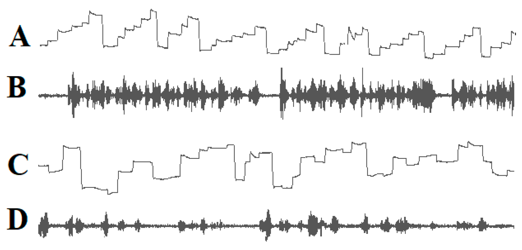
Since the words or word segments to be read must be shifted into the fovea and perifoveal area, the eyes must move in the reading direction, and the saccade amplitude must be adjusted so that after each saccade, the next word to be read is projected onto the fovea and perifoveal region. Good readers complete a succession of staircase-like reading saccades in the reading direction whereas many poor readers execute irregular eye movements that are often directed opposite to the reading direction (Figure 1).
Figure 1. Eye movements (A) and speech spectrogram (B) of a good reader. Ascending lines: eye movements to the right; descending lines: eye movements to the left. The reader exerts a sequence of staircase-like eye movements in the reading direction. The speech spectrogram shows fluent reading without interruptions. Irregular eye movements (C) and the speech spectrogram (D) of a dyslexic child. The child exerts many eye movements opposite to the reading direction. The speech spectrogram demonstrates that the child reads haltingly with many pauses.
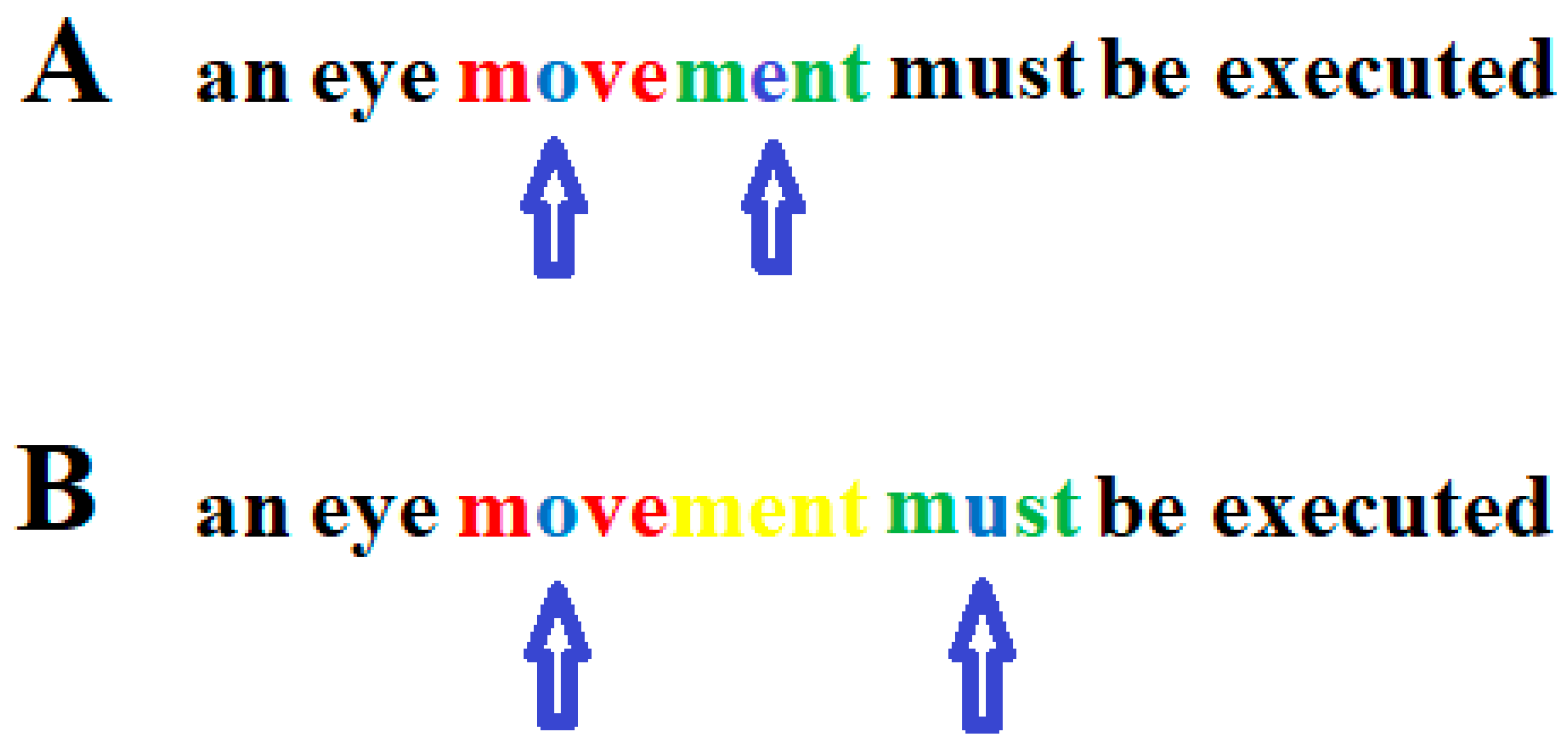
Eye movements were recorded to check whether the readers´ eyes followed the curser. If the reader’s eye movements did not follow the cursor exactly, the text to the left and right of the word being read was erased. In this way, staircase-like eye movements can be induced, such as those seen in good readers. However, inducing staircase eye movements in dyslexic readers, such as those seen in good readers, is not sufficient to improve the reading ability in dyslexic readers. Whether reading eye movements are appropriate does not depend on whether the sequence of saccades matches that of good readers. Rather, it depends on the individual’s ability to simultaneously recognize a string of letters and the duration of the fixation interval required to detect a given string of letters that make up a word [20][21][22][137][138]. Therefore, eye movements cannot be considered a cause of dyslexia if they are unusual and deviate from the norm represented by typical readers. After a word or word segment has been read, a saccade is initiated to the next word or word segment to be read. This saccade should be aimed at approximately the middle of the word or word segment to be read subsequently. The amplitude of the saccade must not exceed the number of letters that can be recognized simultaneously so that the word segment to be read next follows the previous word segment without a gap between the word segments (Figure 2).
Figure 2. Correct (A) and too large (B) saccade during reading. In the upper graph, the arrows indicate on which letter within a word segment a person who can only recognize four letters simultaneously must focus his/her gaze. One letter to the left and two letters to the right (red) of the focused letter (blue) must be recognized simultaneously together with the focused letter. After the first word segment (blue and red) has been read, the gaze only moves so far in the reading direction that the next word segment to be read (blue and green) follows without a gap between the two word segments. (B): After the first word segment (blue and red) was read, the eyes jumped too far to the next word segment (blue and green) so that the letters “ment” (yellow) were overlooked. Arrows indicate the letter on which the gaze is focused.

2.4. DD Is Not Caused by a Phonological Impairment

Reading requires knowing which sounds are associated with which letters or sequences of letters. In languages such as Spanish, German, and Italian, where there is a high grapheme–phoneme correspondence, it is easier to learn the association between sounds and letters than in languages such as English or French in which the pronunciation of the same letter can vary in different words and a sound is often represented by a sequence of letters. The ability to learn the grapheme–phoneme association of a language is not equally developed among all individuals. Even some children with German as their native language have great difficulty learning the grapheme–phoneme correspondence of certain letters, such as “m/n, b/d, or p/q”. Therefore, it is not surprising that children whose native languages have ambiguous grapheme–phoneme correspondence have even greater difficulties. These problems can be easily eliminated in the case of native German speakers by testing whether the grapheme–phoneme connection is mastered for all letters and letter sequences and excluding those children who do not know the phonemes associated with certain letters. The causes of reading problems can then be examined independently of the inability to store the association between some letters and the corresponding sounds in memory. In languages where grapheme–phoneme correspondence is variable and sophisticated, readers must retrieve pronunciation from memory when reading real words or pseudowords. This problem is present in all reading tests with native English and French speakers. Even in three-letter words (e.g., the pronunciation of the letter “a” in the words ‘tar’, ‘raw’, ‘tan’ or the pronunciation of the letter “e” in words like “sea”, “set”, “sew”), the pronunciation of letters can vary. In languages such as Spanish, German, or Italian, the problem of variable pronunciation of letters is almost non-existent. In these languages, the causes of reading problems can be investigated independent of the impaired ability to master the grapheme–phoneme correspondence. It has been demonstrated that children may have severe DD even if there is no impairment in their knowledge of grapheme–phoneme correspondence.

References

  1. Berkhan, O. Über Störungen der Sprache und der Schriftsprache: Für Ärzte und Lehrer Dargestellt; August Hirschwald: Berlin, Germany, 1889.
  2. Berlin, R. Über Dyslexie. Arch. Psychiat. Nervenkrankh. 1884, 15, 276–278.
  3. Helland, T. Trends in dyslexia research during the period 1950 to 2020—Theories, definitions, and publications. Brain Sci. 2022, 12, 1323.
  4. Stein, J. Theories about developmental dyslexia. Brain Sci. 2023, 13, 208.
  5. Livingstone, M.S.; Rosen, G.D.; Drislane, F.W.; Galaburda, A.M. Physiological and anatomical evidence for a magnocellular defect in developmental dyslexia. Proc. Natl. Acad. Sci. USA 1991, 88, 7943–7947.
  6. Stein, J.; Walsh, V. To see but not to read; the magnocellular theory of dyslexia. Trends Neurosci. 1997, 20, 147–152.
  7. Stein, J.; Walsh, V. Impaired neural timing in developmental dyslexia—The magnocellular hypothesis. Dyslexia 1999, 5, 59–77.
  8. Stein, J. The magnocellular theory of dyslexia. In Developmental Dyslexia, Early Precursors, Neurobehavioral Markers, and Biological Substrates; Benasich, A.A., Fitch, R.H., Eds.; Brooks: Baltimore, MD, USA; London, UK; Sydney, Australia, 2012.
  9. Stein, J. The current status of the magnocellular theory of developmental dyslexia. Neuropsychologia 2019, 130, 66–77.
  10. Geiger, G.; Lettvin, J.Y. Peripheral vision in persons with dyslexia. N. Engl. J. Med. 1987, 316, 1238–1243.
  11. Geiger, G.; Lettvin, J.Y.; Fahle, M. Dyslexic children learn a new visual strategy for reading: A controlled experiment. Vis. Res. 1993, 34, 1223–1233.
  12. Spinelli, D.; De Luca, M.; Judica, A.; Zoccolotti, P. Crowding effects on word identification in developmental dyslexia. Cortex 2002, 38, 179–200.
  13. Lorusso, M.L.; Facoetti, A.; Pesenti, S.; Cattaneo, C.; Molteni, M.; Geiger, G. Wider recognition in peripheral vision common to different subtypes of dyslexia. Vis. Res. 2004, 44, 2413–2424.
  14. Whitney, D.; Levi, D.M. Visual crowding: A fundamental limit on conscious perception and object recognition. Trends. Cogn. Sci. 2011, 15, 160–168.
  15. Zorzi, M.; Barbiero, C.; Facoetti, A.; Lonciari, I.; Carrozzi, M.; Montico, M.; Bravar, L.; George, F.; Pech-Georgel, C.; Ziegler, J.C. Extra-large letter spacing improves reading in dyslexia. Proc. Natl. Acad. Sci. USA 2012, 109, 11455–11459.
  16. Gori, S.; Facoetti, A. How the visual aspects can be crucial in reading acquisition? The intriguing case of crowding and developmental dyslexia. J. Vis. 2015, 14, 15.1.8.
  17. Strasburger, H. Seven Myths on Crowding and Peripheral Vision. Iperception 2020, 11, 2041669520913052.
  18. Kirkby, J.A.; Barrington, R.S.; Drieghe, D.; Liversedge, S.P. Parafoveal processing and transposed-letter effects in dyslexic reading. Dyslexia 2022, 28, 359–374.
  19. Kewan-Khalayly, B.; Migó, M.; Yashar, A. Transient attention equally reduces visual crowding in radial and tangential axes. J. Vis. 2022, 22, 3.
  20. Werth, R. Rapid improvement of reading performance in children with dyslexia by altering the reading strategy: A novel approach to diagnoses und therapy of reading deficiencies. Restor. Neurol. Neurosci. 2018, 36, 679–691.
  21. Werth, R. What causes Dyslexia? Identifying the causes and effective compensatory therapy. Restor. Neurol. Neurosci. 2019, 37, 591–608.
  22. Werth, R. Dyslexic readers improve without training when using a computer-guided reading strategy. Brain Sci. 2021, 11, 526.
  23. Werth, R. Is developmental dyslexia due to a visual and not a phonological impairment? Brain Sci. 2021, 11, 1313.
  24. Tallal, P. Auditory temporal perception, phonics, and reading disabilities in children. Brain Lang. 1980, 9, 182–198.
  25. Tallal, P.; Miller, S.; Fitch, R.H. Neurobiological basis of speech: A case for the pre-eminence of temporal processing. Ann. N. Y. Acad. Sci. 1993, 682, 27–47.
  26. Merzenich, M.M.; Jenkins, W.M.; Johnston, P.; Schreiner, C.; Miller, S.L.; Tallal, P. Temporal processing deficits of language learning impaired children ameliorated by training. Science 1996, 271, 77–81.
  27. Nagarajan, S.; Mahnke, H.; Salz, T.; Tallal, P.; Roberts, T.; Merzenich, M.M. Cortical auditory signal processing in poor readers. Proc. Natl. Acad. Sci. USA 1999, 96, 6483–6488.
  28. Pavlidis, G.T. Do eye movements hold the key to dyslexia? Neuropsychologia 1981, 19, 57–64.
  29. Rayner, K.; Pollatsek, A. Eye movement control during reading: Evidence for direct control. Q. J. Exp. Psychol. Sect. A 1982, 33, 351–373.
  30. Pavlidis, G.T. Eye movements in dyslexia: Their diagnostic significance. J. Learn. Disabil. 1985, 18, 42–50.
  31. Rayner, K. Do faulty eye movements cause dyslexia? Dev. Neuropsychol. 1985, 1, 3–15.
  32. Rainer, K. Eye movements and the perceptual span in beginning and skilled readers. J. Exp. Child Psychol. 1986, 41, 211–236.
  33. Fischer, B.; Biscaldi, M.; Otto, P. Saccadic eye movements of dyslexic adult subjects. Neuropsychologia 1993, 31, 887–906.
  34. Eden, G.F.F.; Stein, J.F.F.; Wood, H.M.M.; Wood, F.B.B. Differences in eye movements and reading problems in dyslexic and normal children. Vis. Res. 1994, 34, 1345–1358.
  35. Hyönä, J.; Olson, R.K. Eye fixation patterns among dyslexic and normal readers: Effect of word length and word frequency. J. Exp. Psychol. Learn. Mem. Cogn. 1995, 21, 1430–1440.
  36. Biscaldi, M.; Gezeck, S.; Stuhr, V. Poor saccadic control correlates with dyslexia. Neuropsychologia 1998, 36, 1189–1202.
  37. De Luca, M.; Di Pace, E.; Judica, A.; Spinelli, D.; Zoccolotti, P. Eye movement patterns in linguistic and non-linguistic tasks in developmental surface dyslexia. Neuropsychologia 1999, 37, 1407–1420.
  38. Biscaldi, M.; Fischer, B.; Hartnegg, K. Voluntary saccadic control in dyslexia. Perception 2000, 29, 509–521.
  39. Rayner, K.; Slattery, T.J.; Bélanger, N.N. Eye movements, the percepzual span, and reading speed. Bull. Rev. 2010, 17, 834–839.
  40. Ward, L.M.; Kapoula, Z. Dyslexics’ Fragile Oculomotor Control Is Further Destabilized by Increased Text Difficulty. Brain Sci. 2021, 11, 990.
  41. Temelturk, R.D.; Ozer, E. Binocular coordination of children with dyslexia and typically developing children in linguistic and non-linguistic tasks: Evidence from eye movements. Ann. Dyslexia 2022, 72, 426–444.
  42. Premeti, A.; Bucci, M.P.; Isel, F. Evidence from ERP and eye movements as markers of language dysfunction in dyslexia. Brain Sci. 2022, 12, 73.
  43. Caldani, S.; Acquaviva, E.; Moscoso, A.; Peyre, H.; Delorme, R.; Bucci, M.P. Reading performance in children with ADHD: An eye-tracking study. Ann. Dyslexia 2022, 72, 552–565.
  44. Wagner, R.K.; Torgesen, J.K. The nature of phonological processing and its causal role in the acquisition of reading skills. Psychol. Bull. 1987, 101, 192–212.
  45. Bruce, D. An analysis of word sounds by young children. Br. J. Educ. Psychol. 1964, 34, 158–170.
  46. Bryant, P.E.; MacLean, M.; Bradley, L.L.; Crossland, J. Rhyme and alliteration, phoneme detection, and learning to read. Dev. Psychol. 1990, 26, 429–438.
  47. Torgesen, J.K.; Wagner, R.K.; Rashotte, C.A. Longitudinal studies of phonological processing and reading. J. Learn. Disabil. 1994, 27, 276–286.
  48. McBride-Chang, C. What is phonological awareness? J. Educ. Psychol. 1995, 87, 179–192.
  49. Yopp, H.K. A test for assessing phonemic awareness in young children. Read. Teach. 1995, 49, 20–29.
  50. Torgesen, J.K.; Wagner, R.K.; Rashotte, C.A. Prevention and remediation of severe reading disabilities: Keeping the end in mind. Sci. Stud. Read. 1997, 1, 217–234.
  51. Muter, V.; Hulme, C.; Snowling, M.J.; Taylor, S. Segmentation, not rhyming, predicts early progress in learning to read. J. Exp. Child. Psychol. 1998, 71, 3–27.
  52. Ehri, L.C. Research on learning to read and spell: A personal historical perspective. Sci. Stud. Read. 1998, 2, 97–114.
  53. Snowling, M. From language to reading and dyslexia. Dyslexia. Dyslexia 2001, 7, 37–46.
  54. Goswami, U. Phonology, reading development, and dyslexia: A cross-linguistic perspective. Ann. Dyslexia 2002, 52, 139–163.
  55. Vellutino, F.R.; Fletcher, J.M.; Snowling, M.J. Scanlon, D.M. Specific reading disability (dyslexia): What have we learned in the past four decades. J. Child Psychol. Psychiat. 2004, 45, 2–40.
  56. Goswami, U. Phonology, reading and reading difficulties. In Interdisciplinary Perspectives on Learning to Read. Culture, Cognition and Pedagogy; Hall, K., Goswami, U., Harrison, C., Ellis, S., Soler, Eds.; Routledge: London, UK, 2010; pp. 103–116.
  57. Ligges, C.; Lehmann, T. Multiple case study in German children with dyslexia: Characterization of phonological, auditory, visual, and cerebellar processing on the group and individual level. Brain Sci. 2022, 12, 1292.
  58. Mackie, J.L. Causes and conditions. Am. Philos. Q. 1965, 2, 245–264.
  59. Spirtes, P.; Glymour, C.; Scheines, R. Causation, Prediction, and Search; MIT Press: Cambridge, UK, 1992.
  60. Lewis, D. Causation as influence. J. Philos. 2000, 97, 182–197.
  61. Spohn, W. Causation: An alternative. Brit. J. Philos. Sci. 2006, 57, 93–119.
  62. Pearl, J.; Glymour, M.; Jewell, N.P. Causal Inference in Statistics; Wiley: Chichester, UK, 2016.
  63. Pearl, J. Causality—Models, Reasoning and Inference; Cambridge University Press: Cambridge, UK; New York, NY, USA; Port Melbourne, Australia, 2018; pp. 316–320.
  64. Bressler, S.L.; Seth, A.K. Wiener-Granger causality: A well established methodology. Neuroimage 2011, 58, 323–329.
  65. Stokes, P.A.; Purdon, P.L. A study of problems encountered in Granger causality analysis from a neuroscience perspective. Proc. Natl. Acad. Sci. USA 2017, 114, E7063–E7072.
  66. Farah, M.; Stowe, R.M.; Levinson, K.L. Phonological dyslexia. Loss of a reading specific component of cognitive architecture? Cogn. Neuropsychol. 1996, 13, 849–868.
  67. Hari, R.; Renvall, H. Impaired processing of rapid stimulus sequences in dyslexia. Trends Cogn. Sci. 2001, 5, 525–532.
  68. Roach, N.W.; Hogben, J.H. Attentional modulation of visual processing in adult dyslexia: A spatial cuing deficit. Psychol. Sci. 2004, 15, 650–654.
  69. Buchholz, J.; Davis, A.A. Adults with dyslexia demonstrate space-based and object-based covert attention deficits: Shifting attention to the periphery and shifting attention between objects in the left visual field. Brain Cogn. 2005, 57, 30–34.
  70. Heim, S.; Tschierse, J.; Amunts, K.; Wilms, M.; Vossel, S.; Willmes, K.; Grabowska, A.; Huber, W. Cognitive subtypes of dyslexia. Acta Neurobiol. Exp. 2008, 68, 73–82.
  71. Daniel, A.; Abrams, D.A.; Nicol, T.; Zecker, S.; Kraus, N. Abnormal cortical processing of the syllable rate of speech in poor readers. J. Neurosci. 2009, 29, 7686–7693.
  72. Menghini, D.; Finzi, A.; Benassi, M.; Bolzani, R.; Facoetti, A.; Giovagnoli, S.; Ruffino, M.; Vicari, S. Different underlying neurocognitive deficits in developmental dyslexia: A comparative study. Neuropsychologia 2010, 48, 863–872.
  73. Hornickel, J.; Kraus, N. Unstable representation of sound: A biological marker of dyslexia. J. Neurosci. 2013, 33, 3500–3504.67.
  74. Lorusso, M.L.; Cantiani, C.; Molteni, M. Age, dyslexia subtype and comorbidity modulate rapid auditory processing in developmental dyslexia. Front. Hum. Neurosci. 2014, 8, 313.
  75. van Bergen, E.; de Jong, P.F.; Maassen, B.; Krikhaar, E.; Plakas, A.; van der Leij, A. IQ of four-year-olds who go on to develop dyslexia. J. Learn. Disabil. 2014, 47, 475–484.
  76. Hendren, R.L.; Haft, S.L.; Black, J.M.; White, N.C.; Hoeft, F. Recognizing Psychiatric Comorbidity With Reading Disorders. Front. Psychiatry 2018, 9, 101.
  77. Peters, L.; Bulthé, J.; Daniels, N.; Op de Beeck, H.; De Smedt, B. Dyscalculia and dyslexia: Different behavioral, yet similar brain activity profiles during arithmetic. Neuroimage Clin. 2018, 18, 663–674.
  78. Moll, K.; Landerl, K.; Snowling, M.J.; Schulte-Körne, G. Understanding comorbidity of learning disorders: Task-dependent estimates of prevalence. J. Child. Psychol. Psychiat. 2019, 60, 286–294.
  79. Maziero, S.; Tallet, J.; Bellocchi, S.; Jover, M.; Chaix, Y.; Jucla, M. Influence of comorbidity on working memory profile in dyslexia and developmental coordination disorder. J. Clin. Exp. Neuropsychol. 2020, 42, 660–674.
  80. Facoetti, A.; Paganoni, P.; Turatto, M.; Marzola, V.; Mascetti, G.G. Visual spatial attention in developmental dyslexia. Cortex 2000, 36, 109–123.
  81. Carrasco, M.; Williams, P.E.; Yeshurun, Y. Covert attention increases spatial resolution with or without masks: Support for signal enhancement. J. Vision 2002, 2, 467–479.
  82. Bosse, M.L.; Tainturier, M.J.; Valdois, S. Developmental dyslexia: The visual attention span deficit hypothesis. Cognition 2007, 104, 198–230.
  83. Franceschini, S.; Gori, M.; Ruffino, K.; Pedrolli, A.; Facoetti, A. A causal link between visual spatial attention and reading acquisition. Curr. Biol. 2012, 22, 814–819.
  84. Ruffino, M.; Gori, S.; Boccardi, D.; Molteni, M.; Facoetti, A. Spatial and temporal attention in developmental dyslexia. Front. in Human Neurosci. 2014, 8, 8–331.
  85. Chen, C.; Schneps, M.H.; Masyn, K.E.; Thomson, J.M. The effects of visual attention span and phonological decoding in reading comprehension in dyslexia: A path analysis. Dyslexia 2016, 22, 322–344.
  86. Facoetti, A.; Turatto, M.; Lorusso, M.L.; Mascetti, G.G. Orienting of visual attention in dyslexia: Evidence for asymmetric hemispheric control of attention. 2001, 138, 46–53. Exp. Brain Res. 2001, 138, 46–53.
  87. Facoetti, A.; Lorusso, M.L.; Paganoni, P.; Cattaneo, C.; Galli, R.; Umiltà, C.; Mascetti, G.G. Auditory and visual automatic attention deficits in developmental dyslexia. Brain Res. Cogn. Brain Res. 2003, 16, 185–191.
  88. Roach, N.W.; Hogben, J.H. Spatial cueing deficits in dyslexia reflect generalised difficulties with attentional selection. Vision Res. 2008, 48, 193–207.
  89. Facoetti, A.; Zorzi, M.; Cestnick, L.; Lorusso, M.L.; Molteni, L.M.; Paganoni, P.; Umilta, C.; Mascetti, G.G. The relationship between visuo-spatial attention and nonword reading in developmental dyslexia. Cogn. Neuropsych. 2006, 23, 841–855.
  90. Pina Rodrigues, A.; Castelo-Branco, M.; van Asselen, M. Disrupted spatial organization of cued exogenous attention persists into adulthood in developmental dyslexia. Front. Psychol. 2021, 12, 769237.
  91. Skottun, B.C.; Skoyles, J. Dyslexia: Sensory deficits or inattention? Perception 2007, 36, 1084–1088.
  92. Skottun, B.C. On the use of cues to assess attention in dyslexia. Front. Hum. Neurosci. 2014, 8, 983.
  93. De Luca, M.; Burani, C.; Paizi, D.; Spinelli, D.; Zoccolotti, P. Letter and letter-string processing in developmental dyslexia. Cortex 2010, 46, 1272–1283.
  94. Zoccolotti, P.; De Luca, M.; Spinelli, D. Discrete versus multiple word displays: A re-analysis of studies comparing dyslexic and typically developing children. Front. Psychol. 2015, 6, 1530.
  95. Burani, C.; Marcolini, S.; Traficante, D.; Zoccolotti, P. Reading derived words by Italian children with and without dyslexia: The effect of root length. Front. Psychol. 2018, 9, 647.
  96. Franceschini, S.; Bertoni, S.; Puccio, G.; Gori, S.; Termine, C.; Facoetti, A. Visuo-spatial attention deficit in children with reading difficulties. Sci. Rep. 2022, 12, 13930.
  97. Valdois, S. The visual-attention span deficit in developmental dyslexia: Review of evidence for a visual-attention-based deficit. Dyslexia 2022, 28, 397–415.
  98. Perry, C.; Long, H. What is going on with visual attention in reading and dyslexia? A critical review of recent studies. Brain Sci. 2022, 12, 87.
  99. Stenneken, P.; Egetemeir, J.; Schulte-Körne, G.; Müller, H.J.; Schneider, W.X.; Finke, K. Slow perceptual processing at the core of developmental dyslexia: A parameter-based assessment of visual attention. Neuropsychologia 2011, 49, 3454–3465.
  100. Pammer, K.; Lavis, R.; Hansen, P.; Cornelissen, P.L. Symbol-string sensitivity and children’s reading. Brain Lang. 2004, 89, 601–610.
  101. Graham, C.H.; Cook, C. Visual acuity as a function of intensity and exposure-time. Am. J. Psychol. 1937, 49, 654–661.
  102. Barlow, H.B. Temporal and spatial summation in human vision at different background intensities. J. Physiol. Lond. 1958, 141, 337–350.
  103. Niwa, K.; Tokoro, T. Measurement of temporal summation of visual acuity with use of modified tachistoscope. Jpn. J. Ophthalmol. 1997, 41, 403–408.
  104. Sandberg, K.; Bibby, B.M.; Timmermans, B.; Cleeremans, A.; Overgaard, M. Measuring consciousness: Task accuracy and awareness as sigmoid functions of stimulus duration. Conscious. Cogn. 2011, 20, 1659–1675.
  105. McAnany, J.J. The effect of exposure duration on visual acuity for letter optotypes and gratings. Vision Res. 2014, 105, 86–91.
  106. Windey, B.; Vermeiren, A.; Atas, A.; Cleeremans, A. The graded and dichotomous nature of visual awareness. Philos. Trans. R.Soc. B. Biol. Sci. 2014, 369, 20130282.
  107. Mulholland, P.J.; Redmond, T.; Garway-Heath, D.F.; Zlatkova, M.B.; Anderson, R.S. The Effect of Age on the Temporal Summation of Achromatic Perimetric Stimuli. Invest. Ophthalmol. Vis. Sci. 2015, 56, 6467–6472.
  108. Holmes, R.; Victora, M.; Wang, R.F.; Kwiat, P.G. Measuring temporal summation in visual detection with a single-photon source. Vis. Res. 2017, 140, 33–43.
  109. Stigliani, A.; Jeska, B.; Grill-Spector, K. Encoding model of temporal processing in human visual cortex. Proc. Natl. Acad. Sc.i USA 2017, 114, E11047–E11056.
  110. Zhou, J.; Benson, N.C.; Kay, K.N. Winawer, J. Compressive temporal summation in human visual cortex. J. Neurosci. 2018, 38, 691–709.
  111. Beauny, A.; de Heering, A.; Muñoz Moldes, S.; Martin, J.-R.; de Beir, A.; Cleeremans, A. Unconscious categorization of sub-millisecond complex images. PLoS One 2020, 15, e0236467.
  112. Heinrich, S.P.; Blechenberg, T.; Reichel, C.; Bach, M. The "speed" of acuity in scotopic vs. photopic vision. Graefes Arch. Clin. Exp. Ophthalmol. 2020, 258, 2791–2798.
  113. Harvey, B.M.; Dumoulin, S.O.; Fracasso, A.; Paul, J.M. A Network of Topographic Maps in Human Association Cortex Hierarchically Transforms Visual Timing-Selective Responses. Curr. Biol. 2020, 30, 1424–1434.e6.
  114. Hendrikx, E.; Paul, J.M.; van Ackooij, M.; van der Stoep, N.; Harvey, B.M. Visual timing-tuned responses in human association cortices and response dynamics in early visual cortex. Nat. Commun. 2022, 8, 3952.
  115. Paul, J.M.; van Ackooij, M.; Ten Cate, T.C.; Harvey, B.M. Numerosity tuning in human association cortices and local image contrast representations in early visual cortex. Nat. Commun. 2022, 13, 1340.
  116. Weiner, K.S.; Sayres, R.; Vinberg, J.; Grill-Spector, K. fMRI-adaptation and category selectivity in human ventral temporal cortex: Regional differences across time scales. J. Neurophysiol. 2010, 103, 3349–3365.
  117. Fornaciai, M.; Brannon, E.M.; Woldorff, M.G.; Park, J. Numerosity processing in early visual cortex. Neuroimage 2017, 157, 429–438.
  118. DeWind, N.K.; Park, J.; Woldorff, M.G.; Brannon, E.M. Numerical encoding in early visual cortex. Cortex 2019, 114, 76–89.
  119. Mechelli, A.; Humphreys, G.W.; Mayall, K.; Olson, A.; Price, C.J. Differential effects of word length and visual contrast in the fusiform and lingual gyri during reading. Proc. Royal Soc. Lond. Series B. 2000, 267, 1909–1913.
  120. Schurz, M.; Sturm, D.; Richlan, F.; Kronbichler, M.; Ladurner, G.; Wimmer, H. A dual-route perspective on brain activation in response to visual words: Evidence for a length by lexicality interaction in the visual word form area (VWFA). Neuroimage 2010, 49, 2649–2661.
  121. Bálint, D. Seelenlähmung des “Schauens”, optische Ataxie, räumliche Störung der Aufmerksamkeit. Mschr. Psychiat. Neurol. 1909, 25, 51–81.
  122. Poppelreuter, W. Die Psychischen Schädigungen durch Kopfschuß im Kriege 1914–1918. Bd. I. Die Störungen der Niederen und Höheren Sehleistungen durch Verletzungen des Okzipitalhirns; L. Voss: Leipzig, Germany, 1917.
  123. Luria, A.R. Disorders of “simultaneous perception” in a case of bilateral occipito-parietal brain injury. Brain 1959, 82, 437–449.
  124. Baylis, G.C.; Driver, J.; Baylis, L.L.; Rafal, R.D. Reading of letters and words in a patient with Balint’s syndrome. Neuropsychologia 1994, 32, 1273–1286.
  125. Rizzo., M.; Vecera, S.P. Psychoanatomical substrates of Bálint’s syndrome. J. Neurol. Neurosurg. Psychiatry 2002, 72, 162–178.
  126. Moreaud, O. Balint syndrome. Arch. Neurol. 2003, 60, 1329–1331.
  127. Michel, F.; Henaff, M.A. Seeing without the occipito-parietal cortex: Simultagnosia as a shrinkage of the attentional visual field. Behav. Neurol. 2004, 15, 3–13.
  128. Farah, M.J. Visual Agnosia, 2nd ed.; MIT Press: Cambridge, MA, USA; London, UK, 2004; pp. 43–58.
  129. Chechlacz, M.; Rotshtein, P.; Hansen, P.C.; Riddoch, J.M.; Deb, S.; Humphreys, G.W. The neural underpinings of simultanagnosia: Disconnecting the visuospatial attention network. J. Cogn. Neurosci. 2012, 247, 18–35.
  130. Chechlacz, M.; Humphreys, G.W. The enigma of Balint’s syndrome: Neural substrates and cognitive deficits. Front. Hum. Neurosci. 2014, 8, 123.
  131. Chechlacz, M. Bilateral parietal dysfunctions and disconnections in simultanagnosia and Bálint syndrome. Handb. Clin. Neurol. 2018, 151, 249–267.
  132. Webster, R.G.; Haslerud, G.M. Influence on extreme peripheral vision of attention to a visual or auditory task. J. Exp. Psychol. 1964, 68, 269–272.
  133. Engel, F.L. Visual conspicuity, directed attention and retinal locus. Vision Res. 1971, 11, 563–576.
  134. Ikeda, M.; Takeuchi, T. Influence of foveal load on the functional visual field. Perc. Psychophys. 1975, 18, 225–260.
  135. Henderson, J.M.; Ferreira, F. Effects of foveal processing difficulty on the perceptual span in reading: Implication for attention and eye movement control. J. Exp. Psychol. Learn. Mem. Cogn. 1990, 16, 417–429.
  136. Handy, T.C.; Kingstone, A.; Mangun, G.R. Spatial distribution of visual attention: Perceptual sensitivity and response latency. Percept. Psychophys. 1996, 58, 613–627.
  137. Werth, R. Therapie von Lesestörungen durch Erkennen und Beheben der Ursachen. Ergother. Rehabil. 2006, 9, 6–11.
  138. Klische, A. Leseschwächen gezielt beheben. Individuelle Diagnose und Therapie mit dem Programm Celeco; Tectum: Marburg, Germany, 2007.
More
Information
Contributor MDPI registered users' name will be linked to their SciProfiles pages. To register with us, please refer to https://encyclopedia.pub/register :
View Times: 504
Revisions: 2 times (View History)
Update Date: 13 Apr 2023
1000/1000
ScholarVision Creations