Submitted Successfully!
To reward your contribution, here is a gift for you: A free trial for our video production service.
Thank you for your contribution! You can also upload a video entry or images related to this topic.
Version Summary Created by Modification Content Size Created at Operation
1 -- 3279 2023-04-04 10:40:30 |
2 format correct Meta information modification 3279 2023-04-04 10:43:35 |

Video Upload Options

Do you have a full video?

Confirm

Are you sure to Delete?
Cite
If you have any further questions, please contact Encyclopedia Editorial Office.
Kurpiewska, K.; Sławek, J.; Klonecka, A.; Kozak, M. High Pressure Macromolecular Crystallography. Encyclopedia. Available online: https://encyclopedia.pub/entry/42771 (accessed on 07 May 2024).
Kurpiewska K, Sławek J, Klonecka A, Kozak M. High Pressure Macromolecular Crystallography. Encyclopedia. Available at: https://encyclopedia.pub/entry/42771. Accessed May 07, 2024.
Kurpiewska, Katarzyna, Joanna Sławek, Agnieszka Klonecka, Maciej Kozak. "High Pressure Macromolecular Crystallography" Encyclopedia, https://encyclopedia.pub/entry/42771 (accessed May 07, 2024).
Kurpiewska, K., Sławek, J., Klonecka, A., & Kozak, M. (2023, April 04). High Pressure Macromolecular Crystallography. In Encyclopedia. https://encyclopedia.pub/entry/42771
Kurpiewska, Katarzyna, et al. "High Pressure Macromolecular Crystallography." Encyclopedia. Web. 04 April, 2023.
High Pressure Macromolecular Crystallography
Edit

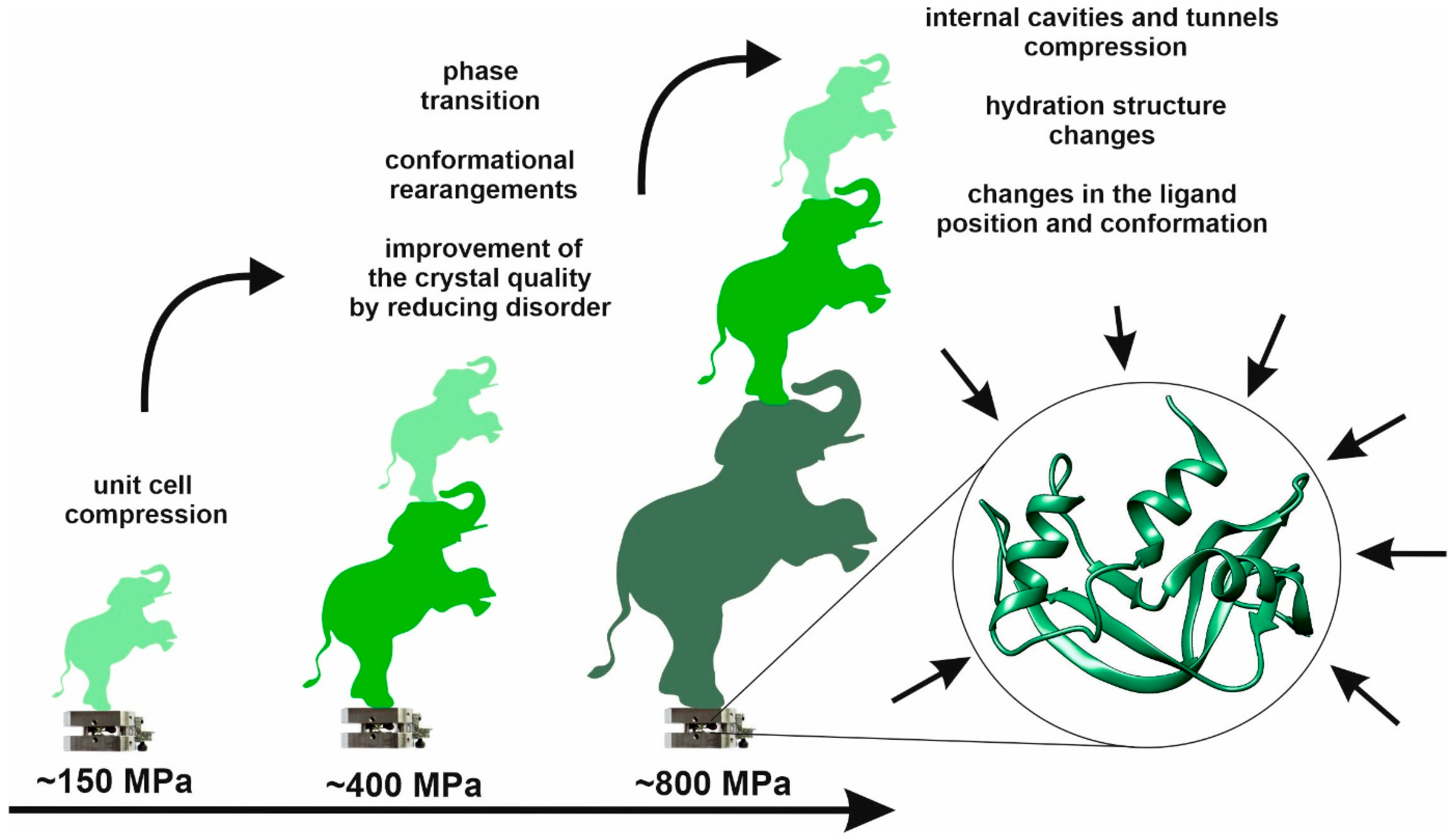
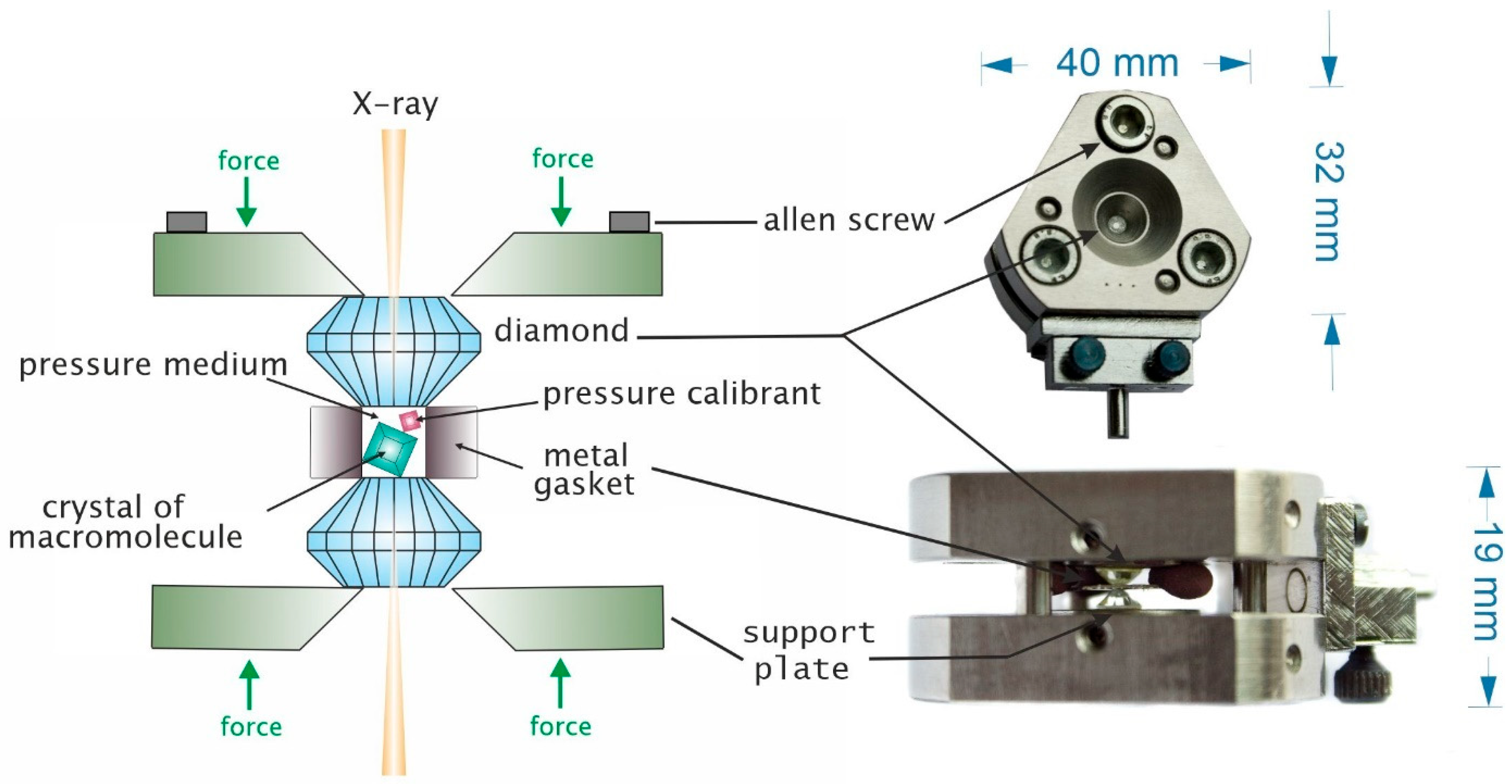
Since its introduction in the early 1970s, high pressure crystallography (HPX) has shown great potential for the investigation of different types of matter. Using diamond anvil cells, HPX is an emerging technique that has been rapidly implemented, making it available to biologists, and there is immense potential for utilizing this technique in biological systems in the future. At the molecular level, high-pressure crystallographic investigation provides information on structural characteristics that not only determine the native conformation of a protein but also the conformations with higher free-energy, thus revealing function-related structural changes and properties that can be modified as a result of pressurization. The increase in the number of crystal structures of different macromolecules determined under high pressure over the last five decades can be ascribed mainly to two factors: the emergence of high-pressure cells with very large, open angles, and the advent of third generation synchrotron sources. The use of high pressure crystallography as a research tool has been shown to contribute to the advancements in the basic fields of biochemistry (protein misfolding and aggregation), biophysics (protein stability), and biotechnology (food processing).

high pressure macromolecular crystallography protein structure unfolding crystal

1. High Pressure in Structural Studies

The origins of high-pressure structural studies can be traced back to geology and astronomy, as a significant portion of condensed matter in the universe exists under high pressure conditions. For instance, a classic study could involve examining the reaction of a mineral structure to increasing pressure levels, up to those present at the corresponding depth within the Earth. As evidenced in previous HPX studies, the structures obtained at various pressure levels are analyzed to reveal the molecular responses to compression. The application of a few tens of MPa may only affect the weak intermolecular interactions, leading to a decrease in the unit cell volume and a possible rearrangement of structural units. Higher pressures may result in changes in the conformation or internal geometry of macromolecules and ligands. At higher compression, notable phenomena include chemical reactions such as polymerization and transformations such as the generation of new polymorphs or reorganization of hydrogen bonding networks in organic and inorganic compounds. Additionally, there may be changes in molecular organization or conformation, closer approach of structural units, and even modifications to the electron configuration [1]. More recently, the scope of materials studied under high pressure has expanded to include biologically relevant molecules spanning from amino acids and DNA fragments to proteins. One significant advantage of applying high pressure in protein studies is the ability to explore intermediate substates between the folded and unfolded states. By combining experimental and theoretical approaches, it has been possible to map the energy landscape and construct p-T phase diagrams for several proteins [2].
Although crystal structures determined under HP comprise less than 0.1% of structures reported in the Protein Data Bank [3], studies at high pressures are becoming more common. The development of apparatus (compact high-pressure cells, high-intensity radiation sources and area detector diffractometers) followed by the improvement of technique (crystal mounting, pressure measurement, and data acquisition) over the past 10 years is remarkable, and HP studies are now conducted for more complex systems. When pressure is applied, different responses can be observed from the macromolecules that form the crystal and the crystal itself (Figure 1). The most important features reported as those that change in the course of the biocrystallographic HP experiment are changes of crystals quality [4][5], crystal symmetry [6], volume of the unit cell [7], and Wilson B factors when crystallographic parameters are concerned. In terms of modification at the molecular level, changes can be observed in the hydration structure [8], the size and localization of cavities and tunnels [9], and the conformation of the polypeptide chain and ligands [10]. All structural rearrangements are often associated with alterations in the stability and/or activity of biomolecules [11].
Figure 1. Features that can change upon pressurization of the macromolecular crystal.
As observed for small organic molecules pressure turned out to be ideally suited also to study the physical behavior of compressed macromolecules. Since weak intermolecular interactions are most likely to be easily compressed and conformations of molecules themselves can be modified it can lead to ordering and structural phase transition [5]. The application of hydrostatic pressure offers a genuine opportunity to manipulate and explore the intermolecular “bonding” present in crystals. For example, the ordering effect of pressure on the crystal lattice was observed for P23 cowpea mosaic virus (CPMV) crystals [4]. In addition, subjecting the crystals to a pressure of 350 MPa resulted in a significant improvement in diffraction, as the crystals underwent a phase transition from an apparently primitive cell to the body-centered I23 space group. Modest pressures significantly enhanced the diffraction quality as the crystal undergoes a phase transition to a space group with enhanced order.
In general, under relatively low pressure (up to 150–200 MPa), due to the high compressibility of protein crystals, compression typically results in a reduction in the unit cell parameters and volume of a few percent [12]. Pressures exceeding 400 MPa can lead to a contraction that is twice as great, highlighting the immense potency of pressure as a thermodynamic variable capable of inducing structural modifications in proteins, without causing denaturation. The reported compressibility of the unit cell volume βv for protein crystals is about 170 MPa−1 at moderate pressure, whereas the compressibility at high pressure (limit determined by loss of diffraction), as reported by Fourme et al. for hen egg-white lysozyme (HEWL) based on crystal structures at pressure as high as 1 GPa was equal 150 MPa−1 [13]. In general, the compressibility of the unit cell volume calculated for macromolecular crystals decreases as pressure increases. It should be underlined that the HPMX also allows the determination of the isothermal compressibility of a protein molecule itself (βM), for example the value of tetragonal HEWL lysozyme is 47 MPa−1 based on X-ray crystallographic data obtained at 0.1 and 100 MPa [14] and for IPMDH from the nonpiezophile S. oneidensis MR-1 (SoIPMDH) dimer is 54 MPa−1 [15]. Furthermore, the analyzed structure revealed that contraction of the molecules in most cases is anisotropic and decreases with increasing pressure. It is worth to mention that with the change of the pressure, the solvent-accessible volume may change, and the solvent molecules can be transferred both sides between pool and the crystals, according to the thermodynamic equilibrium, as it was demonstrated for the CuZnSOD crystals [16].
Analysis of the B-factors can be used as an indicator of the relative vibrational motion of different parts of the structure. Therefore, atoms with low B-factors can be considered as parts of the structure that are well ordered, while atoms with higher B-factors are identified in parts of the structure that are more flexible. Colloc’h et al. in studies on neuroglobin high pressure structures revealed that the increase in the B-factors proved the destabilization of the zone close to the heme, whereas the zone at the back of the protein is stabilized by pressure which was accompanied with a decrease in the B-factors [17]. Furthermore, the general observation of the behavior of secondary structure upon pressurization revealed that beta-sheets are noticeably less deformed than helices.
As aforementioned, the way water interacts with the biomolecules is also rearranged as a result of pressurization. A good example is the analysis of changes in the hydration structure in lysozyme crystals described in [6]. When high pressure is applied, the conformation of the amino acid side chains on the surface of the protein changes and stabilizes the hydrogen bond network, causing more water molecules to be visible. After the pressure exceeded a certain limit and the protein started to unfold, the number of ordered water molecules in the structure decreased.
As pressure is applied, the protein cavities typically are monotonically compressed; however, it was also observed that the volume of a cavity located at the dimer interface can increase, as investigated, for example, in the structure of SoIPMDH where parallel to this volume increase, changes in the hydration shell and water penetration into the cavity were observed [15]. It is also possible that some cavities can shrink sufficiently enough to be undetectable using the 1.2 A radius solvent probe as the pressure increases [18]. Another possible scenario after protein compression describes the generation of a new cleft or tunnels (favorably on the molecular surface) accompanied by water penetration [8]. Such water-penetration phenomena are considered to be initial steps in the pressure-denaturation process. The example of such an event was presented by Hamajima et al. [19], where the authors showed the decrease in DHFR activity under elevated pressure and connected it with the hydrophobic cavity penetration by three water molecules. It was concluded that increasing number of water molecules entering the spaces between subunits of oligomeric proteins can lead to the disruption of quaternary structure, since it is stabilized mainly by hydrophobic forces [20].
As mentioned above, pressures below 200 MPa typically cause only minor disturbances to the overall structure of proteins, resulting in atomic displacements of only a few tenths of an angstrom. At higher pressure shifts of atoms or structural regions are larger, but still can not to be compared in scale with, for example, loops rearrangements or conformational changes observed upon ligand binding. Even though structural perturbations are relatively small, it does not mean that the functional effects are also small. As exemplified in high pressure studies of urate oxidase (UOX), compression of a protein–ligand complex drove the thermodynamic equilibrium towards ligand saturation of the complex and revealed a new binding site [21]. Furthermore, the UOX after decompression displayed a pressure-dependent decrease in specific activity that culminated at 200 MPa when the complete loss of activity was observed [22]. This may be associated with a disruption of the tertiary structure, usually occurring above 200 MPa. At this pressure, internal amino acid residues could be exposed, leading to the disruption of tertiary structure and increasing the surface hydrophobicity [23]. Further increasing of pressure could lead to the breakage of disulfide bonds [24] and rearrangement of the intermolecular hydrogen bonds [25]. Intermolecular hydrogen bonds are usually broken at lower pressures [26], which is sometimes compensated by new intramolecular interactions [27].
Accordingly, the presented examples prove that high-pressure perturbation potentially enables the trapping in crystal states the protein conformations of biological significance. It is worth mentioning that, for different proteins, the level of pressure required for capturing those conformations can greatly vary and cannot be easily predicted, mostly because the architecture of macromolecules may be less and more resistant to external factors.

2. High Pressure Macromolecular Crystallography Instrumentation

The method uses a diamond anvil cell especially configured to maintain protein crystals at a precisely determined pressure. The most widely utilized design for high pressure cells is that of the diamond-anvil cell. The creation of the first DAC chamber dates back to the second half of the 1950s, when it was used for HP IR measurements [28]. A few years later, special DAC cells for powder diffraction [29] and single-crystal X-ray measurements [30] were developed. Placing the metal gasket in between two diamond culets allowed to measure the influence of the applied pressure on the freezing properties of many liquids, but also gave rise to the study of crystals surrounded by hydrostatic medium. Today, DACs are commercially available in various shapes and sizes and can be easily adjusted to different experimental techniques, such as optical spectroscopy (IR, fluorescence, RAMAN), X-ray, magnetic, or electrical measurements [31]. The most popular version of this compact device (ca. 4.0 × 3.2 × 1.9 cm) which can generate pressures up to 10 GPa, and can be effortlessly integrated into a standard diffractometer setup. The essential components of the DAC are shown in Figure 2. Diamonds used in DACs are usually gem quality single crystals, with the carat weight between 1/8 to 1. They are cut and polished along certain crystallographic planes to obtain the brilliant. The culet is usually flat, but other designs such as bevels and toroids are also available [32][33]. To subject the crystal to high pressure diamond cell, it is placed in the sample chamber, a hole drilled in a metal gasket held between the diamond anvils. Pressure is applied to the support plates by tightening screws, which is transferred via the anvils and a hydrostatic medium to the small crystal. In the past, the diamond anvils were supported on beryllium disks. While beryllium is transparent to short-wavelength X-rays, it can introduce a pervasive background that contaminates the diffraction pattern and causes various issues. Therefore, alternative supports are currently being developed. In modern DACs, depending on the application, the supports are made predominantly from steel, tungsten, titanium, or metal alloys.
Figure 2. Diamond anvil cell: scheme of the core body of the 60-degree diamond anvil cell (on the left), a photograph of the core body of the cell once assembled, before it is put in position on the goniometer (on the right). The drawing is scaled for clarity.
Pressures inside the DAC are usually measured by placing a small piece of a calibrant (e.g., ruby) within the chamber and gauging the pressure-dependent shift in its fluorescence band. For the first time ruby was used to determine the pressure in the cell in the 1970s and it revolutionized the field [34]. The procedure requires only a standard Raman or other spectrometer and is non-invasive. Despite the fact that DACs have a relatively simple structure, their use in biocrystallographic studies requires several obstacles to overcome. First, it is necessary to mention that there is no one-size-fits-all strategy that can be used to measure all macromolecular crystals; however, a general workflow of the HPMX experiment can be identified (Figure 3).
Figure 3. Schematic representation of a HPMX experiment.
As already mentioned, macromolecules are complex three-dimensional structures that can be affected to varying degrees by pressure, so the pressure range over which they are studied cannot be standardized. Moreover, from an experimental point of view, there are many factors to consider when planning HPMX measurements. The first challenge, especially at lower pressures and single-crystal measurements, is prevention of crystal movements. Since the DAC is rotated during the measurements, it is important to immobilize the crystals prior to data collection. That can be achieved by adding MPD or PEG in high concentration (20–30%) to the mother liquor [35], but also by placing low absorbing materials, such as cotton fibers [36] or cigarette filter fibers [15] inside the DAC. The crystal space group and its orientation inside the cell are also crucial for the success of the measurements. Low-symmetry crystals require more frames to be collected and are hard to achieve, especially with anisotropic crystals. To overcome this, splinters made from various materials, such as crushed diamonds or boron nitride, can be closed inside DAC together with the sample crystal [37]. Another factor that significantly influences the result of the HPMX experiment is the pressure medium. Although crystallization mother liquor is a natural choice, one should foresee two effects that can occur during sample loading. First, because a very small volume of the mother liquor is used, the most volatile components can evaporate, which may change the composition of the hydrostatic medium and result in destabilization of the crystal before pressure ramping. Second, in case of high concentration of the precipitant, i.e., salt, formation of additional crystals can be observed upon pressurization, as reported by Kurpiewska et al. in studies of RNase A crystallized from the solution containing a high concentration of ammonium sulphate [18]. As a consequence, some undesirable strong reflections can be present on the diffractograms and disturb data processing.
The limit of pressure stability of the crystals is specific to each study and depends on many elements, i.e., composition of the mother liquor, native or mutated form of protein, or complexation with ligand. Loss of diffraction caused by a loss of long-range order in the crystal was observed near 820 MPa for HEWL, at 400 MPa for (CpMV) [38], while crystals of RNase A, as well as those formed by Cu/Zn superoxide dismutase molecules and DNA fragments could be compressed without loss of diffraction beyond 1 GPa and 2 GPa, respectively [11][18][39]. On the opposite side of the pressure sensitivity scale one can find urate oxidase crystals, for which the critical pressure was determined at about 180 MPa [40]. Regardless of pressure limit for the studied system, a high-pressure experiment should commence with the acquisition of a dataset at ambient pressure. This step is primarily taken to ensure that there are no issues with the experimental setup. Subsequently, additional datasets are collected following the gradual application of pressure. The number of data sets collected is determined by the original objective of the experiment, the compression behavior exhibited by the sample during the course of the experiment, and the point at which failure of the crystal or the metal gasket is observed. A preliminary survey involving large increments in pressure may highlight regions of interest that can then be further investigated in detail.
Even though HPMX experiments can be easily carried out by utilization of DAC and in-house diffractometer, synchrotron centers by enabling the tunable wavelengths and adjustable size of the beam significantly facilitate HPMX. There are many synchrotron beamlines that allow DACs to mount, but only a few are suitable for high pressure measurements for biological samples. The main limitation is the weight of the DAC cells. Standard diffractometers and other positioning units are equipped with very precise motors to adjust the sample position. Such motors are not designed to handle heavy loads. Another difficulty is the available space at the sample environment. The DAC, even in its smallest version, is incomparably greater than the sample holder used for regular crystallographic experiment. In many beamlines, due to the proximity of cooling attachments, cameras, and other goniostat components, DAC cell cannot be safely mounted. Nevertheless, some beamlines overcame this by mounting modular diffractometers [41] or by disassembling the attachments surrounding the sample. Examples of such beamlines are BL2S1 at the Aichi Synchrotron (Japan), ID7B2 at CHESS (USA), ID09 and ID27 at ESRF (France), CRISTAL at SOLEIL (France), NW12A of the Photon Factory (Japan), I19 at DIAMOND (UK), and beamline under construction at SOLARIS (Poland). The DACs can be mounted on standard goniometer heads or on specially developed grippers (Figure 4).
Figure 4. High pressure X-ray experiment: (A) mounting DAC with the use of goniometer head (ESRF, beamline ID30B, France); (B) determination of the pressure inside the DAC (green light of the laser is used to excite the ruby fluorescence and the proportional shift of line R1 694.25 nm determines the pressure inside the DAC); (C) the holder produced with 3D printer; (D) mounting DAC with the fitted holder (DIAMOND, beamline I19, UK).
Availability of the new generation synchrotron sources allowed to change the experimental approach by allowing a different data collection strategy. With a smaller beam, it is possible to collect data from multiple crystals loaded to the DAC simultaneously. If the crystals packed in the DAC are oriented differently, by collecting several data series in different places, it is possible to obtain a complete diffraction data set even for crystals with lower symmetry. Moreover, packing several crystals at the same time significantly reduces the sample preparation time prior to the experiment. The problem in this approach is primarily to find the right solution that will prevent the crystals from moving after applying pressure, while not affecting the stability and quality of the crystals. Moreover, it may be difficult to orient crystals in the chamber, especially in the case of anisotropic crystals.
In recent years, interest in measurements of biological samples at high pressures has increased. This is reflected in the development of new techniques for pressure measurements different from DAC implementation. One of them is the technique of freezing crystals under pressure. Crystals are harvested and placed in a drop tubes and pressurized with compressed gas up to 200 MPa prior the flash-cooling in liquid nitrogen [42]. This technique allows to reach pressures much lower than inside the DACs, but does not require access to any specific beamline: crystals are mounted in standard pins compatible with most MX beamlines and home source diffractometers.

References

  1. Rahm, M.; Cammi, R.; Ashcroft, N.W.; Hoffmann, R. Squeezing All Elements in the Periodic Table: Electron Configuration and Electronegativity of the Atoms under Compression. J. Am. Chem. Soc. 2019, 141, 10253–10271.
  2. Roche, J.; Royer, C.A.; Roumestand, C. Monitoring Protein Folding through High Pressure NMR Spectroscopy. Prog. Nucl. Magn. Reson. Spectrosc. 2017, 102–103, 15–31.
  3. Burley, S.K.; Bhikadiya, C.; Bi, C.; Bittrich, S.; Chen, L.; Crichlow, G.V.; Christie, C.H.; Dalenberg, K.; Di Costanzo, L.; Duarte, J.M.; et al. RCSB Protein Data Bank: Powerful New Tools for Exploring 3D Structures of Biological Macromolecules for Basic and Applied Research and Education in Fundamental Biology, Biomedicine, Biotechnology, Bioengineering and Energy Sciences. Nucleic Acids Res. 2021, 49, D437–D451.
  4. Girard, E.; Kahn, R.; Mezouar, M.; Dhaussy, A.C.; Lin, T.; Johnson, J.E.; Fourme, R. The First Crystal Structure of a Macromolecular Assembly under High Pressure: CpMV at 330 MPa. Biophys. J. 2005, 88, 3562–3571.
  5. Lin, T.; Schildkamp, W.; Brister, K.; Doerschuk, P.C.; Somayazulu, M.; Mao, H.K.; Johnson, J.E. The Mechanism of High-Pressure-Induced Ordering in a Macromolecular Crystal. Acta Crystallogr. Sect. D Biol. Crystallogr. 2005, 61, 737–743.
  6. Yamada, H.; Nagae, T.; Watanabe, N. High-Pressure Protein Crystallography of Hen Egg-White Lysozyme. Acta Crystallogr. Sect. D Biol. Crystallogr. 2015, 71, 742–753.
  7. Kurpiewska, K.; Miłaczewska, A.; Lewiński, K. Insulin Conformational Changes under High Pressure in Structural Studies and Molecular Dynamics Simulations. J. Mol. Struct. 2020, 1202, 127251.
  8. Collins, M.D.; Hummer, G.; Quillin, M.L.; Matthews, B.W.; Gruner, S.M. Cooperative Water Filling of a Nonpolar Protein Cavity Observed by High-Pressure Crystallography and Simulation. Proc. Natl. Acad. Sci. USA 2005, 102, 16668–16671.
  9. Collins, M.D.; Quillin, M.L.; Hummer, G.; Matthews, B.W.; Gruner, S.M. Structural Rigidity of a Large Cavity-Containing Protein Revealed by High-Pressure Crystallography. J. Mol. Biol. 2007, 367, 752–763.
  10. Prangé, T.; Girard, E.; Fourme, R.; Dhaussy, A.C.; Edwards, B.; Vaishnav, A.; Patel, C.; Guy-Evans, H.; Hervé, G.; Evans, D.R. Pressure-Induced Activation of Latent Dihydroorotase from Aquifex Aeolicus as Revealed by High Pressure Protein Crystallography. FEBS J. 2019, 286, 1204–1213.
  11. Ascone, I.; Savino, C.; Kahn, R.; Fourme, R. Flexibility of the Cu,Zn Superoxide Dismutase Structure Investigated at 0.57 GPa. Acta Crystallogr. Sect. D 2010, 66, 654–663.
  12. Girard, E.; Dhaussy, A.C.; Couzinet, B.; Chervin, J.C.; Mezouar, M.; Kahn, R.; Ascone, I.; Fourme, R. Toward Fully Fledged High-Pressure Macromolecular Crystallography. J. Appl. Crystallogr. 2007, 40, 912–918.
  13. Fourme, R.; Kahn, R.; Mezouar, M.; Girard, E.; Hoerentrup, C.; Prangé, T.; Ascone, I. High-Pressure Protein Crystallography (HPPX): Instrumentation, Methodology and Results on Lysozyme Crystals. J. Synchrotron Radiat. 2001, 8, 1149–1156.
  14. Kundrot, C.E.; Richards, F.M. Crystal Structure of Hen Egg-White Lysozyme at a Hydrostatic Pressure of 1000 Atmospheres. J. Mol. Biol. 1987, 193, 157–170.
  15. Nagae, T.; Kawamura, T.; Chavas, L.M.G.; Niwa, K.; Hasegawa, M.; Kato, C.; Watanabe, N. High-Pressure-Induced Water Penetration into 3-Isopropylmalate Dehydrogenase. Acta Crystallogr. Sect. D Biol. Crystallogr. 2012, 68, 300–309.
  16. Ascone, I.; Kahn, R.; Girard, E.; Prangé, T.; Dhaussy, A.C.; Mezouar, M.; Ponikwicki, N.; Fourme, R. Isothermal Compressibility of Macromolecular Crystals and Macromolecules Derived from High-Pressure X-Ray Crystallography. J. Appl. Crystallogr. 2010, 43, 407–416.
  17. Colloc’h, N.; Sacquin-Mora, S.; Avella, G.; Dhaussy, A.C.; Prangé, T.; Vallone, B.; Girard, E. Determinants of Neuroglobin Plasticity Highlighted by Joint Coarse-Grained Simulations and High Pressure Crystallography. Sci. Rep. 2017, 7, 1858.
  18. Kurpiewska, K.; Dziubek, K.; Katrusiak, A.; Font, J.; Ribò, M.; Vilanova, M.; Lewiński, K. Structural Investigation of Ribonuclease A Conformational Preferences Using High Pressure Protein Crystallography. Chem. Phys. 2016, 468, 53–62.
  19. Hamajima, Y.; Nagae, T.; Watanabe, N.; Ohmae, E.; Kato-Yamada, Y.; Kato, C. Pressure Adaptation of 3-Isopropylmalate Dehydrogenase from an Extremely Piezophilic Bacterium Is Attributed to a Single Amino Acid Substitution. Extremophiles 2016, 20, 177–186.
  20. Zhang, Z.; Yang, Y.; Zhou, P.; Zhang, X.; Wang, J. Effects of High Pressure Modification on Conformation and Gelation Properties of Myofibrillar Protein. Food Chem. 2017, 217, 678–686.
  21. Prangé, T.; Carpentier, P.; Dhaussy, A.-C.; van der Linden, P.; Girard, E.; Colloc’h, N. Comparative Study of the Effects of High Hydrostatic Pressure per Se and High Argon Pressure on Urate Oxidase Ligand Stabilization. Acta Crystallogr. Sect. D 2022, 78, 162–173.
  22. Girard, E.; Marchal, S.; Perez, J.; Finet, S.; Kahn, R.; Fourme, R.; Marassio, G.; Dhaussy, A.-C.; Prangé, T.; Giffard, M.; et al. Structure-Function Perturbation and Dissociation of Tetrameric Urate Oxidase by High Hydrostatic Pressure. Biophys. J. 2010, 98, 2365–2373.
  23. Cao, Y.; Xia, T.; Zhou, G.; Xu, X. The Mechanism of High Pressure-Induced Gels of Rabbit Myosin. Innov. Food Sci. Emerg. Technol. 2012, 16, 41–46.
  24. Guo, Z.; Huang, Z.; Guo, Y.; Li, B.; Yu, W.; Zhou, L.; Jiang, L.; Teng, F.; Wang, Z. Effects of High-Pressure Homogenization on Structural and Emulsifying Properties of Thermally Soluble Aggregated Kidney Bean (Phaseolus Vulgaris L.) Proteins. Food Hydrocoll. 2021, 119, 106835.
  25. Chen, Y.; Xu, A.; Yang, R.; Jia, R.; Zhang, J.; Xu, D.; Yang, W. Myofibrillar Protein Structure and Gel Properties of Trichiurus Haumela Surimi Subjected to High Pressure or High Pressure Synergistic Heat. Food Bioprocess Technol. 2020, 13, 589–598.
  26. Li, Z.; Liu, H.; Ma, R.; Tang, B.; Pan, D.; Peng, Y.; Ling, X.; Wang, Y.; Wu, X.; Che, L.; et al. Changes to the Tropomyosin Structure Alter the Angiotensin-Converting Enzyme Inhibitory Activity and Texture Profiles of Eel Balls under High Hydrostatic Pressure. Food Funct. 2018, 9, 6535–6543.
  27. Cepero-Betancourt, Y.; Opazo-Navarrete, M.; Janssen, A.E.M.; Tabilo-Munizaga, G.; Pérez-Won, M. Effects of High Hydrostatic Pressure (HHP) on Protein Structure and Digestibility of Red Abalone (Haliotis Rufescens) Muscle. Innov. Food Sci. Emerg. Technol. 2020, 60, 102282.
  28. Sun, Y.S.; Zhao, Z.; Yang, Z.N.; Xu, F.; Lu, H.J.; Zhu, Z.Y.; Shi, W.; Jiang, J.; Yao, P.P.; Zhu, H.P. Risk Factors and Preventions of Breast Cancer. Int. J. Biol. Sci. 2017, 13, 1387–1397.
  29. Piermarini, G.J.; Weir, C.E. A Diamond Cell for X-Ray Diffraction Studies at High Pressures. J. Res. Natl. Bur. Stand. Sect. A Phys. Chem. 1962, 66, 325.
  30. Block, S.; Weir, C.W.; Piermarini, G.J. High-Pressure Single-Crystal Studies of Ice VI. Science 1965, 148, 947–948.
  31. Takele, S.; Hearne, G.R. Magnetic-Electronic Properties of FeS and Fe7S8 Studied by 57Fe Mössbauer and Electrical Measurements at High Pressure and Variable Temperatures. J. Phys. Condens. Matter 2001, 13, 10077–10088.
  32. O’Bannon, E.F.; Jenei, Z.; Cynn, H.; Lipp, M.J.; Jeffries, J.R. Contributed Review: Culet Diameter and the Achievable Pressure of a Diamond Anvil Cell: Implications for the Upper Pressure Limit of a Diamond Anvil Cell. Rev. Sci. Instrum. 2018, 89, 111501.
  33. Anzellini, S.; Boccato, S. A Practical Review of the Laser-Heated Diamond Anvil Cell for University Laboratories and Synchrotron Applications. Crystals 2020, 10, 459.
  34. Forman, R.A.; Piermarini, G.J.; Dean Barnett, J.; Block, S. Pressure Measurement Made by the Utilization of Ruby Sharp-Line Luminescence. Science 1972, 176, 284–285.
  35. Prangé, T.; Colloc’h, N.; Dhaussy, A.C.; Lecouvey, M.; Migianu-Griffoni, E.; Girard, E. Behavior of B-and Z-DNA Crystals under High Hydrostatic Pressure. Crystals 2022, 12, 871.
  36. Katrusiak, A.; Dauter, Z. Compressibility of Lysozyme Protein Crystals by X-Ray Diffraction. Acta Crystallogr. Sect. D Biol. Crystallogr. 1996, 52, 607–608.
  37. Fourme, R.; Girard, E.; Dhaussy, A.C.; Medjoubi, K.; Prangé, T.; Ascone, I.; Mezouar, M.; Kahn, R. A New Paradigm for Macromolecular Crystallography Beamlines Derived from High-Pressure Methodology and Results. J. Synchrotron Radiat. 2011, 18, 31–36.
  38. Fourme, R.; Ascone, I.; Kahn, R.; Mezouar, M.; Bouvier, P.; Girard, E.; Lin, T.; Johnson, J.E. Opening the High-Pressure Domain beyond 2 Kbar to Protein and Virus Crystallography: Technical Advance. Structure 2002, 10, 1409–1414.
  39. Girard, E.; Prangé, T.; Dhaussy, A.C.; Migianu-Griffoni, E.; Lecouvey, M.; Chervin, J.C.; Mezouar, M.; Kahn, R.; Fourme, R. Adaptation of the Base-Paired Double-Helix Molecular Architecture to Extreme Pressure. Nucleic Acids Res. 2007, 35, 4800–4808.
  40. Colloc’h, N.; Girard, E.; Dhaussy, A.C.; Kahn, R.; Ascone, I.; Mezouar, M.; Fourme, R. High Pressure Macromolecular Crystallography: The 140-MPa Crystal Structure at 2.3 Å Resolution of Urate Oxidase, a 135-KDa Tetrameric Assembly. Biochim. Biophys. Acta-Proteins Proteom. 2006, 1764, 391–397.
  41. Meents, A.; Wiedorn, M.O.; Srajer, V.; Henning, R.; Sarrou, I.; Bergtholdt, J.; Barthelmess, M.; Reinke, P.Y.A.; Dierksmeyer, D.; Tolstikova, A.; et al. Pink-Beam Serial Crystallography. Nat. Commun. 2017, 8, 1281.
  42. Quirnheim Pais, D.; Rathmann, B.; Koepke, J.; Tomova, C.; Wurzinger, P.; Thielmann, Y. A Standardized Technique for High-Pressure Cooling of Protein Crystals. Acta Crystallogr. Sect. D Struct. Biol. 2017, 73, 997–1006.
More
Information
Subjects: Crystallography
Contributors MDPI registered users' name will be linked to their SciProfiles pages. To register with us, please refer to https://encyclopedia.pub/register : , , ,
View Times: 1.6K
Revisions: 2 times (View History)
Update Date: 04 Apr 2023
1000/1000