Submitted Successfully!
To reward your contribution, here is a gift for you: A free trial for our video production service.
Thank you for your contribution! You can also upload a video entry or images related to this topic.
Version Summary Created by Modification Content Size Created at Operation
1 -- 3622 2023-03-09 15:14:18 |
2 Main text format revised. Meta information modification 3622 2023-03-10 06:52:31 |

Video Upload Options

Do you have a full video?

Confirm

Are you sure to Delete?
Cite
If you have any further questions, please contact Encyclopedia Editorial Office.
Santarsiero, D.; Aiello, S. The Complement System in Kidney Transplantation. Encyclopedia. Available online: https://encyclopedia.pub/entry/42029 (accessed on 09 September 2024).
Santarsiero D, Aiello S. The Complement System in Kidney Transplantation. Encyclopedia. Available at: https://encyclopedia.pub/entry/42029. Accessed September 09, 2024.
Santarsiero, Donata, Sistiana Aiello. "The Complement System in Kidney Transplantation" Encyclopedia, https://encyclopedia.pub/entry/42029 (accessed September 09, 2024).
Santarsiero, D., & Aiello, S. (2023, March 09). The Complement System in Kidney Transplantation. In Encyclopedia. https://encyclopedia.pub/entry/42029
Santarsiero, Donata and Sistiana Aiello. "The Complement System in Kidney Transplantation." Encyclopedia. Web. 09 March, 2023.
The Complement System in Kidney Transplantation
Edit

Kidney transplantation is the therapy of choice for patients who suffer from end-stage renal diseases. Despite improvements in surgical techniques and immunosuppressive treatments, long-term graft survival remains a challenge. The complement cascade, a part of the innate immune system, plays a crucial role in the deleterious inflammatory reactions that occur during the transplantation process, such as brain or cardiac death of the donor and ischaemia/reperfusion injury. In addition, the complement system also modulates the responses of T cells and B cells to alloantigens, thus playing a crucial role in cellular as well as humoral responses to the allograft, which lead to damage to the transplanted kidney. 

complement activation kidney transplantation complement therapeutics ischaemia/reperfusion injury delayed graft function alloresponse antibody-mediated rejection

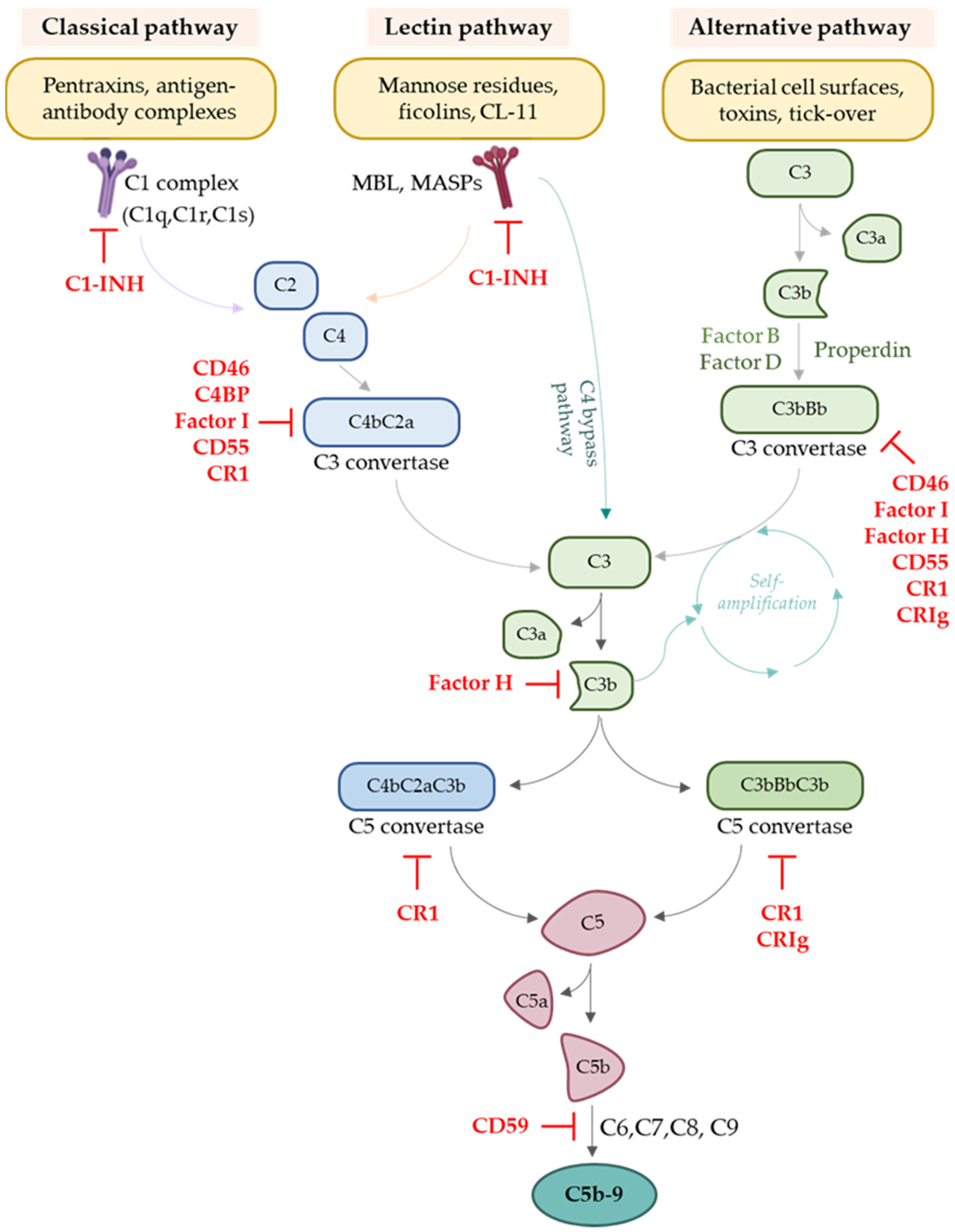
1. Complement System

The complement system is an essential part of innate immunity. It consists of a family of soluble proteins and membrane-expressed receptors and regulators (as summarised in Figure 1) that are widely distributed and operate in the circulation, in tissues, on cell surfaces, and within cells [1][2].
Figure 1. Schematic overview of the complement cascade and of its major ligands and regulators.
Complement is involved in host defence against infection, through: (1) opsonisation of pathogens by C3b, iC3b, C3d, and C4b fragments that are covalently bound to target surfaces to boost phagocytosis [3][4][5][6]; (2) chemotaxis and the activation of leucocytes through the production of potent proinflammatory molecules (the anaphylatoxins C3a and C5a); and (3) direct lysis of bacteria or infected self-cells through the terminal membrane attack complex (MAC, C5b-9) [4]. Secondly, complement can be considered a bridge between innate and adaptive immunity [7]: for example, complement can increase antibody responses and strengthen the immunological memory because C3 receptors are expressed on B cells, antigen-presenting cells (APC), and follicular dendritic cells [8][9][10][11]. Third, complement is essential for the clearance of apoptotic/necrotic, ischaemic, or damaged self-cells (by C1q or C3 binding to cell surfaces) throughout the resolution of the inflammatory reaction, but also during many physiological processes, including development, tissue remodelling, and the maintenance of homeostasis [12][13][14].
In serum and interstitial fluids, complement proteins largely circulate in an inactive form. However, in response to pathogen-associated molecular patterns (PAMPs) or damage-associated molecular patterns (DAMPs), they become activated through a sequential cascade of reactions [15][16]. Indeed, despite the lack of specificity that characterises the innate immune system, complement can selectively recognise pathogens and damaged self-cells through different types of pattern recognition molecules (PRMs), which trigger the initiation of the three pathways of complement activation: the classical (CP), the lectin (LP), and the alternative pathway (AP) [4]. All of these pathways converge in the formation of C3 convertase, independently of the initial danger signal, leading to the production of C3a and C3b. The three pathways of complement activation are schematically overviewed in Figure 1.
The CP initiates when C1q of the C1 complex (which comprises six C1q molecules and two molecules of each of the serine proteases, C1r and C1s) recognises pentraxins, apoptotic, and necrotic cells, or the Fc portion of IgM or IgG antibodies of circulating immune complexes, or of pathogen- or cell-bound immunoglobulins. These interactions lead to the sequential activation of C1r and C1s, which cleave C4 and C2, generating the CP C3 convertase (termed either C4bC2b or C4bC2a in the literature) [17].
The activation of LP relies on the recognition of molecular patterns—such as carbohydrates or other ligands that are expressed on microorganisms but also injured cells [18]—by mannose-binding lectin (MBL), ficolins, or collectins [19]. These PRMs are complexed with mannose-associated serine proteases (MASP-1, MASP-2, and MASP-3), whose activation leads to the cleavage of C4 and C2 to form the LP C3 convertase, in a reaction analogous to CP [4].
Unlike the CP and LP, the AP is continuously and non-specifically activated at low levels in plasma through a process called “tick-over” that allows the system to remain ready for rapid activation when needed [4]. In blood, during “tick-over”, low levels of C3 undergo spontaneous hydrolysis to form C3(H2O). This form of C3 is able to bind Factor B (FB), which then becomes a substrate for serine protease Factor D (FD). The cleavage of FB to Bb and Ba by FD results in the formation of the fluid phase AP C3 convertase C3(H2O)Bb, which, similarly to the CP and LP C3 convertases, can cleave C3 to C3b and C3a. C3b binds covalently to cell surfaces to form membrane-bound C3 convertase, C3Bb [4]. This process is accelerated and sustained by the presence of properdin, which prolongs the survival of C3b [20]. Under physiological conditions, the deposition of C3b via AP is tightly regulated and not allowed on the surface of self-cells, which are protected by complement regulators. On the other hand, AP activation is permitted on activating surfaces, such as the surfaces of pathogens, which are not protected by complement regulators [4][21]. It should be pointed out that the C3b fragment generated as a consequence of the activation of each of the three pathways can interact with FB and, with the action of FD, can generate the AP C3 convertase C3bBb, which therefore is responsible for an amplification loop of the entire complement system, by increasingly converting a large amount of C3 into its split products, C3a and C3b. Through this powerful positive feedback loop, the AP is often the dominant contributor to the overall complement response, even after CP and LP initiation [22][23], since the C3b produced by the CP and LP provides a platform for new AP C3 convertases.
The addition of a C3b molecule to the C3 convertases generates C5 convertases (C4b2b3b for CP and LP, C3bBb3b for AP), which cleave C5 into C5a and C5b, initiating the terminal pathway. Sequential binding of C5b to the components C6, C7, C8, and multiple copies of C9 molecules forms the membrane attack complex (MAC, C5b-9), which is responsible for the direct lysis of the pathogen or, on target cells, culminates in cell activation or lysis [24].
In addition to the C5b-9 complex, which strikes target cells directly, the other fragments generated in the course of complement activation carry out additional functions upon binding to their receptors on target cells. These essential components of the complement system could be classified into complement receptors (CR1, CR2, CR3, and CR4) and anaphylatoxin receptors (C3aR, C5aR1, and C5aR2) [25]. The CR1 molecule has multiple functions, depending on where it is expressed. For example, it was observed as being able to control the activity of T and B cells, to increase the opsonisation activity of phagocytes, and to promote the clearance of immunocomplexes through binding to C3b and C4b [26]. In addition, CR1 also blocks complement activation in different ways: it destabilises and enhances the decay of the C3 and C5 convertases through binding C3b and C4b, it acts as a cofactor for Factor I-mediated inactivation of C3b, and it may regulate the MBL pathway, acting as an MBL receptor [26]. Together with CR1, CR2 signalling is closely related to the activity of B cells and, in its absence, memory B cell survival is markedly impaired [27]. CR3 and CR4 are often co-expressed on the myeloid subset of leucocytes, but they are also found on natural killer cells and activated T and B cells [28]. They play crucial roles in opsonisation by binding to their ligands (C3b and C3 degradation fragments iC3b, C3dg, and C3d), and in cell adhesion [29].
C3a and C5a receptors are expressed mainly on myeloid cells, but also on endothelial and some parenchymal cells, such as tubular epithelial cells (TECs) [30]. These receptors trigger systemic inflammatory responses, including vascular changes and the chemotaxis of immune cells [31]. The C5a/C5aR1 axis was also shown to be a key player in endothelial thromboresistance loss in several pathological conditions, from genetic rare diseases to viral infections [32].
The powerful effects of complement activation have the harmful potential to also damage the host. Indeed, this defence system may be damaging in certain situations (such as during ischaemia/reperfusion injury) in which complement activation can cause autologous injury. Hence, strict regulation of all pathways and steps in complement activation is necessary to ensure that healthy host cells are spared an aberrant complement-mediated attack. Several complement negative regulators circulate in the blood. C1 inhibitor (C1-inh) and C4b-binding protein (C4BP) control the activation of CP and LP. Factor H (FH) is the primary regulator of the AP, both in the fluid phase and on cell surfaces, and it is a cofactor of Factor I and essential for protecting the host from spontaneous AP activation [19]. Complement negative regulators also include membrane-bound molecules, such as complement receptor 1 (CR1/CD35), decay accelerating factor (DAF/CD55), and membrane cofactor protein (MCP/CD46) [4]. All these regulators mediate the degradation of complement convertases or the inactivation of complement split products, preventing the formation of complement effectors C3a, C3b, C5a, and C5b-9.
A stressful event, such as transplantation, can induce an imbalance in the activation/regulation components of the complement system, leading to a proinflammatory milieu that could eventually lead to graft injury.

2. Involvement of the Complement System in Transplantation

During the whole process of transplantation, several events crucially impact graft function and survival and can potentially undermine the overall outcome. Even the steps that must be taken before surgical kidney implantation play a significant role. Indeed, the initial condition of both the donor and the recipient, as well as the organ preservation techniques used, are closely associated with graft quality and outcome. Firstly, the different types of donors, namely living or deceased donors—depending on whether donation occurs after brain death or cardiac death—result in differing organ quality. Regarding kidney transplant recipients, their medical condition and pharmacological treatment until a suitable transplant becomes available also significantly influence graft outcomes. Before surgical implantation, the time and method of graft preservation are crucial and delicate steps in the process of kidney transplantation. Once the organ is implanted in the recipient, the graft finally undergoes reperfusion. From this moment onwards, the graft encounters and inevitably activates the recipient innate and adaptive immune systems, which can potentially induce graft injury and rejection of the donor organ.

2.1. Complement Activation in Donor Kidneys

The clinical condition of the donor and graft have a considerable impact on graft quality and transplant outcomes. In living donation, organs are obtained from healthy people and transplanted following rigorously planned and synchronised surgical procedures. With transplants from deceased donors, which follow brain death (DBD) and cardiac death (DCD), there are frequently pre-existing medical problems, and the organs undergo serious haemodynamic changes, such as prolonged warm ischaemia (in DCD donors) and cell injury with subsequent release of DAMPs and inflammatory responses, which may result in the activation of complement.
Transcriptomic analysis of kidney biopsies revealed activation of the complement cascade in both DBD and in DCD donors before organ retrieval and before the cessation of blood circulation. Kidneys from healthy donors did not exhibit such complement activation [33]. Early studies demonstrated the presence of complement C3 in kidneys from DBD rats [34]. C3d deposition was detected in renal biopsies from human DBD donors before reperfusion, suggesting that C3d was deposited as a result of brain death [35].

2.2. Complement Activation in Transplant Candidates

Complement activation before transplantation may also affect the recipient. Indeed, abnormal complement activity in kidney transplant candidates may be the result of complement-driven kidney diseases (such as atypical haemolytic uremic syndrome (aHUS), C3 glomerulopathy, anti-neutrophil cytoplasmic antibodies (ANCA)-associated glomerulonephritis) [36], and other conditions, including diabetes [37].
Moreover, until a kidney transplant becomes available, many patients are on maintenance haemodialysis. Complement activation can occur during each haemodialysis session. Indeed, upon coming into contact with blood, the surfaces of the biomaterial of the tubing sets and of the dialysis filters are immediately covered with a layer of plasma proteins through passive adsorption. Negatively charged surfaces tend to bind C1q and properdin, which initiate or modulate complement activation via the CP and AP, respectively [38]. C1q binds to immunoglobulins IgG, which are adsorbed by the membrane dialyser and can activate the complement response by the CP [39].

2.3. Ischaemia/Reperfusion Injury (IRI) and Complement Activation

Ischaemia is one of the most common causes of complement activation in kidney transplantation and—combined with reperfusion, which triggers the production of reactive oxygen species [40]—is a major cause of inflammation, graft damage, and DGF.
Organs may undergo ischaemic events in the donor, during organ procurement, transportation and preservation (unless some form of ex vivo organ perfusion is performed), as well as during implantation, until reperfusion is achieved in the recipient [41]. During ischaemia, the kidneys undergo oxygen deprivation and shift to anaerobic metabolism. The resulting acidic conditions interfere with complement regulation, enhancing AP activation [42]. Ischaemia also provokes damage to tubular, endothelial and perivascular cells, with a subsequent massive release of DAMPs, such as hyaluronic acid, fibronectin, and DNA, which can be detected and bound by C1q, MBL, collectins, ficolins, and C3b, thus promoting the activation of the complement system [4][16].
The key contribution of the complement system in IRI is widely studied and well documented in animal models. Some studies showed that administering siRNA against C3 protects the kidney against IRI, thus improving renal function [43][44]. While most of the circulating complement components are produced by hepatic synthesis, smaller amounts are generated at extrahepatic sites, such as the kidney tubular epithelium. The local synthesis of C3, the cross point of the three complement pathways, is essential for complement-mediated reperfusion damage, whereas circulating C3 had a negligible effect [45]. This was demonstrated by the fact that kidney isografts from C3-positive donor kidneys transplanted into C3-negative recipients developed widespread tissue damage and severe acute renal failure, whereas C3-deficient mice exhibited only moderate reperfusion damage when transplanted into wild type recipient mice [45].

2.4. Complement Activation Modulates the Adaptive Immune Response against the Graft

Once in the recipient, the graft is attacked by the host adaptive immune system, which, if left uncontrolled, leads to acute transplant rejection. Several studies highlighted how the complement system and adaptive immunity are intimately interconnected, including in the context of transplantation. Indeed, the complement system can play a role in modulating both the cellular and the humoral immune response against the graft.

2.4.1. The Role of Complement in Regulating T Cell Responses

Through its ability to influence T cell functions, both directly and indirectly, by modulating the activities of antigen-presenting cells (APCs), complement acts as a bridge between the innate and adaptive immune response.
It was shown that DCs are able to synthesise C3 and that C3 synthesis is required for the DC’s capability to stimulate the alloreactive T cell response in vitro and in vivo [46]. Indeed, C3 deficiency in donor DCs favours the polarisation of CD4+ T cells toward a Th2 phenotype and leads to delayed rejection of the allograft in a mouse model of skin transplantation [46]. In addition, in a mouse model of kidney transplantation, it was reported that the absence of local synthesis of C3 (achieved by using grafts from C3-deficient donors) determines defective T cell priming and a subsequent weakened immune response against donor antigens, thus resulting in prolonged graft survival [47].
In resting T cells, Liszewski et al. documented that the protease cathepsin L (CTSL) continuously processes intracellular C3 into bioactive C3a and C3b. When intracellular C3 activation is abrogated by CTSL inhibition, T cells succumb to apoptosis [48]. Following the activation of the T cell receptor and the co-stimulatory CD28 receptor, intracellular C3a and C3b rapidly translocate to the cell surface, where they can bind to C3aR and CD46, respectively [48]. In activated human T cells, autocrine stimulation of complement receptor CD46, and specifically of its intracellular domain CYT-1, by intracellular cleaved C3b, was required for the induction of the amino acid transporter LAT1 and enhanced the expression of the glucose transporter GLUT1, resulting in a metabolic switch toward glycolysis and the activation of the checkpoint kinase mTORC1, both of which are necessary for a Th1 response [49]. In addition, the CD46 transduction signal delivers co-stimulatory signals for optimal cytolytic T cell activity by augmenting the nutrient influx and fatty acid synthesis in CD8+ T cells [50].

2.4.2. The Role of Complement in Antibody-Mediated Rejection

Humoral rejection is a major/the main cause of long-term kidney graft loss [51][52], and complement can help antibody-mediated rejection on several levels.
ABO and HLA system incompatibilities are the main source of immunological risk in allogeneic transplantation. Natural antibodies that react to ABO blood group antigens are involved in humoral immunity to allografts. These pre-existing antibodies, which are produced without previous exposure to the cognate antigen in a T cell-independent manner [53], are primarily IgM that are strong activators of complement [54]. In addition to natural antibodies, the development of anti-HLA antibodies in transplant recipients can occur in a T cell-dependent manner in HLA-mismatched transplant patients who are already immunised, such as multiparous women or patients with a long history of transfusion [55]. Recipients with pre-formed anti-HLA antibodies have an increased risk of hyperacute or acute antibody-mediated rejection (ABMR) and graft loss [56][57].

3. Complement as a Therapeutic Target in Kidney Transplantation

Among the complement inhibitors that are under clinical evaluation in different clinical settings, eculizumab and C1 esterase inhibitor (C1-inh) are the most extensively tested.
Eculizumab is a recombinant humanised hybrid IgG2/IgG4 monoclonal antibody that targets C5. The binding of eculizumab to C5 prevents its cleavage, therefore inhibiting both the production of C5a and the assembly of MAC [58]. Eculizumab was approved for treating patients with paroxysmal nocturnal haemoglobinuria (PNH) [59] and atypical haemolytic uremic syndrome (aHUS) [60][61][62], diseases characterised by abnormal and unrestrained complement activation. Studies showed that patients with aHUS who were waitlisted for kidney transplant and received prophylactic eculizumab treatment underwent successful renal transplantation and experienced a reduced incidence of post-transplant dialysis [63][64]. Eculizumab treatment was also helpful in achieving su
Another complement inhibitor that is widely tested in kidney transplantation is C1-inh. This endogenous complement regulatory protein inactivates the serine proteases that operate at the beginning of CP (C1r, C1s) and LP (MASPs). Therefore, unlike eculizumab, which impedes C5 cleavage, C1-inh acts at the first step of the complement cascade, preventing the production of C3 and C4 split products [65]. A double-blinded, randomised clinical trial investigated the safety and efficacy of C1-inh in reducing DGF in patients who receive a kidney transplant from deceased donors. Perioperative treatment for recipients with C1-inh showed promising results, since it was associated with a lower incidence of graft failure and with an improvement in the estimated glomerular filtration rate (eGFR), even 3 years after transplantation [66]. Clinical trials are planned to test the efficacy of C1-inh in transplant patients who are at high risk of DGF. C1-inh will be given perioperatively to adult subjects who receive kidney allografts from deceased donors [67][68] or to DBD donors as a pre-explant treatment [69].
To attenuate ABMR in transplanted patients with high levels of preformed anti-HLA antibodies, a distinct strategy based on the use of IdeS is worth mentioning. IdeS, a recombinant Streptococcus pyogenes-derived endopeptidase, cleaves all IgG subclasses in the central region. The first step results in the single cleavage of the IgG molecule, in which one heavy chain remains intact. The second step generates a fully cleaved product that cannot mediate complement-dependent cytotoxicity or antibody-dependent cell-mediated cytotoxicity [70]. Promising results from a combined phase I–II clinical trial report that IdeS reduced or eliminated DSAs and permitted HLA-incompatible transplantation in 24 of the 25 highly sensitised patients recruited [70][71][72][73].

4. Blocking Complement to Help Pro-Tolerogenic Cell Therapies

Novel approaches that aim to modulate anti-donor immune responses and induce transplant tolerance are based on the use of cellular therapies. Among the cellular therapies studied, one based on mesenchymal stromal cells (MSCs) recently emerged as very promising.
In human recipients of kidney transplants from living donors, autologous bone marrow-derived MSC infusion was found to be a safe and practicable strategy to promote a pro-tolerogenic environment [74][75][76] and to prevent acute rejection with lower than conventional doses of immunosuppressive therapy [76][77][78][79]. However, to be effective, autologous MSCs should be administered before transplantation [74][80]. Indeed, since systemically infused MSCs tend to migrate to damaged tissues, such as those exposed to ischaemia/reperfusion injury [81][82], MSC infusion performed a few days after transplant led to intragraft MSC recruitment, subsequent graft inflammation, neutrophil infiltration, and C3 deposition, with no advantage for graft survival [74]. However, a pretransplant approach is not applicable to deceased donor transplantation since the surgical transplant procedure takes places a few hours after a donor kidney becomes available.

Casiraghi et al. recently investigated how to render MSCs a therapeutic option for recipients of organs from deceased donors as well. Based on evidence that human MSCs express C3aR and C5aR [83], and that the chemoattractant effects of anaphylatoxins could guide MSCs toward injured tissues [83][84], the scholars evaluated the effect of antagonising complement receptors on MSCs given on day +2 post-transplant in preventing their recruitment into the graft and in prolonging graft survival [85]. Post-transplant MSC infusion combined with a short course of C3aR or C5aR antagonist, or the administration of MSCs pre-treated with C3aR and C5aR antagonists, prevented intragraft recruitment of MSCs [85]. Notably, antagonising C3aR or C5aR allowed MSCs to home the secondary lymphoid organs, and led to diminished C3 deposition and neutrophil recruitment, with a subsequent reduction in graft inflammation [85].

In conclusion, inhibiting the complement cascade not only might help prevent IRI and DGF, as well as alleviating both T cell- and antibody-mediated rejection (as discussed above) but could also be used to make the promising pro-tolerogenic MSC therapy feasible in the context of transplantation with organs from deceased donors.

References

  1. Luo, S.; Hu, D.; Wang, M.; Zipfel, P.F.; Hu, Y. Complement in Hemolysis- and Thrombosis- Related Diseases. Front. Immunol. 2020, 11, 1212.
  2. Arbore, G.; Kemper, C.; Kolev, M. Intracellular complement − the complosome − in immune cell regulation. Mol. Immunol. 2017, 89, 2–9.
  3. Merle, N.S.; Noé, R.; Halbwachs-Mecarelli, L.; Fremeaux-Bacchi, V.; Roumenina, L.T. Complement System Part II: Role in Immunity. Front. Immunol. 2015, 6, 257.
  4. Noris, M.; Remuzzi, G. Overview of Complement Activation and Regulation. Semin. Nephrol. 2013, 33, 479–492.
  5. De la O Becerra, K.I.; Oosterheert, W.; Bos, R.M.V.D.; Xenaki, K.T.; Lorent, J.H.; Ruyken, M.; Schouten, A.; Rooijakkers, S.H.M.; Henegouwen, P.M.P.V.B.E.; Gros, P. Multifaceted Activities of Seven Nanobodies against Complement C4b. J. Immunol. 2022, 208, 2207–2219.
  6. Zhou, T.; Li, Y.; Li, X.; Zeng, F.; Rao, Y.; He, Y.; Wang, Y.; Liu, M.; Li, D.; Xu, Z.; et al. Microglial debris is cleared by astrocytes via C4b-facilitated phagocytosis and degraded via RUBICON-dependent noncanonical autophagy in mice. Nat. Commun. 2022, 13, 1–22.
  7. Walport, M.J. Complement. New Engl. J. Med. 2001, 344, 1140–1144.
  8. Tew, J.G.; Kosco, M.H.; Burton, G.F.; Szakal, A.K. Follicular Dendritic Cells as Accessory Cells. Immunol. Rev. 1990, 117, 185–211.
  9. Thieblemont, N.; Haeffner-Cavaillon, N.; Haeffner, A.; Weiss, L.; Kazatchkine, M.D. Triggering of Complement Receptors CR1 (CD35) and CR3 (CD11b/CD18) Induces Nuclear Translocation of NF-Kappa B (P50/P65) in Human Monocytes and Enhances Viral Replication in HIV-Infected Monocytic Cells. J. Immunol. 1950, 155, 4861–4867.
  10. Phan, T.; Grigorova, I.; Okada, T.; Cyster, J.G. Subcapsular encounter and complement-dependent transport of immune complexes by lymph node B cells. Nat. Immunol. 2007, 8, 992–1000.
  11. Kopf, M.; Abel, B.; Gallimore, A.; Carroll, M.; Bachmann, M.F. Complement component C3 promotes T-cell priming and lung migration to control acute influenza virus infection. Nat. Med. 2002, 8, 373–378.
  12. Soteros, B.M.; Sia, G.M. Complement and microglia dependent synapse elimination in brain development. Wiley Interdiscip. Rev. Syst. Biol. Med. 2021, 14, e1545.
  13. Mevorach, D. Clearance of dying cells and systemic lupus erythematosus: The role of C1q and the complement system. Apoptosis 2010, 15, 1114–1123.
  14. Defendi, F.; Thielens, N.M.; Clavarino, G.; Cesbron, J.-Y.; Dumestre-Pérard, C. The Immunopathology of Complement Proteins and Innate Immunity in Autoimmune Disease. Clin. Rev. Allergy Immunol. 2019, 58, 229–251.
  15. Franzin, R.; Stasi, A.; Fiorentino, M.; Stallone, G.; Cantaluppi, V.; Gesualdo, L.; Castellano, G. Inflammaging and Complement System: A Link Between Acute Kidney Injury and Chronic Graft Damage. Front. Immunol. 2020, 11, 734.
  16. Nauser, C.L.; Farrar, C.A.; Sacks, S.H. Complement Recognition Pathways in Renal Transplantation. J. Am. Soc. Nephrol. 2017, 28, 2571–2578.
  17. Ricklin, D.; Reis, E.S.; Lambris, J.D. Complement in disease: A defence system turning offensive. Nat. Rev. Nephrol. 2016, 12, 383–401.
  18. Grafals, M.; Thurman, J.M. The Role of Complement in Organ Transplantation. Front. Immunol. 2019, 10, 2380.
  19. Moore, S.R.; Menon, S.S.; Cortes, C.; Ferreira, V.P. Hijacking Factor H for Complement Immune Evasion. Front. Immunol. 2021, 12, 602277.
  20. Lesher, A.M.; Nilsson, B.; Song, W.-C. Properdin in complement activation and tissue injury. Mol. Immunol. 2013, 56, 191–198.
  21. Blaum, B.; Hannan, J.; Herbert, A.P.; Kavanagh, D.; Uhrín, D.; Stehle, T. Structural basis for sialic acid–mediated self-recognition by complement factor H. Nat. Chem. Biol. 2014, 11, 77–82.
  22. Harboe, M.; Ulvund, G.; Vien, L.; Fung, M.; Mollnes, T.E. The quantitative role of alternative pathway amplification in classical pathway induced terminal complement activation. Clin. Exp. Immunol. 2004, 138, 439–446.
  23. Harboe, M.; Garred, P.; Karlstrøm, E.; Lindstad, J.K.; Stahl, G.L.; Mollnes, T.E. The down-stream effects of mannan-induced lectin complement pathway activation depend quantitatively on alternative pathway amplification. Mol. Immunol. 2009, 47, 373–380.
  24. Morgan, B.P.; Walters, D.; Serna, M.; Bubeck, D. Terminal complexes of the complement system: New structural insights and their relevance to function. Immunol. Rev. 2016, 274, 141–151.
  25. Qi, R.; Qin, W. Role of Complement System in Kidney Transplantation: Stepping From Animal Models to Clinical Application. Front. Immunol. 2022, 13, 811696.
  26. Khera, R.; Das, N. Complement Receptor 1: Disease associations and therapeutic implications. Mol. Immunol. 2009, 46, 761–772.
  27. Roozendaal, R.; Carroll, M.C. Complement receptors CD21 and CD35 in humoral immunity. Immunol. Rev. 2007, 219, 157–166.
  28. Vorup-Jensen, T.; Jensen, R.K. Structural Immunology of Complement Receptors 3 and 4. Front. Immunol. 2018, 9, 2716.
  29. Wagner, C.; Hänsch, G.M.; Stegmaier, S.; Denefleh, B.; Hug, F.; Schoels, M. The complement receptor 3, CR3 (CD11b/CD18), on T lymphocytes: Activation-dependent up-regulation and regulatory function. Eur. J. Immunol. 2001, 31, 1173–1180.
  30. Fayyazi, A.; Scheel, O.; Werfel, T.; Schweyer, S.; Oppermann, M.; Götze, O.; Radzun, H.J.; Zwirner, J. The C5a receptor is expressed in normal renal proximal tubular but not in normal pulmonary or hepatic epithelial cells. Immunology 2000, 99, 38–45.
  31. Thurman, J.M.; Nester, C.M. All Things Complement. Clin. J. Am. Soc. Nephrol. 2016, 11, 1856–1866.
  32. Aiello, S.; Gastoldi, S.; Galbusera, M.; Ruggenenti, P.L.; Portalupi, V.; Rota, S.; Rubis, N.; Liguori, L.; Conti, S.; Tironi, M.; et al. C5a and C5aR1 are key drivers of microvascular platelet aggregation in clinical entities spanning from aHUS to COVID-19. Blood Adv. 2022, 6, 866–881.
  33. Damman, J.; Bloks, V.W.; Daha, M.R.; Van Der Most, P.J.; Sanjabi, B.; Van Der Vlies, P.; Snieder, H.; Ploeg, R.J.; Krikke, C.; Leuvenink, H.G.D.; et al. Hypoxia and Complement-and-Coagulation Pathways in the Deceased Organ Donor as the Major Target for Intervention to Improve Renal Allograft Outcome. Transplantation 2015, 99, 1293–1300.
  34. Damman, J.; Nijboer, W.N.; Schuurs, T.A.; Leuvenink, H.G.; Morariu, A.M.; Tullius, S.G.; Van Goor, H.; Ploeg, R.J.; Seelen, M.A. Local renal complement C3 induction by donor brain death is associated with reduced renal allograft function after transplantation. Nephrol. Dial. Transplant. 2011, 26, 2345–2354.
  35. Poppelaars, F.; Seelen, M.A. Complement-mediated inflammation and injury in brain dead organ donors. Mol. Immunol. 2017, 84, 77–83.
  36. Mizuno, M.; Suzuki, Y.; Ito, Y. Complement regulation and kidney diseases: Recent knowledge of the double-edged roles of complement activation in nephrology. Clin. Exp. Nephrol. 2017, 22, 3–14.
  37. Jiang, S.; Jiao, Y.; Zou, G.; Gao, H.; Zhuo, L.; Li, W. Activation of Complement Pathways in Kidney Tissue May Mediate Tubulointerstitial Injury in Diabetic Nephropathy. Front. Med. 2022, 9, 845679.
  38. Ekdahl, K.N.; Soveri, I.; Hilborn, J.; Fellström, B.; Nilsson, B. Cardiovascular disease in haemodialysis: Role of the intravascular innate immune system. Nat. Rev. Nephrol. 2017, 13, 285–296.
  39. Wang, Y.; Gao, L. Inflammation and Cardiovascular Disease Associated With Hemodialysis for End-Stage Renal Disease. Front. Pharmacol. 2022, 13, 800950.
  40. Malek, M.; Nematbakhsh, M. Renal ischemia/reperfusion injury; from pathophysiology to treatment. J. Ren. Inj. Prev. 2015, 4, 20–27.
  41. Heylen, L.; Pirenne, J.; Naesens, M.; Sprangers, B.; Jochmans, I. “Time is tissue”—A minireview on the importance of donor nephrectomy, donor hepatectomy, and implantation times in kidney and liver transplantation. Am. J. Transplant. 2021, 21, 2653–2661.
  42. Sim, E.; Sim, R.B. Enzymic assay of C3b receptor on intact cells and solubilized cells. Biochem. J. 1983, 210, 567–576.
  43. Zheng, X.; Feng, B.; Chen, G.; Zhang, X.; Li, M.; Sun, H.; Liu, W.; Vladau, C.; Liu, R.; Jevnikar, A.M.; et al. Preventing Renal IschemiaReperfusion Injury Using Small Interfering RNA by Targeting Complement 3 Gene. Am. J. Transplant. 2006, 6, 2099–2108.
  44. Zheng, X.; Zhang, X.; Sun, H.; Feng, B.; Li, M.; Chen, G.; Vladau, C.; Chen, D.; Suzuki, M.; Min, L.; et al. Protection of Renal Ischemia Injury using Combination Gene Silencing of Complement 3 and Caspase 3 Genes. Transplantation 2006, 82, 1781–1786.
  45. Farrar, C.A.; Zhou, W.; Lin, T.; Sacks, S.H. Local extravascular pool of C3 is a determinant of postischemic acute renal failure. FASEB J. 2006, 20, 217–226.
  46. Peng, Q.; Li, K.; Patel, H.; Sacks, S.H.; Zhou, W. Dendritic Cell Synthesis of C3 Is Required for Full T Cell Activation and Development of a Th1 Phenotype. J. Immunol. 2006, 176, 3330–3341.
  47. Pratt, J.R.; Basheer, S.A.; Sacks, S.H. Local synthesis of complement component C3 regulates acute renal transplant rejection. Nat. Med. 2002, 8, 582–587.
  48. Liszewski, M.K.; Kolev, M.; Le Friec, G.; Leung, M.; Bertram, P.G.; Fara, A.F.; Subias, M.; Pickering, M.C.; Drouet, C.; Meri, S.; et al. Intracellular Complement Activation Sustains T Cell Homeostasis and Mediates Effector Differentiation. Immunity 2013, 39, 1143–1157.
  49. Kolev, M.; Dimeloe, S.; Le Friec, G.; Navarini, A.; Arbore, G.; Povoleri, G.A.; Fischer, M.; Belle, R.; Loeliger, J.; Develioglu, L.; et al. Complement Regulates Nutrient Influx and Metabolic Reprogramming during Th1 Cell Responses. Immunity 2015, 42, 1033–1047.
  50. Arbore, G.; West, E.E.; Rahman, J.; Le Friec, G.; Niyonzima, N.; Pirooznia, M.; Tunc, I.; Pavlidis, P.; Powell, N.; Li, Y.; et al. Complement receptor CD46 co-stimulates optimal human CD8+ T cell effector function via fatty acid metabolism. Nat. Commun. 2018, 9, 1–15.
  51. Einecke, G.; Sis, B.; Reeve, J.; Mengel, M.; Campbell, P.M.; Hidalgo, L.G.; Kaplan, B.; Halloran, P.F. Antibody-Mediated Microcirculation Injury Is the Major Cause of Late Kidney Transplant Failure. Am. J. Transplant. 2009, 9, 2520–2531.
  52. Halloran, P.F.; Chang, J.; Famulski, K.; Hidalgo, L.G.; Salazar, I.D.; Lopez, M.M.; Matas, A.; Picton, M.; de Freitas, D.; Bromberg, J.; et al. Disappearance of T Cell-Mediated Rejection Despite Continued Antibody-Mediated Rejection in Late Kidney Transplant Recipients. J. Am. Soc. Nephrol. 2015, 26, 1711–1720.
  53. Biglarnia, A.-R.; Huber-Lang, M.; Mohlin, C.; Ekdahl, K.N.; Nilsson, B. The multifaceted role of complement in kidney transplantation. Nat. Rev. Nephrol. 2018, 14, 767–781.
  54. Ugurlar, D.; Howes, S.C.; de Kreuk, B.-J.; Koning, R.I.; de Jong, R.N.; Beurskens, F.J.; Schuurman, J.; Koster, A.J.; Sharp, T.H.; Parren, P.W.H.I.; et al. Structures of C1-IgG1 provide insights into how danger pattern recognition activates complement. Science 2018, 359, 794–797.
  55. De Clippel, D.; Baeten, M.; Torfs, A.; Emonds, M.-P.; Feys, H.B.; Compernolle, V.; Vandekerckhove, P. Screening for HLA antibodies in plateletpheresis donors with a history of transfusion or pregnancy. Transfusion 2014, 54, 3036–3042.
  56. Lederer, S.R.; Schneeberger, H.; Albert, E.; Johnson, J.P.; Gruber, R.; Land, W.; Burkhardt, K.; Hillebrand, G.; Feucht, H.E. Early renal graft dysfunction: The Role of Preformed Antibodies to DR-Typed Lymphoblastoid Cell Lines: 1. Transplantation 1996, 61, 313–319.
  57. Bentall, A.; Cornell, L.D.; Gloor, J.M.; Park, W.D.; Gandhi, M.J.; Winters, J.L.; Chedid, M.F.; Dean, P.G.; Stegall, M.D. Five-Year Outcomes in Living Donor Kidney Transplants With a Positive Crossmatch. Am. J. Transplant. 2012, 13, 76–85.
  58. Frémeaux-Bacchi, V.; Legendre, C.M. The emerging role of complement inhibitors in transplantation. Kidney Int. 2015, 88, 967–973.
  59. Hillmen, P.; Young, N.S.; Schubert, J.; Brodsky, R.A.; Socié, G.; Muus, P.; Röth, A.; Szer, J.; Elebute, M.O.; Nakamura, R.; et al. The Complement Inhibitor Eculizumab in Paroxysmal Nocturnal Hemoglobinuria. N. Engl. J. Med. 2006, 355, 1233–1243.
  60. Legendre, C.M.; Licht, C.; Muus, P.; Greenbaum, L.A.; Babu, S.; Bedrosian, C.; Bingham, C.; Cohen, D.J.; Delmas, Y.; Douglas, K.; et al. Terminal Complement Inhibitor Eculizumab in Atypical Hemolytic–Uremic Syndrome. N. Engl. J. Med. 2013, 368, 2169–2181.
  61. Noris, M.; Galbusera, M.; Gastoldi, S.; Macor, P.; Banterla, F.; Bresin, E.; Tripodo, C.; Bettoni, S.; Donadelli, R.; Valoti, E.; et al. Dynamics of complement activation in aHUS and how to monitor eculizumab therapy. Blood 2014, 124, 1715–1726.
  62. Fakhouri, F.; Schwotzer, N.; Golshayan, D.; Frémeaux-Bacchi, V. The Rational Use of Complement Inhibitors in Kidney Diseases. Kidney Int. Rep. 2022, 7, 1165–1178.
  63. Weitz, M.; Amon, O.; Bassler, D.; Koenigsrainer, A.; Nadalin, S. Prophylactic eculizumab prior to kidney transplantation for atypical hemolytic uremic syndrome. Pediatr. Nephrol. 2011, 26, 1325–1329.
  64. Siedlecki, A.M.; Isbel, N.; Walle, J.V.; Eggleston, J.J.; Cohen, D.J.; Licht, C.; Frémeaux-Bacchi, V.; Ariceta, G.; Ardissino, G.; Fakhouri, F.; et al. Eculizumab Use for Kidney Transplantation in Patients With a Diagnosis of Atypical Hemolytic Uremic Syndrome. Kidney Int. Rep. 2018, 4, 434–446.
  65. Davis, A.E.; Mejia, P.; Lu, F. Biological activities of C1 inhibitor. Mol. Immunol. 2008, 45, 4057–4063.
  66. Huang, E.; Vo, A.; Choi, J.; Ammerman, N.; Lim, K.; Sethi, S.; Kim, I.; Kumar, S.; Najjar, R.; Peng, A.; et al. Three-Year Outcomes of a Randomized, Double-Blind, Placebo-Controlled Study Assessing Safety and Efficacy of C1 Esterase Inhibitor for Prevention of Delayed Graft Function in Deceased Donor Kidney Transplant Recipients. Clin. J. Am. Soc. Nephrol. 2019, 15, 109–116.
  67. MD, S.J. A Phase I/II, Double-Blind, Placebo-Controlled Study: Assessing Safety and Efficacy of Preoperative Renal Allograft Infusions of C1 Inhibitor (Berinert®) (Human) (C1INH) vs. Placebo Administration in Recipients of a Renal Allograft From Deceased High Risk Donors and Its Impact on Delayed Graft Function (DGF) and Ischemia/Reperfusion Injury (IRI). 2022. Available online: https://clinicaltrials.gov/ct2/show/NCT04696146 (accessed on 26 October 2022).
  68. University of Wisconsin, Madison A Phase I/II, Single Center, Randomized, Double-Blind, Placebo-Controlled Study to Evaluate the Feasibility of Using Human Recombinant C1 Inhibitor(RUCONEST®) as a Therapeutic Strategy to Reduce the Incidence of Delayed Graft Function in Recipients of Kidneys From Donation After Cardio-Circulatory Death. 2020. Available online: https://clinicaltrials.gov/ct2/show/NCT03791476 (accessed on 26 October 2022).
  69. University of Wisconsin, Madison A Phase I, Single Center, Randomized, Double-Blind, Placebo-Controlled Study to Evaluate Tolerability of C1 Inhibitor (CINRYZE) as a Donor Pre-Treatment Strategy in Brain Dead Donors Who Meet a Kidney Donor Risk Index (KDRI) Above 60%. 2020. Available online: https://clinicaltrials.gov/ct2/show/NCT02435732 (accessed on 26 October 2022).
  70. Jordan, S.C.; Lorant, T.; Choi, J.; Kjellman, C.; Winstedt, L.; Bengtsson, M.; Zhang, X.; Eich, T.; Toyoda, M.; Eriksson, B.-M.; et al. IgG Endopeptidase in Highly Sensitized Patients Undergoing Transplantation. N. Engl. J. Med. 2017, 377, 442–453.
  71. Hansa Biopharma, A.B. A Phase II Study to Evaluate the Safety, Tolerability, Pharmacokinetics and Efficacy of Intravenous IdeS After Administration of Ascending Doses in Chronic Kidney Disease Patients. 2017. Available online: https://clinicaltrials.gov/ct2/show/NCT02224820 (accessed on 26 October 2022).
  72. Hansa Biopharma, A.B. A Phase II Study to Evaluate the Safety, Tolerability, Efficacy and Pharmacokinetics of Intravenous Ascending Doses of IdeS in Kidney Transplantation. 2018. Available online: https://clinicaltrials.gov/ct2/show/NCT02475551 (accessed on 26 October 2022).
  73. MD, S.J. A Phase I/II Trial to Evaluate the Safety and Tolerability of Ides® (IgG Endopeptidase) to Eliminate Donor Specific HLA Antibodies (DSAs) and Prevent Antibody-Mediated Rejection Post-Transplant in Highly-HLA Sensitized Patients. 2022. Available online: https://clinicaltrials.gov/ct2/show/NCT02426684 (accessed on 26 October 2022).
  74. Perico, N.; Casiraghi, F.; Introna, M.; Gotti, E.; Todeschini, M.; Cavinato, R.A.; Capelli, C.; Rambaldi, A.; Cassis, P.; Rizzo, P.; et al. Autologous Mesenchymal Stromal Cells and Kidney Transplantation. Clin. J. Am. Soc. Nephrol. 2011, 6, 412–422.
  75. Perico, N.; Casiraghi, F.; Gotti, E.; Introna, M.; Todeschini, M.; Cavinato, R.A.; Capelli, C.; Rambaldi, A.; Cassis, P.; Rizzo, P.; et al. Mesenchymal stromal cells and kidney transplantation: Pretransplant infusion protects from graft dysfunction while fostering immunoregulation. Transpl. Int. 2013, 26, 867–878.
  76. Casiraghi, F.; Perico, N.; Cortinovis, F.C.M.; Remuzzi, G. Mesenchymal stromal cells in renal transplantation: Opportunities and challenges. Nat. Rev. Nephrol. 2016, 12, 241–253.
  77. Tan, J.; Wu, W.; Xu, X.; Liao, L.; Zheng, F.; Messinger, S.; Sun, X.; Chen, J.; Yang, S.; Cai, J.; et al. Induction Therapy With Autologous Mesenchymal Stem Cells in Living-Related Kidney Transplants. JAMA 2012, 307, 1169–1177.
  78. Peng, Y.; Ke, M.; Xu, L.; Liu, L.; Chen, X.; Xia, W.; Li, X.; Chen, Z.; Ma, J.; Liao, D.; et al. Donor-Derived Mesenchymal Stem Cells Combined With Low-Dose Tacrolimus Prevent Acute Rejection After Renal Transplantation. Transplantation 2013, 95, 161–168.
  79. Reinders, M.; Van Kooten, C.; Rabelink, T.; De Fijter, J.W. Mesenchymal Stromal Cell Therapy for Solid Organ Transplantation. Transplantation 2018, 102, 35–43.
  80. Casiraghi, F.; Azzollini, N.; Todeschini, M.; Cavinato, R.A.; Cassis, P.; Solini, S.; Rota, C.; Morigi, M.; Introna, M.; Maranta, R.; et al. Localization of Mesenchymal Stromal Cells Dictates Their Immune or Proinflammatory Effects in Kidney Transplantation. Am. J. Transplant. Off. J. Am. Soc. Transplant. Am. Soc. Transpl. Surg. 2012, 12, 2373–2383.
  81. Herrera, M.; Bussolati, B.; Bruno, S.; Morando, L.; Mauriello-Romanazzi, G.; Sanavio, F.; Stamenkovic, I.; Biancone, L.; Camussi, G. Exogenous mesenchymal stem cells localize to the kidney by means of CD44 following acute tubular injury. Kidney Int. 2007, 72, 430–441.
  82. Dong, F.; Harvey, J.; Finan, A.; Weber, K.; Agarwal, U.; Penn, M.S. Myocardial CXCR4 Expression Is Required for Mesenchymal Stem Cell Mediated Repair Following Acute Myocardial Infarction. Circulation 2012, 126, 314–324.
  83. Ingrid U. Schraufstatter, Richard G. DiScipio, Ming Zhao, Sophia K. Khaldoyanidi; C3a and C5a Are Chemotactic Factors for Human Mesenchymal Stem Cells, Which Cause Prolonged ERK1/2 Phosphorylation1. J Immunol 2009, 182 (6): 3827–3836.
  84. Hengartner, N.-E.; Fiedler, J.; Schrezenmeier, H.; Huber-Lang, M.; Brenner, R.E. Crucial Role of IL1beta and C3a in the In Vitro-Response of Multipotent Mesenchymal Stromal Cells to Inflammatory Mediators of Polytrauma. PLoS ONE 2015, 10, e0116772.
  85. Casiraghi, F.; Todeschini, M.; Azzollini, N.; Cravedi, P.; Cassis, P.; Solini, S.; Fiori, S.; Rota, C.; Karachi, A.; Carrara, C.; et al. Effect of Timing and Complement Receptor Antagonism on Intragraft Recruitment and Protolerogenic Effects of Mesenchy-mal Stromal Cells in Murine Kidney Transplantation. Transplantation 2019, 103, 1121–1130.
More
Information
Subjects: Immunology
Contributors MDPI registered users' name will be linked to their SciProfiles pages. To register with us, please refer to https://encyclopedia.pub/register : ,
View Times: 599
Revisions: 2 times (View History)
Update Date: 10 Mar 2023
1000/1000
ScholarVision Creations