Submitted Successfully!
To reward your contribution, here is a gift for you: A free trial for our video production service.
Thank you for your contribution! You can also upload a video entry or images related to this topic.
Version Summary Created by Modification Content Size Created at Operation
1 -- 2314 2023-02-23 10:18:23 |
2 format correction Meta information modification 2314 2023-02-27 02:17:06 |

Video Upload Options

Do you have a full video?

Confirm

Are you sure to Delete?
Cite
If you have any further questions, please contact Encyclopedia Editorial Office.
Cassano, R.; Perri, P.; Esposito, A.; Intrieri, F.; Sole, R.; Curcio, F.; Trombino, S. Expanded Polytetrafluoroethylene Membranes. Encyclopedia. Available online: https://encyclopedia.pub/entry/41581 (accessed on 25 April 2024).
Cassano R, Perri P, Esposito A, Intrieri F, Sole R, Curcio F, et al. Expanded Polytetrafluoroethylene Membranes. Encyclopedia. Available at: https://encyclopedia.pub/entry/41581. Accessed April 25, 2024.
Cassano, Roberta, Paolo Perri, Antonio Esposito, Francesco Intrieri, Roberta Sole, Federica Curcio, Sonia Trombino. "Expanded Polytetrafluoroethylene Membranes" Encyclopedia, https://encyclopedia.pub/entry/41581 (accessed April 25, 2024).
Cassano, R., Perri, P., Esposito, A., Intrieri, F., Sole, R., Curcio, F., & Trombino, S. (2023, February 23). Expanded Polytetrafluoroethylene Membranes. In Encyclopedia. https://encyclopedia.pub/entry/41581
Cassano, Roberta, et al. "Expanded Polytetrafluoroethylene Membranes." Encyclopedia. Web. 23 February, 2023.
Expanded Polytetrafluoroethylene Membranes
Edit

Coated stents are defined as innovative stents surrounded by a thin polymer membrane based on polytetrafluoroethylene (PTFE)useful in the treatment of numerous vascular pathologies. Endovascular methodology involves the use of such devices to restore blood flow in small-, medium- and large-calibre arteries, both centrally and peripherally. These membranes cross the stent struts and act as a physical barrier to block the growth of intimal tissue in the lumen, preventing so-called intimal hyperplasia and late stent thrombosis. PTFE for vascular applications is known as expanded polytetrafluoroethylene (e-PTFE) and it can be rolled up to form a thin multilayer membrane expandable by 4 to 5 times its original diameter. This membrane plays an important role in initiating the restenotic process because wrapped graft stent could be used as the treatment option for trauma devices during emergency situations and to treat a number of pathological vascular disease.

PTFE membrane covered stent vascular disease

1. Introduction

Expanded polytetrafluoroethylene (e-PTFE) is a hydrophobic polymer obtained by stretching PTFE, a fluoropolymer commonly used in the biomedical field for the manufacture of catheters for angioplasty, orbital implants, orthopedic joint implants, and membranes for stent coating [1]. Expansion occurs during the manufacturing process, when the solid material is modified into a porous lattice. The presence of negative charges on the polymer blocks the coagulation of blood proteins on the tissue surface and limits the activation of platelets [2].
e-PTFE is reasonably tough, and chemically inert; for this reason, in the biomedical field and in tissue engineering, this polymer has been used to make synthetic membranes capable of wrapping metal stentgrafts for the treatment of vascular diseases such as popliteal aneurysms, arterial perforations, iatrogenic perforations, and vessel stenosis [3]. The presence of a synthetic membrane, stretching between the stent struts and covering both the luminal and abluminal portions of the stent, is essential to create a physical barrier that can effectively prevent plaque protrusion, successfully sealing aneurysms, arterial perforations, or simply restoring blood flow in vessels prone to occlusion [4][5]. e-PTFE membranes have a porous and flexible structure and are chemically stable, biocompatible and inert, properties that enable them to resist degradation produced by microbiological or enzymatic reactions [6][7]. Fundamental characteristics of e-PTFE-membranes are non-thrombogenicity, the same viscoelasticity as native vessels and resistance to high blood pressure. In addition, when covered stent, they must be able to provide biocompatibility, accelerate the endothelization process, minimize vascular damage and reduce the proliferative response of the native artery. e-PTFE membranes are manufactured through stretching, spinning and pore-forming techniques [8]. Electrospinning can originate fibres with diameters ranging from a few nanometres to a few microns, presenting a high specific surface area and high porosity [9] that can be easily adjusted by changing the concentration of the spinning solution or the spinning parameters [10]. This characteristic also applies to the mechanical properties of the membrane [11]. e-PTFE membranes are often functionalized through covalent and non-covalent bonds, chemical impregnation, chemical surface modification, autologous vascularization, etc. These strategies are used to improve their properties and compatibility with blood tissue [12][13]. There are several commercially available e-PTFE covered stents for clinical use. The most commonly used ones consist of stainless steel (SS) metal frames with diameters ranging from 2. 5 to 16 mm with an e-PTFE film cover (iCAST, Atrium, Ekkatuthangal, Chennai, India), with an SS frame and struts embedded in an ePTFE film (Advanta V12, Atrium) [14], with an ePTFE film sandwiched between the SS matrix (JoStentGraftmaster Coronary Stent Graft or JoStent Peripheral Stent Graft, Abbott Vascular, Rome, Italy) [15] and with a Nitinol (NiTi) frame sandwiched between ePTFE (Symbiot Covered Stent, Boston Scientific, Pennsylvania Avenue, Washington, DC) [16]. There are also stents with a Nitinol frame covered with thin ePTFE films surrounding the metal frame (Viabahn Endoprosthesis, GORE, Bergheim, Austria) [17], or encapsulated with two thin ePTFE layers (Fluency + Vascular Stent Graft, BARD Peripheral Vascular, Becton, NJ, USA) [18]. The stents listed above are used in clinical conditions such as: treatment of tracheobronchial structures caused by the growth of cancerous tissue; treatment of iliac and renal arteries; treatment of aortic coarctation; treatment of aneurysms, fistulas, ruptures and perforations; and finally degenerated saphenous vein grafts [19].

2. e-PTFE Membrane Preparation

e-PTFE membrane preparation started mainly in the early 1960s. Today, various techniques including stretching, pore-forming, sintering, wrapping, electrospinning and near field electrospinningare used to produce them.

2.1. Stretching and Pore-Forming Process

A fine e-PTFE powder is homogeneously dispersed in an oil lubricant (processing aid) to become a paste. The first step, called pre-forming, typically takes place between 21 and 25 °C and serves to align the PTFE particles for packing in the best possible way. The presence of lubricants is essential to promote rapid absorption of the mixture [20]. The resulting paste is pressed into a mould and ram extruded into sheets or rods that are passed several times through two heated rolling mills to obtain a precise thickness. The resulting membranes are then stretched and hardened at room temperature and sintered at 340–365 °C, maintaining the stretching condition to fix the porous structure and prevent shrinkage [21] (Figure 1).
Figure 1. Stretching process.

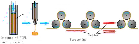
2.2. Sintering Process

In the sintering process, the polymer particles are only fully melted on the outer surface, so there are still many microchannels in the material that form a network of through holes [22]. As thetemperature, the surface of the attached particles melts further and firmly due to the movement and diffusion of the molecular chain segments, resulting in a porous film [23]. Sintering is a process that influences the final properties of the membrane as it improves its porosity usinginorganic agents (e.g., ZnAc2, NaCl and BaCl2) that are used as additives [24]. These additives are introduced and uniformly mixed with the polymeric materials, favouring their formation upon removal through post-processing or chemical attack [25].

2.3. Wrapping Process

The wrapping technique is used to make e-PTFE hollow-fibre membranes whose porosity increases as the stretching ratio increases. Membranes with large pore sizes are not suitable for use in various areas of technology or in the biomedical sector, as they do not guarantee adequate microfiltration [26]. Instead, by means of the wrapping method, it is possible to wrap ane-PTFE membrane with a small pore diameter on the outer surface of a PTFE hollow membrane with a large pore diameter to form a double layer that generates an asymmetric hollow membrane with 81% porosity and a pore size of 0.2 μm [27].

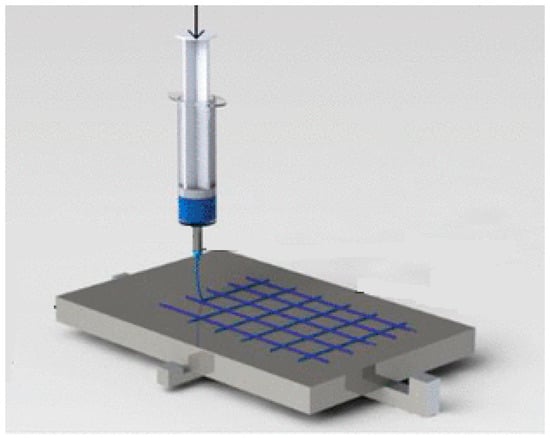
2.4. Near-Field Electrospinning Process

Near-field electrospinning (NFES) is a technique that allows membranes to be made by layering polymers on a planar substrate under the action of an electric field. Compared to conventional electrospinning technology, it favours a shorter spinning distance, a lower supply voltage, a more complex structure and controlled deposition [28] (Figure 2). The advantage of using this technique is the realization of membranes with a regular geometric pore structure [29]. In the biomedical field, electrospun fibrous scaffolds with adequate porosity, nanoscale topography and interconnectivity provide an ideal model for biomedical engineering as human tissues and/or cells are able to attach and organize themselves appropriately around fibres with diameters smaller than their own [30].
Figure 2. Near-field electrospinning process.

2.5. Electrospinning Process

Electrospinning is a technique that allows polymeric fibres with nano/submicron dimensions to be produced using an instrument consisting of an injector with a blunt tip, a booster pump to control the extrusion speed of a polymer solution, a DC electric field and a grounded collector [31]. In detail, a polymer fluid is able to pass through a high-voltage electric field generating a microjet that is solidified on a substrate as a fibre membrane layer [32]. In the case of e-PTFE membranes, it is necessary to add additives to the emulsion to be able to be produced by this technique because PTFE is very viscoelastic, and it is difficult to spin it into fibrils [33]. In an electrospun PTFE membrane, the pores are generated by the accumulation of nanofibrils, so it is easier to control their thickness, porosity, and diameter. To obtain a pure e-PTFE membrane, a sintering process is required to remove the additives after formation [34].

3. e-PTFE Membrane Used in the Design of Covered Stents

A covered stent consists of a thin synthetic e-PTFE membrane sleeve that may cover the inner lumen or abluminal surface (outer surface against the vessel wall) of the metal scaffold, or completely cover the stent in a sandwich-like configuration [35]. This thin membrane decreases the radial pressure of the stent and reduces the incidence of SRI and re-embolization by sealing the endoluminal layer with a physical barrier between the vessel wall and blood flow to limit tissue growth and prevent the release of thromboemboli [36]. Metal stents and e-PTFE membranes must be elastic enough to adequately expand to resist or overcome external forces from the vessel wall to avoid so-called metal scaffold recoil (a major cause of stent malposition and late thrombosis due to lack of coverage on the stent surface) [37]. For this reason, the total length of the stent should correspond to the length of the membrane [38]. The membranes covering the stent must withstand high pressures (>500 mm H), have low water permeability (to seal the perforated artery or prevent aneurysm growth), be hemocompatible (so as not to cause inflammatory or unwanted reactions), and be capable of undergoing surface modification to allow presentation of biomimetic peptides, antibodies and growth factors or to incorporate nanomaterials and therapeutic drugs for localized administration [39]. In this regard, stents covered with synthetic membranes can also serve as efficient drug delivery platforms, providing uniform coverage of the arterial wall and increased surface area for lesion coverage [40]. Furthermore, the presence of an e-PTFE coating on the surface of the stent can reduce the need for emergency surgery as its deployment time is relatively short (4 to 15 min), thus avoiding fluid spillage. The deposition of a layer of e-PTFE on a metallic stent can take place either by electrospinning, exploiting the electrostatic attraction between the needle of the syringe and the ground so as to generate an electrically charged jet of polymer solution in the form of nanofibres while the stent is rotated axially; or through immersion techniques in which the stent mounted on a rotating stainless steel or glass support is introduced into a mould filled with polymer and the polymer is poured over it at a controlled and predefined rate; or it may be through wrapping methods that consist of covering the outer surface of the stent with a previously prepared polymeric thin film, followed by suturing and bonding to the metal surface [41][42].

4. The Advantages and Disadvantages of e-PTFE Membranes as Stent Coatings

e-PTFE membranes used for the coating of metallic stents are relatively inert materials with a low friction coefficient and non-adhesion characteristics that provide durable and degradation-resistant properties [43]. They are highly biocompatible, and their inertness often prevents cells from adhering to the prosthesis, while their microporous structure prevents cell penetration and tissue growth [44]. However, under certain circumstances biological reactions such as adhesion of blood components (serum albumin, fibrin proteins, platelets) responsible for graft occlusion through activation of neointimal hyperplasia may occur on them [45]. For example, e-PTFE is 40–50% effective for large-diameter arterial grafts, while smaller calibre grafts only guarantee 20–25% permeable as it can cause thrombosis [46]. Today, one of the advantages of enhancing the properties of e-PTFE and making them favourable for coating vascular stents is their chemical functionalization through the formation of covalent and non-covalent bonds, with drugs and/or substances that can prevent the process of restenosis, bacterial infections, etc. [47]. In particular, chemical impregnation, chemical surface modification, autologous vascularisation and cell seeding can be promoted [48][49]. In order to give these membranes an antibacterial and anti-inflammatory action, it is possible to carry out a chemical reduction in the e-PTFE surface by UV-grafting polymerisation [50], a surface irradiation with gamma rays for immobilization of silver nanoparticles [51], a surface oxidation with grafting and covalent immobilisation of PLGA nanoparticles [52], and finally it is possible to subject them to an antibiotic pre-soaking [53]. To avoid the process of restenosis, endothelialization, platelet and/or cell adhesion typical of coated stents, e-PTFE membranes can be subjected to autologous vascularisation [54], spin-coating with polyurethane (PU) followed by deposition of PU nanoparticles [55], poly(1, 8-octanediol-co-citrate) spin-shearing method and covalent bonding of heparin (drug eluting stent) [56], seeding of autologous endothelial cells on the luminal surface [57], coating with a thin layer of thermoplastic styrene-ethylene propylene styrene copolymer (SEPS) [58] and/or grafting of the SEPS layer and covalent bonding with heparan-like semi-synthetic molecules sulphate (Patent EP 1501565 B1 [59].In this regard, it is important to emphasise the use of drug eluting stents, devices used in the prevention of restenosis that are capable of releasing bioactive agents into the blood stream that can be deposited in the tissues adjacent to the stent [60][61]. These drugs of an anti-inflammatory, immunomodulatory or anti-proliferative and antithrombotic nature (heparin, sirolimus, paclitaxel, etc.), may simply be bound to the surface of the stent, incorporated and released within polymeric materials (e-PTFE membranes) covering the stent (strutadherent), or they may cross (strut-spanning) the stent struts and/or be released by vectors [62]. In the case reviewed by Gohbara et al., good apposition of the e-PTFE-covered stent was confirmed in a patient with restenosis caused by DES. In particular, the possibility of stent thrombosis was found as the mechanical valve used was a VKA together with a P2Y12 inhibitor (prasugrel) [63].Compared to stents covered with materials such as PU, PTFEP (poly(bis (trifluoroethoxy) phosphazene), poly(ethylene-co-vinyl acetate) (PEVA), poly(n-butyl methacrylate) (PBMA), and poly(styrene block polymers -b-isobutylene-b-styrene) (SIBS) [64], those coated with e-PTFE possess the advantage of having better endothelialization depending on the pore size. The greater conformability of e-PTFE should allow the material to collapse easily which does not happen with other polymers whose stiffness may promote the possibility of kinking and disrupting flow [65]. They also possess a significantly smaller void than other grafts due to its inherent hydrophobic nature that produces a natural barrier to water that prevents blood permeation [66]. However, PU, PTFEP, PEVA, PBMA couplings have been developed to improve the low flexibility and poor adherence of ePTFE ones (as they require a high post-expansion pressure). Furthermore, they are biocompatible and relatively elastomeric, and have a low coefficient of friction and a non-stick surface [66].

References

  1. Roina, Y.; Auber, F.; Hocquet, D.; Herlem, G. ePTFE functionalization for medical applications. Mater. Today Chem. 2021, 20, 100412.
  2. Aronson, J.K. (Ed.) Polytetrafluoroethylene. In Meyler’s Side Effects of Drugs, 16th ed.; Elsevier: Amsterdam, The Netherlands, 2016; pp. 872–873.
  3. Huang, H.; Yao, Q.; Zhang, X.; Wang, H. Microporous expanded polytetrafluoroethylene layer functionalized hydrophilic groups for excellent mechanical durability and superior performance in proton exchange membrane fuel cell. J. Power Sources 2022, 526, 231130.
  4. Weaver, J.D.; Ku, D.N. A study on the effects of covered stents on tissue prolapse. J. Biomech. Eng. 2012, 134, 024505.
  5. Farhatnia, Y.; Tan, A.; Motiwala, A.; Cousins, B.G.; Seifalian, A.M. Evolution of covered stents in the contemporary era: Clinical application, materials and manufacturing strategies using nanotechnology. Biotechnol. Adv. 2013, 31, 524.
  6. Soldatos, N.K.; Stylianou, P.; Angelov, N.; Koidou, P.; Yukna, R.; Romanos, G.E. Limitations and options using resorbable versus nonresorbable membranes for successful guided bone regeneration. Quintessence Int. 2017, 48, 131.
  7. Alauddin, M.S.; Ayman, N.; Hayei, A.; Sabarudin, M.A.; Haliza, N.; Baharin, M. Barrier Membrane in Regenerative Therapy: A Narrative Review. Membranes 2022, 12, 444.
  8. Shiohara, A.; Prieto-Simon, B.; Voelcker, N.H. Porous polymeric membranes: Fabrication techniques and biomedical applications. J. Mater. Chem. B 2021, 9, 2129–2154.
  9. Jain, R.; Shetty, S.; Yadav, K.S. Unfolding the electrospinning potential of biopolymers for preparation of nanofibers. J. Drug Deliv. Sci. Technol. 2020, 57, 101604.
  10. Sun, M.; Han, K.; Hu, R.; Liu, D.; Fu, W.; Liu, W. Advances in micro/nanoporous membranes for biomedical engineering. Adv. Healthc. Mater. 2021, 10, 2001545.
  11. Bottino, A.; Capannelli, G.; Comite, A.; Costaetal, C. Novel polytetrafluoroethylene tubular membranes for membrane distillation. Distill. Water Treat. 2015, 53, 6.
  12. Guney, A.; Kara, F.; Ozgen, O.; Aksoy, E.A.; Hasirci, V.; Hasirci, N. Surface modification of polymeric biomaterials. Biomater. Surface Sci. 2013, 4, 89–158.
  13. Weinbaum, J.S.; Haskett, D.G.; Mandelkern, T.F.; Vorp, D.A. Advances in cell seeding of tissue engineered vascular grafts. In Tissue-Engineered Vascular Grafts; Walpoth, B., Bergmeister, H., Bowlin, G., Kong, D., Rotmans, J., Zilla, P., Eds.; Springer: Berlin/Heidelberg, Germany, 2020; pp. 295–319.
  14. Zhang, L.L.; Ham, S.W.; Weaver, F.A.; Rowe, V.L.; Ziegler, K.R.; Magee, G.A.; Sukgu, M. Performance of Gore Viabahn VBX Compared with Atrium iCast as Bridging Stents During Fenestrated Endovascular Aortic Repairs. J. Vasc. Surg. 2019, 69, e215–e216.
  15. Patel, N.J.; Agasthi, P.; Mhatre, A.U.; Heuser, R.R. Out of the Mind of Edward B. Diethrich: The Development of the PolytetrafluoroethyleneCovered Coronary Stent. J. Endovasc. Ther. 2020, 27, 157–159.
  16. Zhou, X.; Qiu, C.; Li, D.; Zhang, H. Treatment of posttraumatic infrapopliteal pseudoaneurysm with a physician modified covered stent. Vasc. Investig. Ther. 2021, 4, 95.
  17. Patel, D.V. The Value of the GORE® VIABAHN® Endoprosthesis. Supplement to endovascular. 2019.
  18. Tenorio, E.R.; Oderich, G.S.; Kölbel, T.; Dias, N.V.; Sonesson, B.; Karelis, A.; A Farber, M.; Parodi, F.E.; Timaran, C.H.; Scott, C.K.; et al. Multicenter global early feasibility study to evaluate total endovascular arch repair using three-vessel inner branch stent-grafts for aneurysms and dissections. J. Vasc. Surg. 2021, 74, 1055–1065.
  19. Jamshidi, P.; Mahmoody, K.; Erne, P. Covered stents: A Review. Int. J. Cardiol. 2008, 130, 28.
  20. Fenga, S.; Zhonga, Z.; Wanga, Y.; Xinga, W.; Drioli, E. Progress and perspectives in PTFE membrane: Preparation, modification, and applications. J. Membr. Sci. 2018, 549, 332–349.
  21. Tan, X.M.; Rodrigue, D. A Review on Porous Polymeric Membrane Preparation. Part II: Production Techniques with Polyethylene, Polydimethylsiloxane, Polypropylene, Polyimide, and Polytetrafluoroethylene. Polymers 2019, 11, 1310.
  22. Otto, C.; Handge, U.A.; Georgopanos, P.; Aschenbrenner, O.; Kerwitz, J.; Abetz, C.; Metze, A.L.; Abetz, V. Porous UHMWPE membranes and composites filled with carbon nanotubes: Permeability, mechanical, and electrical properties. Macromol. Mater. Eng. 2017, 302, 1600405.
  23. Remanan, S.; Sharma, M.; Bose, S.; Das, N.C. Recent advances in preparation of porous polymeric membranes by unique techniques and mitigation of fouling through surface modification. ChemistrySelect 2018, 609–633.
  24. Van Rooyen, L.J.; Bissett, H.; Khoathane, M.C.; Karger-Kocsis, J. Preparation of PTFE/graphene nanocomposites by compression moulding and free sintering: A guideline. J. Appl. Polym. Sci. 2016, 133, 43369.
  25. Zhang, Y.-Y.; Ge, Q.; Yang, L.-L.; Shi, X.-J.; Li, J.-J.; Yang, D.-Q.; Sacher, E. Durable superhydrophobic PTFE films through the introduction of micro-and nanostructured pores. Appl. Surf. Sci. 2015, 339, 151–157.
  26. Li, K.; Zhang, Y.; Xu, L.; Zeng, F.; Hou, D.; Wang, J. Optimizing stretching conditions in fabrication of PTFE hollow fber membrane for performance improvement in membrane distillation. J. Membr. Sci. 2018, 550, 126–135.
  27. Wang, F.; Li, J.; Zhu, H.; Zhang, H.; Tang, H.; Chen, J.; Guo, Y. Effect of the highly asymmetric structure on the membrane characteristics and microfiltration performance of PTFE wrapped hollow fiber membrane. J. Water Proc. Eng. 2015, 7, 36–45.
  28. He, X.-X.; Zheng, J.; Yu, G.-F.; You, M.-H.; Yu, M.; Ning, X.; Long, Y.-Z. Near-feld electrospinning: Progress and applications. J. Phys. Chem. C 2017, 121, 8663–8678.
  29. Nazemi, M.M.; Khodabandeh, A.; Hadjizadeh, A. Near-Field Electrospinning: Crucial Parameters, Challenges, and Applications. ACS Appl. Bio Mater. 2022, 5, 394.
  30. King, W.E.; Bowlin, G.L. Near-Field Electrospinning and Melt Electrowriting of Biomedical Polymers—Progress and Limitations. Polymers 2021, 13, 1097.
  31. Kang, W.; Li, F.; Zhao, Y.; Qiao, C.; Ju, J.; Cheng, B. Fabrication of porous Fe2O3/PTFE nanofiber membranes and their application as a catalyst for dye degradation. RSC Adv. 2016, 6, 32646.
  32. Dong, Z.-Q.; Ma, X.-h.; Xu, Z.-L.; You, W.-T.; Li, F. Superhydrophobic PVDF–PTFE electrospun nanofibrous membranes for desalination by vacuum membrane distillation. Desalination 2014, 347, 175.
  33. Feng, Y.; Xiong, T.; Jiang, S.; Liu, S.; Hou, H. Mechanical properties and chemical resistance of electrospunpolyterafluoroethylenefibres. RSC Adv. 2016, 6, 24250.
  34. Liu, G.; Gao, C.; Li, X.; Guo, C.; Chen, Y.; Lv, J. Preparation and properties of porous polytetrafluoroethylenehollow fiber membrane through mechanical operations. J. Appl. Polym. Sci. 2015, 132, 1.
  35. Ghorbani, M.; Griessenauer, C.J.; Shojaei, H.; Wipplinger, C.; Hejazian, E. Endovascular reconstruction of iatrogenic internal carotid artery injury following endonasal surgery: A systematic review. Neurosurg. Rev. 2020, 44, 1797–1804.
  36. McKavanagh, P.; Zawadowski, G.; Ahmed, N.; Kutryk, M. Expert Review of Cardiovascular The evolution of coronary stents. Therapy 2018, 16, 219–228.
  37. Stefanini, G.; Byrne, R.A.; Windecker, S.; Kastrati, A. State of the art: Coronary artery stents—Past, present and future. EuroIntervention 2017, 13, 706.
  38. Sakamoto, A.; Torii, S.; Jinnouchi, H.; Virmani, R.; Finn, A.V. Histopathologic and physiologic effect of overlapping vs single coronary stents: Impact of stent evolution. Expert Rev. Med. Devices 2018, 9, 1–18.
  39. Weekes, A.; Bartnikowski, N.; Pinto, N.; Jenkins, J.; Meinert, C.; Klein, T.J. Biofabrication of small diameter tissue-engineered vascular grafts. Acta Biomater. 2022, 138, 92–111.
  40. Uthamaraj, S.; Tefft, B.J.; Jana, S.; Hlinomaz, O.; Kalra, M.; Lerman, A.; Dragomir-Daescu, D.; Sandhu, G.S.J. Fabrication of small caliber stent-grafts using electrospinning and balloon expandable bare metal stents. Vis. Exp. 2016, 116, e54731.
  41. Dalton, P.D.; Vaquette, C.; Farrugia, B.L.; Dargaville, T.R.; Brown, T.D.; Hutmacher, D.W. Electrospinning and additive manufacturing: Converging technologies. Biomater. Sci. 2013, 1, 171.
  42. Cronenwett, J.L.; Johnston, K.W. Rutherford’s Vascular Surgery; Elsevier: Amsterdam, The Netherlands, 2014.
  43. Li, C.; Guo, C.; Fitzpatrick, V.; Ibrahim, A.; Zwierstra, M.J.; Hanna, P.; Lechtig, A.; Nazarian, A.; Lin, S.J.; Kaplan, D.L. Design of biodegradable, implantable devices towards clinical translation. Nat. Rev. Mater. 2020, 5, 61.
  44. Gao, F.; Hu, Y.; Li, G.; Liu, S.; Quan, L.; Yang, Z.; Wei, Y.; Pan, C. Layer-by-layer deposition of bioactive layers on magnesium alloy stent materials to improve corrosion resistance and biocompatibility. Bioact. Mater. 2020, 5, 611.
  45. Labarrere, C.A.; Dabiri, A.E. Thrombogenic and Inflammatory Reactions to Biomaterials in Medical Devices. Front. Bioeng. Biotechnol. 2020, 8, 123.
  46. Lee, K.S.; Kayumov, M.; Emechebe, G.A.; Kim, D.W.; Cho, H.J.; Jeong, Y.J.; Lee, D.W.; Park, J.K.; Park, C.H.; Kim, C.S.; et al. A Comparative Study of an Anti-Thrombotic Small-Diameter Vascular Graft with Commercially Available e-PTFE Graft in a Porcine Carotid Model. Tissue Eng. Regen. Med. 2022, 19, 537–551.
  47. Ray, P.; Chakraborty, R.; Banik, O.; Banoth, E.; Kumar, P. Surface Engineering of a Bioartificial Membrane for Its Application in Bioengineering Devices. ACS Omega 2023, 8, 3606–3629.
  48. Sun, W.; Liu, W.; Wu, Z.; Chen, H. Chemical surface modification of polymeric biomaterials for biomedical applications. Macromol. Rapid Commun. 2020, 41, 1900430.
  49. Gupta, P.; Mandal, B.B. Tissue-engineered vascular grafts: Emerging trends and technologies. Adv. Funct. Mater. 2021.
  50. Jin, Y.J.; Kang, S.; Park, P.; Choi, D.; Kim, D.W.; Jung, D.; Koh, J.; Jeon, J.; Lee, M.; Ham, J.; et al. Anti-inflammatory and antibacterial effects of covalently attached biomembrane-mimic polymer grafts on gore-teximplants. ACS Appl. Mater. Interfaces 2017, 9, 5–30.
  51. Lopez-Saucedo, F.; Flores-Rojas, G.; Magarinos, B.; Concheiro, A.; Alvarez, C.; Bucio, E. Radiation grafting of poly (methyl methacrylate) and poly(vinylimidazole) onto polytetrafluoroethylene films and silver immobilization for antimicrobial performance. Appl. Surf. Sci. 2019, 473, 951–959.
  52. Al Meslmani, B.; Mahmoud, G.; Bakowsky, U. Development of expanded polytetrafluoroethylene cardiovascular graft platform based on immobilization of poly lactic-co-glycolic acid nanoparticles using a wet chemical modification technique. Int. J. Pharm. 2017, 529, 238–244.
  53. Herten, M.; Idelevich, E.A.; Sielker, S.; Becker, K.; Scherzinger, A.S.; Osada, N.; Torsello, G.B.; Bisdas, T. Vascular graft impregnation with antibiotics: The influence of high concentrations of rifampin, vancomycin, daptomycin, and bacteriophage endolysin HY-133 on viability of vascular cells. Med. Sci. Monit. Basic Res. 2017, 23, 250–257.
  54. Geelhoed, W.; van der Bogt, K.; Rothuizen, T.; Damanik, F.; Hamming, J.; Mota, C.; van Agen, M.; de Boer, H.; Restrepo, M.T.; Hinz, B.; et al. A novel method for engineering autologous nonthrombogenic in situ tissue-engineered blood vessels for arteriovenous grafting. Biomaterials 2020, 229, 119577.
  55. Zhang, J.; Wang, Y.; Liu, C.; Feng, F.; Wang, D.; Mo, H.; Si, L.; Wei, G.; Shen, J. Polyurethane/polyurethane nanoparticle-modified expanded poly(tetrafluoroethylene) vascular patches promote endothelialization. J. Biomed. Mater. Res. A 2018, 106, 2131–2140.
  56. Táborská, J.; Riedelová, Z.; Brynda, E.; Májek, P.; Riedel, T. Endothelialization of an ePTFE vessel prosthesis modified with an antithrombogenic fibrin/heparin coating enriched with bound growth factors. RSC Adv. 2021, 11, 5903–5913.
  57. Tzchori, I.; Falah, M.; Shteynberg, D.; Ashkenazi, D.L.; Loberman, Z.; Perry, L.; Flugelman, M.Y. Improved patency of ePTFE grafts as a hemodialysis access site by seeding autologous endothelial cells expressing fibulin-5 and VEGF. Mol. Ther. 2018, 26, 1660–1668.
  58. Cheng, B.; Ishihara, K. Formation of stable polydopamine layer on polytetrafluoroethylene substrate by hybrid process involved plasma treatment and spontaneous chemical reactions. Mater. Today Commun. 2020, 22, 100774.
  59. Wulff, B.; Stahlhoff, S.; Vonthein, R.; Schmidt, A.; Sigler, M.; Torsello, G.B.; Herten, M. Biomimetic heparan sulfate-like coated ePTFE grafts reduce in-graft neointimal hyperplasia in ovine carotids. Ann. Vasc. Surg. 2017, 40, 274–284.
  60. Aoki, J.; Tanabe, K. Mechanisms of drug eluting stent restenosis. Cardiovasc. Interv. Ther. 2021, 36, 23–29.
  61. Gouëffic, Y.; Sauguet, A.; Desgranges, P.; Feugier, P.; Rosset, E.; Ducasse, E.; Kaladji, A.; Salomon du Mont, L.; Pernès, J.M.; Commeau, P.; et al. A Polymer-Free Paclitaxel-Eluting Stent Versus a Bare-Metal Stent for De Novo Femoropopliteal Lesions: The BATTLE Trial. J. Am. Coll. Cardiol. Intv. 2020, 447–457.
  62. Kuramitsu, S. Drug-eluting stent thrombosis: Current and future perspectives. Cardiovasc. Interv. Ther. 2021, 36, 158–168.
  63. Gohbara, M.; Sugano, T.; Ishikawa, T.; Tamura, K.; Kimura, K. A case of a coronary covered stent for repeated restenosis at the anastomosis site between saphenous vein graft and graft prosthesis. J. Cardiol. Cases 2021, 25, 110–114.
  64. Wang, Y.; Li, G.; Yang, L.; Luo, R.; Guo, G. Development of Innovative Biomaterials and Devices for the Treatment of Cardiovascular Disease. Adv. Mater. 2022, 34, 2201971.
  65. Radu, E.R.; Voicu, S.I.; Thakur, V.K. Polymeric Membranes for Biomedical Applications. Polymers 2023, 15, 619.
  66. Smelt, H.; Cheng, W.; Tyler, S.; Virmani, R.J. TCT-125 Ultrahigh Molecular Weight Polyethylene Membrane for Use in Vascular Stent Graft Applications—Preliminary Evidence from an Ovine Peripheral Implantation Model. Am. Coll. Cardiol. 2017, 70, B56.
More
Information
Subjects: Surgery
Contributors MDPI registered users' name will be linked to their SciProfiles pages. To register with us, please refer to https://encyclopedia.pub/register : , , , , , ,
View Times: 407
Revisions: 2 times (View History)
Update Date: 27 Feb 2023
1000/1000