Submitted Successfully!
To reward your contribution, here is a gift for you: A free trial for our video production service.
Thank you for your contribution! You can also upload a video entry or images related to this topic.
Version Summary Created by Modification Content Size Created at Operation
1 -- 2608 2023-01-19 00:26:36 |
2 update references and layout -5 word(s) 2603 2023-01-19 03:28:18 |

Video Upload Options

Do you have a full video?

Confirm

Are you sure to Delete?
Cite
If you have any further questions, please contact Encyclopedia Editorial Office.
Galper, J.;  Kim, W.S.;  Dzamko, N. LRRK2 and Lipid Pathways. Encyclopedia. Available online: https://encyclopedia.pub/entry/40386 (accessed on 06 July 2024).
Galper J,  Kim WS,  Dzamko N. LRRK2 and Lipid Pathways. Encyclopedia. Available at: https://encyclopedia.pub/entry/40386. Accessed July 06, 2024.
Galper, Jasmin, Woojin S. Kim, Nicolas Dzamko. "LRRK2 and Lipid Pathways" Encyclopedia, https://encyclopedia.pub/entry/40386 (accessed July 06, 2024).
Galper, J.,  Kim, W.S., & Dzamko, N. (2023, January 19). LRRK2 and Lipid Pathways. In Encyclopedia. https://encyclopedia.pub/entry/40386
Galper, Jasmin, et al. "LRRK2 and Lipid Pathways." Encyclopedia. Web. 19 January, 2023.
LRRK2 and Lipid Pathways
Edit

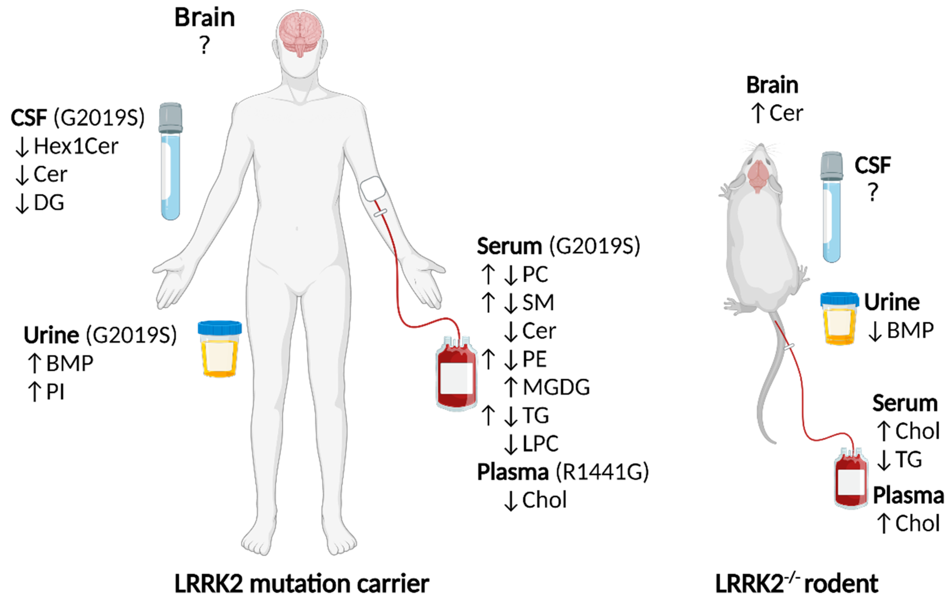
Genetic alterations in the LRRK2 gene, encoding leucine-rich repeat kinase 2, are a common risk factor for Parkinson’s disease. How LRRK2 alterations lead to cell pathology is an area of ongoing investigation, multiple lines of evidence suggest a role for LRRK2 in lipid pathways. It is increasingly recognized that in addition to being energy reservoirs and structural entities, some lipids, including neural lipids, participate in signaling cascades. Early investigations revealed that LRRK2 localized to membranous and vesicular structures, suggesting an interaction of LRRK2 and lipids or lipid-associated proteins. LRRK2 substrates from the Rab GTPase family play a critical role in vesicle trafficking, lipid metabolism and lipid storage, all processes which rely on lipid dynamics. In addition, LRRK2 is associated with the phosphorylation and activity of enzymes that catabolize plasma membrane and lysosomal lipids. Furthermore, LRRK2 knockout studies have revealed that blood, brain and urine exhibit lipid level changes, including alterations to sterols, sphingolipids and phospholipids, respectively. In human LRRK2 mutation carriers, changes to sterols, sphingolipids, phospholipids, fatty acyls and glycerolipids are reported in multiple tissues.

LRRK2 lipid Parkinson’s disease glucocerebrosidase

1. Introduction

1.1. Overview

Missense mutations in leucine-rich repeat kinase 2 (LRRK2) are the most common risk factor for autosomal dominantly inherited Parkinson’s disease (PD) [1][2]. Polymorphisms in LRRK2 are also associated with an increased risk of the more common sporadic form of PD [3]. Although the biological role of LRRK2 remains to be fully defined, studies have indicated that LRRK2 can be localized to membranous and vesicular structures, including lysosomes, endosomes, synaptic vesicles and mitochondria, suggesting that LRRK2 may have an affinity for lipids or lipid-associated proteins [4][5][6][7]. Indeed, increasing evidence from LRRK2 knockout rodents and LRRK2 mutation carriers further support a role for LRRK2 in processes relying on lipid membrane dynamics such as the endosome-lysosome system, the synaptic vesicle cycle, intracellular trafficking and lipid metabolism.

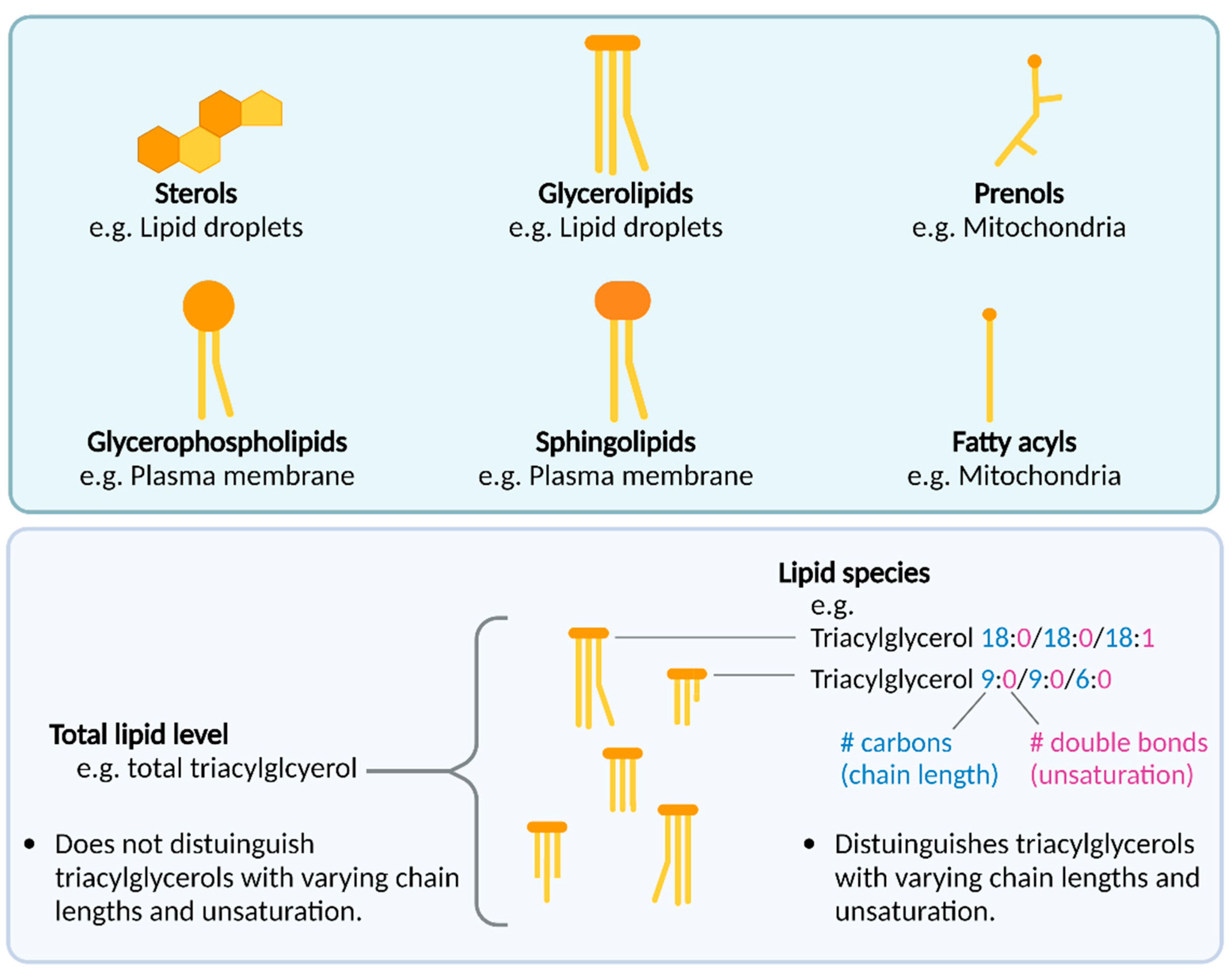
1.2. An Introduction to Lipids

Lipids are commonly known for their roles as storage silos for energy, and for being important membrane components surrounding cells and organelles. However, it is increasingly recognized that some lipids, including neural lipids, are also bioactive signaling molecules that can directly facilitate signal transduction by means such as binding to receptors to initiate signaling cascades (reviewed in [8][9]). In contrast to proteins, lipids are not encoded by genes. Instead, lipids can be endogenously biosynthesized by enzymes using lipid precursors downstream of acetyl coenzyme A, a metabolite from glucose, fatty acid and amino acid catabolism [10]. Alternatively, some lipids are obtained from the diet [11]. The thousands of endogenous lipids that exist in humans have been organized into six categories based on their structure: glycerophospholipids, glycerolipids, sphingolipids, sterol lipids, prenol lipids and fatty acyls (Figure 1). Each category has a hierarchy of classes and sub-classes, which can be viewed online on the LIPID MAPS database (https://www.lipidmaps.org/data/classification/LM_classification_exp.php) [12]. With sensitive enough analytical methods such as mass spectrometry, information on the carbon chain length and double bond number (unsaturation) of lipid molecules are provided in following lipid nomenclature (extensively defined in [12]). For example, triacylglycerol 18:0/18:0/18:1 refers to a triacylglycerol with three carbon chains (separated by /), the first and second chain containing 18 carbons and 0 double bonds, and the third chain containing 18 carbons and 1 double bond (Figure 1). Lipid studies with and without such carbon chain and saturation resolution, which may have functional significance.
Figure 1. Top panel: Lipid categories and examples of their cellular location. Bottom panel: Enzymatic and chemical assays can measure total lipid levels, while mass spectrometry techniques can discern the levels of specific lipid species, which have varying degrees of unsaturation and carbon chain lengths. Created with BioRender.com (accessed on 26 October 2022).

1.3. Lipid Alterations in LRRK2 Knockouts

Studies from rodents suggest that a loss of LRRK2 can result in both central and systemic changes in lipids (Figure 2) [13][14][15][16][17]. Sphingolipid changes found in the brains of sporadic PD patients [18] have spurred the interest in investigating potential lipid changes associated with PD risk genes such as LRRK2. In a small study examining brain sphingolipids via liquid chromatography-mass spectrometry (LC-MS), brains from LRRK2-/- mice were found to have significantly increased levels of ceramide compared to WT (n = 3 in each genotype) [13]. The ceramide level comprised a summation of 13 long and very long chain ceramides, which were the only chain lengths detected. Levels of sphingomyelin, hexosylceramide (glucosylceramide/galactosylceramide) and sulfatide were unchanged, however, the researchers noted that sphingomyelin and sulfatide from LRRK2-/- brains trended to increase. Another targeted study specifically investigated a biomarker of lysosomal dysregulation, di-docosahexaenoyl (22:6) bis(monoacylglycerol) phosphate (di-22:6-BMP). Significantly decreased urine di-22:6-BMP was found in LRRK2-/- mice compared to WT, as measured by LC-MS/MS [14]. LRRK2-/- rats have also been found to have higher serum [15] and plasma [16] total cholesterol (cholesterol from low and high-density lipoprotein) compared to WT (Figure 2). In addition, LRRK2-/- rats have lower total serum triacylglycerols compared to WT (ceramide and BMP were not investigated) [16]. The difference between total levels of lipid and specific lipid species is depicted in Figure 1. In summary, LRRK2-/- rodents show changes in lipid levels from the sphingolipid, phospholipid, sterol and glycerolipid categories.

1.4. Lipid Alterations in LRRK2 Mutation Carrier Humans

In contrast to LRRK2 knockouts, the main pathogenic PD-associated LRRK2 mutations found in humans increase the kinase activity of LRRK2 [19][20], whereas loss-of-function LRRK2 variants are not strongly associated with disease states in humans [21]. A metabolomics study found that LRRK2 R1441G PD patients had decreased plasma total cholesterol, whereas LRRK2 G2019S PD patients showed no change in cholesterol compared to controls, as assessed by MS (n = 8 participants per group) [22]. Whether this is linked to the R1441G mutation increasing the kinase activity of LRRK2 to a greater extent than the G2019S mutation remains to be elucidated [23]. Building on the findings that urine levels of di-22:6 BMP were increased in LRRK2-/- mice, another two studies investigating phospholipids in urine via LC-MS/MS indicated that LRRK2 G2019S carriers showed significantly increased di-22:6 BMP and di-18:1 BMP compared to non-carriers [24] (Figure 2). Phosphatidylinositol (PI) 16:0/20:4 and PI 18:0/20:4 were also significantly increased in urine from the same cohort, while globotriaosylceramides were unchanged (N = 80). Significantly increased levels of di-18:1 BMP and di-22:6 BMP in urine were then validated in a second cohort (N = 116). One study also found these BMP isoforms were increased in LRRK2 R1441G/C carriers [25]. To better understand the extent to which lipid dysregulation occurs in LRRK2 mutation carriers, a recent, untargeted lipidomics study in LRRK2 G2019S carriers found significantly decreased levels of CSF hexosylceramide m38:0 (glucosylceramide/galactosylceramide), ceramide d32:1 and diacylglycerol 22:1e compared to non-carriers, as assessed by LC-MS/MS in N = 88 participants [26]. Whether such changes are reflected in the brains of LRRK2 mutation carriers remains to be elucidated. The same study reported serum lipid species from phosphatidylcholine, ceramide, sphingomyelin, phosphatidylethanolamine, monogalactosyldiacylglycerol, triacylglycerol and lysophosphatidylcholine sub-classes could significantly distinguish LRRK2 G2019S carriers from non-carriers in two separate cohorts of N = 221 and N = 315 participants [26] (Figure 2). Critically, this study demonstrated that in contrast to the total level of each lipid class changing in serum from LRRK2 G2019S carriers, different lipid species within each of these lipid classes were significantly altered in opposing directions of change [26]. One explanation is that lipid species within a class do not all behave the same and may even have inverse regulatory functions. For example, studies have found that very long chain ceramide exhibits an opposing effect on long chain ceramide in apoptosis [27][28][29]. However, the functional consequences of varying lipid length, saturation and linkage type are an area of development. Indeed, functional databases are mostly protein-centric, and the functional databases that include lipids (such as KEGG [30]) largely do not account for potential functional variation in lipids of varying unsaturation, chain length or linkage type.
Figure 2. Lipid level changes in tissues from LRRK2 mutation carrier humans and LRRK2-/- rodents. Human LRRK2 G2019S mutation carriers have alterations in serum phospholipid, sphingolipid and glycerolipid species, and CSF sphingolipid and glycerolipid species compared to those without a LRRK2 mutation [26]. Human LRRK2 G2019S mutation carriers have increased BMP and PI species in urine compared to those without the mutation [24]. Human LRRK2 R1441G mutation carriers have decreased plasma cholesterol [22]. LRRK2-/- rats have higher serum [15] and plasma [16] cholesterol, and lower serum triacylglycerol [15] compared to WT. LRRK2-/- mice have increased brain ceramide [13] and decreased urine BMP [14] compared to WT. For simplicity, specific lipid species are not depicted. Hex1Cer = hexosylceramide, Cer = ceramide, DG = diacylglycerol, BMP = bis (monoacylglycerol) phosphate, PI = phosphatidylinositol, PC = phosphatidylcholine, SM = sphingomyelin, PE = phosphatidylethanolamine, MGDG = monogalactosyldiacylglycerol, TG = triacylglycerol, LPC = lysophosphatidylcholine, Chol = cholesterol. Created with BioRender.com (accessed on 26 October 2022).
Triacylglycerol and cholesterol levels in the blood have been widely studied due to their relevance to cardiovascular disease [31]. In blood from pre-clinical or manifesting PD patients, the levels of “total” lipid such as triacylglycerol and cholesterol are controversial [32][33]. For example, one study on genotyped subjects reported that LRRK2-associated PD patients exhibited higher total serum triacylglycerol levels than other PD genotypes [34], while another study found no such difference [35]. Although the reason for this inconsistency is unclear, variability may arise due to unaccounted genotypic backgrounds that may influence lipid levels, for example, variations in apolipoprotein encoding genes such as APOC2, APOA5 and APOB can influence triacylglycerol levels [36]. In addition, possible lipid level variability may arise from the capacity of analytical methods to detect different lipid species within the crude/total lipid level. Specifically, enzymatic and chemical assays can measure total lipid levels [37], whereas mass spectrometry techniques can provide more detailed information on specific lipid species of varying chain lengths and degrees of unsaturation, which may be important (Figure 1). In particular, information on lipid species can capture detail regarding levels of different species within a class changing in opposing directions. Future high-resolution studies in genetically well-characterized cohorts will aid in better understanding the nature of lipid alterations in PD. The majority of lipidomics studies have been conducted at the tissue or cellular level at one time point, however, sub-cellular studies would help clarify which organelles may exhibit lipid changes that reflect the tissue or whole cell changes found in LRRK2 studies. As evidence indicates that some intracellular lipids oscillate with the time of day [38], the sample collection time may be another covariate to consider in future study.
To what extent peripheral changes in lipids are reflected in the brain depends on the lipid in question, as rates of peripheral exchange may differ across lipid types and in addition, lipids may be modified to cross the blood–brain barrier. For example, cholesterol is converted to hydroxycholesterol to pass across the blood–brain barrier [39]. Further, other brain lipids may be peroxidized to form metabolites such as aldehydes, which can be measured in the blood [40]. Future investigations measuring lipids and their associated derivatives, using matching brain and peripheral samples, could help clarify the concordance between brain and peripheral lipids.

2. LRRK2 and Lysosomal Lipid Metabolism

LRRK2 can be localized to lysosomes and LRRK2 mutations and inhibition of LRRK2 alter lysosomal function [41]. In addition, LRRK2 mutations result in enlarged lysosome size and a reduced number of lysosomes [42]. Studies on LRRK2-/- rodent kidneys, revealed abnormal accumulation of lipofuscin [17][43][44], which is an aggregation of mainly protein and lipid, and to a lesser extent carbohydrate and metal [45]. Lipofuscin forms within lysosomes from undigested lysosomal material, and an accumulation of lipofuscin in LRRK2-/- rodents may therefore indicate lysosomal dysfunction [17][43][44][45]. Genetic alterations to LRRK2 also affect the lysosomal lipid BMP (also known as lysobisphosphatidic acid) [14][24]. BMP is localized in late endosomes/lysosomes and plays a role in the formation, structure and trafficking of endolysosomal compartments [46][47][48]. Interestingly, manipulation of BMP in cell lines and human fibroblasts indicates that BMP controls endolysosomal cholesterol levels [49][50] by enhancing the secretion of cholesterol-containing exosomes [50]. Another phospholipid which may be associated with genetic alterations to LRRK2 is phosphatidylinositol [24]. Phosphatidylinositol is a precursor to phosphoinositides, which are implicated in several lysosomal functions, including lysosomal cholesterol transport [51].
Intriguingly, recent studies [13][52][53][54][55] have pointed to a role for LRRK2 in regulating the lysosomal enzyme β-glucocerebrosidase (GCase), encoded by another common PD risk gene, GBA1 [56][57][58][59]. GCase catabolizes the sphingolipids glucosylceramide and glucosylsphingosine into glucose and ceramide and glucose and sphingosine, respectively [60]. Mutations to GCase commonly associated with PD are loss-of-function, diminishing the enzyme’s hydrolytic activity [53][61]. GCase protein level is significantly decreased in LRRK2-/- mouse brain [13] and human LRRK2 G2019S and I2020T frontal cortex [62]. Perhaps as compensation for reduced GCase protein levels, LRRK2-/- mice brains, including striata, display significantly increased GCase activity compared to WT [13][52]. GCase activity from LRRK2 G2019S knock-in mice midbrain is also significantly higher compared to WT [55]. In human LRRK2 G2019S mutation carrier PBMCs [55] and fibroblasts [55], significantly increased GCase activity has been found compared to non-carriers, whereas a significant increase [53] and no change [63] in GCase activity have been reported in dried blood spots. A further study has indicated that GCase activity from dried blood spot samples decreases over time, which may explain reported inconsistencies [64]. In iPSC-derived dopaminergic neurons from LRRK2 mutation carriers (n = 2 G2019S and R1441C, n = 1 R1441G), significantly decreased GCase activity compared to controls (n = 2) has been reported, which could be restored upon treatment with a LRRK2 inhibitor [54]. However, another study using a different enzyme activity assay found significantly increased activity in dopaminergic neurons from LRRK2 G2019S mutation carriers (n = 2) compared to controls (n = 4) [55]. It has been suggested that the variability in the GCase results reported may be due to different GCase substrates utilized to measure enzyme activity, as commercial GCase substrates differ in their specificity for lysosomal and cytoplasmic GCase and sensitivity to other endogenous factors [55][65]. In addition, GCase activity may be affected in a tissue-specific manner [55]. Notably, inconsistencies may also arise from the low sample sizes available for iPSC-derived neuron investigations. Further large-scale studies separating lysosomal and total GCase readouts in situ will help clarify the nature of potential GCase activity alterations in LRRK2 mutation carriers. In addition, a mechanism behind a potential LRRK2-mediated effect on GCase warrants further exploration.
A key implication of LRRK2 affecting GCase activity is that LRRK2 could ultimately affect GCase substrates, specifically glucosylceramide and glucosylsphingosine breakdown. An alteration of GCase activity by LRRK2 could therefore result in altered metabolism or levels of ceramide, glucose, sphingosine, glucosylceramide and glucosylsphingosine in the lysosome. This altered lysosomal lipid metabolism may disrupt the lysosomal breakdown of proteins and other lipids, contributing to pathology.
In addition to being a metabolite of glucosylceramide breakdown, ceramide is also a metabolic product of sphingomyelin breakdown by acid sphingomyelinase in the lysosome [66]. Ceramide species are decreased in LRRK2 G0219S carrier CSF and serum and increased in LRRK2-/- brains, while serum from LRRK2 G0219S carriers exhibits both increases and decreases in different sphingomyelin species (Figure 2). Whether potential alterations to serum, CSF and brain sphingolipids reflect alterations to lysosomal sphingolipid metabolism needs further investigation, as sphingolipids are found throughout the cell. Notably, however, mutations to GBA1, GALC, SMPD1 and ASAH1 have been linked to PD and all encode enzymes that catabolize sphingolipids in the lysosome [67][68][69][70]. Whether LRRK2 may directly or indirectly affect these enzymes and their ability to metabolize sphingolipids is an area that warrants further investigation.
In summary, genetic alterations in LRRK2 in humans and rodent models show consequences for lysosomal GCase [13][52][53][54][55] and endolysosomal BMP [14][24]. Lysosomal GCase catabolizes glucosphingolipids [61], while lysosomal BMP appears to control cholesterol levels [49][50]. In addition to BMP, the levels of cholesterol and sphingolipids are altered in LRRK2 mutation carrier humans and LRRK2-/- rodents (Figure 2). Collectively, these data strongly support a role for LRRK2 in modulating lysosomal function, particularly the lysosomal metabolism of glucosphingolipids and sterols.

References

  1. Monfrini, E.; Di Fonzo, A. Leucine-Rich Repeat Kinase (LRRK2) Genetics and Parkinson’s Disease. Adv. Neurobiol. 2017, 14, 3–30.
  2. Healy, D.G.; Falchi, M.; O’Sullivan, S.S.; Bonifati, V.; Durr, A.; Bressman, S.; Brice, A.; Aasly, J.; Zabetian, C.P.; Goldwurm, S.; et al. Phenotype, genotype, and worldwide genetic penetrance of LRRK2-associated Parkinson’s disease: A case-control study. Lancet Neurol. 2008, 7, 583–590.
  3. Gilks, W.P.; Abou-Sleiman, P.M.; Gandhi, S.; Jain, S.; Singleton, A.; Lees, A.J.; Shaw, K.; Bhatia, K.P.; Bonifati, V.; Quinn, N.P.; et al. A common LRRK2 mutation in idiopathic Parkinson’s disease. Lancet 2005, 365, 415–416.
  4. Biskup, S.; Moore, D.J.; Celsi, F.; Higashi, S.; West, A.B.; Andrabi, S.A.; Kurkinen, K.; Yu, S.-W.; Savitt, J.M.; Waldvogel, H.J.; et al. Localization of LRRK2 to membranous and vesicular structures in mammalian brain. Ann. Neurol. 2006, 60, 557–569.
  5. Shin, N.; Jeong, H.; Kwon, J.; Heo, H.Y.; Kwon, J.J.; Yun, H.J.; Kim, C.H.; Han, B.S.; Tong, Y.; Shen, J.; et al. LRRK2 regulates synaptic vesicle endocytosis. Exp. Cell Res. 2008, 314, 2055–2065.
  6. Piccoli, G.; Condliffe, S.B.; Bauer, M.; Giesert, F.; Boldt, K.; De Astis, S.; Meixner, A.; Sarioglu, H.; Vogt-Weisenhorn, D.M.; Wurst, W.; et al. LRRK2 Controls Synaptic Vesicle Storage and Mobilization within the Recycling Pool. J. Neurosci. 2011, 31, 2225–2237.
  7. Alegre-Abarrategui, J.; Christian, H.; Lufino, M.M.; Mutihac, R.; Venda, L.L.; Ansorge, O.; Wade-Martins, R. LRRK2 regulates autophagic activity and localizes to specific membrane microdomains in a novel human genomic reporter cellular model. Hum. Mol. Genet. 2009, 18, 4022–4034.
  8. Piomelli, D.; Astarita, G.; Rapaka, R. A neuroscientist’s guide to lipidomics. Nat. Rev. Neurosci. 2007, 8, 743–754.
  9. Yoon, J.H.; Seo, Y.; Jo, Y.S.; Lee, S.; Cho, E.; Cazenave-Gassiot, A.; Shin, Y.-S.; Moon, M.H.; An, H.J.; Wenk, M.R.; et al. Brain lipidomics: From functional landscape to clinical significance. Sci. Adv. 2022, 8, eadc9317.
  10. Shi, L.; Tu, B.P. Acetyl-CoA and the regulation of metabolism: Mechanisms and consequences. Curr. Opin. Cell. Biol. 2015, 33, 125–131.
  11. Holthuis, J.C.M.; Menon, A.K. Lipid landscapes and pipelines in membrane homeostasis. Nature 2014, 510, 48–57.
  12. Fahy, E.; Subramaniam, S.; Murphy, R.C.; Nishijima, M.; Raetz, C.R.; Shimizu, T.; Spener, F.; van Meer, G.; Wakelam, M.J.; Dennis, E.A. Update of the LIPID MAPS comprehensive classification system for lipids. J. Lipid Res. 2009, 50, S9–S14.
  13. Ferrazza, R.; Cogo, S.; Melrose, H.; Bubacco, L.; Greggio, E.; Guella, G.; Civiero, L.; Plotegher, N. LRRK2 deficiency impacts ceramide metabolism in brain. Biochem. Biophys. Res. Commun. 2016, 478, 1141–1146.
  14. Fuji, R.N.; Flagella, M.; Baca, M.; Baptista, M.A.S.; Brodbeck, J.; Chan, B.K.; Fiske, B.K.; Honigberg, L.; Jubb, A.M.; Katavolos, P.; et al. Effect of selective LRRK2 kinase inhibition on nonhuman primate lung. Sci. Transl. Med. 2015, 7, 273ra215.
  15. Baptista, M.A.S.; Dave, K.D.; Frasier, M.A.; Sherer, T.B.; Greeley, M.; Beck, M.J.; Varsho, J.S.; Parker, G.A.; Moore, C.; Churchill, M.J.; et al. Loss of Leucine-Rich Repeat Kinase 2 (LRRK2) in Rats Leads to Progressive Abnormal Phenotypes in Peripheral Organs. PLoS ONE 2013, 8, e80705.
  16. Ness, D.; Ren, Z.; Gardai, S.; Sharpnack, D.; Johnson, V.J.; Brennan, R.J.; Brigham, E.F.; Olaharski, A.J. Leucine-Rich Repeat Kinase 2 (LRRK2)-Deficient Rats Exhibit Renal Tubule Injury and Perturbations in Metabolic and Immunological Homeostasis. PLoS ONE 2013, 8, e66164.
  17. Boddu, R.; Hull, T.D.; Bolisetty, S.; Hu, X.; Moehle, M.S.; Daher, J.P.L.; Kamal, A.I.; Joseph, R.; George, J.F.; Agarwal, A.; et al. Leucine-rich repeat kinase 2 deficiency is protective in rhabdomyolysis-induced kidney injury. Hum. Mol. Genet. 2015, 24, 4078–4093.
  18. Abbott, S.K.; Li, H.; Muñoz, S.S.; Knoch, B.; Batterham, M.; Murphy, K.E.; Halliday, G.M.; Garner, B. Altered ceramide acyl chain length and ceramide synthase gene expression in Parkinson’s disease. Mov. Disord. 2014, 29, 518–526.
  19. Bardien, S.; Lesage, S.; Brice, A.; Carr, J. Genetic characteristics of leucine-rich repeat kinase 2 (LRRK2) associated Parkinson’s disease. Park. Relat. Disord. 2011, 17, 501–508.
  20. Yu, M.; Arshad, M.; Wang, W.; Zhao, D.; Xu, L.; Zhou, L. LRRK2 mediated Rab8a phosphorylation promotes lipid storage. Lipids Health Dis. 2018, 17, 34.
  21. Whiffin, N.; Armean, I.M.; Kleinman, A.; Marshall, J.L.; Minikel, E.V.; Goodrich, J.K.; Quaife, N.M.; Cole, J.B.; Wang, Q.; Karczewski, K.J.; et al. The effect of LRRK2 loss-of-function variants in humans. Nat. Med. 2020, 26, 869–877.
  22. Yakhine-Diop, S.M.S.; Morales-García, J.A.; Niso-Santano, M.; González-Polo, R.A.; Uribe-Carretero, E.; Martinez-Chacon, G.; Durand, S.; Maiuri, M.C.; Aiastui, A.; Zulaica, M.; et al. Metabolic alterations in plasma from patients with familial and idiopathic Parkinson’s disease. Aging (Albany NY) 2020, 12, 16690–16708.
  23. Taylor, M.; Alessi, D.R. Advances in elucidating the function of leucine-rich repeat protein kinase-2 in normal cells and Parkinson’s disease. Curr. Opin. Cell. Biol. 2020, 63, 102–113.
  24. Alcalay, R.N.; Hsieh, F.; Tengstrand, E.; Padmanabhan, S.; Baptista, M.; Kehoe, C.; Narayan, S.; Boehme, A.K.; Merchant, K. Higher Urine bis (Monoacylglycerol) Phosphate Levels in LRRK2 G2019S Mutation Carriers: Implications for Therapeutic Development. Mov. Disord. 2020, 35, 134–141.
  25. Gomes, S.; Garrido, A.; Tonelli, F.; Obiang, D.; Tolosa, E.; Marti, M.J.; Ruiz-Martinez, J.; Aragon, A.V.; Hernandez-Eguiazu, H.; Croitoru, I.; et al. Elevated urine BMPs—bis (Monoacylglycerol) Phosphates—in LRRK2 G2019S, R1441G/C and VPS35 D620N mutation carriers with and without Parkinson’s disease. medRxiv 2022.
  26. Galper, J.; Dean, N.J.; Pickford, R.; Lewis, S.J.G.; Halliday, G.M.; Kim, W.S.; Dzamko, N. Lipid pathway dysfunction is prevalent in patients with Parkinson’s disease. Brain J. Neurol. 2022.
  27. Stiban, J.; Perera, M. Very long chain ceramides interfere with C16-ceramide-induced channel formation: A plausible mechanism for regulating the initiation of intrinsic apoptosis. Biochim. Biophys. Acta (BBA) Biomembr. 2015, 1848, 561–567.
  28. Hartmann, D.; Wegner, M.-S.; Wanger, R.A.; Ferreirós, N.; Schreiber, Y.; Lucks, J.; Schiffmann, S.; Geisslinger, G.; Grösch, S. The equilibrium between long and very long chain ceramides is important for the fate of the cell and can be influenced by co-expression of CerS. Int. J. Biochem. Cell Biol. 2013, 45, 1195–1203.
  29. Abou-Ghali, M.; Stiban, J. Regulation of ceramide channel formation and disassembly: Insights on the initiation of apoptosis. Saudi J. Biol. Sci. 2015, 22, 760–772.
  30. Kanehisa, M.; Furumichi, M.; Sato, Y.; Ishiguro-Watanabe, M.; Tanabe, M. KEGG: Integrating viruses and cellular organisms. Nucleic Acids Res. 2020.
  31. McGranaghan, P.; Kirwan, J.A.; Garcia-Rivera, M.A.; Pieske, B.; Edelmann, F.; Blaschke, F.; Appunni, S.; Saxena, A.; Rubens, M.; Veledar, E.; et al. Lipid Metabolite Biomarkers in Cardiovascular Disease: Discovery and Biomechanism Translation from Human Studies. Metabolites 2021, 11, 621.
  32. Fu, X.; Wang, Y.; He, X.; Li, H.; Liu, H.; Zhang, X. A systematic review and meta-analysis of serum cholesterol and triglyceride levels in patients with Parkinson’s disease. Lipids Health Dis. 2020, 19, 97.
  33. Jiang, Z.; Xu, X.; Gu, X.; Ou, R.; Luo, X.; Shang, H.; Song, W. Effects of Higher Serum Lipid Levels on the Risk of Parkinson’s Disease: A Systematic Review and Meta-Analysis. Front. Neurol. 2020, 11, 597.
  34. Thaler, A.; Shenhar-Tsarfaty, S.; Shaked, Y.; Gurevich, T.; Omer, N.; Bar-Shira, A.; Gana-Weisz, M.; Goldstein, O.; Kestenbaum, M.; Cedarbaum, J.M.; et al. Metabolic syndrome does not influence the phenotype of LRRK2 and GBA related Parkinson’s disease. Sci. Rep. 2020, 10, 9329.
  35. Macías-García, D.; Periñán, M.T.; Muñoz-Delgado, L.; Jimenez-Jaraba, M.V.; Labrador-Espinosa, M.Á.; Jesús, S.; Buiza-Rueda, D.; Méndez-Del Barrio, C.; Adarmes-Gómez, A.; Gómez-Garre, P.; et al. Serum lipid profile among sporadic and familial forms of Parkinson’s disease. Npj Park. Dis. 2021, 7, 59.
  36. Johansen, C.T.; Hegele, R.A. Genetic bases of hypertriglyceridemic phenotypes. Curr. Opin. Lipidol. 2011, 22, 247–253.
  37. Li, L.-H.; Dutkiewicz, E.P.; Huang, Y.-C.; Zhou, H.-B.; Hsu, C.-C. Analytical methods for cholesterol quantification. J. Food Drug Anal. 2019, 27, 375–386.
  38. Aviram, R.; Manella, G.; Kopelman, N.; Neufeld-Cohen, A.; Zwighaft, Z.; Elimelech, M.; Adamovich, Y.; Golik, M.; Wang, C.; Han, X.; et al. Lipidomics Analyses Reveal Temporal and Spatial Lipid Organization and Uncover Daily Oscillations in Intracellular Organelles. Mol. Cell 2016, 62, 636–648.
  39. Vance, J.E. Dysregulation of cholesterol balance in the brain: Contribution to neurodegenerative diseases. Dis. Model. Mech. 2012, 5, 746–755.
  40. Phan, K.; He, Y.; Pickford, R.; Bhatia, S.; Katzeff, J.S.; Hodges, J.R.; Piguet, O.; Halliday, G.M.; Kim, W.S. Uncovering pathophysiological changes in frontotemporal dementia using serum lipids. Sci. Rep. 2020, 10, 3640.
  41. Roosen, D.A.; Cookson, M.R. LRRK2 at the interface of autophagosomes, endosomes and lysosomes. Mol. Neurodegener. 2016, 11, 73.
  42. Henry, A.G.; Aghamohammadzadeh, S.; Samaroo, H.; Chen, Y.; Mou, K.; Needle, E.; Hirst, W.D. Pathogenic LRRK2 mutations, through increased kinase activity, produce enlarged lysosomes with reduced degradative capacity and increase ATP13A2 expression. Hum. Mol. Genet. 2015, 24, 6013–6028.
  43. Tong, Y.; Yamaguchi, H.; Giaime, E.; Boyle, S.; Kopan, R.; Kelleher, R.J.; Shen, J. Loss of leucine-rich repeat kinase 2 causes impairment of protein degradation pathways, accumulation of α-synuclein, and apoptotic cell death in aged mice. Proc. Natl. Acad. Sci. 2010, 107, 9879–9884.
  44. Tong, Y.; Giaime, E.; Yamaguchi, H.; Ichimura, T.; Liu, Y.; Si, H.; Cai, H.; Bonventre, J.V.; Shen, J. Loss of leucine-rich repeat kinase 2 causes age-dependent bi-phasic alterations of the autophagy pathway. Mol. Neurodegener. 2012, 7, 2.
  45. Keller, J.N.; Dimayuga, E.; Chen, Q.; Thorpe, J.; Gee, J.; Ding, Q. Autophagy, proteasomes, lipofuscin, and oxidative stress in the aging brain. Int. J. Biochem. Cell Biol. 2004, 36, 2376–2391.
  46. Kobayashi, T.; Beuchat, M.-H.; Chevallier, J.; Makino, A.; Mayran, N.; Escola, J.-M.; Lebrand, C.; Cosson, P.; Kobayashi, T.; Gruenberg, J. Separation and Characterization of Late Endosomal Membrane Domains*. J. Biol. Chem. 2002, 277, 32157–32164.
  47. Kobayashi, T.; Stang, E.; Fang, K.S.; de Moerloose, P.; Parton, R.G.; Gruenberg, J. A lipid associated with the antiphospholipid syndrome regulates endosome structure and function. Nature 1998, 392, 193–197.
  48. Kobayashi, T.; Beuchat, M.-H.; Lindsay, M.; Frias, S.; Palmiter, R.D.; Sakuraba, H.; Parton, R.G.; Gruenberg, J. Late endosomal membranes rich in lysobisphosphatidic acid regulate cholesterol transport. Nat. Cell Biol. 1999, 1, 113–118.
  49. Chevallier, J.; Chamoun, Z.; Jiang, G.; Prestwich, G.; Sakai, N.; Matile, S.; Parton, R.G.; Gruenberg, J. Lysobisphosphatidic Acid Controls Endosomal Cholesterol Levels*. J. Biol. Chem. 2008, 283, 27871–27880.
  50. Ilnytska, O.; Jeziorek, M.; Lai, K.; Altan-Bonnet, N.; Dobrowolski, R.; Storch, J. Lysobisphosphatidic acid (LBPA) enrichment promotes cholesterol egress via exosomes in Niemann Pick type C1 deficient cells. Biochim. Biophys. Acta (BBA) Mol. Cell Biol. Lipids 2021, 1866, 158916.
  51. Ebner, M.; Koch, P.A.; Haucke, V. Phosphoinositides in the control of lysosome function and homeostasis. Biochem. Soc. Trans. 2019, 47, 1173–1185.
  52. Albanese, F.; Mercatelli, D.; Finetti, L.; Lamonaca, G.; Pizzi, S.; Shimshek, D.R.; Bernacchia, G.; Morari, M. Constitutive silencing of LRRK2 kinase activity leads to early glucocerebrosidase deregulation and late impairment of autophagy in vivo. Neurobiol. Dis. 2021, 159, 105487.
  53. Alcalay, R.N.; Levy, O.A.; Waters, C.H.; Fahn, S.; Ford, B.; Kuo, S.-H.; Mazzoni, P.; Pauciulo, M.W.; Nichols, W.C.; Gan-Or, Z.; et al. Glucocerebrosidase activity in Parkinson’s disease with and without GBA mutations. Brain J. Neurol. 2015, 138, 2648–2658.
  54. Ysselstein, D.; Nguyen, M.; Young, T.J.; Severino, A.; Schwake, M.; Merchant, K.; Krainc, D. LRRK2 kinase activity regulates lysosomal glucocerebrosidase in neurons derived from Parkinson’s disease patients. Nat. Commun. 2019, 10, 5570.
  55. Kedariti, M.; Frattini, E.; Baden, P.; Cogo, S.; Civiero, L.; Ziviani, E.; Zilio, G.; Bertoli, F.; Aureli, M.; Kaganovich, A.; et al. LRRK2 kinase activity regulates GCase level and enzymatic activity differently depending on cell type in Parkinson’s disease. Npj Park. Dis. 2022, 8, 92.
  56. Sidransky, E.; Nalls, M.A.; Aasly, J.O.; Aharon-Peretz, J.; Annesi, G.; Barbosa, E.R.; Bar-Shira, A.; Berg, D.; Bras, J.; Brice, A.; et al. Multicenter Analysis of Glucocerebrosidase Mutations in Parkinson’s Disease. New Engl. J. Med. 2009, 361, 1651–1661.
  57. Wang, Y.; Liu, L.; Xiong, J.; Zhang, X.; Chen, Z.; Yu, L.; Chen, C.; Huang, J.; Zhang, Z.; Mohmed, A.A.; et al. Glucocerebrosidase L444P mutation confers genetic risk for Parkinson’s disease in central China. Behav. Brain Funct. 2012, 8, 57.
  58. Guimarães Bde, C.; Pereira, A.C.; Rodrigues Fda, C.; dos Santos, A.V.; Campos, M., Jr.; dos Santos, J.M.; dos Santos, F.L.; de Rosso, A.L.; Nicaretta, D.H.; Pereira, J.S.; et al. Glucocerebrosidase N370S and L444P mutations as risk factors for Parkinson’s disease in Brazilian patients. Park. Relat. Disord 2012, 18, 688–689.
  59. Aharon-Peretz, J.; Rosenbaum, H.; Gershoni-Baruch, R. Mutations in the Glucocerebrosidase Gene and Parkinson’s Disease in Ashkenazi Jews. N. Engl. J. Med. 2004, 351, 1972–1977.
  60. Vaccaro, A.M.; Muscillo, M.; Suzuki, K. Characterization of human glucosylsphingosine glucosyl hydrolase and comparison with glucosylceramidase. Eur. J. Biochem. 1985, 146, 315–321.
  61. Smith, L.; Schapira, A.H.V. GBA Variants and Parkinson Disease: Mechanisms and Treatments. Cells 2022, 11, 1261.
  62. Zhao, Y.; Perera, G.; Takahashi-Fujigasaki, J.; Mash, D.C.; Vonsattel, J.P.G.; Uchino, A.; Hasegawa, K.; Jeremy Nichols, R.; Holton, J.L.; Murayama, S.; et al. Reduced LRRK2 in association with retromer dysfunction in post-mortem brain tissue from LRRK2 mutation carriers. Brain J. Neurol. 2017, 141, 486–495.
  63. Omer, N.; Giladi, N.; Gurevich, T.; Bar-Shira, A.; Gana-Weisz, M.; Glinka, T.; Goldstein, O.; Kestenbaum, M.; Cedarbaum, J.M.; Mabrouk, O.S.; et al. Glucocerebrosidase Activity is not Associated with Parkinson’s Disease Risk or Severity. Mov. Disord. 2022, 37, 190–195.
  64. Ortega, R.A.; Bodamer, O.; Peake, R.W.A.; Raymond, D.; Bressman, S.B.; Saunders-Pullman, R. Assessment of Glucocerebrosidase Enzyme Activity in Parkinson Disease Using Multiple Approaches. Mov. Disord. 2022, 37, 655–656.
  65. Ysselstein, D.; Young, T.J.; Nguyen, M.; Padmanabhan, S.; Hirst, W.D.; Dzamko, N.; Krainc, D. Evaluation of Strategies for Measuring Lysosomal Glucocerebrosidase Activity. Mov. Disord. Off. J. Mov. Disord. Soc. 2021, 36, 2719–2730.
  66. Stoffel, W. Functional analysis of acid and neutral sphingomyelinases in vitro and in vivo. Chem. Phys. Lipids 1999, 102, 107–121.
  67. Chang, D.; Nalls, M.A.; Hallgrímsdóttir, I.B.; Hunkapiller, J.; van der Brug, M.; Cai, F.; Kerchner, G.A.; Ayalon, G.; Bingol, B.; Sheng, M.; et al. A meta-analysis of genome-wide association studies identifies 17 new Parkinson’s disease risk loci. Nat. Genet. 2017, 49, 1511–1516.
  68. Gan-Or, Z.; Ozelius, L.J.; Bar-Shira, A.; Saunders-Pullman, R.; Mirelman, A.; Kornreich, R.; Gana-Weisz, M.; Raymond, D.; Rozenkrantz, L.; Deik, A.; et al. The p.L302P mutation in the lysosomal enzyme gene SMPD1 is a risk factor for Parkinson disease. Neurology 2013, 80, 1606–1610.
  69. Parveen, F.; Bender, D.; Law, S.-H.; Mishra, V.K.; Chen, C.-C.; Ke, L.-Y. Role of Ceramidases in Sphingolipid Metabolism and Human Diseases. Cells 2019, 8, 1573.
  70. Robak, L.A.; Jansen, I.E.; van Rooij, J.; Uitterlinden, A.G.; Kraaij, R.; Jankovic, J.; Heutink, P.; Shulman, J.M.; Consortium, I.P.s.D.G. Excessive burden of lysosomal storage disorder gene variants in Parkinson’s disease. Brain J. Neurol. 2017, 140, 3191–3203.
More
Information
Subjects: Cell Biology
Contributors MDPI registered users' name will be linked to their SciProfiles pages. To register with us, please refer to https://encyclopedia.pub/register : , ,
View Times: 529
Revisions: 2 times (View History)
Update Date: 19 Jan 2023
1000/1000
Video Production Service