Submitted Successfully!
To reward your contribution, here is a gift for you: A free trial for our video production service.
Thank you for your contribution! You can also upload a video entry or images related to this topic.
Version Summary Created by Modification Content Size Created at Operation
1 -- 3431 2022-12-12 13:12:29 |
2 update references and layout -54 word(s) 3377 2022-12-13 02:18:16 | |
3 update layout -2 word(s) 3375 2022-12-13 02:19:24 |

Video Upload Options

We provide professional Video Production Services to translate complex research into visually appealing presentations. Would you like to try it?

Confirm

Are you sure to Delete?
Cite
If you have any further questions, please contact Encyclopedia Editorial Office.
Buglak, A.A.;  Kapitonova, M.A.;  Vechtomova, Y.L.;  Telegina, T.A. Biochemical and Physiological Application of Pterin Photochemistry. Encyclopedia. Available online: https://encyclopedia.pub/entry/38600 (accessed on 02 December 2024).
Buglak AA,  Kapitonova MA,  Vechtomova YL,  Telegina TA. Biochemical and Physiological Application of Pterin Photochemistry. Encyclopedia. Available at: https://encyclopedia.pub/entry/38600. Accessed December 02, 2024.
Buglak, Andrey A., Marina A. Kapitonova, Yulia L. Vechtomova, Taisiya A. Telegina. "Biochemical and Physiological Application of Pterin Photochemistry" Encyclopedia, https://encyclopedia.pub/entry/38600 (accessed December 02, 2024).
Buglak, A.A.,  Kapitonova, M.A.,  Vechtomova, Y.L., & Telegina, T.A. (2022, December 12). Biochemical and Physiological Application of Pterin Photochemistry. In Encyclopedia. https://encyclopedia.pub/entry/38600
Buglak, Andrey A., et al. "Biochemical and Physiological Application of Pterin Photochemistry." Encyclopedia. Web. 12 December, 2022.
Biochemical and Physiological Application of Pterin Photochemistry
Edit

Pterins are low-molecular weight heterocyclic compounds widely distributed in living organisms, primarily as reduced coenzymes. Structurally, pterins are a conjugated system of pyrazine and pyrimidine rings, the so-called pteridine, which is accompanied by a carbonyl group (C=O) at the C4 position and an amino group (NH2) at the C2 position. The pteridine structure is also characteristic of folates (folic acid and its derivatives) and flavins, or benzopteridines, which are derivatives of isoalloxazine. Folates are usually called “conjugated pterins” since they possess a para-aminobenzoilglutamine residue, whereas pterins are called “unconjugated pterins”. In addition to pterin, folates include a para-aminobenzoic acid (p-ABA) residue and one to five glutamic acid (Glu) residues.

pteridines photonics photosensitization

1. Evidence of Pteridine Participation in Photoreception

Flavins, or benzopteridines, are the nearest “relatives” of unconjugated pterins. Among pteridines, flavins are the most widespread photosensor molecules primarily because of the flavin adenine dinucleotide (FAD) and flavin mononucleotide (FMN) cofactors involved in light, oxygen, and voltage (LOV) blue light sensing using flavins (BLUF) domains containing photoreceptor proteins. Also, FAD is the main chromophore of the cryptochrome photolyase family (CPF) proteins.
The common structural features of pteridines and flavins determine the similarity of the electronic structure and chemical properties of their excited molecules. The photochemical properties of flavins are determined by the presence of an isoalloxazine (2,4-dioxo-benzo-[g]-pteridine) ring within a developed system of conjugated double bonds, which allows the formation of stable radicals. In flavins (FMN or FAD), the light absorption band corresponding to the lower singlet level (S1) of excitation is in the blue region of the spectrum and has a maximum at 450 nm. The other two bands have an absorption maximum at 260 and 365 nm. The absorption of a photon increases the energy of the flavin by 265.8 kJ mol−1, making it a highly electrophilic excited molecule (Fl*). When an electron passes from a donor molecule to an excited flavin, a free radical (FlH· or Fl·hich plays a key role in some flavin pho) is formed, which plays a key role in some flavin photocycles (for example, the proposed mechanism of the BLUF domain, see below). The addition of one more electron transforms it into dihydroflavin (FlH2 or FlH). In the dark, the photoreduced flavin undergoes oxidation, returning to its original state, which is a process that can proceed in a cyclic mode. The flavin radical can also be formed as a result of the oxidation of the photoexcited flavin dihydroform; this reaction is the basis for the functioning of DNA photolyases (see below) [1][2]. Additional stability of the flavin radical can be imparted by amino acids that surround the flavin in the reaction center of the protein [3][4].
The BLUF and LOV domains are minimal modules (about 100–110 a.a.) that are part of various regulatory proteins capable of perceiving and reacting to blue light [5][6]. The BLUF domain non-covalently binds the FMN or FAD chromophores. When light is absorbed, photoreceptor proteins with associated chromophores (flavins) undergo conformational changes that allow them to transmit signals to other proteins. The photocycles of BLUF photoreceptors are thought to involve PCET.
It is assumed that the photoexcited flavin takes an electron from the tyrosine molecule located in close proximity (at a distance of the hydrogen bond) in the reaction center, forming the flavin radical, FlH·. Herein, the proton (H+) is taken from the glutamine (Gln), which is also in close proximity to the flavin molecule. A redistribution of the H-bonding network rearranged in the reaction center occurs, which forces Gln to change its spatial orientation (Gln as an intermediate of a proton relay), which, in turn, leads to conformational changes in the photoreceptor and the formation of a protein signal form. The mechanisms of further signal transduction to acceptor proteins are not completely clear. The reverse process of electron transfer to tyrosine (PCET) leads to the restoration of the original form of the photoreceptor and the closure of the photocycle [7][8][9][10][11].
The photocycle of LOV domains begins with the dark state of LOV, in which FMNox is non-covalently bound to the protein. As a result of the photocatalytic process, a covalent bond is formed between FMN-C(4a) and conserved cysteine (LOV390 form) (Figure 1).
Figure 1. Тhe blue light-induced formation of a covalent adduct for LOV domains that thermally reverts to the parental state or can partially be photoreverted with UVA/violet light [5].
It is assumed that the excited flavin passes into the triplet state, which leads to the formation of the FMNH·-H2CS·radical pair. FMNH· and H2CS· rapidly interact with each other and form an adduct. Since a covalent bond is formed, the time it takes for the reverse process to occur (from several seconds to several hours) depends on various factors: temperature, amino acids of the active center and so on. UVA/blue light can also accelerate this process [5].
The cryptochrome photolyase family (CPF) includes: DNA photolyases—enzymes that repair DNA damaged by UVB (280–320 nm) under the action of near UV and blue light (320–480 nm) [2][12][13][14] and cryptochromes—receptor proteins for near UV and blue light. Cryptochromes carry out photoregulation of transcription for various genes and also participate in circadian rhythms [12][14][15] and magnitoreception [16].
The main FAD chromophore is located in the active center of all CPF proteins. In DNA photolyases, flavin in the active center is in the FADH form. In cryptochromes, depending on the type of cryptochrome and the functions it performs, flavin can be found in almost any form: FADox, FAD·, FADH·, or FADH [17][18]. FAD is responsible for substrate binding and basic photoreceptor function. The second chromophore performs the function of a “light-collecting antenna”, which captures additional light and transfers the excitation energy to FAD [14][19].
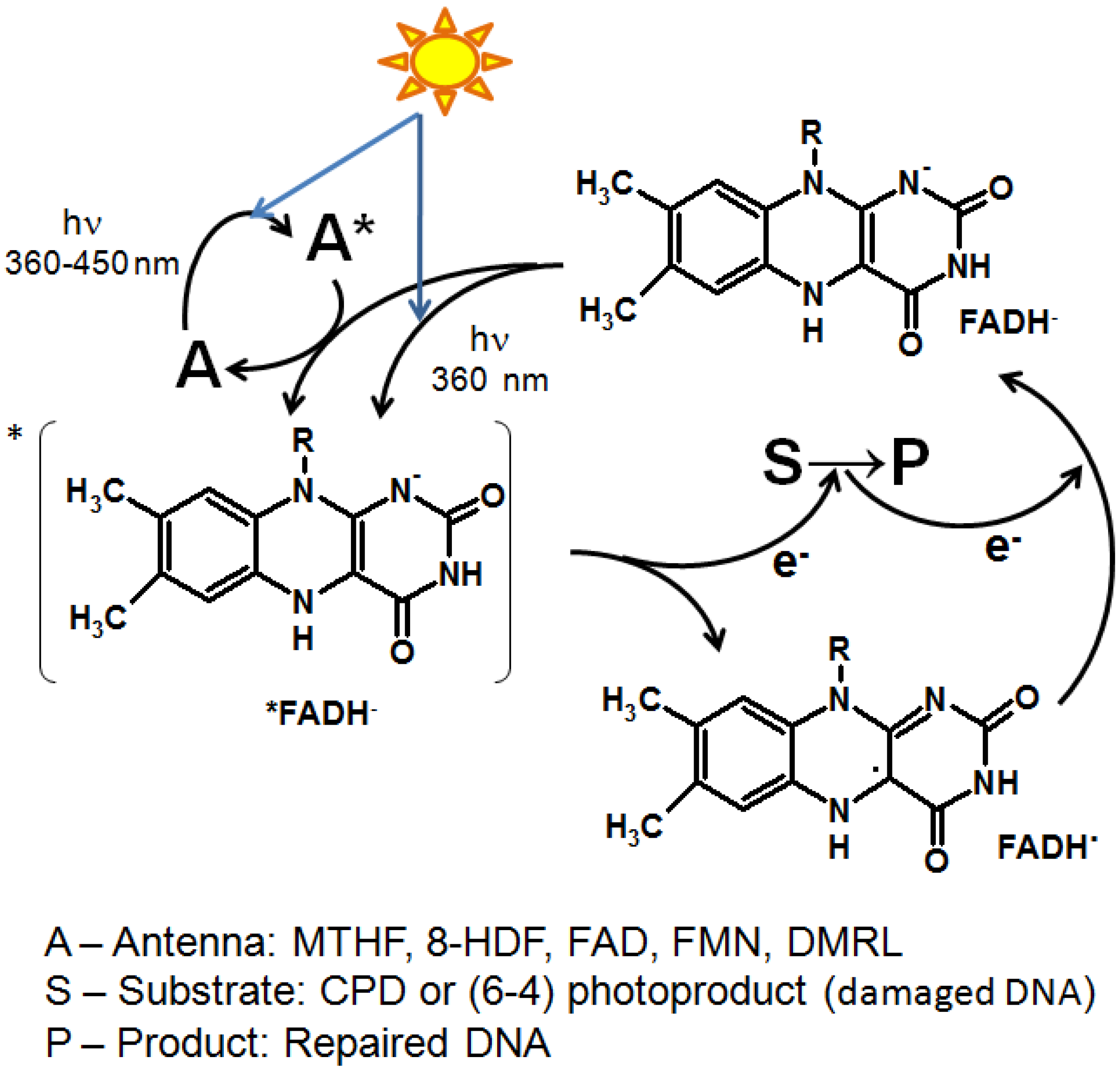
The antenna molecule absorbs a UV-A/blue light photon (360–450 nm) and transfers the excitation energy by the Förster dipole-dipole resonance interaction to FADH, forming a photoexcited *FADH molecule [20]. The latter can also be formed by direct irradiation of FADH in the region of the absorption maximum at 360 nm. The excited FADH then donates an electron to the substrate cyclobutane pyrimidine dimers (CPD) or pyrimidine-pyrimidone (6-4) photoproducts ((6-4)PP) to form FADH·. Furthermore, the electron density is redistributed within the damaged DNA molecule and the original structure is restored (Figure 2). After that, the electron returns to the flavin, regenerating the FADH form [12]. The photorepair quantum yield for CPD photolyases equals 0.7–1 (depending on the antenna and the efficiency of energy transfer from it to the flavin) and for (6-4) photolyases it is ca. 0.3 due to the more complex splitting mechanism of (6-4)PP [2][14][21].
Figure 2. The photocycle of DNA photolyases.
The mechanism of functioning of cryptochromes is not fully understood yet. It is now generally accepted that the photocycle of cryptochromes involves the photoreduction of the FAD molecule, which is usually in a fully or partially oxidized state in the dark, to a partially or fully reduced form, which puts the protein into an active signaling state. Photoreduction can occur due to the transfer of an electron to the photoexcited flavin from the conserved aromatic amino acids, tryptophan and tyrosine, located in the active center of the photoreceptor. Aspartic acid can serve as a hydrogen donor [17][22]. At the same time, conformational changes occur in the protein structure, which are due to the redistribution of hydrogen bonds or a change in its surface charge. Also, phosphorylation of some amino acids allows cryptochrome to interact with other proteins with which it could not interact in the ground state [23][24].
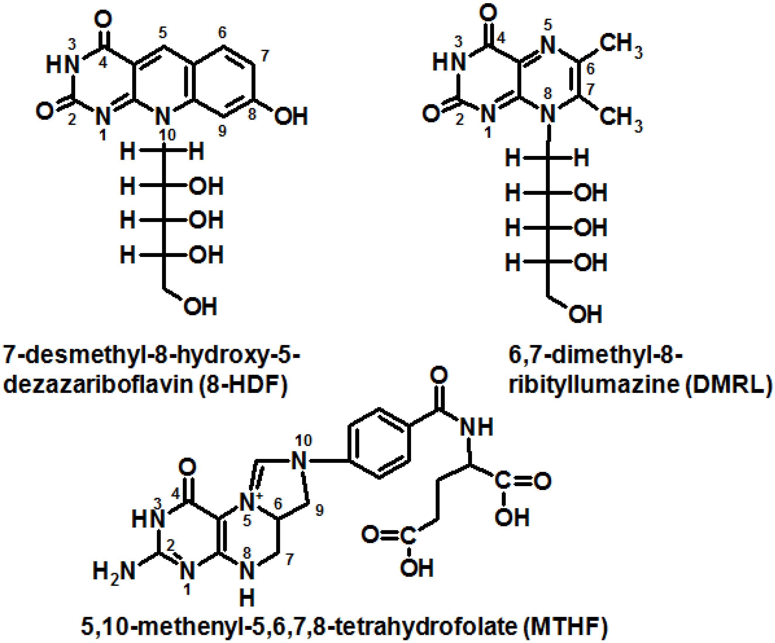
In CPF proteins, in addition to the main FAD chromophore, there are pigments (derivatives of flavins and pterins) that act as a “light-harvesting antenna” (Figure 3). Five molecules have been described as antennas of CPF [13]:
Figure 3. Antenna molecules of CPF proteins.
5,10-methenyl-5,6,7,8-tetrahydrofolate (MTHF) acts as an antenna in most eukaryotes and some prokaryotes [25][26];
7-desmethyl-8-hydroxy-5-deazariboflavin (8-HDF)—in some prokaryotes and protozoa eukaryotes, which have a biosynthetic pathway for this compound [27];
FMN or the second FAD can also function as an antenna [28];
it has recently been shown that 6,7-dimethyl-8-ribityllumazine can function as an antenna in some prokaryotic (6-4)-photolyases (Figure 3) [29].
The antenna chromophore has a higher extinction coefficient and a wider absorption band in the UVA region (ε 385 nm 25,000 M−1 cm−1 for MTHF or ε 440 nm 40,000 M−1 cm−1 for 8-HDF) compared with the flavin chromophore (FADH ε 360 nm 5600 M−1 cm−1) [1].
Until recently, there was no evidence of unconjugated pterin photochemical activity and its participation in photoreception. Only in the last few years has it been shown that some pterin compounds are involved in the reception of UV-B. It is assumed that H4pterins can act as chromophores of some UV photoreceptors [30][31]. The photoreceptors themselves have not yet been isolated and studied, however, it is known that H4cyanopterin (6-[1-(4-O-methyl-(α-D-glucuronyl)-(1,6)-(β-D-galactosyloxy]α-methyl-5,6,7,8-tetrahydropterin) is responsible for the phototaxis of cyanobacteria in response to UV radiation. The photoreceptor containing cyanopterin has been shown to suppress negative phototaxis in response to UV and blue light [32]. The pgtA mutants (the pgtA mutant lacks the pteridine glucosyltransferase enzyme, which is related to the cyanopterin biosynthetic pathway) have the same positive phototaxis in response to red and green light as in the wild-type Synechocystis sp. PCC 6803. However, in response to the effect of white light, pgtA mutants are disoriented, cells move in a fan-like manner: in all directions with a slight positive phototaxis. A similar reaction is observed when exposed to UV and blue light. Notably, when Synechocystis sp. PCC 6803 is exposed to UV, cyanobacteria remain still, while pgtA mutants exhibit negative phototaxis [32].
Evidently, H4Bip is also a photoreceptor molecule: the action spectra of UV-B-induced anthocyanin accumulation in carrot cells indicated that H4Bip is involved in the regulation of anthocyanin synthesis [31]. In addition, the UV-B-induced activity of phenyalanine ammonium lyase (PAL), an enzyme that catalyzes the conversion of phenylalanine to ammonia and cinnamic acid, was suppressed by N-acetylserotonin (an inhibitor of tetrahydrobiopterin biosynthesis). The addition of H4Bip or Bip partially restored the UV-B-induced PAL activity in cells treated with N-acetylserotonin. It was assumed that there is a UV-B photoreceptor different from the UVR8 photoreceptor protein, in which the role of a chromophore is played by tetrahydropterin [31].
Thus, it has been established that H4pterins play the role of chromophores in certain UV-B receptors; however, these UV-B receptors themselves have not yet been isolated and studied. According to a hypothesis, the DASH cryptochrome is responsible for UV reception in cyanobacteria [30], whereas H4cyanopterin plays the role of a chromophore along with flavin [33]. The action spectrum of Synechocystis sp. PCC 6803 coincides with the action spectrum of the DASH cryptochrome. The action spectrum has three main peaks: the peaks at 300 nm and 380 nm correspond to pterins, the peak at 440 nm is characteristic for flavins. The peaks at 380 nm and 440 nm are also found in the fluorescence excitation spectrum of cryptochrome Ccry1 (the DASH cryptochrome of cyanobacteria) from Synechocystis sp. PCC 6803 [33]. If this hypothesis is not confirmed, it could be assumed that a new UV photoreceptor with H4pterin as a chromophore will be discovered in the near future.
As is known, H4pterins do not fluoresce and, therefore, cannot transmit a light signal by means of dipole-dipole energy transfer according to the Foster mechanism. H4pterins most likely do not form triplet forms and excited states with a long lifetime [34]. It could be assumed that the transmission of the light signal occurs as a change of molecular conformation followed by structural changes in the UV-B receptor apoprotein similar to the UVR8, for example [35].

2. The Role of Pterin Coenzymes in the Photoregulation of Metabolism
The significance of pterin photochemistry for the metabolism regulation will be assessed using vitiligo pathology as an example. Vitiligo is a pigmentation disorder, which is expressed in the disappearance of melanin and the appearance of depigmented skin areas. The etiology of vitiligo is still not known, but, it is believed to be associated with the metabolic functions of phenylalanine hydroxylase (PAH), an H4Bip-dependent enzyme of the initial stage of melanogenesis, and tyrosinase (EC 1.14.18.1)) [36][37], as well as with photochemical reactions of H4Bip and oxidized pterins [38][39]. Moreover, it has been shown that pterin can oxidize α-melanocyte-stimulating hormone [40].
One of the most likely causes of vitiligo is a disorder of tyrosine (Tyr) metabolism (Tyr is a precursor of melanin). During the catalytic oxidation of Phe to Tyr, H4Bip is oxidized to 4a-OH-tetrahydrobiopterin (carbinolamine). Dehydration of carbinolamine to quinoid dihydrobiopterin (qH2Bip) is catalyzed by pterin-4a-carbinolamine dehydratase (PCD) (EC 4.2.1.96). qH2Bip has a strong inhibitory effect on PCD, while 7,8-dihydrobiopterin (H2Bip) does not. In the absence of PCD, dehydration of carbinolamine proceeds non-enzymatically and leads to the formation of both H2Bip and 7-H2biopterin, or dihydroprimapterin [41]. 7-H2Bip, in contrast to 4a-OH-tetrahydrobiopterin, has an inhibitory effect on PAH [42]. Finally, the conversion of qH2Bip to H4Bip occurs with the participation of dihydropteridine reductase in a NADH-dependent reaction. Thus, the regeneration of H4Bip is necessary for the metabolism of phenylalanine, since: (1) a constant supply of H4Bip is required for the functioning of PAH; and (2) the accumulation of metabolites resulting from the non-ezymatic rearrangement of 4a-OH-tetrahydrobiopterin is unfavorable.
H4Bip directly regulates tyrosinase activity. The H4Bip binding site in tyrosinase has a sequence homologous to the H4Bip binding sites in PAH and PCD [44]. Under the low concentrations of Phe, H4Bip inhibits PAH [45]. In order to control tyrosinase activity by H4Bip, the presence of L-tyrosine is required. If L-DOPA acts as a substrate for tyrosinase, H4Bip has no inhibitory effect on the enzyme. H2Bip and Bip (products of H4Bip oxidation) do not have a significant inhibitory effect on tyrosinase, which means that the reaction of tyrosine hydroxylation to DOPA is controlled by the H4Bip/Bip ratio and can be initiated by H4Bip photooxidation [39][46]. It has been shown that H4Bip can function as a UV-B switch for de novo melanogenesis, since photoinduced oxidation of H4Bip can “remove” its inhibitory effect on tyrosinase [47].
It has been established that oxidative stress develops in vitiligo cells [48][49][50], and hydrogen peroxide accumulates at millimolar concentrations. Under oxidative stress conditions, tyrosinase is activated by low concentrations of hydrogen peroxide (<0.3 × 10⁻³ M), but is deactivated when the peroxide concentration is in the range of 0.5–5.0 × 10⁻³ M [51]. Under the oxidative stress conditions, the work of PCD can be inhibited by hydrogen peroxide, which leads to disruption of the H4Bip regeneration cycle [43]. The hypothesis that vitiligo is caused by a violation of the H4Bip regeneration cycle is one of the most developed and substantiated to date. Peroxide concentrations of less than 30 μM increase DHPR activity, but concentrations above 30 μM inactivate DHPR, which occurs through the oxidation of Met146 and Met151 protein sequences and leads to a disruption of the NADH-dependent active site of the enzyme [52]. PCD inactivation leads to nonenzymatic dehydration of 4a-OH-tetrahydrobiopterin, which proceeds with the formation of H4Bip and 7-H2biopterin [41]. Enzymatic reduction of H2Bip and 7-H2biopterin with the participation of dihydropteridine reductase (DHPR) proceeds with the formation of 6(R,S)-5,6,7,8-tetrahydrobiopterin and 7(R,S)-5,6,7,8-tetrahydrobiopterin, accordingly, since DHPR has low stereospecificity [53]. The Michaelis constant for the interaction of (6S)-5,6,7,8-tetrahydro-L-biopterin (6S-H4Bip) and PAH is 2 times higher than for (6R)-5,6,7,8-tetrahydro-L-biopterin [54]; 7(R,S)-5,6,7,8-tetrahydro-L-biopterin (7-H4Bip) inhibits PAH [55].
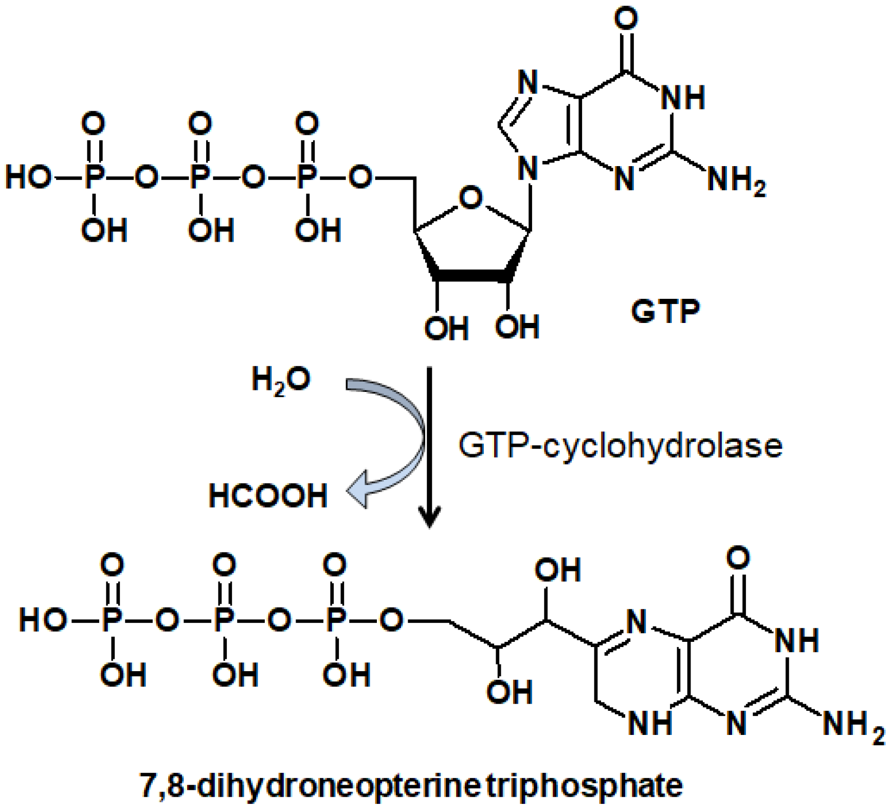
GTP-cyclohydrolase I (GTPCH) converts GTP to 7,8-dihydroneopterin-3’-phosphate (Figure 4), which is the limiting reaction for H4Bip biosynthesis. Inhibition of PAH by the feedback mechanism leads to a three to five-fold increase in the activity of GTPCH. An increase in GTPCH activity leads to excessive de novo synthesis of (6R)-5,6,7,8-tetrahydrobiopterin [56][57]. Excessive synthesis of H4Bip, in turn, leads to the complete inhibition of tyrosinase [44], thus, tyrosine is not formed and, as a result, melanogenesis in epidermal cells stops.
Figure 4 A simplified scheme of guanosine triphosphate (GTP) transformation to 7,8-dihydroneopterinphosphate, a precursor of H4Bip [42].
The products of H4Bip oxidation, biopterin, and primapterin, accumulate in the depigmented epidermis cells (evidently, as a result of nonenzymatic oxidation of H2Bip and H4Bip by molecular oxygen) and exhibit characteristic fluorescence under ultraviolet irradiation [58][59]. Photolysis of biopterin under aerobic conditions leads to the formation of additional amounts of peroxide in vitiligo [60][61]. In addition to Bip and primapterin, epidermal cells accumulate 6-carboxypterin, a product of biopterin oxidation, which also effectively sensitizes the formation of ROS under UV exposure [62]. This circumstance makes H4Bip (H4Bip accumulates in depigmented skin as a result of excessive de novo synthesis) sensitive to UV radiation and leads to the addition generation of ROS sensitized by oxidized pterins—A “snowball” effect. The study of the process of photooxidation and photosensitized oxidation of H4Bip is significant for understanding the etiology and course of the disease, as well as for developing methods of vitiligo therapy [38][39][46].

3. Evolutionary Aspects of Pterin Photochemistry

Recently, it has become known that pteridines (pterins and flavins) are chromophores of photoreceptor proteins: photolyases, cryptochromes and so on. Pteridines can participate in redox processes and, on the other hand, function as ultraviolet and blue light receptors [63]. In the first instance, such wide functionality can be associated with their resistance to UV radiation. UV radiation and the blue part of the spectrum of the Sun were the most important sources of free energy during the period of pre-biological and early biological evolution on Earth. During this period, those substances that were available from abiogenesis were probably used. It has been shown that pterins and isoalloxazines (flavins) can be formed upon thermal condensation of abiogenic amino acids [64][65][66]. In the period of pre-biological evolution, chromoproteinoids containing pteridines (pterins and flavins) as chromophores could function as catalysts in dark redox reactions and as photosensitizers in photoinduced processes. The availability of this group of compounds may indicate their antiquity and possible participation at all stages of evolution.
The second important evolutionary aspect of pterin photochemistry is the pteridines and the “RNA world” [67][68]. The existing set of bases in nucleic acids is the product of evolution and selection. A variety of heterocycles could participate in the selection. It has been shown that pteridines are sterically suitable and can be inserted into nucleotide sequences [69]. Flavins and pterins could enrich the catalytic and photocatalytic capabilities of primitive polyribonucleotides lacking redox functions. Under conditions of abiogenesis, pteridines could absorb UV and function as photocatalysts of free radical processes leading to the synthesis of compounds for further pre-biological evolution [70]. Under non-oxidative conditions of pre-biological and early biological evolution, the structural similarity of pterins and purines may have allowed pterins to be incorporated into proto-RNA. In the absence of significant amounts of free oxygen in the atmosphere, H2pterins and H4pterins conjugated into proto-RNA could function as electron and hydrogen donors in various processes of the complication of carbon compounds on the way to life, including processes associated with the storage of free energy.
With the oxygenation of the environment and appearance of the ozone layer, the functions of pteridines also changed. The pyrazine part of the pterin structure became oxidized, and the π-electrons of its double bonds began to significantly affect the electronic configuration and redox properties of pterins. Oxidized pterins are dominated by the processes of fluorescence, S-T intercombination conversion, as well as the processes of energy transfer to molecular oxygen with the formation of singlet oxygen, which is dangerous for polyribonucleotides and other molecules. Herein, the main photoreception of UV energy and its storage as the reaction products have passed to more specialized chromophores—porphyrins, which possessed hydrophobicity, localization in membranes, and absorption of light in the visible part of the spectrum. At the same time, pteridines retained their UV chromophoric functions as a part of photoregulatory proteins. The catalytic redox functions of pteridines also appear to be conserved throughout the evolution.
Another evolutionary aspect of pterin photochemistry is the possibility of the participation of H4pterins in the photoprotection of cyanobacteria during the early stages of biological evolution. Prior to the Great Oxidation Event, oxygen was absent in the environment, whereas cyanobacteria were among the first living organisms on the Earth more than 2.4 billion years ago [71][72]. H4pterins are found in high concentrations (at a ratio of 1:1.6 towards chlorophyll a) in cyanobacterial cells [73]. Since H4pterins have high photostability and an ultrashort lifetime of the excited states along with low ionization potential [74], this makes them an ideal candidate for the role of photoprotectors and antioxidants.

References

  1. Kritsky, M.S.; Telegina, T.A.; Vechtomova, Y.L.; Buglak, A.A. Why flavins are not competitors of chlorophyll in the evolution of biological converters of solar energy. Int. J. Mol. Sci. 2013, 14, 575.
  2. Kavakli, I.H.; Baris, I.; Tardu, M.; Gül, Ş.; Öner, H.; Çal, S.; Bulut, S.; Yarparvar, D.; Berkel, Ç.; Ustaoğlu, P.; et al. The Photolyase/Cryptochrome Family of Proteins as DNA Repair Enzymes and Transcriptional Repressors. Photochem. Photobiol. 2017, 93, 93–103.
  3. Hosokawa, Y.; Müller, P.; Kitoh-Nishioka, H.; Iwai, S.; Yamamoto, J. Limited solvation of an electron donating tryptophan stabilizes a photoinduced charge-separated state in plant (6–4) photolyase. Sci. Rep. 2022, 12, 5084.
  4. Cellini, A.; Shankar, M.K.; Wahlgren, W.Y.; Nimmrich, A.; Furrer, A.; James, D.; Wranik, M.; Aumonier, S.; Beale, E.V.; Dworkowski, F.; et al. Structural basis of the radical pair state in photolyases and cryptochromes. Chem. Commun. 2022, 58, 4889–4892.
  5. Losi, A.; Mandalari, C.; Gärtner, W. From Plant Infectivity to Growth Patterns: The Role of Blue-Light Sensing in the Prokaryotic World. Plants 2014, 3, 70–94.
  6. Park, S.-Y.; Tame, J.R.H. Seeing the light with BLUF proteins. Biophys. Rev. 2017, 9, 169–176.
  7. Gauden, M.; Yeremenko, S.; Laan, W.; van Stokkum, I.H.M.; Ihalainen, J.A.; van Grondelle, R.; Hellingwerf, K.J.; Kennis, J.T.M. Photocycle of the Flavin-Binding Photoreceptor AppA, a Bacterial Transcriptional Antirepressor of Photosynthesis Genes. Biochemistry 2005, 44, 3653–3662.
  8. Goings, J.J.; Li, P.; Zhu, Q.; Hammes-Schiffer, S. Formation of an unusual glutamine tautomer in a blue light using flavin photocycle characterizes the light-adapted state. Proc. Natl. Acad. Sci. USA 2020, 117, 26626–26632.
  9. Tokonami, S.; Onose, M.; Nakasone, Y.; Terazima, M. Slow Conformational Changes of Blue Light Sensor BLUF Proteins in Milliseconds. J. Am. Chem. Soc. 2022, 144, 4080–4090.
  10. Shibata, K.; Nakasone, Y.; Terazima, M. Selective Photoinduced Dimerization and Slow Recovery of a BLUF Domain of EB1. J. Phys. Chem. B 2022, 126, 1024–1033.
  11. Kang, X.-W.; Chen, Z.; Zhou, Z.; Zhou, Y.; Tang, S.; Zhang, Y.; Zhang, T.; Ding, B.; Zhong, D. Direct Observation of Ultrafast Proton Rocking in the BLUF Domain. Angew. Chem. Int. Ed. 2022, 61, e202114423.
  12. Sancar, A. Mechanisms of DNA Repair by Photolyase and Excision Nuclease (Nobel Lecture). Angew. Chem. Int. Ed. 2016, 55, 8502–8527.
  13. Kavakli, I.H.; Ozturk, N.; Gul, S. DNA repair by photolyases. Adv. Protein Chem. Struct. Biol. 2019, 115, 1–19.
  14. Vechtomova, Y.L.; Telegina, T.A.; Kritsky, M.S. Evolution of Proteins of the DNA Photolyase/Cryptochrome Family. Biochemistry 2020, 85, 131–153.
  15. Ponnu, J.; Hoecker, U. Signaling Mechanisms by Arabidopsis Cryptochromes. Front. Plant Sci. 2022, 13, 844714.
  16. Pooam, M.; El-Esawi, M.A.; Aguida, B.; Ahmad, M. Arabidopsis cryptochrome and Quantum Biology: New insights for plant science and crop improvement. J. Plant Biochem. Biotechnol. 2020, 29, 636–651.
  17. Brazard, J.; Usman, A.; Lacombat, F.; Ley, C.; Martin, M.M.; Plaza, P.; Mony, L.; Heijde, M.; Zabulon, G.; Bowler, C. Spectro−Temporal Characterization of the Photoactivation Mechanism of Two New Oxidized Cryptochrome/Photolyase Photoreceptors. J. Am. Chem. Soc. 2010, 132, 4935–4945.
  18. Hense, A.; Herman, E.; Oldemeyer, S.; Kottke, T. Proton transfer to flavin stabilizes the signaling state of the blue light receptor plant cryptochrome. J. Biol. Chem. 2015, 290, 1743–1751.
  19. Partch, C.L.; Sancar, A. Photochemistry and Photobiology of Cryptochrome Blue-light Photopigments: The Search for a Photocycle. Photochem. Photobiol. 2005, 81, 1291–1304.
  20. Tan, C.; Guo, L.; Ai, Y.; Li, J.; Wang, L.; Sancar, A.; Luo, Y.; Zhong, D. Direct Determination of Resonance Energy Transfer in Photolyase: Structural Alignment for the Functional State. J. Phys. Chem. A 2014, 118, 10522–10530.
  21. Vechtomova, Y.; Telegina, T.; Buglak, A.; Kritsky, M. UV Radiation in DNA Damage and Repair Involving DNA-Photolyases and Cryptochromes. Biomedicines 2021, 9, 1564.
  22. Kiontke, S.; Göbel, T.; Brych, A.; Batschauer, A. DASH-type cryptochromes—Solved and open questions. Biol. Chem. 2020, 401, 1487–1493.
  23. Lin, C.; Schneps, C.M.; Chandrasekaran, S.; Ganguly, A.; Crane, B.R. Mechanistic insight into light-dependent recognition of Timeless by Drosophila Cryptochrome. Structure 2022, 30, 851–861.e5.
  24. Palayam, M.; Ganapathy, J.; Guercio, A.M.; Tal, L.; Deck, S.L.; Shabek, N. Structural insights into photoactivation of plant Cryptochrome-2. Commun. Biol. 2021, 4, 28.
  25. Scheerer, P.; Zhang, F.; Kalms, J.; von Stetten, D.; Krauß, N.; Oberpichler, I.; Lamparter, T. The Class III Cyclobutane Pyrimidine Dimer Photolyase Structure Reveals a New Antenna Chromophore Binding Site and Alternative Photoreduction Pathways. J. Biol. Chem. 2015, 290, 11504–11514.
  26. Petersen, J.; Rredhi, A.; Szyttenholm, J.; Oldemeyer, S.; Kottke, T.; Mittag, M. The World of Algae Reveals a Broad Variety of Cryptochrome Properties and Functions. Front. Plant Sci. 2021, 12, 766509.
  27. Chen, S.; Liu, C.; Zhou, C.; Wei, Z.; Li, Y.; Xiong, L.; Yan, L.; Lv, J.; Shen, L.; Xu, L. Identification and characterization of a prokaryotic 6-4 photolyase from Synechococcus elongatus with a deazariboflavin antenna chromophore. Nucleic Acids Res. 2022, 50, 5757–5771.
  28. Terai, Y.; Sato, R.; Matsumura, R.; Iwai, S.; Yamamoto, J. Enhanced DNA repair by DNA photolyase bearing an artificial light-harvesting chromophore. Nucleic Acids Res. 2020, 48, 10076–10086.
  29. Geisselbrecht, Y.; Frühwirth, S.; Schroeder, C.; Pierik, A.J.; Klug, G.; Essen, L.-O. CryB from Rhodobacter sphaeroides: A unique class of cryptochromes with new cofactors. EMBO Rep. 2012, 13, 223–229.
  30. Moon, Y.-J.; Kim, S.I.; Chung, Y.-H. Sensing and responding to UV-A in cyanobacteria. Int. J. Mol. Sci. 2012, 13, 16303–16332.
  31. Takeda, J.; Nakata, R.; Ueno, H.; Murakami, A.; Iseki, M.; Watanabe, M. Possible involvement of a tetrahydrobiopterin in photoreception for UV-B-induced anthocyanin synthesis in carrot. Photochem. Photobiol. 2014, 90, 1043–1049.
  32. Moon, Y.-J.; Kim, S.-J.; Park, Y.M.; Chung, Y.-H. Sensing UV/blue: Pterin as a UV-A absorbing chromophore of cryptochrome. Plant Signal. Behav. 2010, 5, 1127–1130.
  33. Moon, Y.-J.; Lee, E.-M.; Park, Y.M.; Park, Y.S.; Chung, W.-I.; Chung, Y.-H. The role of cyanopterin in UV/blue light signal transduction of cyanobacterium Synechocystis sp. PCC 6803 phototaxis. Plant Cell Physiol. 2010, 51, 969–980.
  34. Buglak, A.A. Photobiochemistry of Pterin Coenzymes; Research Centre of Biotechnology RAS: Moscow, Russian, 2016.
  35. Wu, D.; Hu, Q.; Yan, Z.; Chen, W.; Yan, C.; Huang, X.; Zhang, J.; Yang, P.; Deng, H.; Wang, J.; et al. Structural basis of ultraviolet-B perception by UVR8. Nature 2012, 484, 214–219.
  36. Schallreuter, K.U.; Chavan, B.; Rokos, H.; Hibberts, N.; Panske, A.; Wood, J.M. Decreased phenylalanine uptake and turnover in patients with vitiligo. Mol. Genet. Metab. 2005, 86 (Suppl. 1), S27–S33.
  37. Niu, C.; Aisa, H.A. Upregulation of Melanogenesis and Tyrosinase Activity: Potential Agents for Vitiligo. Molecules 2017, 22, 1303.
  38. Telegina, T.A.; Lyudnikova, T.A.; Buglak, A.A.; Vechtomova, Y.L.; Biryukov, M.V.; Demin, V.V.; Kritsky, M.S. Transformation of 6-tetrahydrobiopterin in aqueous solutions under UV-irradiation. J. Photochem. Photobiol. A Chem. 2018, 354, 155–162.
  39. Telegina, T.A.; Vechtomova, Y.L.; Kritsky, M.S.; Madirov, E.I.; Nizamutdinov, A.S.; Obuhov, Y.N.; Buglak, A.A. Tetrahydrobiopterin Photooxidation: A Key Process in Vitiligo Phototherapy. Appl. Biochem. Microbiol. 2021, 57, 571–578.
  40. Castaño, C.; Lorente, C.; Martins-Froment, N.; Oliveros, E.; Thomas, A.H. Degradation of α-melanocyte-stimulating hormone photosensitized by pterin. Org. Biomol. Chem. 2014, 12, 3877–3886.
  41. Davis, M.D.; Kaufman, S.; Milstien, S. Conversion of 6-substituted tetrahydropterins to 7-isomers via phenylalanine hydroxylase-generated intermediates. Proc. Natl. Acad. Sci. USA 1991, 88, 385–389.
  42. Thöny, B.; Auerbach, G.; Blau, N. Tetrahydrobiopterin biosynthesis, regeneration and functions. Biochem. J. 2000, 347 Pt 1, 1–16.
  43. Buglak, A.A.; Telegina, T.A.; Vechtomova, Y.L.; Kritsky, M.S. Autoxidation and photooxidation of tetrahydrobiopterin: A theoretical study. Free Radic. Res. 2021, 55, 499–509.
  44. Wood, J.M.; Schallreuter-Wood, K.U.; Lindsey, N.J.; Callaghan, S.; Gardner, M.L. A specific tetrahydrobiopterin binding domain on tyrosinase controls melanogenesis. Biochem. Biophys. Res. Commun. 1995, 206, 480–485.
  45. Eichinger, A.; Danecka, M.K.; Möglich, T.; Borsch, J.; Woidy, M.; Büttner, L.; Muntau, A.C.; Gersting, S.W. Secondary BH4 deficiency links protein homeostasis to regulation of phenylalanine metabolism. Hum. Mol. Genet. 2018, 27, 1732–1742.
  46. Buglak, A.A.; Telegina, T.A.; Lyudnikova, T.A.; Vechtomova, Y.L.; Kritsky, M.S. Photooxidation of tetrahydrobiopterin under UV irradiation: Possible pathways and mechanisms. Photochem. Photobiol. 2014, 90, 1017–1026.
  47. Schallreuter, K.U.; Wood, J.M.; Körner, C.; Harle, K.M.; Schulz-Douglas, V.; Werner, E.R. 6-Tetrahydrobiopterin functions as a UVB-light switch for de novo melanogenesis. Biochim. Biophys. Acta 1998, 1382, 339–344.
  48. Jain, A.; Mal, J.; Mehndiratta, V.; Chander, R.; Patra, S.K. Study of oxidative stress in vitiligo. Indian J. Clin. Biochem. 2011, 26, 78–81.
  49. Wang, Y.; Li, S.; Li, C. Perspectives of New Advances in the Pathogenesis of Vitiligo: From Oxidative Stress to Autoimmunity. Med. Sci. Monit. Int. Med. J. Exp. Clin. Res. 2019, 25, 1017–1023.
  50. Xuan, Y.; Yang, Y.; Xiang, L.; Zhang, C. The Role of Oxidative Stress in the Pathogenesis of Vitiligo: A Culprit for Melanocyte Death. Oxid. Med. Cell. Longev. 2022, 2022, 8498472.
  51. Wood, J.M.; Chavan, B.; Hafeez, I.; Schallreuter, K.U. Regulation of tyrosinase by tetrahydropteridines and H2O2. Biochem. Biophys. Res. Commun. 2004, 325, 1412–1417.
  52. Hasse, S.; Gibbons, N.C.J.; Rokos, H.; Marles, L.K.; Schallreuter, K.U. Perturbed 6-Tetrahydrobiopterin Recycling via Decreased Dihydropteridine Reductase in Vitiligo: More Evidence for H2O2 Stress. J. Investig. Dermatol. 2004, 122, 307–313.
  53. Rebrin, I.; Bailey, S.W.; Boerth, S.R.; Ardell, M.D.; Ayling, J.E. Catalytic characterization of 4a-hydroxytetrahydropterin dehydratase. Biochemistry 1995, 34, 5801–5810.
  54. Haavik, J.; Døskeland, A.P.; Flatmark, T. Stereoselective effects in the interactions of pterin cofactors with rat-liver phenylalanine 4-monooxygenase. Eur. J. Biochem. 1986, 160, 1–8.
  55. Davis, M.D.; Ribeiro, P.; Tipper, J.; Kaufman, S. “7-tetrahydrobiopterin,” a naturally occurring analogue of tetrahydrobiopterin, is a cofactor for and a potential inhibitor of the aromatic amino acid hydroxylases. Proc. Natl. Acad. Sci. USA 1992, 89, 10109–10113.
  56. Bendall, J.K.; Douglas, G.; McNeill, E.; Channon, K.M.; Crabtree, M.J. Tetrahydrobiopterin in Cardiovascular Health and Disease. Antioxid. Redox Signal. 2013, 20, 3040–3077.
  57. Schallreuter, K.U.; Wood, J.M.; Ziegler, I.; Lemke, K.R.; Pittelkow, M.R.; Lindsey, N.J.; Gütlich, M. Defective tetrahydrobiopterin and catecholamine biosynthesis in the depigmentation disorder vitiligo. Biochim. Biophys. Acta 1994, 1226, 181–192.
  58. Estébanez, S.; Lorente, C.; Tosato, M.G.; Miranda, M.A.; Marín, M.L.; Lhiaubet-Vallet, V.; Thomas, A.H. Photochemical formation of a fluorescent thymidine-pterin adduct in DNA. Dye. Pigment. 2019, 160, 624–632.
  59. Schallreuter, K.U.; Wood, J.M.; Pittelkow, M.R.; Gütlich, M.; Lemke, K.R.; Rödl, W.; Swanson, N.N.; Hitzemann, K.; Ziegler, I. Regulation of melanin biosynthesis in the human epidermis by tetrahydrobiopterin. Science 1994, 263, 1444–1446.
  60. Dantola, M.L.; Reid, L.O.; Castaño, C.; Lorente, C.; Oliveros, E.; Thomas, A.H. Photosensitization of peptides and proteins by pterin derivatives. Pteridines 2017, 28, 105–114.
  61. Rokos, H.; Beazley, W.D.; Schallreuter, K.U. Oxidative stress in vitiligo: Photo-oxidation of pterins produces H(2)O(2) and pterin-6-carboxylic acid. Biochem. Biophys. Res. Commun. 2002, 292, 805–811.
  62. Lorente, C.; Thomas, A.H. Photophysics and photochemistry of pterins in aqueous solution. Acc. Chem. Res. 2006, 39, 395–402.
  63. Kritsky, M.S.; Telegina, T.A.; Vechtomova, Y.L.; Kolesnikov, M.P.; Lyudnikova, T.A.; Golub, O.A. Excited flavin and pterin coenzyme molecules in evolution. Biochemistry 2010, 75, 1200–1216.
  64. Heinz, B.; Ried, W.; Dose, K. Thermal Generation of Pteridines and Flavines from Amino Acid Mixtures. Angew. Chemie Int. Ed. 1979, 18, 478–483.
  65. Heinz, B.; Ried, W. The formation of chromophores through amino acid thermolysis and their possible role as prebiotic photoreceptors. Biosystems 1981, 14, 33–40.
  66. Schwartz, W. S. W. Fox and K. Dose, Molecular Evolution and the Origin of Life. Z. Allg. Mikrobiol. 1973, 13, 732.
  67. Kirschning, A. Coenzymes and Their Role in the Evolution of Life. Angew. Chem. Int. Ed. 2021, 60, 6242–6269.
  68. Kritsky, M.S.; Telegina, T.A.; Lyudnikova, T.A.; Zemskova, Y.L. Coenzymes in Evolution of the RNA World. In Life in the Universe; Seckbach, J., Chela-Flores, J., Owen, T., Raulin, F., Eds.; Springer: Dordrecht, The Netherlands, 2004; pp. 115–118. ISBN 978-94-007-1003-0.
  69. Hawkins, M.E. Fluorescent pteridine probes for nucleic acid analysis. Methods Enzymol. 2008, 450, 201–231.
  70. Schmidt, W.; Butler, W.L. Flavin-mediated photoreactions in artificial systems: A possible model for the blue-light photoreceptor pigment in living systems. Photochem. Photobiol. 1976, 24, 71–75.
  71. Schirrmeister, B.E.; Sanchez-Baracaldo, P.; Wacey, D. Cyanobacterial evolution during the Precambrian. Int. J. Astrobiol. 2016, 15, 187–204.
  72. Chen, S.-C.; Sun, G.-X.; Yan, Y.; Konstantinidis, K.T.; Zhang, S.-Y.; Deng, Y.; Li, X.-M.; Cui, H.-L.; Musat, F.; Popp, D.; et al. The Great Oxidation Event expanded the genetic repertoire of arsenic metabolism and cycling. Proc. Natl. Acad. Sci. USA 2020, 117, 10414–10421.
  73. Lee, H.W.; Oh, C.H.; Geyer, A.; Pfleiderer, W.; Park, Y.S. Characterization of a novel unconjugated pteridine glycoside, cyanopterin, in Synechocystis sp. PCC 6803. Biochim. Biophys. Acta 1999, 1410, 61–70.
  74. Buglak, A.A.; Telegina, T.A. A theoretical study of 5,6,7,8-tetrahydro-6-hydroxymethylpterin: Insight into intrinsic photoreceptor properties of 6-substituted tetrahydropterins. Photochem. Photobiol. Sci. 2019, 18, 516–523.
More
Information
Contributors MDPI registered users' name will be linked to their SciProfiles pages. To register with us, please refer to https://encyclopedia.pub/register : , , ,
View Times: 604
Revisions: 3 times (View History)
Update Date: 13 Dec 2022
1000/1000
ScholarVision Creations