Your browser does not fully support modern features. Please upgrade for a smoother experience.
Submitted Successfully!
 +1 credit
Thank you for your contribution! You can also upload a video entry or images related to this topic.
For video creation, please contact our Academic Video Service.
| Version | Summary | Created by | Modification | Content Size | Created at | Operation |
|---|---|---|---|---|---|---|
| 1 | Joydip Sengupta | -- | 1538 | 2022-12-09 03:41:01 | | | |
| 2 | Vivi Li | + 21 word(s) | 1559 | 2022-12-09 04:32:07 | | |
Video Upload Options
We provide professional Academic Video Service to translate complex research into visually appealing presentations. Would you like to try it?
Cite
If you have any further questions, please contact Encyclopedia Editorial Office.
Sengupta, J.; Hussain, C.M. CNT-Based Analytical Biosensing Platforms in Viruses Detection. Encyclopedia. Available online: https://encyclopedia.pub/entry/38401 (accessed on 26 June 2026).
Sengupta J, Hussain CM. CNT-Based Analytical Biosensing Platforms in Viruses Detection. Encyclopedia. Available at: https://encyclopedia.pub/entry/38401. Accessed June 26, 2026.
Sengupta, Joydip, Chaudhery Mustansar Hussain. "CNT-Based Analytical Biosensing Platforms in Viruses Detection" Encyclopedia, https://encyclopedia.pub/entry/38401 (accessed June 26, 2026).
Sengupta, J., & Hussain, C.M. (2022, December 09). CNT-Based Analytical Biosensing Platforms in Viruses Detection. In Encyclopedia. https://encyclopedia.pub/entry/38401
Sengupta, Joydip and Chaudhery Mustansar Hussain. "CNT-Based Analytical Biosensing Platforms in Viruses Detection." Encyclopedia. Web. 09 December, 2022.
Copy Citation
It has been proven that viral infections pose a serious hazard to humans and also affect social health, including morbidity and mental suffering, as illustrated by the COVID-19 pandemic. The early detection and isolation of virally infected people are, thus, required to control the spread of viruses. Due to the outstanding and unparalleled properties of nanomaterials, numerous biosensors were developed for the early detection of viral diseases via sensitive, minimally invasive, and simple procedures. To that aim, viral detection technologies based on carbon nanotubes (CNTs) are being developed as viable alternatives to existing diagnostic approaches.
carbon nanotube (CNT)
biosensor
virus
SARS-CoV-2
toxicity
biocompatibility
1. Introduction
1.1. Carbon Nanotubes and Their Applicability in Biosensing
The carbon nanotube (CNT) is the 1D allotrope of carbon; the experimental evidence was first reported by L.V. Radushkevich and V.M. Lukyanovich [1] from the Institute of Physical Chemistry and Electrochemistry of the Russian Academy of Sciences, in 1952. However, after Ijima’s paper [2] in 1991, research interest in CNT escalated rapidly. Structurally, CNTs can be divided into two major categories based on the number of graphitic layers, namely, single-wall CNT (SWCNT) and multi-wall CNT (MWCNT). Depending on the direction of the roll-up, SWCNTs can have different structures, namely, the zigzag, armchair, or chiral formations. While depending on the nature of the wrapping i.e., whether a graphitic sheet is rolled around itself multiple times (Swiss roll) or if the graphitic sheets are arranged as concentric cylinders (Russian doll), MWCNT can also be categorized, as in Scheme 1.
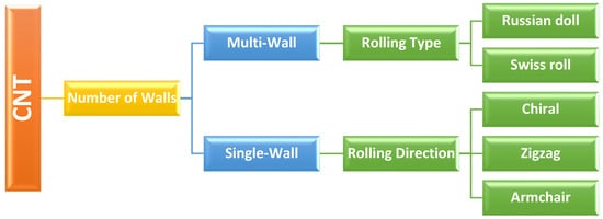
Scheme 1. Different categories of carbon nanotube (CNT).
CNT can be synthesized through various means, such as arc discharge, laser ablation, chemical vapor deposition (CVD), etc. However, because of the scalability, CVD-based approaches appear to be the most appropriate for large-scale CNT synthesis [3]. A wide range of prospective applications in important industrial fields, including nanoelectronics and biotechnology are promised by the distinctive mix of electrical, thermal, mechanical, and chemical characteristics that CNTs display. Additionally, among the many nanomaterials, CNTs are particularly intriguing because they provide an exceedingly tiny inner hollow core, virtually a one-dimensional space, for material storage. Thus, a novel structure can also be formed by filling the core of a CNT with the components necessary for specific applications.
Previous research has discovered a linkage between biomolecules in living beings and illnesses. The monitoring of aberrant physical parameters and the early diagnosis of illnesses help to minimize mortality and ensure organisms’ physical health. Conventional laboratory approaches for assessing pathogenic variables are typically time-consuming, expensive, and complicated. Biosensors can facilitate the reliable and rapid analysis of metabolites in the body to help the current therapeutic procedure. CNTs have unique physicochemical and photoelectric qualities that can improve the performance of biosensors, such as a greater surface area for better catalyst adhesion; CNT-modified electrodes offer quicker electron transfer, resulting in enhanced sensitivity of detection for biosensors. CNT’s unrivaled electronic features, such as quantum wire-like behavior, ballistic-type electronic conduction, remarkable thermal properties derived from phonon quantization, excellent flexibility, and high breaking stress despite its low density make CNT one of the best transducer materials for the transmission of signals related to the recognition of analytes, metabolites, or disease biomarkers. The curvature of the tube contributes to CNTs’ high reactivity and sensitivity to chemical or environmental interactions. Moreover, as the carbon atom near the end of an open-ended tube has only two bonds, foreign molecules can easily enter the structure, thereby helping in the preferential addition of one or more species for functionalization. More importantly, from the viewpoint of biosensors, CNTs can act effectively as scaffolds for the immobilization of biomolecules at their surface. These fascinating characteristics have led to CNTs being widely used in biosensor applications.
2. Types of Biosensors Used for Virus Detection
2.1. Immunosensors
Because of its capacity to handle information, the immune system is an appealing subject in scientific studies. The major purpose of an immune system, as part of the system’s defensive mechanism, is to accredit and ascertain all cells and molecules in the assembly and classify these biological substances as either toxic or non-toxic. When exposed to foreign substances (i.e., antigens), specialized immune system cells make immunoglobulins (i.e., antibodies) that attach to these antigens precisely. An immunosensor (Figure 1, top left), an affinity-based biosensing device, exploits the concept of immunology and employs an antibody for the specific molecular identification of antigens that are immobilized on a transducer surface, and then develops a stable immunocomplex. The immunocomplex is calculated and quantified by connecting the antibody and antigen interactions to the surface of a transducer. The transducer detects the response and transforms it into an electrical signal, which may then be processed, recorded, and examined. The detection of the target analyte in immunosensors might be direct, by witnessing the production of immunocomplexes, or indirect, by using a label. Immunosensors can be categorized into several categories, based on various methodologies, such as electrochemical, impedimetric, potentiometric, amperometric, voltammetric, conductometric, capacitive, and surface plasmon resonance (SPR)-based methodologies.
2.2. Optical Biosensor
An optical biosensor, a compact analytical instrument, combines an optical transducer system with a biorecognition-sensing element (Figure 1, bottom left). An optical biosensor’s primary goal is to provide a signal that is proportional to the concentration of the material being analyzed (the analyte). Optical detection is made possible by using the interplay between the optical field and a biorecognition element. Label-free and label-based optical biosensing are the main two categories of optical biosensors. In a label-free mode, the interaction between the substances is analyzed, and the transducer directly generates the measured signal. In contrast, label-based sensing makes use of a label to assess the biorecognition event and generates an optical signal using a colorimetric, fluorescent, or luminescent approach. However, in some cases, such as antibody-antigen interactions, when a label is coupled with one of the bio-reactants, then this labeling might modify the binding characteristics, introducing systematic inaccuracy into biosensor analysis.
2.3. Electrochemical Biosensor
Due to the direct conversion of a biological event to an electrical signal, electrochemical biosensors provide an appealing technique for analyzing the content of a biological sample. The measurement of electrical characteristics in biosensing, for extracting information from biological systems, is generally electrochemical in nature, with a bio-electrochemical component serving as the major transduction aspect (Figure 1, bottom right). While biosensing devices use a variety of recognition components, electrochemical detection approaches mostly involve enzymes. This is mainly owing to their unique binding properties and biocatalytic activity. In bio-electrochemistry, the reaction under examination would typically create a quantifiable current (amperometric), a measurable potential or charge buildup (potentiometric), or a measurable impedance (impedimetric). The electrodes are essential components for the operation of electrochemical biosensors since reactions are generally observed near the electrode’s surface. Depending on the electrode’s parameters, the material, the surface modification, or the electrode’s size have a significant impact on the capability of detection. In general, three electrodes, namely, the reference electrode, counter or auxiliary electrode, and working electrode are needed for electrochemical sensing. To maintain a known and constant voltage, the reference electrode is kept away from the reaction site. The counter electrode creates a link to the electrolytic solution so that a current may be supplied to the working electrode, while the working electrode acts as the transduction element in the biological reaction. These electrodes ought to be chemically stable and conductible to achieve a faithful analysis.
2.4. Field-Effect Transistor (FET)-Based Biosensor
FET biosensors, which have the characteristics of being quick, inexpensive, and straightforward, stood out among a wide spectrum of electrical sensing devices as one of the most promising options for biosensing (Figure 1, top right). This cutting-edge technology, which has evolved since 1970 [4] in various forms, is the easiest method for the quick and accurate detection of numerous analytes. Specific probes on the conducting channel of FET-based biosensors can be embedded to provide real-time and label-free analysis. A FET is a type of solid-state device that controls the semiconductor’s electron conductivity between its source and drain terminals by the application of a third gate electrode, via an insulator. To recognize specific analytes, biological receptors are immobilized on the sensing channels, which are linked to the source and drain electrodes. After exposing the biosensor to target analytes and forming specific biological complexes, the transducer system converts biochemical changes into a measurable signal. The addition of charged biomolecules to the surface of the gate dielectric is equivalent to the application of voltage by the use of a gate electrode and results in threshold voltage variations. Therefore, the FET biosensors’ underlying method relies on the conductance of the species that have been adsorbed. The two main types of FETs are n-type and p-type devices, wherein electrons and holes, respectively, serve as the principal charge carriers. An n-type FET sensor will respond by increasing the conductance if the target molecule is positively charged as a result of electron aggregation. Conversely, the conductance will be reduced if the target is a molecule with a negative charge. When it comes to the p-type FET system, the opposite tendency is applicable.
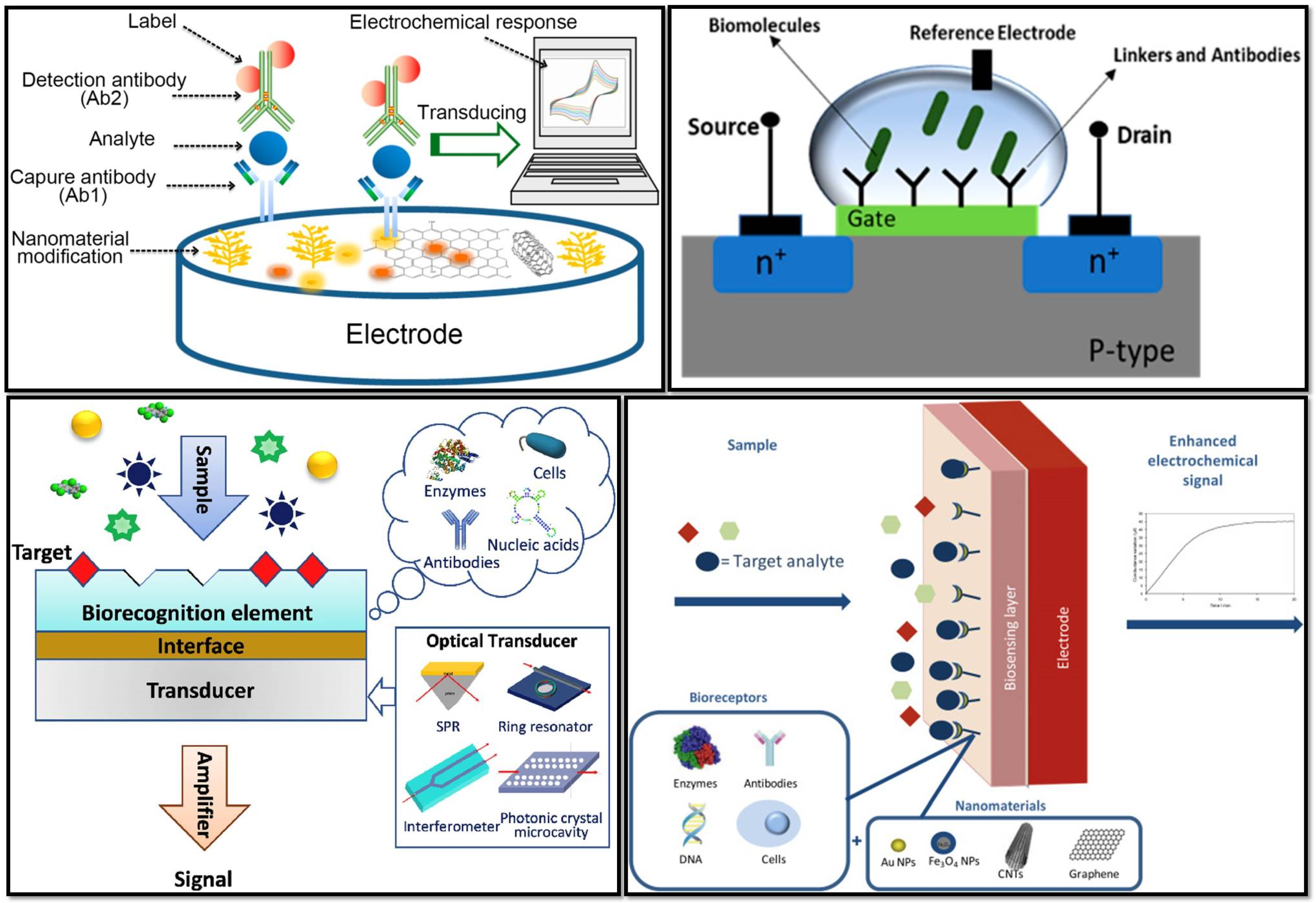
Figure 1. (Top left) A schematic representation of an electrochemical immunosensor (Reprinted with permission from Ref. [5]); (top right) schematic diagram of a field effect transistor (FET)-based biosensor with a source and drain (Reprinted with permission from Ref. [6]); (bottom left) schematic diagram of optical biosensor constitution (Reprinted with permission from Ref. [7]); (bottom right) the main constituents of a nanomaterial-based electrochemical biosensor (Reprinted with permission from Ref. [8]).
References
- Radushkevich, L.V.; Lukyanovich, V.M. The Structure of Carbon Produced by Thermal Decomposition of Carbon Monoxide on an Iron Catalyst. Russ. J. Phys. Chem. 1952, 26, 88–95.
- Iijima, S. Helical Microtubules of Graphitic Carbon. Nature 1991, 354, 56–58.
- Chemistry (IUPAC), T.I.U. of P. and A. IUPAC-Biosensor (B00663). Available online: https://goldbook.iupac.org/terms/view/B00663 (accessed on 18 October 2022).
- Bergveld, P. Development of an Ion-Sensitive Solid-State Device for Neurophysiological Measurements. IEEE Trans. Biomed. Eng. 1970, BME-17, 70–71.
- Zhang, Z.; Cong, Y.; Huang, Y.; Du, X. Nanomaterials-Based Electrochemical Immunosensors. Micromachines 2019, 10, 397.
- Masurkar, N.; Varma, S.; Mohana Reddy Arava, L. Supported and Suspended 2D Material-Based FET Biosensors. Electrochem 2020, 1, 260–277.
- Chen, Y.; Liu, J.; Yang, Z.; Wilkinson, J.S.; Zhou, X. Optical Biosensors Based on Refractometric Sensing Schemes: A Review. Biosens. Bioelectron. 2019, 144, 111693.
- Dridi, F.; Marrakchi, M.; Gargouri, M.; Saulnier, J.; Jaffrezic-Renault, N.; Lagarde, F. 5-Nanomaterial-Based Electrochemical Biosensors for Food Safety and Quality Assessment. In Nanobiosensors; Grumezescu, A.M., Ed.; Academic Press: Cambridge, MA, USA, 2017; pp. 167–204. ISBN 978-0-12-804301-1.
More
Information
Subjects:
Nanoscience & Nanotechnology
Contributors
MDPI registered users' name will be linked to their SciProfiles pages. To register with us, please refer to https://encyclopedia.pub/register
:
View Times:
957
Revisions:
2 times
(View History)
Update Date:
13 Dec 2022
Table of Contents


Notice
You are not a member of the advisory board for this topic. If you want to update advisory board member profile, please contact office@encyclopedia.pub.
OK
Confirm
Only members of the Encyclopedia advisory board for this topic are allowed to note entries. Would you like to become an advisory board member of the Encyclopedia?
Yes
No
${ textCharacter }/${ maxCharacter }
Submit
Cancel
Back
Comments
${ item }
|
${ item.createdUser.fullName }
${ item.createdAt }
${ item.vote }
${ item.reply }
Delete
${ reply.createdUser.fullName }
${ reply.createdAt }
${ reply.vote }
Delete
There is no reply to this comment~
${ item.replyTextCharacter }/${ item.replyMaxCharacter }
Submit
Cancel
More
No more~
There is no comment~
${ textCharacter }/${ maxCharacter }
Submit
Cancel
${ selectedItem.replyTextCharacter }/${ selectedItem.replyMaxCharacter }
Submit
Cancel
Confirm
Are you sure to Delete?
Yes
No