You're using an outdated browser. Please upgrade to a modern browser for the best experience.
Submitted Successfully!
Thank you for your contribution! You can also upload a video entry or images related to this topic. For video creation, please contact our Academic Video Service.
Version Summary Created by Modification Content Size Created at Operation
1 Nikos E. Papaioannou -- 2590 2022-11-23 10:07:56 |
2 Reference format revised. Lindsay Dong -2 word(s) 2588 2022-11-24 14:07:16 | |
3 Reference format revised. Lindsay Dong Meta information modification 2588 2022-11-24 14:08:02 |

Video Upload Options

We provide professional Academic Video Service to translate complex research into visually appealing presentations. Would you like to try it?
Cite
If you have any further questions, please contact Encyclopedia Editorial Office.
Kerdidani, D.;  Papaioannou, N.E.;  Nakou, E.;  Alissafi, T. Dendritic Cells as Multifaceted Orchestrators of Immune Responses. Encyclopedia. Available online: https://encyclopedia.pub/entry/36054 (accessed on 20 December 2025).
Kerdidani D,  Papaioannou NE,  Nakou E,  Alissafi T. Dendritic Cells as Multifaceted Orchestrators of Immune Responses. Encyclopedia. Available at: https://encyclopedia.pub/entry/36054. Accessed December 20, 2025.
Kerdidani, Dimitra, Nikos E. Papaioannou, Evangelia Nakou, Themis Alissafi. "Dendritic Cells as Multifaceted Orchestrators of Immune Responses" Encyclopedia, https://encyclopedia.pub/entry/36054 (accessed December 20, 2025).
Kerdidani, D.,  Papaioannou, N.E.,  Nakou, E., & Alissafi, T. (2022, November 23). Dendritic Cells as Multifaceted Orchestrators of Immune Responses. In Encyclopedia. https://encyclopedia.pub/entry/36054
Kerdidani, Dimitra, et al. "Dendritic Cells as Multifaceted Orchestrators of Immune Responses." Encyclopedia. Web. 23 November, 2022.
Dendritic Cells as Multifaceted Orchestrators of Immune Responses
Edit

Immune-mediated inflammatory diseases (IMIDs) are a group of autoimmune and chronic inflammatory disorders with constantly increasing prevalence in the modern world. The vast majority of IMIDs develop as a consequence of complex mechanisms dependent on genetic, epigenetic, molecular, cellular, and environmental elements, that lead to defects in immune regulatory guardians of tolerance, such as dendritic (DCs) cells.

immune-mediated inflammatory disorders autoimmune diseases immune regulation dendritic cells

1. Introduction

Immune-mediated inflammatory diseases (IMIDs) are a diverse group of incurable clinical disorders that constitute a unique conceptual and medical challenge for the scientific community. Under the umbrella of the broad term IMIDs, many autoimmune as well as chronic inflammatory diseases, such as rheumatoid arthritis (RA), inflammatory bowel disease (IBD), systemic lupus erythematosus (SLE), type 1 diabetes (T1D), cutaneous inflammatory disorders (including psoriasis and atopic dermatitis (AD)), asthma and autoimmune neurological diseases such as multiple sclerosis (MS), can be incorporated. IMIDs develop as a consequence of complex mechanisms that depend on genetic, epigenetic, molecular, cellular, and environmental elements and result in defects in immune regulatory checkpoints of tolerance [1][2]. This breakdown of self-tolerance leads to the aberrant activation of lymphocytes against otherwise harmless self or foreign antigens causing chronic unrestrained inflammation that destroys self-organs and tissues.
The key checkpoints of self-tolerance and decision-makers of the type and magnitude of the immune response is dendritic (DC) cells. On the one side, DCs, by up-taking environmental cues, self or foreign antigens and translating them into signals for the proper initiation of the immune response, constitute the sensors of the immune system and the link between innate and adaptive immunity [3][4]. On the other side are Tregs, that respond to signals of DCs, regulating and restraining exacerbated inflammation, thus comprising the brakes of the immune response [5][6]. During IMIDs, both cell types have been reported to be dysregulated, with altered frequencies in the periphery of patients, overt activation, and certain degrees of imbalance in their phenotype and function [7][8][9][10], thus leading to the breakdown of self-tolerance. Although the previous two decades have been transformative for the understanding of the mechanisms that govern immune dysregulation in IMIDs, effective and highly targeted treatments have proven to be elusive. Evidently, IMIDs remain a major burden on health systems around the world, accounting annually for several billion EUR in medical costs and lost income. Deciphering in depth the cellular and molecular mechanisms that contribute to the breakdown of immune tolerance is thus an important goal, with the prospect that this knowledge will pave the way to new clinical advances in the treatment of IMIDs.

2. Dendritic Cells as Multifaceted Orchestrators of Immune Responses

The multifaceted role of DCs in immune responses is a derivative of their heterogeneity. Notably, the DC term functions as an umbrella that encloses several cell subsets, each possessing distinct developmental requirements, phenotype and functional properties [11][12]. While DCs have initially been studied more extensively in mice, with the help of multi-omics approaches, recent publications have elegantly dissected the human DC compartment, elucidating in parallel a high interspecies conservation of their development, phenotype and function [12][13][14]. Among DCs, two main distinct lineages can be distinguished, namely conventional DCs (cDCs) and plasmacytoid DCs (pDCs).

In both mice and humans, pDCs have a prominent role in anti-viral defense due to their ability to secrete copious amounts of type I interferons (IFN) in response to virally derived nucleic acids [15]. The efficiency of pDCs in antigen presentation and T cell activation is still not clearly defined due to controversial findings between different experimental settings [16][17][18][19]. While their exact developmental trajectory has also been a highly debated topic in recent years, [20][21][22] the consensus is that their differentiation is dependent on the transcription factor E2-2 in both species [14][15]. On the contrary, their major defining phenotypic markers seem to be not so well-conserved. Despite MHC-II/HLA-DR expression being a common trait, murine pDCs are characterized as B220+, SiglecH+, CD317+, Ly6C+, CD11cintermediate,while in humans characteristic pDC markers are CD123, CD303, CD304, combined with a lack of CD11c and CD5 expression [14][15][23].
cDCs excel in the activation of adaptive immune responses by presenting antigens to T cells [12]. They are subsequently divided into cDC1 and cDC2 and exhibit a remarkable division of labor when it comes to their role in immune responses [12]. Both cDC subsets are characterized by the expression of CD11c and MHC-II/HLA-DR but are distinct in dependence on transcription factors and the expression of other surface markers. Continuous and high expression of the transcription factors IRF8 and BATF3 is a prerequisite for maintaining the developmental and functional program of both human and murine cDC1 [13][24][25][26]. Genetic approaches have additionally elucidated the role of ID2 [27] and NFIL3 [28][29] in mouse cDC1 development, however, their implication in humans has yet to be determined. In terms of their phenotype, murine cDC1 can be reliably identified across tissues by the expression of XCR-1, CLEC9A, CD24 and CD205 [12]. Moreover, CD8α and CD103 are used as cDC1 characteristic markers in lymphoid and non-lymphoid tissues, respectively, despite the latter also being expressed in an intestinal cDC2 population [12]. In addition to XCR-1 and CLEC9A, human cDC1 in both blood and non-lymphoid tissues have characteristic expression of CD141 and CADM1 [14][23]. Functionally, cDC1 play a dominant role in inducing cytotoxic CD8+ T and Th1 polarized CD4+ T cell responses against intracellular pathogens, such as viruses and bacteria, but also participate majorly in antitumor immunity [12]. They do so via producing ample amounts of IL-12 that activates T cells both directly and indirectly by promoting a Th1-favorable cytokine milieu from bystander cells [26][30][31]. Added to the above, their remarkable potential as CD8+ T cell activators is extended by their ability to cross-present extracellular antigens on MHC-I molecules [26][30][31]. In contrast to the pro-inflammatory role described above, especially in mice, the high potential of cDC1 to induce peripheral regulatory T cells has also been proposed [32][33].
In contrast to pDCs and cDC1 subtypes, the phenotype and developmental requirements of cDC2 between humans and mice seem to overlap the least. In mice, studies have identified transcription factors IRF-4, ZEB2, KLF4 and RELB as central mediators of cDC2 development [11][12][29] as well as pathways with more tissue-specific context such as NOTCH and retinoic acid signaling [34]. While human cDC2 distinctively expresses IRF-4, its role in their development is not yet elucidated. Characteristic murine cDC2 surface markers include CD11b, CD172a, CD4 and CLEC4A4 [12] of which only CD172a is a common defining marker with their human counterparts. The latter are additionally identified by their expression of CD1c, FcεR1α and CLEC10A [14][23]. Functionally, human and murine cDC2 align and are believed to be more efficient in inducing CD4+ T cell activation and polarization towards Tfh, Th2 or Th17 effector responses, crucial for T cell-dependent antibody production by B cells, defense against multicellular pathogens such as helminths or extracellular bacteria and fungi, respectively [35][36][37][38][39][40][41][42]. Their CD4+ T cell activation pattern also extends to regulatory directions via the induction of Tregs both in the thymus and in peripheral tissues [43][44]. Remarkably, cDC2 have been found to exhibit the highest intra-subset diversity compared to pDCs and cDC1. This heterogeneity, despite being ever-growing, has been studied in detail in mice [45][46][47], however, it has only recently been appreciated in humans.

2.1. Elucidating the Role of Dendritic Cells in IMIDs Utilizing Multi-Omics Approaches

Given their role in maintaining the balance between protective immune responses and self-tolerance, DCs play a critical part in IMID manifestations in which this balance is by default perturbed. Their detailed role has been extensively reviewed elsewhere [7], and in brief entails the dysregulation of one or more of the following functional properties: (a) perturbation in the pattern of secreted cytokines, quantitatively and qualitatively, that promote pro-inflammatory responses from other innate and adaptive immune system cells; (b) enhanced antigen presentation of primarily self-antigens; and (c) altered distribution in terms of both frequency and spatial arrangement, often related to differences in their migratory capacity, that affects especially the inflamed tissues but also peripheral blood. Here, cases in which the role of DCs in IMIDs has been refined or enriched by the advent of recent omics approaches (Figure 1) are reported.
Figure 1. Multi-omics approaches utilized in IMIDs research focusing on dendritic cells. The Pie chart depicts omics technologies that have been used to study the contribution of dendritic cells in the pathology of IMIDs. Mainly scRNA-seq but also proteomic and metabolomic studies have highlighted dendritic cell subsets and inflammatory signatures that drive pathogenic responses in the disease spectrum of IMIDs. scRNA-seq, single cell RNA-sequencing; RA, Rheumatoid Arthritis; SLE, Systemic Lupus Erythematosus; T1D, Type I Diabetes; AD, Atopic Dermatitis; PsO, Psoriasis; Ssc, Systemic sclerosis.

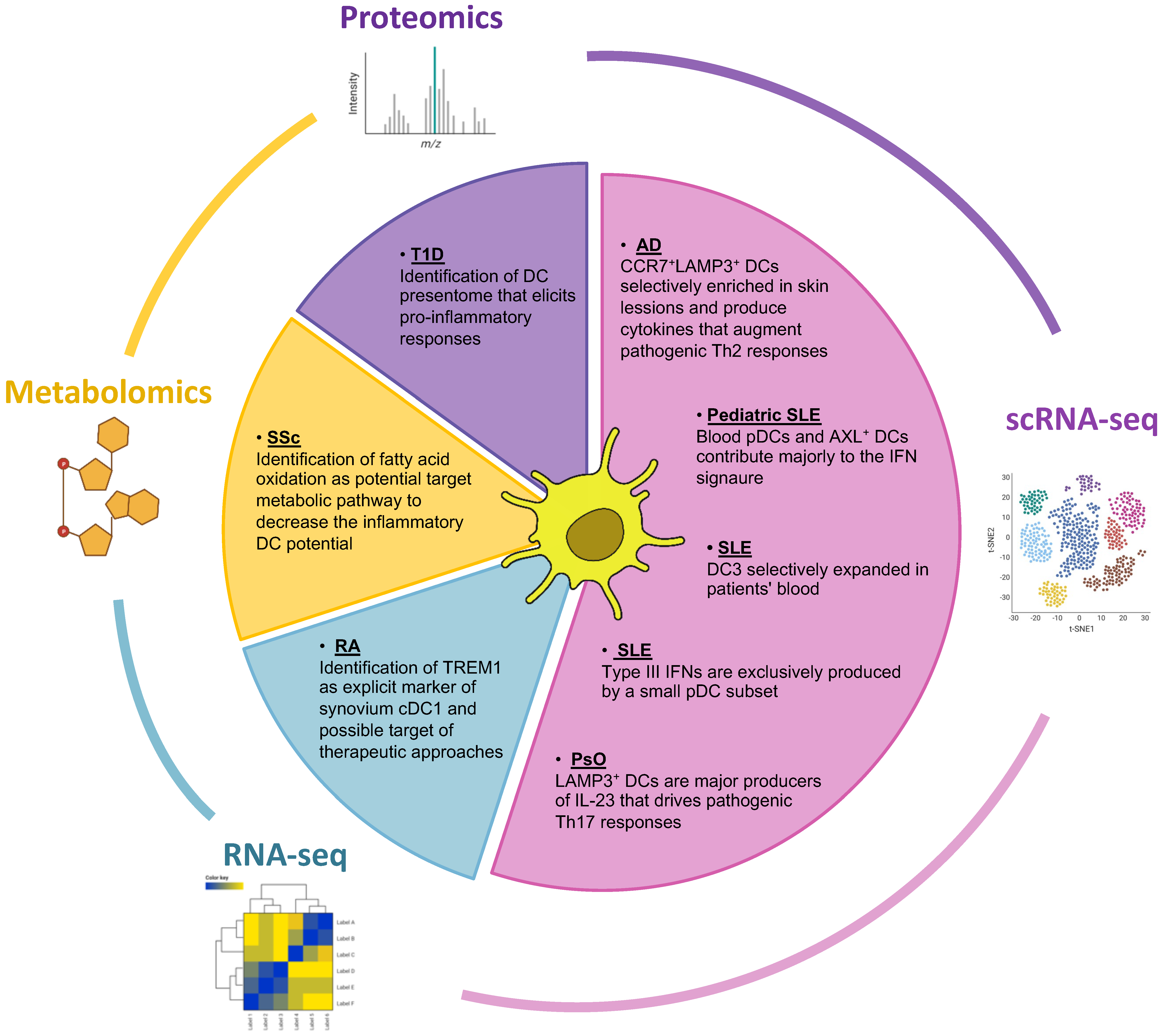
2.2. Bulk and Single-Cell RNA Sequencing Have Expanded the Portfolio of DC Subsets and Illuminated Their Role in IMIDs Perturbations

One remarkable advantage of multi-omics approaches is their potential for single-cell resolution. This was made apparent especially for human cDC2, as recent studies identified novel subsets within the CD1c+ cDC2 population using scRNA-seq coupled with index sorting [48][49]. The subdivision of these new subpopulations, namely DC2 (CD5+/−CD163CD14) and DC3 (CD5CD163+CD14+/−), based on their immunophenotype was also found to be accompanied by functional differences [49][50]. In the context of IMIDs, CD163+ DC3s were found to be expanded in the blood of SLE patients and presented a highly activated phenotype compared to healthy controls. Interestingly, their frequency in blood was highly correlated to clinical scores. Secretome analysis showed that, among cDC2 subsets, DC3s uniquely produced many pro-inflammatory mediators when activated by the serum of SLE patients [49]. Given the above, it would be intriguing to investigate the performance of these cells as disease biomarkers and establish whether manipulating their function could ameliorate disease progression. Additionally, their role in other IMIDs such as RA and Psoriatic Arthritis (PsA) warrants further investigation due to their increased potential for induction of IL-17A producing T cells [49].
DC3s were also found selectively expanded, among cDC2, as assessed by scRNA-seq in pediatric SLE (cSLE) patients’ peripheral blood mononuclear cells (PBMCs), compared to age-matched healthy individuals [51]. Interestingly, overtaking even DC3s, the majorly expanded cDC cluster resembled the AXL+ DCs first identified by Villani et al. [48]. Additionally, although pDCs were found decreased as a total population in cSLE samples, further analysis revealed four distinct subclusters, one of which was profoundly expanded in SLE compared to healthy individuals. Notably, the defining markers of this expanded pDCs subcluster consisted primarily of interferon-induced genes, accompanied by genes connected to transcription factors (e.g., STAT1, IRF7) and antigen presentation (e.g., CD74, HLA-DRA, CTSB) [51].
In most cases, peripheral blood has been used as a mirror to study DC properties in IMIDs, however, analyses from inflamed tissues are equally or even more important as suggested by the expected effect of tissue microenvironments in DC transcriptional and functional signatures [52][53]. This site-specific analysis has been bolstered by recent omics advances, since their high throughput performance automatically decreases the required cell numbers to conduct meaningful experiments. As an example, Caravan and colleagues, studied the impact of the synovial microenvironment in cDCs from RA patients [54][55]. Using multiparameter flow cytometry and RNA sequencing, they found not only that cDCs were enriched in the synovial tissue of RA patients, compared to the blood of the same individuals as well as that of healthy controls, but that they also exhibited a highly activated phenotype as assessed by expression of costimulatory molecules [54][55]. Regarding the CD1c+ cDC2, the synovial microenvironment was shown to induce metabolic alterations, polarizing them to a more glycolytic phenotype while a more detailed analysis was performed for CD141+ cDC1. For the latter, the hypoxic synovium was shown to specifically induce the expression of TREM-1 as part of a site and disease-specific signature. Interestingly, in vitro crosslinking of TREM-1 in cDC1 isolated from synovial tissue could induce their activated phenotype in parallel to an increased ability to induce pro-inflammatory cytokine production from heterologous and autologous T cells [54].
Omics analysis targeted to the inflamed tissue is additionally essential for another IMID, namely AD. Two recent studies have attempted to interrogate the immune and non-immune skin compartments of patients with AD and healthy controls using scRNA-seq [56][57]. Both studies found that DC populations were expanded in the pathogenic samples in relation to healthy skin, with cDC2 probably being the more over-represented population due to their characteristic expression pattern of surface markers. Alongside the “typical” cDC populations, another smaller cluster expressing CCR7 and LAMP3 was identified. Despite their small numbers, these cells exhibited some very interesting traits such as clear characteristics of mature and migratory behavior and selective enrichment in the lesional skin of AD patients combined with their almost complete absence from healthy samples [56][57].

2.3. Contribution of Proteomics in the Identification of DC-Presented Epitopes in IMIDs

As mentioned above, a prominent DC function lies in the processing and presentation of autoantigens to autoreactive T cells. However, an up-to-the-point question concerns whether specific epitopes dominate and are preferentially presented, even in cases that a particulate cell population is targeted. In the example of T1D, a recent study aimed to delineate the naturally processed and presented epitopes by DCs as derived from pancreatic beta cells [58]. The authors isolated peripheral blood CD14+ monocytes from healthy donors and cultured them in vitro with GM-CSF and IL-4 in order to generate moDCs. Then, these moDCs were pulsed in vitro with various pancreatic islet autoantigens and then their “presentome” was analyzed (the eluted epitopes presented in the surface HLA-DR molecules) with mass spectrometry. Their experimental set up also held augmented clinical relevance as they selectively used moDCs from individuals possessing the alleles HLA-DR3 and HLA-DR4, associated with high-risk of disease emergence. Among their findings was the addition of new epitopes to those already characterized for some peptide autoantigens as well as the discovery of some derived from pancreatic islet peptides for which epitope generation had not been previously reported. Interestingly, they were able to show that not all the discovered epitopes induce a pro-inflammatory reaction, evident by the response magnitude and its induced IL-10 or IFN-α signature upon incubation of PBMCs from T1D patients with them. Importantly, the most immunodominant epitopes were generated by moDCs when compared to B cells, another immune system cell with antigen presentation capacity, in an HLA-DR allele independent manner. Thus, such approaches could help increase the efficacy of peptide-based tolerogenic immunotherapies as well as their patient-specific tailoring by using epitopes preferentially presented by each HLA haplotype in moDCs.

2.4. Bridging the Metabolic Profile and the Function of DCs in IMIDs: An Emerging Field of Research

The relevance of metabolomics in IMIDs is constantly increasing, however, its targeted application in DCs is only lately gaining attention. Towards that point, a recent study aimed to identify metabolic pathways exhibiting similar dysregulation in the circulation and DCs of patients with systemic sclerosis [59]. To this end, they first performed metabolite analysis using plasma of patients and healthy individuals and discovered evidence of imbalanced fatty acid and carnitine levels in systemic sclerosis samples. In line with this, they also found increased levels of L-carnitine in moDCs, derived from GM-CSF and IL-4 cultures of peripheral blood monocytes from systemic sclerosis patients, after their stimulation with TLR agonists. As a continuation of their observations, the authors tested the effect of etoposide, a carnitine transporter inhibitor widely used for cancer treatment, on the activation of patient-derived moDCs after TLR stimulation and showed secretion of reduced levels of pro-inflammatory cytokines such as IL-6 in its presence. As carnitine transports fatty acids into the mitochondria in order to facilitate their oxidation, the above observations suggest that targeted suppression of fatty acid oxidation in DCs could be helpful in decreasing the inflammation related to the particular IMID.

References

  1. Wahren-Herlenius, M.; Dörner, T. Immunopathogenic Mechanisms of Systemic Autoimmune Disease. Lancet 2013, 382, 819–831.
  2. Bluestone, J.A.; Anderson, M. Tolerance in the Age of Immunotherapy. N. Engl. J. Med. 2020, 383, 1156–1166.
  3. Steinman, R.M.; Hemmi, H. Dendritic Cells: Translating Innate to Adaptive Immunity. Curr. Top. Microbiol. Immunol. 2006, 311, 17–58.
  4. Mellman, I.; Steinman, R.M. Dendritic Cells Specialized and Regulated Antigen Processing Machines. Cell 2001, 106, 255–258.
  5. Sakaguchi, S.; Yamaguchi, T.; Nomura, T.; Ono, M. Regulatory T Cells and Immune Tolerance. Cell 2008, 133, 775–787.
  6. Fontenot, J.D.; Gavin, M.A.; Rudensky, A.Y. Foxp3 Programs the Development and Function of CD4+CD25+ Regulatory T Cells. Nat. Immunol. 2003, 4, 330–336.
  7. Coutant, F.; Miossec, P. Altered Dendritic Cell Functions in Autoimmune Diseases: Distinct and Overlapping Profiles. Nat. Rev. Rheumatol. 2016, 12, 703–715.
  8. Sakaguchi, S.; Mikami, N.; Wing, J.B.; Tanaka, A.; Ichiyama, K.; Ohkura, N. Regulatory T Cells and Human Disease. Annu. Rev. Immunol. 2020, 38, 541–566.
  9. Dominguez-Villar, M.; Hafler, D.A. Regulatory T Cells in Autoimmune Disease. Nat. Immunol. 2018, 19, 665–673.
  10. Hatzioannou, A.; Boumpas, A.; Papadopoulou, M.; Papafragkos, I.; Varveri, A.; Alissafi, T.; Verginis, P. Regulatory T Cells in Autoimmunity and Cancer: A Duplicitous Lifestyle. Front. Immunol. 2021, 12, 731947.
  11. Durai, V.; Murphy, K.M. Functions of Murine Dendritic Cells. Immunity 2016, 45, 719–736.
  12. Cabeza-Cabrerizo, M.; Cardoso, A.; Minutti, C.M.; da Costa, M.P.; Reis e Sousa, C. Dendritic Cells Revisited. Annu. Rev. Immunol. 2021, 39, 131–166.
  13. Guilliams, M.; Dutertre, C.-A.; Scott, C.L.; McGovern, N.; Sichien, D.; Chakarov, S.; Gassen, S.V.; Chen, J.; Poidinger, M.; Prijck, S.D.; et al. Unsupervised High-Dimensional Analysis Aligns Dendritic Cells across Tissues and Species. Immunity 2016, 45, 669–684.
  14. Patente, T.A.; Pinho, M.P.; Oliveira, A.A.; Evangelista, G.C.M.; Bergami-Santos, P.C.; Barbuto, J.A.M. Human Dendritic Cells: Their Heterogeneity and Clinical Application Potential in Cancer Immunotherapy. Front. Immunol. 2019, 9, 3176.
  15. Reizis, B. Plasmacytoid Dendritic Cells: Development, Regulation, and Function. Immunity 2019, 50, 37–50.
  16. See, P.; Dutertre, C.-A.; Chen, J.; Günther, P.; McGovern, N.; Irac, S.E.; Gunawan, M.; Beyer, M.; Händler, K.; Duan, K.; et al. Mapping the Human DC Lineage through the Integration of High-Dimensional Techniques. Science 2017, 356, eaag3009.
  17. Matsui, T.; Connolly, J.E.; Michnevitz, M.; Chaussabel, D.; Yu, C.I.; Glaser, C.; Tindle, S.; Pypaert, M.; Freitas, H.; Piqueras, B.; et al. CD2 Distinguishes Two Subsets of Human Plasmacytoid Dendritic Cells with Distinct Phenotype and Functions. J. Immunol. 2009, 182, 6815.
  18. Villadangos, J.A.; Young, L. Antigen-Presentation Properties of Plasmacytoid Dendritic Cells. Immunity 2008, 29, 352–361.
  19. Hoeffel, G.; Ripoche, A.C.; Matheoud, D.; Nascimbeni, M.; Escriou, N.; Lebon, P.; Heshmati, F.; Guillet, J.G.; Gannagé, M.; Caillat-Zucman, S.; et al. Antigen Crosspresentation by Human Plasmacytoid Dendritic Cells. Immunity 2007, 27, 481–492.
  20. Rodrigues, P.F.; Alberti-Servera, L.; Eremin, A.; Grajales-Reyes, G.E.; Ivanek, R.; Tussiwand, R. Distinct Progenitor Lineages Contribute to the Heterogeneity of Plasmacytoid Dendritic Cells. Nat. Immunol. 2018, 19, 711–722.
  21. Dress, R.J.; Dutertre, C.-A.; Giladi, A.; Schlitzer, A.; Low, I.; Shadan, N.B.; Tay, A.; Lum, J.; Kairi, M.F.B.M.; Hwang, Y.Y.; et al. Plasmacytoid Dendritic Cells Develop from Ly6D+ Lymphoid Progenitors Distinct from the Myeloid Lineage. Nat. Immunol. 2019, 20, 852–864.
  22. Feng, J.; Pucella, J.N.; Jang, G.; Alcántara-Hernández, M.; Upadhaya, S.; Adams, N.M.; Khodadadi-Jamayran, A.; Lau, C.M.; Stoeckius, M.; Hao, S.; et al. Clonal Lineage Tracing Reveals Shared Origin of Conventional and Plasmacytoid Dendritic Cells. Immunity 2022, 55, 405–422.e11.
  23. Mair, F.; Liechti, T. Comprehensive Phenotyping of Human Dendritic Cells and Monocytes. Cytom. Part A 2021, 99, 231–242.
  24. Poulin, L.F.; Reyal, Y.; Uronen-Hansson, H.; Schraml, B.U.; Sancho, D.; Murphy, K.M.; Hakansson, U.K.; Moita, L.F.; Agace, W.W.; Bonnet, D.; et al. DNGR-1 Is a Specific and Universal Marker of Mouse and Human Batf3-Dependent Dendritic Cells in Lymphoid and Nonlymphoid Tissues. Blood 2012, 119, 6052–6062.
  25. Sichien, D.; Scott, C.L.; Martens, L.; Vanderkerken, M.; Gassen, S.V.; Plantinga, M.; Joeris, T.; Prijck, S.D.; Vanhoutte, L.; Vanheerswynghels, M.; et al. IRF8 Transcription Factor Controls Survival and Function of Terminally Differentiated Conventional and Plasmacytoid Dendritic Cells, Respectively. Immunity 2016, 45, 626–640.
  26. Hildner, K.; Edelson, B.T.; Purtha, W.E.; Diamond, M.; Matsushita, H.; Kohyama, M.; Calderon, B.; Schraml, B.U.; Unanue, E.R.; Diamond, M.S.; et al. Batf3 Deficiency Reveals a Critical Role for CD8α+ Dendritic Cells in Cytotoxic T Cell Immunity. Science 2008, 322, 1097–1100.
  27. Hacker, C.; Kirsch, R.D.; Ju, X.S.; Hieronymus, T.; Gust, T.C.; Kuhl, C.; Jorgas, T.; Kurz, S.M.; Rose-John, S.; Yokota, Y.; et al. Transcriptional Profiling Identifies Id2 Function in Dendritic Cell Development. Nat. Immunol. 2003, 4, 380–386.
  28. Kashiwada, M.; Pham, N.L.; Pewe, L.L.; Harty, J.T.; Rothman, P.B. NFIL3/E4BP4 Is a Key Transcription Factor for CD8α+ Dendritic Cell Development. Blood 2011, 117, 6193–6197.
  29. Liu, T.-T.; Kim, S.; Desai, P.; Kim, D.-H.; Huang, X.; Ferris, S.T.; Wu, R.; Ou, F.; Egawa, T.; Dyken, S.J.V.; et al. Ablation of cDC2 Development by Triple Mutations within the Zeb2 Enhancer. Nature 2022, 607, 142–148.
  30. Reis e Sousa, C.; Hieny, S.; Scharton-Kersten, T.; Jankovic, D.; Charest, H.; Germain, R.N.; Sher, A. In Vivo Microbial Stimulation Induces Rapid CD40 Ligand-Independent Production of Interleukin 12 by Dendritic Cells and Their Redistribution to T Cell Areas. J. Exp. Med. 1997, 186, 1819–1829.
  31. Mashayekhi, M.; Sandau, M.M.; Dunay, I.R.; Frickel, E.M.; Khan, A.; Goldszmid, R.S.; Sher, A.; Ploegh, H.L.; Murphy, T.L.; Sibley, L.D.; et al. CD8α+ Dendritic Cells Are the Critical Source of Interleukin-12 That Controls Acute Infection by Toxoplasma Gondii Tachyzoites. Immunity 2011, 35, 249–259.
  32. Idoyaga, J.; Fiorese, C.; Zbytnuik, L.; Lubkin, A.; Miller, J.; Malissen, B.; Mucida, D.; Merad, M.; Steinman, R.M. Specialized Role of Migratory Dendritic Cells in Peripheral Tolerance Induction. J. Clin. Investig. 2013, 123, 844–854.
  33. Tabansky, I.; Keskin, D.B.; Watts, D.; Petzold, C.; Funaro, M.; Sands, W.; Wright, P.; Yunis, E.J.; Najjar, S.; Diamond, B.; et al. Targeting DEC-205-DCIR2+ Dendritic Cells Promotes Immunological Tolerance in Proteolipid Protein-Induced Experimental Autoimmune Encephalomyelitis. Mol. Med. 2018, 24, 17.
  34. Bosteels, C.; Scott, C.L. Transcriptional Regulation of DC Fate Specification. Mol. Immunol. 2020, 121, 38–46.
  35. Dudziak, D.; Kamphorst, A.O.; Heidkamp, G.F.; Buchholz, V.R.; Trumpfheller, C.; Yamazaki, S.; Cheong, C.; Liu, K.; Lee, H.W.; Park, C.G.; et al. Differential Antigen Processing by Dendritic Cell Subsets in Vivo. Science 2007, 315, 107–111.
  36. Shin, C.; Han, J.-A.; Choi, B.; Cho, Y.-K.; Do, Y.; Ryu, S. Intrinsic Features of the CD8α− Dendritic Cell Subset in Inducing Functional T Follicular Helper Cells. Immunol. Lett. 2016, 172, 21–28.
  37. Persson, E.K.; Uronen-Hansson, H.; Semmrich, M.; Rivollier, A.; Hägerbrand, K.; Marsal, J.; Gudjonsson, S.; Håkansson, U.; Reizis, B.; Kotarsky, K.; et al. IRF4 Transcription-Factor-Dependent CD103+CD11b+ Dendritic Cells Drive Mucosal T Helper 17 Cell Differentiation. Immunity 2013, 38, 958–969.
  38. Schlitzer, A.; McGovern, N.; Teo, P.; Zelante, T.; Atarashi, K.; Low, D.; Ho, A.W.S.; See, P.; Shin, A.; Wasan, P.S.; et al. IRF4 Transcription Factor-Dependent CD11b+ Dendritic Cells in Human and Mouse Control Mucosal IL-17 Cytokine Responses. Immunity 2013, 38, 970–983.
  39. Tussiwand, R.; Everts, B.; Grajales-Reyes, G.E.; Kretzer, N.M.; Iwata, A.; Bagaitkar, J.; Wu, X.; Wong, R.; Anderson, D.A.; Murphy, T.L.; et al. Klf4 Expression in Conventional Dendritic Cells Is Required for T Helper 2 Cell Responses. Immunity 2015, 42, 916–928.
  40. Rojas, I.M.L.; Mok, W.-H.; Pearson, F.E.; Minoda, Y.; Kenna, T.J.; Barnard, R.T.; Radford, K.J. Human Blood CD1c+ Dendritic Cells Promote Th1 and Th17 Effector Function in Memory CD4+ T Cells. Front. Immunol. 2017, 8, 971.
  41. Binnewies, M.; Mujal, A.M.; Pollack, J.L.; Combes, A.J.; Hardison, E.A.; Barry, K.C.; Tsui, J.; Ruhland, M.K.; Kersten, K.; Abushawish, M.A.; et al. Unleashing Type-2 Dendritic Cells to Drive Protective Antitumor CD4+ T Cell Immunity. Cell 2019, 177, 556–571.e16.
  42. Mayer, J.U.; Hilligan, K.L.; Chandler, J.S.; Eccles, D.A.; Old, S.I.; Domingues, R.G.; Yang, J.; Webb, G.R.; Munoz-Erazo, L.; Hyde, E.J.; et al. Homeostatic IL-13 in Healthy Skin Directs Dendritic Cell Differentiation to Promote TH2 and Inhibit TH17 Cell Polarization. Nat. Immunol. 2021, 22, 1538–1550.
  43. Bamboat, Z.M.; Stableford, J.A.; Plitas, G.; Burt, B.M.; Nguyen, H.M.; Welles, A.P.; Gonen, M.; Young, J.W.; DeMatteo, R.P. Human Liver Dendritic Cells Promote T Cell Hyporesponsiveness. J. Immunol. 2009, 182, 1901.
  44. Hu, Z.; Li, Y.; Nieuwenhuijze, A.V.; Selden, H.J.; Jarrett, A.M.; Sorace, A.G.; Yankeelov, T.E.; Liston, A.; Ehrlich, L.I.R. CCR7 Modulates the Generation of Thymic Regulatory T Cells by Altering the Composition of the Thymic Dendritic Cell Compartment. Cell Rep. 2017, 21, 168–180.
  45. Brown, C.C.; Gudjonson, H.; Pritykin, Y.; Deep, D.; Lavallée, V.-P.; Mendoza, A.; Fromme, R.; Mazutis, L.; Ariyan, C.; Leslie, C.; et al. Transcriptional Basis of Mouse and Human Dendritic Cell Heterogeneity. Cell 2019, 179, 846–863.e24.
  46. Lewis, K.L.; Caton, M.L.; Bogunovic, M.; Greter, M.; Grajkowska, L.T.; Ng, D.; Klinakis, A.; Charo, I.F.; Jung, S.; Gommerman, J.L.; et al. Notch2 Receptor Signaling Controls Functional Differentiation of Dendritic Cells in the Spleen and Intestine. Immunity 2011, 35, 780–791.
  47. Bosteels, C.; Neyt, K.; Vanheerswynghels, M.; Helden, M.J.V.; Sichien, D.; Debeuf, N.; Prijck, S.D.; Bosteels, V.; Vandamme, N.; Martens, L.; et al. Inflammatory Type 2 cDCs Acquire Features of cDC1s and Macrophages to Orchestrate Immunity to Respiratory Virus Infection. Immunity 2020, 52, 1039–1056.e9.
  48. Villani, A.-C.; Satija, R.; Reynolds, G.; Sarkizova, S.; Shekhar, K.; Fletcher, J.; Griesbeck, M.; Butler, A.; Zheng, S.; Lazo, S.; et al. Single-Cell RNA-Seq Reveals New Types of Human Blood Dendritic Cells, Monocytes, and Progenitors. Science 2017, 356, eaah4573.
  49. Dutertre, C.-A.; Becht, E.; Irac, S.E.; Khalilnezhad, A.; Narang, V.; Khalilnezhad, S.; Ng, P.Y.; van den Hoogen, L.L.; Leong, J.Y.; Lee, B.; et al. Single-Cell Analysis of Human Mononuclear Phagocytes Reveals Subset-Defining Markers and Identifies Circulating Inflammatory Dendritic Cells. Immunity 2019, 51, 573–589.e8.
  50. Bourdely, P.; Anselmi, G.; Vaivode, K.; Ramos, R.N.; Missolo-Koussou, Y.; Hidalgo, S.; Tosselo, J.; Nuñez, N.; Richer, W.; Vincent-Salomon, A.; et al. Transcriptional and Functional Analysis of CD1c+ Human Dendritic Cells Identifies a CD163+ Subset Priming CD8+CD103+ T Cells. Immunity 2020, 53, 335–352.e8.
  51. Nehar-Belaid, D.; Hong, S.; Marches, R.; Chen, G.; Bolisetty, M.; Baisch, J.; Walters, L.; Punaro, M.; Rossi, R.J.; Chung, C.-H.; et al. Mapping Systemic Lupus Erythematosus Heterogeneity at the Single-Cell Level. Nat. Immunol. 2020, 21, 1094–1106.
  52. Liu, K.; Victora, G.D.; Schwickert, T.A.; Guermonprez, P.; Meredith, M.M.; Yao, K.; Chu, F.-F.; Randolph, G.J.; Rudensky, A.Y.; Nussenzweig, M. In Vivo Analysis of Dendritic Cell Development and Homeostasis. Science 2009, 324, 392–397.
  53. Granot, T.; Senda, T.; Carpenter, D.J.; Matsuoka, N.; Weiner, J.; Gordon, C.L.; Miron, M.; Kumar, B.V.; Griesemer, A.; Ho, S.-H.; et al. Dendritic Cells Display Subset and Tissue-Specific Maturation Dynamics over Human Life. Immunity 2017, 46, 504–515.
  54. Canavan, M.; Walsh, A.M.; Bhargava, V.; Wade, S.M.; McGarry, T.; Marzaioli, V.; Moran, B.; Biniecka, M.; Convery, H.; Wade, S.; et al. Enriched Cd141+ DCs in the Joint Are Transcriptionally Distinct, Activated, and Contribute to Joint Pathogenesis. JCI Insight 2018, 3, e95228.
  55. Canavan, M.; Marzaioli, V.; McGarry, T.; Bhargava, V.; Nagpal, S.; Veale, D.J.; Fearon, U. Rheumatoid Arthritis Synovial Microenvironment Induces Metabolic and Functional Adaptations in Dendritic Cells. Clin. Exp. Immunol. 2020, 202, 226–238.
  56. He, H.; Suryawanshi, H.; Morozov, P.; Gay-Mimbrera, J.; Duca, E.D.; Kim, H.J.; Kameyama, N.; Estrada, Y.; Der, E.; Krueger, J.G.; et al. Single-Cell Transcriptome Analysis of Human Skin Identifies Novel Fibroblast Subpopulation and Enrichment of Immune Subsets in Atopic Dermatitis. J. Allergy Clin. Immun. 2020, 145, 1615–1628.
  57. Rojahn, T.B.; Vorstandlechner, V.; Krausgruber, T.; Bauer, W.M.; Alkon, N.; Bangert, C.; Thaler, F.M.; Sadeghyar, F.; Fortelny, N.; Gernedl, V.; et al. Single-Cell Transcriptomics Combined with Interstitial Fluid Proteomics Defines Cell Type–Specific Immune Regulation in Atopic Dermatitis. J. Allergy Clin. Immun. 2020, 146, 1056–1069.
  58. Van Lummel, M.; van Veelen, P.A.; de Ru, A.H.; Janssen, G.M.C.; Pool, J.; Laban, S.; Joosten, A.M.; Nikolic, T.; Drijfhout, J.W.; Mearin, M.L.; et al. Dendritic Cells Guide Islet Autoimmunity through a Restricted and Uniquely Processed Peptidome Presented by High-Risk HLA-DR. J. Immunol. 2016, 196, 3253–3263.
  59. Ottria, A.; Hoekstra, A.T.; Zimmermann, M.; van der Kroef, M.; Vazirpanah, N.; Cossu, M.; Chouri, E.; Rossato, M.; Beretta, L.; Tieland, R.G.; et al. Fatty Acid and Carnitine Metabolism Are Dysregulated in Systemic Sclerosis Patients. Front. Immunol. 2020, 11, 822.
More
Upload a video for this entry
Information
Subjects: Immunology
Contributors MDPI registered users' name will be linked to their SciProfiles pages. To register with us, please refer to https://encyclopedia.pub/register : Dimitra Kerdidani , Nikos E. Papaioannou , Evangelia Nakou , Themis Alissafi
View Times: 712
Revisions: 3 times (View History)
Update Date: 24 Nov 2022
Notice
You are not a member of the advisory board for this topic. If you want to update advisory board member profile, please contact office@encyclopedia.pub.
OK
Confirm
Only members of the Encyclopedia advisory board for this topic are allowed to note entries. Would you like to become an advisory board member of the Encyclopedia?
Yes
No
${ textCharacter }/${ maxCharacter }
Submit
Cancel
There is no comment~
${ textCharacter }/${ maxCharacter }
Submit
Cancel
${ selectedItem.replyTextCharacter }/${ selectedItem.replyMaxCharacter }
Submit
Cancel
Confirm
Are you sure to Delete?
Yes No
Academic Video Service