Submitted Successfully!
To reward your contribution, here is a gift for you: A free trial for our video production service.
Thank you for your contribution! You can also upload a video entry or images related to this topic.
Version Summary Created by Modification Content Size Created at Operation
1 -- 4465 2022-11-21 13:13:20 |
2 update references and layout -183 word(s) 4282 2022-11-22 02:05:05 | |
3 update layout + 3 word(s) 4285 2022-11-22 02:07:46 |

Video Upload Options

Do you have a full video?

Confirm

Are you sure to Delete?
Cite
If you have any further questions, please contact Encyclopedia Editorial Office.
Beňačka, R.;  Szabóová, D.;  Guľašová, Z.;  Hertelyová, Z.;  Radoňák, J. Markers in Diagnostics and Classification of Breast Cancer. Encyclopedia. Available online: https://encyclopedia.pub/entry/35574 (accessed on 27 July 2024).
Beňačka R,  Szabóová D,  Guľašová Z,  Hertelyová Z,  Radoňák J. Markers in Diagnostics and Classification of Breast Cancer. Encyclopedia. Available at: https://encyclopedia.pub/entry/35574. Accessed July 27, 2024.
Beňačka, Roman, Daniela Szabóová, Zuzana Guľašová, Zdenka Hertelyová, Jozef Radoňák. "Markers in Diagnostics and Classification of Breast Cancer" Encyclopedia, https://encyclopedia.pub/entry/35574 (accessed July 27, 2024).
Beňačka, R.,  Szabóová, D.,  Guľašová, Z.,  Hertelyová, Z., & Radoňák, J. (2022, November 21). Markers in Diagnostics and Classification of Breast Cancer. In Encyclopedia. https://encyclopedia.pub/entry/35574
Beňačka, Roman, et al. "Markers in Diagnostics and Classification of Breast Cancer." Encyclopedia. Web. 21 November, 2022.
Markers in Diagnostics and Classification of Breast Cancer
Edit

Breast cancer (BC)is etiologically, histopathologically and genetically a heterogeneous disease with both hereditary predispositions and non-hereditary factors. This is certainly true for BC as it refers to mammary carcinoma from ductal or lobular cells in the mammary epithelial tissue. Only a minor portion are sarcomas transformed from connective tissue and vessels. Malignant transformation in BC is the product of accumulations of consecutive mutations in critical regions of the genome that are normally involved in control of cell growth and division, DNA repair and programmed cell death. These mutations are partly inherited but mostly spontaneous. Contribution of genetic factors in BC has been indicated by familial occurrence which is estimated as 5–10% of all cases. High-penetrance genes which are linked with inherited BC susceptibility include BRCA1 and BRCA2, and more rarely TP53, PTEN, CDH1 and SKT11. Heterozygotic mutations in DNA repair genes BRCA1 (locus Ch17q21.31) or BRCA2 (Ch13q13.1) are the most common inherited conditions associated with BC. Absolute risk of BC for BRCA1 mutations reads ~50–65% in females and ~1% in males, while in BRCA2 mutations female risk ranges from 40% to 55% and reaches up to 9% in males. Familiar susceptibility to BC is also associated with mutations of lower penetrance genes as ATM (Ch11q22.3), PALB2 (Ch16p12.2) and CHEK2 (Ch22q12.1). Mutation in the androgen receptor gene (AR) has been found in cases of male BC. Susceptibility to sporadic BC cases can be linked with many more genes: e.g., sporadic invasive ductal variant of BC and lobular BC are associated with somatic mutation of genes RAD54L (Ch1p34.1) and CDH1 (Ch16q22.1), respectively. Other genes candidates associated with sporadic BC include: TP53 (Ch17p13.1), SLC22A1 (Ch11p15.4), PIK3CA (Ch3q26.32), ESR1 (Ch6q25.1-q25.2), RB1CC1 (Ch8q11.23), KRAS (12p12.1), AKT1 (14q32.33), RB1 (Ch13q14.2), PPM1D (Ch17q23.2), MYC (Ch8q24.21), FGFR1 and eventually ERBB2 (Ch17q12), CCND1 (Ch11q13.3), GATA3 (Ch10p14), MAP3K1 (Ch5q11.2) in certain lineages.

breast cancer histological and molecular subtypes molecular biomarkers breast cancer gene candidates

1. Diagnostics and Surveillance of the Breast Cancer (BC)

Most of current breast-cancer-related diagnostic procedures fall into one or more of the following categories:
(a) Screening tests are done routinely to people who are not suspected of having BC. Self-performed manual palpation of breast (BSE) guided by professionals is considered an effective way of early detection of breast tumour. Findings of lumps, redness, thickenings or asymmetries in breasts and enlargement of axillary lymph nodes should be examined by medical professionals as the obvious next step. Regular screening has been one of the major tools in mortality decline over the past decade [1][2][3][4];
(b) Diagnostic tests are done in those suspected of having BC, either because of symptoms or screening results. These tests include mammography [5], or MRI [6], or biopsy, or a combination of all in uncertain cases [7]:
  • Mammography and breast NMRI (Magnetic Resonance Imaging) are useful non-invasive ways of how to exclude eventual other palpable breast lumps as abscess, cysts or fibroadenomas [5];
  • Biopsy is a preferred diagnostic tool. It can be done either as fine needle aspiration (FNA) or ultrasound-guided or stereotactic-navigated core needle biopsy (CNB). More recent minimally invasive breast biopsy or vacuum-assisted biopsies allow collection of several samples in one insertion instead of several punctures, which minimizes the spread of potentially malignant cells into surrounding tissue. Larger samples of tissue are obtained by classical surgery (probatory incisions or partial excisions or mamaectomy), as is done with tissue from regional lymph nodes. Tissue collected from breast tumour and sentinel lymph nodes is examined microscopically to determine the pathomorphological features and to classify them [7];
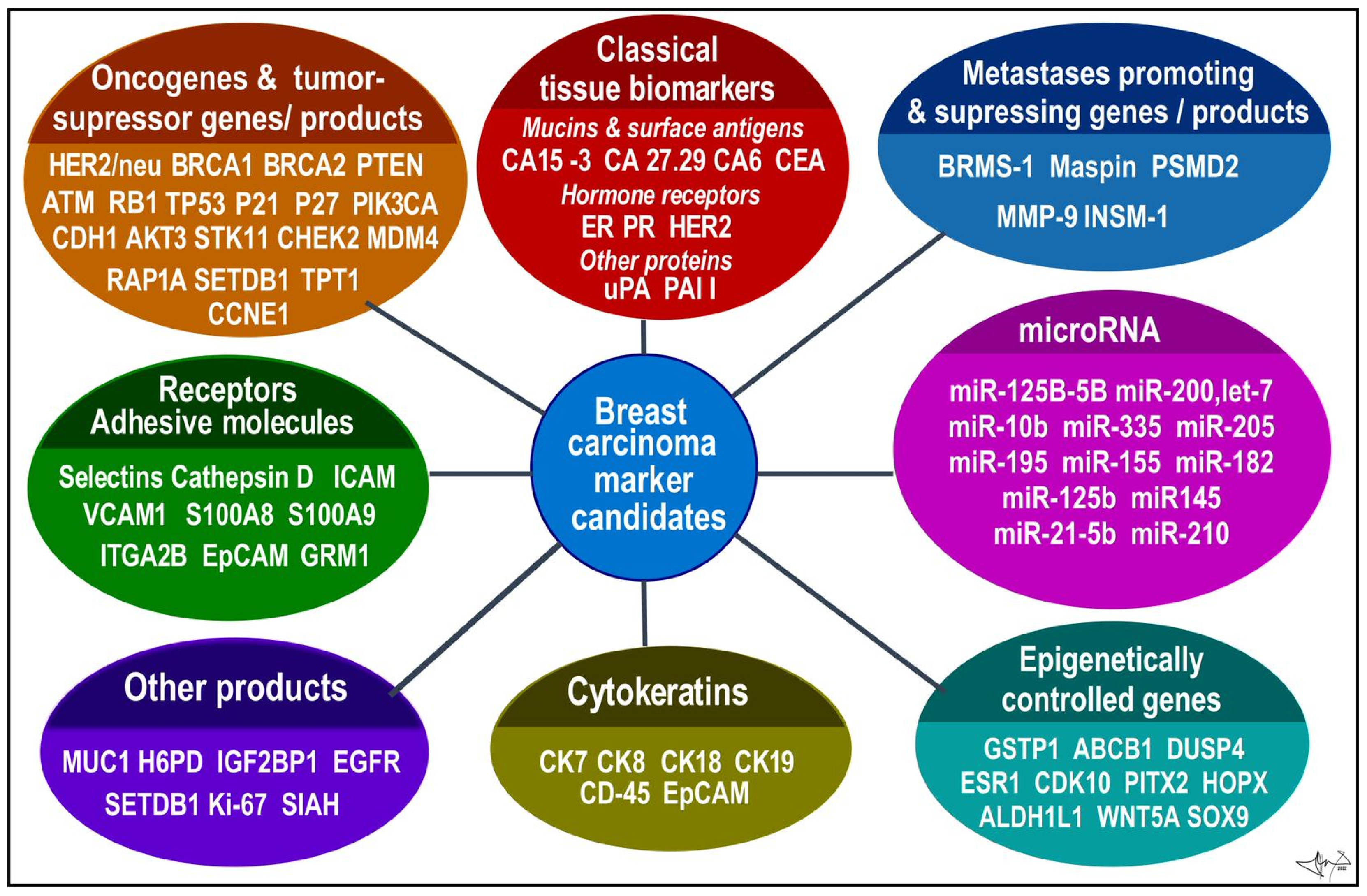
  • Histological proof of malignancy and assignment of histopathological phenotype has been a principal diagnostic method for a long time. It is supplemented by analysis of specific tumour cells products or markers to determine a molecular subtype of BC. Common biomarkers currently include oestrogen (ER) [8] and progesterone (PR) receptors [9], cytokeratins (CK) [10][11][12], human epidermal growth factor type 2 receptor (HER2) [13][14][15]. The BC samples obtained by biopsy and/or from post-surgery specimen can be currently processed by various methods (described in the following section). Genomic tests using individual or multigene assays can detect expression patterns of candidate genes associated with BC. All these methods should determine whether cancer is present, and if so, to identify the type of tumour, location, shape and spread of masses within or outside of the breast, respectively [3][10][16][17].
Therapeutic and prognostic factors include tumour size, grade and lymph node infiltration [4]. Tumour size (≤2 cm vs. >2 cm) is a prognostic factor for local or regional recurrence and overall survival and the probability increases with a tumour diameter [4]. Nottingham cancer grading system (score from 1 to 9) scores nuclear pleomorphism, mitotic activity and other features [4]. Lymph node status (positive or negative) refers to a presence or absence of tumour cells in close or distant sentinel nodes. Control of sentinel lymph nodes is a standard procedure (through a surgery or later by PET) to predict a recurrence of disease. It should be considered that up to 1/3 of untreated patients with negative lymph node status will develop recurrent or metastatic disease within 10 years after diagnosis [4]. All above data and other clinical evidence of eventual distant organ metastasis are a part of tumour–node–metastasis staging system. BC can be divided into four clinical stages—I, II, III and IV [1]. Cancer staging provides information useful for individual patient prognosis (aggressiveness of cancer and the extent of its invasion) [18] and for large scale analysis [19];
(c) Monitoring tests during and after treatment are done to determine the benefit of therapy and may be used to check for any signs of recurrence. Examination of blood samples for increased levels of serum biomarkers from tumour cells are commonly used in this step, e.g., carcinoma antigen 15-3 (CA 15.3), Carcinoma antigen 27.29 (CA27.29), Carcinoembryonic antigen (CEA) and others [20]. Periodical NMRI and mammography tests should be used in controlling potential relapse of tumours in their original site, new tumours in other breast or potential spread into distant metastases. Patients using aromatase inhibitors undertake regular densitometry examination of bone density. Regular check-ups are usually scheduled every 3–4 months during the first two years, every 6–8 months during 3–5 years’ period after the treatment, and after that once a year [9][21].

2. Histopathological Forms of BC

Determination of histopathological features of tumour mass is a principal step in diagnosis and in determining suitable therapeutic algorithm and prognosis. In many cases, non-invasive procedures such as mammography and MRI can provide enough data to distinguish non-tumour lesions from tumours masses and benign from malignant breast tumours, respectively [5][6][17]. Nevertheless, tumour samples obtained by multifocal biopsy, incision, or post-surgery samples (after partial or total ablation of breast) are inevitable for diagnostic conclusion about malignant histological phenotype [7]. Breasts are made up of fatty tissue, fibrous tissue forming the stroma and glandular tissue. Pathogenically, the most important structure for understanding development of BC is terminal ductal-lobular segment [16][17]. BCs arise from multipotent breast stem cell precursors which give rise to myoepithelial basal cells (allowing milk ejection) and luminal epithelial cells (milk production). These two histological phenotypes—basal and luminal—differ also by their biological functions and expression of specific genes, which also largely determine therapeutic responsiveness of specific tumour cell lineages. Mixed basoluminal type also exist and share certain features of both main types [10]. There are several histopathological subtypes of pre-cancerous and invasive BCs; their main features are as follows:
(1) Non-invasive (in-situ) types of breast tumours remain in a particular location of the breast, without being spread into surrounding breast tissue, lobules or ducts [1]. The two main types of in situ cancers are recognized: ductal carcinoma, which represent 80% of pre-cancerous forms, and lobular carcinoma accounting for the next 20% [23].
(a) Ductal carcinoma in situ (DCIS), also called intraductal carcinoma (or stage 0 BC), represents nearly 20% of newly diagnosed breast tumours altogether. Recurrence is less than 30% within 5 to 10 years after the diagnosis [1]. DCIS is a pre-invasive form of BC (pre-cancer), that may turn into adenocarcinoma. Tumour is derived from the epithelial cells lining the milk duct. Cell mass grows within the ductus but over the time it can break through the ductal walls into the surrounding tissue. DCIS is divided by histology into papillary, solid, micropapillary, cribriform and comedo-like subtypes, respectively. The low-grade DCSI is characterized by small, uniform-looking cells with uniform nuclei and a normal chromatin pattern. The intermediate-grade DCSI is similar to the low-grade, the difference between them being the intraluminal necrosis found in some of the ducts. The high-grade DCSI has atypical pleomorphic cells with very distinct nuclei. Necrosis also occurs and is mostly surrounded by proliferating tumour pleomorphic cells. Lumpectomy without BC radiation therapy has 25–35% chance of recurrence. Adding radiation therapy to the treatment decreases this risk to about 15% [23].
(b) Lobular carcinoma in situ (LCIS) grows from adenomatous cells inside milk producing lobules of breast and tends to remain within lobules [1]. As being deeper in the breast it is rarely felt as a lump. It is rather detected randomly in preventive mammogram or in needle or excisional biopsy samples due to other reasons. Histologically, LCIS is described as classic, pleomorphic, and florid (with central necrosis) subtypes, respectively. The latest two show an increasingly higher rate of anaplasias and signal increased risk of malignant transformation (7–12 times higher as compared to classic type) [23]. Another benign lobular pathology is atypical lobular hyperplasia (ALH). Both increase risk for invasive BC.
(2) Invasive BCs grow invasively into surrounding breast tissue, spread into lymph nodes and other organs. Two main types of malignancies can be classified as invasive lobular carcinoma (ILC) and invasive ductal carcinoma (IDC). There are also tumours which share the features and cell types of designated cases as mixed forms of carcinomas.
(a) IDC (also called infiltrating ductal carcinoma) is the most common type of invasive breast tumour (accounts for 70-80% of all cases). It occurs mostly in women older than 50 years and mainly those with inherited BRCA1 and/or BRCA2 mutations [24]. The risk of IDC is elevated if the first menstrual period came before age 12 or if the woman entered menopause after age 55, due to prolonged exposure to female sex hormones. The 5-year survival rate estimate is 99% when cancer has spread only within the breast or 86% if cancer has spread into neighbouring lymph nodes [20]. Prognosis is getting dramatically worse (falls to 28%) if it has metastasized to distant parts of the body. IDC outgrowths outside of the milk ducts to other parts of the breast making a solid mass that the patient feels as the breast lump. It spreads through the lymph vasculature or bloodstream. It is a heterogeneous group of tumours with several subtypes based on morphological properties of tumour cells [23]:
(i)
The classical nonspecific subtype is typical by pleomorphic cells with different shapes, sizes, and large non-uniform nuclei. In most cases, squamous and apocrine metaplasias, tissue necrosis and calcification are observed;
(ii)
The apocrine subtype is associated with a very poor prognosis. Cells are large, with typically strongly acidophilic granular cytoplasm. The nuclei are distinct and vesicular [23];
(iii)
Medullary carcinoma accounts for 3–5% of BC. Typically, women in their late 40s and early 50s are affected, and most commonly those who carry a BRCA1 gene mutation. It is often of triple-negative molecular pattern, but more responsive to chemotherapy and better prognosis than other ductal cancer;
(iv)
Mucinous carcinoma, also called colloid carcinoma, accounts for less than 2% of BC. Tumours contains clusters of uniform epithelial tumour cells with mildly atypical nuclei that are loosely surrounded by excessive mucus;
(v)
Papillary ductal carcinoma accounts for less than 1% of invasive BC. It is typical for older, postmenopausal women. Under a microscope, these cells resemble tiny fingers (papillae). Cells are typically small;
(vi)
Tubular ductal carcinoma accounts for less than 2% of BC and is more common in women older than 50. The tumour cells are oval or elongated, well differentiated, randomly arranged, and lined with a single layer of epithelial cells and without the outer layer of myoepithelial cells. In all these last three phenotypes tumour cells are positive for ER and/or PR receptors and negative for the HER2 receptor [9].
(b) ILC is the 2nd most common type of invasive cancer (~10% of cases). ILC starts in lobules, in 1/5 of cases in both breasts, and is harder to detect on mammograms. Cells are small, relatively uniform and of rounded shape, and have a typical stromal infiltration pattern. Like ductal carcinoma, lobular carcinoma can also be divided into subtypes according to closer histological characterization:
(i)
Classic (non-specific) subtype carries typical morphological features of lobular invasive carcinoma. Cells are small and uniformly distributed across the stroma, forming a typical Indian pattern. All, or at least part, of the pleomorphic subtype cells are considerably larger than those of the classical subtype and are characteristic for their eosinophilic cytoplasm. The nuclei of these cells are hyperchromatic, located eccentrically within the cell and with a very pronounced nucleolus. Absent expression of hormone receptors and high expression of tumour protein p53 and HER-2 receptor are also very typical for this subtype [25];
(ii)
Tubulolobular subtype is a variant of classical lobular carcinoma. It is characterized by small tubular formations with and without a lumen and cells forming a linear pattern similar to the classical subtype. An in situ lesion is often present in this subtype;
(iii)
Histiocytoid subtype consists of cells with a diffused growth pattern. Tumour cells are large, with a foamy cytoplasmic consistency that contains a significant number of granules. E-cadherin expression is negative for this subtype [23].
(3) Special types of breast tumours. This category contains rare and histologically or clinically distinct BCs:
(a) Inflammatory BC (IBC) is typical by erythema occupying at least one third of the breast and “peau d’orange-like” changes on the skin. IBC is considered a specific histological subtype, however, with no specific molecular signature. Thus, the diagnosis is based on clinical signs and symptoms [26]. IBC is a very aggressive, fast-growing type of cancer that accounts for 1% to 6% of invasive BCs. It arises from ductal malignant cells and usually has a high histological nuclear grade. As it grows more superficial into the soft tissue of breast it often blocks lymph vessels causing the breast to get inflamed. Skin upon the lesion becomes erythematous, warm, and swollen due to increased blood flow and accumulation of exudate with white blood cells build-up. Surface is getting thicker and firm with dimpled appearance like an orange peel [27]. The breast is getting tender or painful and itchy. Sometimes there are large patches of redness and red bumps that resemble bug bites (“inflammatory BC rash”). Only 15% cases have a real lump. When spread to areolar-nipple segment it causes the nipple to be flattened or inverted. Typically, axillary lymph nodes and subclavian lymph nodes are early getting swollen. The onset of the symptoms is relatively rapid and has to be distinguished from benign bacterial infections or inflammation around the cysts. Hypothesized genes contributing to IBC´s aggressive phenotype includes ESR1, GATA3, MUC1, ERBB2 and KRT5 [26];
(b) Paget’s disease of breast nipple is a rare BC occurring mostly around 56 years of age, and accounts for 1% to 4% of all invasive cases in woman. Nevertheless, this kind of BC also occurs in males [28]. Tumour originates from the ductal cells (in situ or invasive ductal carcinoma) and spreads to the skin of areola and the nipple with invasion of the overlying epidermis by malignant cells (so called Paget cells). Clinically the tumour is first manifested by eczematous changes, nipple discharge, bleeding, itching that can be misdiagnosed as psoriasis, contact dermatitis, erosive adenomatosis of the nipple etc. [29]. These finding are later followed by palpable mass of tumour [28]. Prognosis for males is worse, as five-year survival rate in males is 20% to 30% compared to 30% to 40% in females [29][30];
(c) Angiosarcoma of breast is a rare tumour accounting for less than 1% of all BCs, and 1% to 2% of all body sarcomas. It is formed up from the endothelial lining lymph or blood vessels within a breast. Angiosarcoma is most common in people older than 70 and it can occur as a complication from radiation therapy to the breast with some 8 to 10 years delay. It is often diagnosed late when it has already spread to other areas of the body [23];
(d) Phylloides tumour is a rare, mostly benign breast tumour (up to 80%) which mostly affects women in their 40s, though it may develop in patients of all ages. The tumour develops from the cells of the connective tissue (stroma) of the breast. Approximately 20% to 25% of phylloides tumours may turn to malignant phenotype. People with Li–Fraumeni syndrome (AR-inherited condition) are at an increased risk for this type of tumour [1].
Metastatic carcinoma. Breast tumour cells spread by lymph and blood. Tissues mostly affected by metastasis include brain, bones, lungs, and liver [1]. BC metastases, similar to other tumours, display certain organotropism, which, in addition to histological origin, is also determined by molecular subtype. BC spreads through lymphatic drainage into closest lymph nodes, mostly to axillar nodes (~30–50%) or mammary internal chain lymph nodes (10–40%), but rarely into supraclavicular nodes (~up to 4%). Peritoneal metastasis incl. ovaria represent near ~10% of all metastases. Most of them originated from lobular carcinoma (40%). If cancer has spread only within the breast, the 5-year survival rate reaches up to 99%. When the neighbouring lymph nodes are affected the survival rate decreases to 86%, and if it has spread to a distant part of the body, the rate decreases to 28% [22].

3. Molecular Subtypes of BC

In a study showing the differences in gene expression in various BCs, Perou and Sorlie proposed for the first time the “Molecular Classification of BC” in 2000 and brought a molecular insight into histopathological classifications [9][35]. Except for normal breast cell, several sub-groups of cancer cells were proposed, as luminal cancer (express genes typical in normal luminal epithelial cells), HER2 positive (overexpress ErbB2/neu oncogene) and basal cancer (express genes typical in normal breast basal and myoepitelial cells) [3][14][36]. The classical, mainly immunohistochemical markers analysed from BC cells used in the classification include ER, PR and HER2 (overexpressed oncogenic variant of EGF-like receptor) receptors. Additional markers used mainly for basal cell carcinoma stratification include Ki-67, EGFR (epidermal growth factor receptor) and basal cytokeratins (CK5/6, CK14, C19). Additionally, Urokinase plasminogen activator (uPA) and plasminogen activator inhibitor (PAI) could be analysed [8][9][13][31][36] .
Molecular classification strengthened the view on BC as histomorphologically and biologically heterogeneous group of tumours that show different growth intensity, invasiveness, and metastatic imprint, which require different therapeutic strategy [36]. As research progresses, the classification is a subject of continuous updates and modifications. In particular, 2–3 subforms of luminal BCs were established [37] and basal cell types of cancers were reclassified into triple negative (TN) BCs with “basal-like” and “non-basal like” features using EGFR and CK5/6 markers, respectively, and their subtyping continues [10][38]. Each BC seems to be morphologically and genetically unique, thus determining that the clinical manifestations of the disease are needed for personalized therapy for each patient [39].
ER and PR are intracellular (nuclear) receptors for oestrogens and progesterone in humans, encoded by gene ESR1 (Ch 6q25.1-q25.2) and PGR (Ch11q22), respectively. After hormone binding complex hormone/receptor is formed and assembled into dimers. After being actively transported into the nucleus, dimers serve as transcription factor and bind to hormone responsive elements of responsive genes to activate their transcription. ER and PR are common constituents of hormone-responsive breast tissue cells, mainly of luminal-epithelial origin as compared to basal myoepithelial lineages [40]. Oestrogen signalling is involved in control over cell growth, proliferation, and differentiation of breast tissue. According to the hormonal receptor status and sensitivity to hormone therapy, BCs are designated either as hormone positive (H+), as seen in luminal types of cancers, or as hormone negative (H−), as typical in basal types of cancers and BCs. Hormone-positive BCs could be targeted by hormonal therapy [17][20]. Mutations of ESR were detected in spontaneous BCs [8].
HER2 (human EGF-like receptor; ERB-B2) is an oncoprotein-surface receptor with tyrosine kinase activity from EFFR (epidermal growth factor receptor) family encoded by oncogene neu/ERBB2 (alternatively erbB-2, CD340, HER-2/neu) on locus Ch17q12 [15][41]. HER2-positive BCs overexpress the HER2 gene via amplification (20-25% of BCs) or other ways (~5%) [14]. Amplification is detected by immunohistochemical and hybridisation techniques. There are likely more than 60% of BCs considered yet as HER2-negative that possess at least some HER2 proteins on the surface (so called HER2-low) [15][36]. HER2 promotes tumour growth by enhancing cell proliferation, invasion, and metastasis by constitutively activating classical ras-dependent signalling pathway (ras-raf-MAPK) and alternative pathways (e.g., PI3K/AKTPKB). HER2+ is marker of possible responsiveness to targeted monoclonal therapy [4][14][36].
Cytokeratins are components of intermediate filaments of cell cytoskeleton and are inevitable in epithelial cells to resist mechanical stress. Their expression is tissue specific are used to identify the origin of human tumours. Presence of CK5/6, CK14, CK18 and CK19 is one of the diagnostics markers of cancer from basal cells, i.e., triple negative BC [12][38][42][43] (Figure 1).
Figure 1. BC candidates arranged according to functional categories. Data were collected from several sources [9][24][31][32][33][36][43][44][45].
Ki-67 is 359-kD nuclear protein associated with ribosomal RNA synthesis (gene MKI67; Ch10q26.2); it is considered a proliferation index and has been associated with poor prognosis [4]. Overactive MKI67 is identified in most proliferative cells (mainly in S phase), and in opposite is absent in the resting cells (G0). Ki-67 as well as EDGF, which is a natural growth promoting factor, are important in subgroupings especially in ER-positive BCs, although appropriateness of these proliferation markers or more detailed mitotic index scoring system over classical histopathologic predictions is still disputed [3][36]. The uPA is an enzyme that like serum tPA converts plasminogen to plasmin, action that is inhibited by PAI-1. This uPA is an extracellular matrix (ECM)-degrading protease, together with plasmin, in concentrations found in tumour tissue, degrading components of ECM in tumour stoma (fibrin, fibronectin, laminin etc.) and enhancing invasive growth and metastasing. Both PAI-1 and uPA belong to progression and metastasis markers of BC to predict the benefit effectiveness of adjuvant chemotherapy in patients with early BC, and are used as prognostic biomarkers in lymph node-negative BC [9][36][46].
According to expression of above-mentioned markers, BC can be classified into several molecular subtypes, which show distinct biological features and also invasiveness, therapeutical sensitivity and affinity to various tissues for distant metastases (Figure 2) [10]:
(a) Luminal A BC accounts for up to 50% of all invasive BCs. They express high levels of hormone receptors (either ER or PR or both) and obvious luminal (low molecular weight) cytokeratins. They have low status of HER2 (negative) and express low levels of Ki-67 protein. Histologically they are identified as tubular carcinomas, cribriform or classic lobular carcinomas, or low grade invasive ductal carcinomas, respectively. The most descriptive molecular feature of lobular carcinoma is the loss of E-cadherin. Luminal A cancers grow more slowly and are show lower grade of malignity than other BCs. They respond better to hormone therapy and also have a better prognosis [23][47];
(b) Luminal B BC accounts for 20–30% of cases. Similar to type A, these tumours express luminal cytokeratins and are ER-positive. They are mostly PR-positive but could be PR-negative, too. Opposite to A-type, Luminal B express high levels of Ki-67 and their HER-2 status is variable; it can be either negative or positive. Luminal type B often manifests genomic instability and the accumulation of TP53 mutations. Cancers of this type have a higher proliferation rate and histologic grade than luminal A type. Histologically, luminal B include mostly invasive ductal carcinomas or micropapillary carcinomas. Their response to endocrine therapy (tamoxifene, aromatase inhibitors) as well as prognosis are not as good as in Luminal A [23][47].
Recently, BCs with mediate-to high-levels of ER and PR-receptors and HER2-positive status were classified as a special subcategory of luminal B type BCs (luminal HER2(+)). These so-called triple-positive cancers (TP) (ER/PR/HER2-positive) can be treated with hormone drugs as well as drugs that target HER2 [48];
(c) HER2-positive BC accounts for 15–20% of invasive BCs. It is characterized by overexpression of the HER2/neu oncogene, obviously with low expression of both ER and PR receptors. As compared to luminal type A, HER2-positive tumours are associated with worse prognosis. Histologically, HER2+ tumours are high grade invasive ductal carcinomas. They show diffused TP53 mutations, high proliferation and histologic grade and nodal positivity. HER2+ cancers show more aggressive behaviour/higher mitotic activity, increased invasiveness, cellular motility, leading to earlier and more frequent relapses of the disease after primary multimodal treatment [36];
(d) Triple-negative (TN) or basal-like BC represents ~15% of invasive BCs and is characterized by ER/PR/HER2-negative profile (triple negative) and high Ki-67 proliferative index. TNBC is more common in younger women and is more frequent in African than Caucasian population. It shows a high proliferation rate, diffused TP53 mutation, BRCA1 mutation (germline, sporadic) and is considered more aggressive than either luminal A or luminal B cancer with very poor prognosis [1][23][47][49]. TNBC is not only a most aggressive subtype, but also stands for the most heterogeneous group as well. Lack of ER, PR removes hormonal feedback control overgrowth a differentiation, rendering TNBC high mitotic activity, high staged nuclear grade and unresponsive to hormonal therapy. Lack of HER2+ makes TNBC tolerant of monoclonal targeted therapy.
Over past decades, TNBC attracted a lot of research interests, and stratification of TNBC is continuously updated. Based on markers as EGFR or CK5/6, TNBC can be subtyped into prevalent basal-like type (approx. 80% of cases) and non-basal like type (approx. 20% of cases) [10]. Several additional categories were proposed considering unique histological features and typical genetic abnormalities caused by cumulative mutations of BC-associated genes (e.g., BRCA1, PTEN, RB1, TP53, NF1, HRAS, MAPK, STAT4, SMAD4, PIK3CA and so on). Subclasses of basal TNBC include basal-like 1 subtype (histologically mostly ductal BC) and basal-like 2 subtype (ductal, squamous and inflammatory BC). Non-basal TNBCs contain cells with mesenchymal or stem-cell like features and immune response genes (so called claudin-low, mesenchymal-like, immunomodulatory subtypes), as well as luminal subtype of TNBC (called LAR) with high androgen -receptor (AR) positivity [45].

References

  1. Akram, M.; Iqbal, M.; Daniyal, M.; Khan, A.U. Awareness and current knowledge of breast cancer. Biol Res. 2017, 50, 33.
  2. Torre, L.A.; Siegel, R.L.; Ward, E.M.; Jemal, A. Global Cancer Incidence and Mortality Rates and Trends--An Update. Cancer Epidemiol. Biomark. Prev. 2016, 25, 16–27.
  3. Hagemann, I.S. Molecular Testing in Breast Cancer: A Guide to Current Practices. Arch. Pathol. Lab. Med. 2016, 140, 815–824.
  4. Nicolini, A.; Ferrari, P.; Duffy, M.J. Prognostic and predictive biomarkers in breast cancer: Past, present and future. Semin Cancer Biol. 2018, 52 (Pt 1), 56–73.
  5. Gøtzsche, P.C.; Jørgensen, K.J. Screening for breast cancer with mammography. Cochrane Database Syst. Rev. 2013, 6, CD001877.
  6. Morrow, M.; Waters, J.; Morris, E. MRI for breast cancer screening, diagnosis, and treatment. Lancet 2011, 378, 1804–1811.
  7. Pesapane, F.; Suter, M.B.; Rotili, A.; Penco, S.; Nigro, O.; Cremonesi, M.; Bellomi, M.; Jereczek-Fossa, B.A.; Pinotti, G.; Cassano, E. Will traditional biopsy be substituted by radiomics and liquid biopsy for breast cancer diagnosis and characterisation? Med. Oncol. 2020, 37, 29.
  8. Sommer, S.; Fuqua, S.A. Estrogen receptor and breast cancer. Semin. Cancer Biol. 2001, 11, 339–352.
  9. Berse, B.; Lynch, J.A. Molecular diagnostic testing in breast cancer. Semin. Oncol. Nurs. 2015, 31, 108–121.
  10. Tsang, J.Y.S.; Tse, G.M. Molecular Classification of Breast Cancer. Adv. Anat. Pathol. 2020, 27, 27–35.
  11. Weissenstein, U.; Schumann, A.; Reif, M.; Link, S.; Toffol-Schmidt, U.D.; Heusser, P. Detection of circulating tumor cells in blood of metastatic breast cancer patients using a combination of cytokeratin and EpCAM antibodies. BMC Cancer 2012, 12, 206.
  12. Xenidis, N.; Perraki, M.; Kafousi, M.; Apostolaki, S.; Bolonaki, I.; Stathopoulou, A.; Kalbakis, K.; Androulakis, N.; Kouroussis, C.H.; Pallis, T.; et al. Predictive and Prognostic Value of Peripheral Blood Cytokeratin-19 mRNA-Positive Cells Detected by Real-Time Polymerase Chain Reaction in Node-Negative Breast Cancer Patients. J. Clin. Oncol. 2006, 24, 3756–3762.
  13. Litton, J.K.; Burstein, H.J.; Turner, N.C. Molecular Testing in Breast Cancer. Am. Soc. Clin. Oncol. Educ. Book. 2019, 39, e1–e7.
  14. Loibl, S.; Gianni, L. HER2-positive breast cancer. Lancet 2017, 389, 2415–2429.
  15. Nitta, H.; Kelly, B.D.; Allred, C.; Jewell, S.; Banks, P.; Dennis, E.; Grogan, T.M. The assessment of HER2 status in breast cancer: The past, the present, and the future. Pathol. Int. 2016, 66, 313–324.
  16. Coleman, C. Early Detection and Screening for Breast Cancer. Semin. Oncol. Nurs. 2017, 33, 141–155.
  17. Tarighati, E.; Keivan, H.; Mahani, H. A review of prognostic and predictive biomarkers in breast cancer. Clin. Exp. Med. 2022. Online ahead of print.
  18. Yao, F.; Yan, C.; Zhang, Y.; Shen, L.; Zhou, D.; Ni, J. Identification of blood protein biomarkers for breast cancer staging by integrative transcriptome and proteome analyses. J. Proteom. 2021, 230, 103991.
  19. Yoon, E.C.; Schwartz, C.; Brogi, E.; Ventura, K.; Wen, H.; Darvishian, F. Impact of biomarkers and genetic profiling on breast cancer prognostication: A comparative analysis of the 8th edition of breast cancer staging system. Breast J. 2019, 25, 829–837.
  20. Patel, B. A review of breast cancer and hormonal therapy. Int. J. Pharm. Sci. Res. 2019, 10, 519–527.
  21. Cardoso, F.; Kyriakides, S.; Ohno, S.; Penault-Llorca, F.; Poortmans, P.; Rubio, I.T.; Zackrisson, S.; Senkus, E. Early breast cancer:ESMO Clinical Practice Guidelines for diagnosis, treatment and follow-up. Ann. Oncol. 2019, 30, 1194–1220.
  22. Gote, V.; Nookala, A.R.; Bolla, P.K.; Pal, D. Drug Resistance in Metastatic Breast Cancer: Tumor Targeted Nanomedicine to the Rescue. Int. J. Mol. Sci. 2021, 22, 4673.
  23. Makki, J. Diversity of Breast Carcinoma: Histological Subtypes and Clinical Relevance. Clin. Med. Insights Pathol. 2015, 8, 23–31.
  24. Santarpia, L.; Bottai, G.; Kelly, C.M.; Győrffy, B.; Szekely, B.; Pusztai, L. Deciphering and Targeting Oncogenic Mutations and Pathways in Breast Cancer. Oncologist. 2016, 21, 1063–1078.
  25. Fumagalli, C.; Ranghiero, A.; Gandini, S.; Corso, F.; Taormina, S.; De Camilli, E.; Rappa, A.; Vacirca, D.; Viale, G.; Guerini-Rocco, E.; et al. Inter-tumor genomic heterogeneity of breast cancers: Comprehensive genomic profile of primary early breast cancers and relapses. Breast Cancer Res. 2020, 22, 107.
  26. Mamouch, F.; Berrada, N.; Aoullay, Z.; Khanoussi, B.E.L.; Errihani, H. Inflammatory Breast Cancer: A Literature Review. World J. Oncol. 2018, 9, 129–135.
  27. Lim, B.; Woodward, W.A.; Wang, X.; Reuben, J.M.; Ueno, N.T. Inflammatory breast cancer biology: The tumour microenvironment is key. Nat. Rev. Cancer. 2018, 18, 485–499.
  28. Dubar, S.; Boukrid, M.; Bouquet, D.E.; Joliniere, J.; Guillou, L.; Vo, Q.D.; Major, A.; Ben Ali, N.; Khomsi, F.; Feki, A. Paget’s Breast Disease: A Case Report and Review of the Literature. Front Surg. 2017, 4, 51.
  29. Adams, S.J.; Kanthan, R. Paget’s disease of the male breast in the 21st century: A systematic review. Breast 2016, 29, 14–23.
  30. Fentiman, I.S.; Fourquet, A.; Hortobagyi, G.N. Male breast cancer. Lancet 2016, 367, 595–604.
  31. Li, J.; Guan, X.; Fan, Z.; Ching, L.M.; Li, Y.; Wang, X.; Cao, W.M.; Liu, D.X. Non-Invasive Biomarkers for Early Detection of Breast Cancer. Cancers 2020, 12, 2767.
  32. He, Z.; Chen, Z.; Tan, M.; Elingarami, S.; Liu, Y.; Li, T.; Deng, Y.; He, N.; Li, S.; Fu, J.; et al. A review on methods for diagnosis of breast cancer cells and tissues. Cell Prolif. 2020, 53, e12822.
  33. Najjar, S.; Allison, K.H. Updates on breast biomarkers. Virchows Arch. 2022, 480, 163–176.
  34. Chen, W.; Hoffmann, A.D.; Liu, H.; Liu, X. Organotropism: New insights into molecular mechanisms of breast cancer metastasis. NPJ Precis. Onc. 2018, 2, 4.
  35. Perou, C.; Sørlie, T.; Eisen, M.; Van De Rijn, M.; Jeffrey, S.S.; Rees, C.A.; Pollack, J.R.; Ross, D.T.; Johnsen, H.; Akslen, L.A.; et al. Molecular portraits of human breast tumours. Nature 2000, 406, 747–752.
  36. Sauter, E.R. Reliable Biomarkers to Identify New and Recurrent Cancer. Eur. J. Breast Health 2017, 13, 162–167.
  37. Mcguire, A.; Brown, J.A.L.; Kerin, M.J. Metastatic breast cancer: The potential of miRNA for diagnosis and treatment monitoring. Cancer Metastasis Rev. 2015, 34, 145–155.
  38. Wang, Z.; Liu, L.; Li, Y.; Song, Z.; Jing, Y.; Fan, Z.; Zhang, S. Analysis of CK5/6 and EGFR and Its Effect on Prognosis of Triple Negative Breast Cancer. Front Oncol. 2021, 10, 575317.
  39. Arancibia, T.; Morales-Pison, S.; Maldonado, E.; Jara, L. Association between single-nucleotide polymorphisms in miRNA and breast cancer risk: An updated review. Biol. Res. 2021, 54, 26.
  40. Daniel, A.R.; Hagan, C.R.; Lange, C.A. Progesterone receptor action: Defining a role in breast cancer. Expert Rev. Endocrinol. Metab. 2011, 6, 359–369.
  41. ONLINE MENDELIAN INHERITANCE IN MAN, OMIM®. Johns Hopkins University, Baltimore, MD. MIM Number:114480, World Wide Web URL. Available online: https://omim.org/ (accessed on 5 May 2022).
  42. Orafa, Z.; Karimi, N.; Keyvani, S.; Oloomi, M. Quantitative CK19 biomarker detection in breast cancer cell lines. J. Med. Life 2022, 15, 188–195.
  43. Fazilat-Panah, D.; Vakili Ahrari Roudi, S.; Keramati, A.; Fanipakdel, A.; Sadeghian, M.H.; Shandiz, F.H.; Shahidsales, S.; Javadinia, S.A. Changes in Cytokeratin 18 during Neoadjuvant Chemotherapy of Breast Cancer: A Prospective Study. Iran J. Pathol. 2020, 15, 117–126.
  44. Lukong, K.E. Understanding breast cancer—The long and winding road. BBA Clin. 2017, 7, 64–77.
  45. Zubair, M.; Wang, S.; Ali, N. Advanced Approaches to Breast Cancer Classification and Diagnosis. Front Pharmacol. 2021, 11, 632079.
  46. Duffy, M.J.; Mcgowan, P.M.; Harbeck, N.; Thomssen, C.; Schmitt, M. uPA and PAI-1 as biomarkers in breast cancer: Validated for clinical use in level-of-evidence-1 studies. Breast Cancer Res. 2014, 16, 428.
  47. Kittaneh, M.; Montero, A.J.; Glück, S. Molecular profiling for breast cancer: A comprehensive review. Biomark Cancer. 2013, 5, 61–70.
  48. Holowatyj, A.N.; Ruterbusch, J.J.; Ratnam, M.; Gorski, D.H.; Cote, M.L. HER2 status and disparities in luminal breast cancers. Cancer Med. 2016, 5, 2109–2116.
  49. Collignon, J.; Lousberg, L.; Schroeder, H.; Jerusalem, G. Triple-negative breast cancer: Treatment challenges and solutions. Breast Cancer 2016, 8, 93–107.
More
Information
Subjects: Oncology
Contributors MDPI registered users' name will be linked to their SciProfiles pages. To register with us, please refer to https://encyclopedia.pub/register : , , , ,
View Times: 465
Revisions: 3 times (View History)
Update Date: 22 Nov 2022
1000/1000
Video Production Service