Submitted Successfully!
To reward your contribution, here is a gift for you: A free trial for our video production service.
Thank you for your contribution! You can also upload a video entry or images related to this topic.
Version Summary Created by Modification Content Size Created at Operation
1 -- 5707 2022-11-03 11:55:16 |
2 format corrected. + 3 word(s) 5710 2022-11-04 04:22:31 | |
3 format corrected. Meta information modification 5710 2022-11-04 04:23:09 |

Video Upload Options

Do you have a full video?

Confirm

Are you sure to Delete?
Cite
If you have any further questions, please contact Encyclopedia Editorial Office.
Sánchez-Pellicer, P.;  Navarro-Moratalla, L.;  Núñez-Delegido, E.;  Agüera-Santos, J.;  Navarro-López, V. The Pathogenesis of Alopecia Areata. Encyclopedia. Available online: https://encyclopedia.pub/entry/32751 (accessed on 23 April 2024).
Sánchez-Pellicer P,  Navarro-Moratalla L,  Núñez-Delegido E,  Agüera-Santos J,  Navarro-López V. The Pathogenesis of Alopecia Areata. Encyclopedia. Available at: https://encyclopedia.pub/entry/32751. Accessed April 23, 2024.
Sánchez-Pellicer, Pedro, Laura Navarro-Moratalla, Eva Núñez-Delegido, Juan Agüera-Santos, Vicente Navarro-López. "The Pathogenesis of Alopecia Areata" Encyclopedia, https://encyclopedia.pub/entry/32751 (accessed April 23, 2024).
Sánchez-Pellicer, P.,  Navarro-Moratalla, L.,  Núñez-Delegido, E.,  Agüera-Santos, J., & Navarro-López, V. (2022, November 03). The Pathogenesis of Alopecia Areata. In Encyclopedia. https://encyclopedia.pub/entry/32751
Sánchez-Pellicer, Pedro, et al. "The Pathogenesis of Alopecia Areata." Encyclopedia. Web. 03 November, 2022.
The Pathogenesis of Alopecia Areata
Edit

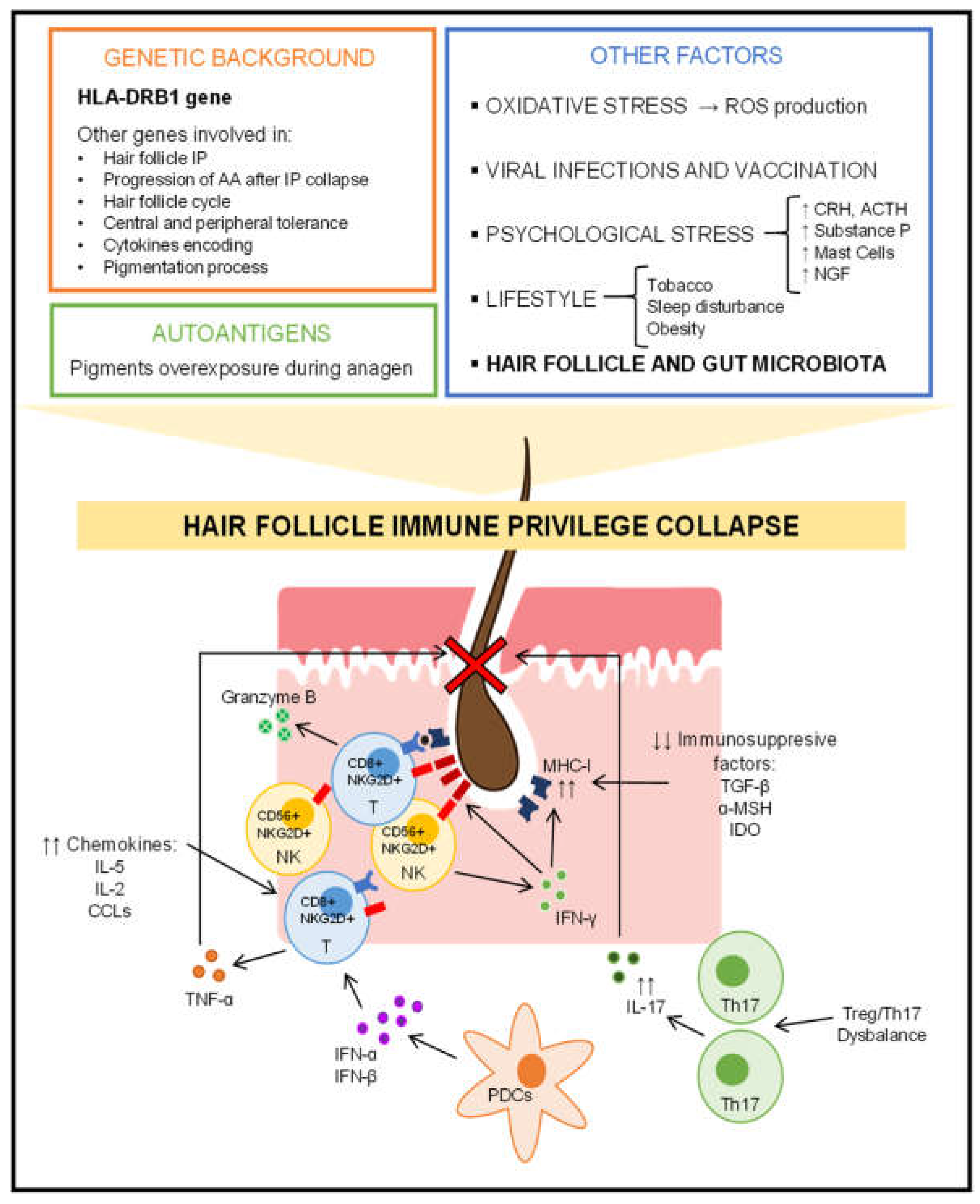
Alopecia areata is a multifactorial autoimmune-based disease with a complex pathogenesis. As in all autoimmune diseases, genetic predisposition is key. The collapse of the immune privilege of the hair follicle leading to scalp loss is a major pathogenic event in alopecia areata. The microbiota considered a bacterial ecosystem located in a specific area of the human body could somehow influence the pathogenesis of alopecia areata, as it occurs in other autoimmune diseases. Moreover, the Next Generation Sequencing of the 16S rRNA bacterial gene and the metagenomic methodology have provided an excellent characterization of the microbiota.

alopecia areata non-scarring alopecia skin microbiota hair follicle microbiota scalp microbiota

1. Hair Cycle Disruption

The pilosebaceous unit is composed of the hair follicle in close relation to the sebaceous gland together with the pili muscles and the capillaries, which supplement it in its lower part through the dermal papilla. The infundibulum is the section of the mature hair follicle that begins with the projection into the epidermis until the entry of the sebaceous gland. Next, in the dermis, the bulge is situated, a region of vital importance in the hair cycle as it harbors the stem cells that will generate the transit-amplifying cells during the growth phase. The bulb is located (a regeneration area) in a region deeper than the bulge. The bulb is composed of the hair follicle matrix, surrounding the dermal papilla. In the hair follicle matrix, there are melanocytes, which produce the hair pigment. In the suprabulbar area the hair follicle epithelium is located, composed of concentric layers forming the outermost outer root sheath, the inner root sheath, and the most internal hair shaft (it will be the visible part of the emerging hair) [1].
Basically, the hair cycle alternates phases of growth, involution, and quiescence. In the active growth or anagen phase, matrix cells proliferate and differentiate into transit-amplifying cells, which mobilize to the bulb and hair shaft. As the nutrition supply to the matrix cells diminishes and the catagen phase is beginning, the lower hair follicle regresses and the hair shaft growth ceases. Moreover, apoptosis is now occurring in the lower hair follicle which causes the separation of the dermal papilla (and blood vessel). Afterwards, the hair follicle enters the quiescent or telogen phase. However, the telogen phase is highly dynamic since the necessary conditions are being generated to start a new cycle with a new anagen phase. When the telogen phase is ending, the hair shaft is shed so that a new hair shaft can begin to grow [2].
Each of the different phases of the hair follicle cycle is regulated by signal molecules and cells involved in complex signaling pathways. When this homeostatic balance is lost, the development of the hair follicle cycle is affected [3]. In alopecia areata patients, different hair cycle disorders are reported. In the acute phase of the disease, peribulbar and intrabulbar inflammatory infiltration are characteristic, although it may not be observed in chronic stages [4]. The infiltrate comprises mainly T-lymphocytes, Langerhans cells, histiocytes, plasma cells, mast cells and eosinophils. T-cells cause apoptosis of the more peripheral matrix cells resulting in thinning of the hair shaft and the appearance of the typical exclamation mark hairs. This inflammation also causes a rapid transition from the anagen phase to the catagen and telogen phases, which leads to a reversal of the anagen/telogen ratio in alopecia areata patients. It is important to highlight that the inflammatory infiltrate does not affect the stem cells, so the hair follicle has the capacity to regenerate if it disappears after a new growth cycle. However, if during the new anagen phase, the hair follicle is once again affected by peribulbar and intrabulbar inflammation, the cycle will be interrupted prematurely and alopecia areata will persist [5].

2. Hair Follicle Immune Privilege Collapse and Autoimmune Response

The hair follicle presents the characteristic of being a region of immune privilege (IP), for instance certain organs such as the brain, eyes, gonads, and placenta [6]. This was firstly hypothesized by Paus et al., in 2003 [7]. The IP is the main mechanism by which tolerance is induced and immune recognition is prevented due to the presence of potential autoantigens in the aforementioned tissues. In the hair follicle, this protects the stem cells and derived cells that constitute its epithelium [8]. There are several forms to maintain the hair follicle with IP in normal conditions. As a physical barrier, an extracellular matrix protects the hair bulb by preventing the infiltration of immune cells [9]. A reduction in the expression of major histocompatibility complex (MHC) class I molecules is observed due to the local production of immunosuppressive factors such as α-melanocyte-stimulating hormone (α-MSH), transforming growth factor-β (TGF-β) and indoleamine-2,3-dioxygenase (IDO) among others [10]. There is also a decrease in the expression of MHC class II molecules on Langerhans cells, which are a type of dendritic cells in the skin, thus there is an impairing of the antigen-presenting function [11]. Moreover, the hair bulb (and bulge, so IP is not only restricted to the bulb) shows a very high expression of the transmembrane glycoprotein CD200, which interacts with its receptor on several immune cells to generate tolerance signals, decrease the activity of antigen-presenting cells (APCs) and decrease the secretion of pro-inflammatory cytokines [12][13]. Moreover, during the crash of the IP there is a secretion of chemokines such as IL-15, IL-2 and various CXCLs (CXC ligands), which act as chemotactics for other immune cells in the hair follicle [11].
The collapse of the IP of the hair follicle is the main condition for the initiation of the mechanisms that cause hair loss in alopecia areata. Therefore, it leads to an overexposure of autoantigens during the anagen phase (mainly), and the hair follicle cycle is corrupted, which explains the dermoscopic findings characteristic of alopecia areata [11]. Some authors defend the hypothesis that the hair follicle autoantigens are related to certain pigments produced by melanocytes (or derived products), which would be immunogenic and due to this the hair follicle is established as a region with IP [14][15].
Two major immunological elements in the pathogenesis of alopecia areata are T-lymphocytes CD8+NKG2D+ and INF-γ (interferon γ). Under normal conditions with an IP in the hair follicle, there is a decreasing in the expression of the NK cell receptor D (NKGD2) and its ligands MICA (MHC class I chain-related A) and ULBPs (UL16-binding proteins). Upon IP collapse, NKG2D receptors in NK cells and T-cells CD8+, and its ligands in the epithelium of hair follicles are upregulated [16]. Xing et al., in 2014, demonstrated that cytotoxic T-cells CD8+NKG2D+ are necessary and sufficient for the establishment of alopecia areata in the susceptible disease murine model C3H/HeJ (principal, genetically susceptible, murine model of alopecia areata). Basically, T-cells CD8+NKG2D+ produce INF-γ through JAK1 and JAK3 signaling pathways, which in turn triggers hair follicle epithelial cells to produce IL-15, which interacts with T-cells CD8+NKG2D+ to produce more INF-γ through JAK1 and JAK2, thus establishing a positive feedback loop [17]. T-lymphocytes CD8+NKG2D+ are the major contributors to hair loss in alopecia areata and are the first cells to infiltrate the hair follicle [18]. These cytotoxic T-cells CD8+ attack the hair follicle through granzyme B, which is elevated in the hair follicle of humans with alopecia areata [19]. Similarly, high expression of migration inhibitory factor (MIF) prevents the infiltration of NK cells CD56+NKG2D+, but when the IP collapses, these cells infiltrate the hair follicle [20]. In this regard, Gilhar et al., in 2013, demonstrated the importance of the human peripheral blood mononuclear cells (PBMCs) enriched for NKG2D+ and CD56+ cells (including NK cells). They performed several normal hairy human scalp engraftments in 25 SCID mice (mice that are severely deficient in functional B and T-lymphocytes) and injected them with a cell suspension of PBMCs enriched for NKG2D+ and CD56+ cells. At 3–5 weeks they observed histological and dermoscopic findings of alopecia areata where the injections were administered in the scalp transfer. Thus, as in alopecia areata in humans, a peribulbar infiltrate of T-lymphocytes and a collapse of the IP were observed [21].
INF-γ breaks down the IP of the hair follicle and is a key player in the pathogenesis of alopecia areata. INF-γ is mainly produced by NK and NKT cells. In addition to being a key element in the vicious cycle between T-lymphocytes CD8+NKGD2+ and hair follicle epithelial cells with the intervention of the intracellular JAK signaling pathway system, INF-γ induces the expression of MHC molecules, NKGD2 and its ligands in the hair bulb during the anagen phase, thus promoting autoimmunity against the hair follicle [9]. In this context, Gilhar et al., in 2005, observed that INF-γ accelerated the development of alopecia areata in C3H/HeJ mice. This was associated with increased expression of MHC class I and II molecules in the hair follicle epithelium [22]. Moreover, Freyschmidt-Paul et al., in 2006, engrafted skin with alopecia areata from C3H/HeJ mice into other C3H/HeJ mice with (knockout-type) and without (wild-type) deletion in the gene encoding INF-γ. While 90% of the wild-type mice developed alopecia areata, none of the knockout mice developed hair loss. Thus, knockout mice fail to activate Th1 cells in response to transplanted autoantigens, implicating the key role of INF-γ in the induction of alopecia areata [23].
Plasmacytoid dendritic cells (PDCs) have become of recent interest in the pathogenesis of alopecia areata. Ito et al., in 2020, observed that INF-α produced by PDCs cause alopecia areata in C3H/HeJ mice by stimulating apoptosis and increasing the secretion of chemokines such as CXCL10, infiltrating Th1/Thc1 cells [24]. PDCs are not found in healthy skin; however, they can infiltrate when an infectious or autoimmune pathology is established, such as viral infections or psoriasis [25]. PDCs produce large amounts of type I interferons such as INF-α or INF-β and have been shown to participate in the antiviral response. In fact, some studies have reported that alopecia areata can be established after a viral infection [26].
T-lymphocytes CD4+ Th17 and Treg are also involved in the pathogenesis of alopecia areata, as Th17 infiltrates the dermis and around the hair follicle (T-cells CD4+ were abundant after upregulation of MHC class II molecules in the hair follicle) [27]. Moreover, in patients with alopecia areata, there is a systemic imbalance between Treg and Th17 cells, a not uncommon finding in autoimmune diseases [28]. Moreover, Th17-derived cytokines are increased both in alopecia areata lesions and in the blood of alopecia areata patients [29].
Plasmatic TNF-α (tumor necrosis factor α) is also increased in patients with alopecia areata [30]. In alopecia areata lesions, TNF-α is derived from T-lymphocytes (CD4+ and CD8+) [31]. TNF-α acts by interfering with the hair follicle cycle, but its mechanism could be paradoxical since it could inhibit the overexpression of MHC class I molecules in the hair follicle, as well as suppress the development of PDCs responsible for the coordination of T-cells CD4+, CD8+ and NK cells in the establishment of alopecia areata [18].
Other elements that could be involved in the pathogenesis of alopecia areata are mast cells that can drive an antigen-specific cytotoxic T-cell CD8+ response in the hair follicle [32], neuropeptides (secreted by the sensory nerve fibers that innervate the hair follicle), and neurohormones such as substance P, vasoactive intestinal peptide (VIP), calcitonin gene-related peptide (CGRP), somatostatin, corticotropin-releasing hormone (CRH), adrenocorticotropic hormone (ACTH), cortisol, etc. [11]. It is beyond the scope of this research to discuss in detail these molecular mechanisms involved.
These multiple possible pathogenic immunological pathways involved in alopecia areata could explain the clinical heterogeneity observed in these patients with different developments such as spontaneous remission without relapses versus severe progressive evolution towards universal alopecia, as well as different associations with other diseases (comorbidities) and differences in response to different treatments.

3. Role of Genetics in Alopecia Areata

Observational studies have shown a genetic component in alopecia areata, demonstrating a high incidence in first-degree relatives and a high concordance in twins [33]. This implies that family history is a risk factor for alopecia areata. Nevertheless, there is variability in the prevalence and development of alopecia areata in patients with a family history and it is difficult to predict a pattern, suggesting that other factors such as environmental or lifestyle factors are necessary for alopecia areata to develop [18].
Alopecia areata is a complex polygenic disease with hundreds of single nucleotide polymorphisms (SNPs) found in patients. Many of these polymorphisms are found in genomic regions involved in the phenotype of the immune system, including the activation and functionality of Treg and cytotoxic T-lymphocytes, cytokine expression and antigen presentation [16]. Many of these genes are also associated with other autoimmune or immune-based diseases such as inflammatory bowel disease, multiple sclerosis, type 1 diabetes mellitus and psoriasis [34].
Several genes encoding HLA (human leukocyte antigen) variants have been correlated with alopecia areata, among them, polymorphisms in HLA-DRB1 are a major contributor to the disease phenotype. A recent meta-analysis has shown that HLA-DRB1*04 and HLA-DRB1*16 variants increase the risk of alopecia areata, while HLA-DRB1*0301, HLA-DRB1*09 and HLA-DRB1*13 variants are protective [35]. These polymorphisms tend to occur in the peptide-binding region of the APCs, thus affecting their binding affinity and therefore are related to autoimmune processes [36].
Regarding genes involved in hair follicle IP and hair follicle collapse that occurs in alopecia areata, the genes encoding NKG2D and its ligands such as MICA and ULBP are also relevant, and they are only involved in the pathogenesis of alopecia areata and not in other autoimmune diseases. Some polymorphisms in these genes may modify the receptor–ligand affinity or increase expression, which would lead to greater activation [37]. SNPs in the genes encoding some heat shock proteins (HSPs) [38] and genes involved in the elimination of reactive oxygen species (ROS) such as PRDX5 and ACOX have also been linked to alopecia areata [39]. It is noteworthy that some of these genes encode stress-inducible proteins or are associated with a greater effect of stress in alopecia areata [40], which will be discussed later in one of the next sections, which is another factor that influences the development of the disease.
As genes are involved in central and peripheral tolerance, there are several associated with alopecia areata. Polymorphisms in the autoimmune regulator gene (AIRE), whose complete loss of function produces polyendocrinopathy-candidiasis-ectodermal dystrophy (APECED) where alopecia areata is a common sign, continue to be associated with alopecia areata in patients without APECED [41]. Other genes implicated include forkhead box P3 (FOX3P) [42], ikaros family zinc finger 4 (IKZF4) [16], glycoprotein A repetitions predominant (GARP) [39], cytotoxic T lymphocyte-associated antigen 4 (CTL4) [16], IL-2A receptor chain (IL-2RA) [16], protein tyrosine phosphatase no receptor 22 (PTPN22) [43] and transporter associated with antigen processing 1 (TAP1) [44] among others [40].
SNPs in genes encoding cytokines involved in the progression of alopecia areata after the IP collapse are also associated. These genes encode IL-1 family, IL-4, IL-13, IL-17, IL-6, IL-26, IL-12B and TNF family [40]. Other genes are related to the progression of alopecia areata after the IP collapse of hair follicles such as the suppressor of cytokine signaling-1 (SOCS1) gene [16], FAS and FAS ligand genes [45], the neurogenic locus homolog 4 (NOTCH4) [46] and some chemokines encoding genes [47].
Likewise, some genes involved in the pigmentation process have also been associated with alopecia areata, such as the gene encoding the melanin-concentrating hormone receptor 2 (MHCR2) [48]. In addition, autophagy-related genes that in the context of alopecia areata act at the level of melanogenesis, autoimmunity and hair follicle cycling have also been linked, such as gene encoding syntaxin 17 (STX17) [16].
Finally, some alopecia areata-associated genes encode proteins involved in the hair follicle cycle that operate as structural proteins or transcription factors. These include, for example, the SMARCA2 gene [49].

4. Additional Influential Factors in the Development of Alopecia Areata

Oxidative stress could play a role in the pathogenesis of alopecia areata as it is a disease where an inflammatory condition is established. The ROS generated could exceed the cellular antioxidant capacity and this could influence the development of the disease. Biomarkers of oxidation/anti-oxidation status have provided inconsistent results in some studies [50][51]. In this context, a recent meta-analysis of 18 studies published in 2020 concluded that current evidence suggests that alopecia areata is associated with oxidative stress, but that further studies are required to provide robustness to this association. Furthermore, oxidative stress biomarkers are more increased in severe forms of the disease compared to mild or moderate forms [52]. Recently, Sachdeva et al., in 2022, found that total antioxidant status (TAS) and superoxide dismutase (SOD) levels were decreased and malondialdehyde (MDA) increased in 40 patients with alopecia areata compared to 40 age- and sex-matched healthy controls [53]. Furthermore, TAS and SOD decreased, and MDA increased with respect to disease severity. Therefore, the data suggest that oxidative stress seems to influence the development of alopecia in terms of its severity. On the other hand, in the previous section about genetics and alopecia areata, it has been mentioned that some associated polymorphisms also act at the oxidative stress level. Polymorphisms in stress-inducible protein genes such as HSPs [38] and NKGD2 receptor ligands [37], as well as in genes involved in ROS clearance such as PRDX5 and ACOX [39], have been linked to alopecia areata [40]. It is also noteworthy that ROS can occur in the context of situations other than alopecia areata as a consequence of treatment, infections or established comorbidities [54][55].
Although some patients who develop alopecia areata describe a previous episode of psychological stress, other studies have not found this event. So, it is certain that a close causal relationship between alopecia areata and emotional stress has never been established [18][56]. However, there is relative evidence that the central nervous system (CNS) activation in response to psychological stress influences the pathogenesis of alopecia areata to some degree. Much of this information has been obtained from murine models [56]. Firstly, it is important to remark that the hair follicle is a producer and target of hormones related to psychological stress. Thus, in the skin, there is a system analogous to the hypothalamic–pituitary–adrenal (HPA) axis that becomes activated during physical aggression or psychological stress in the same way as in the CNS [57]. Psychological stress activates both the HPA and the brain–hair follicle (BHF) axis. This could lead to some findings that would be involved in the pathogenesis of alopecia areata. Increased secretion of substance P by the sensory nerve fibers of the hair follicle causes perifollicular activation of mast cells, which leads to inhibition of hair follicle growth in the anagen phase [58], as well as causing cutaneous neuroinflammation [59]. Nerve growth factor (NGF), a neurotrophin that is increased during stress, also contributes to the disruption of the hair follicle cycle and the collapse of its IP [59]. Likewise, in the hair follicle, CRH promotes mast cell degranulation, which also causes perifollicular neuroinflammation [60]. Moreover, an increase in CRH receptors in response to emotional stress has been observed in the skin of patients with alopecia areata [61].
Viral infections, as with other autoimmune diseases [62], have also been implicated in the pathogenesis of alopecia areata, especially infections of Epstein-Barr virus, hepatitis B and C [63], and even recently SARS-CoV-2 [64]. As discussed in previous sections, the activation of IFN-α-producing PDCs by viral infection is an important mechanism that establishes this relationship [24][26]. Supposedly, in a similar mechanobiological manner, cases of alopecia areata have been reported following vaccination [65].
Certain lifestyle factors play a role in the onset and development of alopecia areata [66]. Smokers have a higher risk of developing alopecia than non-smokers [67]. Although the details of the influence of tobacco on the pathogenesis of alopecia areata are unknown, it could cause a Th17-mediated inflammation in the hair follicle [68]. Moreover, sleep quality could also influence the development of alopecia areata, although there are observational studies that have shown that patients with sleep disorders have an increased risk of developing alopecia areata [69], while others have not found a relationship between sleep quality and the development of alopecia areata [70]. Sleep disturbance affects the immune system on several levels and due to the autoimmune nature of alopecia areata, it is possible that it somehow influences its pathogenesis [71]. Furthermore, obesity increases the risk of developing alopecia areata [72], as with other inflammatory skin diseases [73]. A significant change in obesity is the dysregulation of adipokine production. Its dysregulation contributes to chronic low-grade inflammation and affects the modulation of the immune response, intervening in the pathogenesis of various autoimmune diseases, such as alopecia areata [74].
Figure 1 provides a summary of the factors that influence the complex pathogenesis of alopecia areata.
Figure 1. Overview of the pathogenesis of alopecia areata. IP: Immune privilege; AA: Alopecia Areata; ROS: Reactive Oxygen Species; CRH: Corticotropin-Releasing Hormone; ACTH: Adrenocorticotropic Hormone; NGF: Nerve Growth Factor; IL-2: Interleukin 2: IL-5: Interleukin 5; IL-17: Interleukin 17; CXCLs: CXC Ligands; TNF-α: Tumor Necrosis Factor α; INF-α: Interferon α; INF-β: Interferon β. INF-γ: Interferon γ; PDCs: Plasmacytoid dendritic cells; NK: Natural Killers; Th17: T-cells Th17; Treg: T-cells Treg; MHC-I: Major Histocompatibility Complex Class I; NKGD2D: NK Cell Receptor D; TGF-β: Transforming Growth Factor β; α-MSH: α-Melanocyte-Stimulating Hormone; IDO: Indoleamine-2,3-Dioxygenase.

5. Alopecia Areata and Skin Microbiota

The hair follicle contains a variety of bacteria that can reach deeper compartments of the skin [75]. In addition, many bacteria are often organized in biofilms [76] and therefore their study by usual swab sampling may have limitations because it may not be a representative sample of this area. Matard et al., in 2013, reported for the first time the presence of biofilms in the deep zone of scalp hair follicles in both patients with folliculitis decalvans and healthy controls, suggesting their ubiquity [77].
Thus, the hair follicle allows the superficial skin environment and its microbiota to be linked to all skin layers, creating environmental conditions that are highly favorable for persistent bacterial colonization. These favorable environmental conditions are moisture, vascularization, relatively greater protection from ultraviolet light and a more optimal pH for bacterial growth [78]. The establishment of microbiota in the hair follicle is also favored by its IP status.
It is important to emphasize that given the size of the bacteria, access through the infundibular area should be limited to the deep infundibulum and the sebaceous gland. However, the deeper regions of the infundibulum are permeable (epithelium not as tight as the more superficial infundibulum) [79]. In addition, some skin bacteria have enzyme endowments facilitating this penetration and tissue invasion [80].
Given these mentioned conditions, the colonization of the hair follicle is not erratic or random but is regulated and balanced under physiological conditions where the immune system plays an important role. Under normal conditions, therefore, the system that is established is stable, mutually beneficial and under homeostasis control. Communication is established between the bacteria and the underlying hair follicle tissue, and this process is dynamic since both the microbiota influences the immune microenvironment (functionality and composition of the cells of the immune system) and the characteristics of this habitat modulate the composition of the microbiota present [81]. Several mechanisms are involved in the shaping of the hair follicle microbiota. An efficient control system of the healthy hair follicle is the production of antimicrobial peptides (AMPs) such as β-defensin 1 and 2, psoriasin and RNase 7 [79] of different specificities in response to bacterial metabolites that would inhibit pathogenic bacteria. These AMPs are secreted by keratinocytes located in different regions of the hair follicle [82]. This mechanism involves activation of the innate immune system [83]. On the other hand, outer root sheath keratinocytes can also recruit immune system cells in case of excessive colonization or dysbiosis in the hair follicle by secreting cytokines and chemokines [78]. In addition, the connective tissue of the hair follicle is heavily populated with macrophages and mast cells. Bacteria-derived metabolites stimulate keratinocytes, as mentioned above, and these can secrete growth factors for mast cells [84], which are cells with antibacterial capacity, and so they influence the modeling of the microbiota of the hair follicle [85]. In addition, Langerhans cells located mainly in the upper part of the hair follicle (a region where the IP is not yet widely evident) extend their dendrites between the keratinocytes to capture and process bacteria-derived antigens and present them to T-cells. This triggers an adaptive immune response that shapes the composition of the hair follicle microbiota. Therefore, the access of APCs to bacteria has immunomodulatory implications that affect hair follicle homeostasis [86].
The microbiota of the hair follicle under normal conditions in a healthy state is quite similar to that of the skin [80]. There is an abundance of the phylum Actinobacteria, especially of the Propionibacteriaceae and Corynebacteriaceae families, and Firmicutes (Staphylococcus and Streptococcus genera) and Proteobacteria phylum [78][87]. This healthy hair follicle microbiota prevents colonization by pathogens, stimulates the production of cytokines related to the initiation and maintenance of the immune response [79], and influences the reduction in inflammation and tissue repair [88]. However, there is significant uncertainty about how dysbiosis in the microbiota of the hair follicle affects the growth cycle, regeneration, and immune environment of the hair follicle itself.
The studies detailed below reporting changes in the microbiota of the scalp of patients with alopecia areata do not answer the question of whether these changes are a causal factor or a secondary phenomenon to alopecia areata. However, alterations in the microbiota of the hair follicle or the penetration of material of bacterial origin and immunogenic could influence the modulation of cutaneous immune reactions and inflammatory processes. A peribulbar inflammation is a key process in the pathogenesis of alopecia areata. Moreover, during the anagen phase, the hair follicle is more vulnerable, and growth depends on a strong and robust IP around the bulb [9][11]. In this context, the epithelium around the bulge may be exposed to external (bacteria, metabolites, or bacterial products) or internal pro-inflammatory stimuli (immune system signals triggered by bacteria). Therefore, the microbiota of the hair follicle could be an influential factor in the pathogenesis of alopecia areata.
In relation to this, a higher prevalence of atopic dermatitis has been found in patients with alopecia areata [89]. In the pathogenesis of atopic dermatitis, an important factor is the functionality of T-cells Th17 and Th2, which in turn may be influenced by the skin microbiota [90]. Polak-Witka et al. formulate the hypothesis that the dysbiosis established in alopecia areata and atopic dermatitis could corrupt the IP of the hair follicle and the alteration of the barrier function of the skin in atopic dermatitis could facilitate the penetration of bacterial antigens into deep compartments of the hair follicle [79]. In fact, studies have recently been carried out on the efficacy of drugs that act at the level of the immune mechanisms that are common in alopecia areata and atopic dermatitis [91].

6. Alopecia Areata and Gut Microbiota

The human gastrointestinal tract harbors 100 trillion bacteria, most of which inhabit the large intestine, including about 1000 species [92]. These bacteria have cooperatively co-evolved with humans. This translates into a 1.3 to 1 ratio of bacteria to human cells, so there are more bacteria than human cells in human's organisms. The gut microbiota encodes more than 4 million genes (for approximately 25,000 genes contained in the human genome) and is involved in numerous metabolic reactions that influence the host’s physiology and metabolism to a substantial degree [93].

The composition of the gut microbiota tends to be modulated to a greater degree by environmental or lifestyle factors, for example diet or exposure to antibiotics (or other drugs, such as proton pump inhibitors such as omeprazole), rather than by host genetic factors [94]. Western diet, rich in saturated fat and poor in fiber, leads to an unhealthy metabolic profile and markedly influences microbiota composition and diversity [95].
The gut microbiota is stable in healthy adults and consists of a highly adaptive microbial community [96]. A major challenge is to define a “healthy” gut microbiota due to its high gut variability, as it is affected by a variety of known and unknown factors, as well as this, some variability may be stochastic [97]. Despite this high interindividual variability and temporal intraindividual variability in the gut microbiota composition, the metabolic functionality of the gut microbiota is much less diverse and more conserved [96]. This connects with the concept of functional redundancy. It has even been suggested that the functional definition may be superior to the taxonomic definition in distinguishing "abnormal" from "normal" gut microbiota [98].
Furthermore, a huge portion of the human body’s immune function is involved in maintaining the balance of the gut microbiota, as 70% of lymphocytes are in gut-associated lymphoid tissue. Given this scenario, it is evident that the gut microbiota is closely involved in the inflammatory response and commensal bacteria act as immune regulators that are critically implicated in the processes of immune tolerance [99]. Disruption of the regulation of these immune responses contributes to the development of inflammatory-based diseases [100]. It has even been observed that changes in the composition and diversity of the gut microbiota can lead to alterations in immunity and inflammation in organs at a distance from the gut [101].
In recent years, the immunomodulatory role of the gut microbiota in distant organs has acquired considerable relevance. Specifically in the gut–skin axis, the gut microbiota basically modulates the functionality and composition of the innate and adaptive immune system, and vice versa [102]. This fact explains why certain skin diseases manifest gut comorbidities [103] and suggests a relationship between the presence of gut dysbiosis and the imbalance of skin homeostasis, with a special role of the gut microbiota in the pathogenesis of several inflammatory skin diseases [104]. On the other hand, although there is still no robust evidence of the role of the gut microbiota in the context of the pathogenesis of alopecia areata, a connection with other autoimmune diseases has been well established [105][106][107]. On this basis, some authors have recently proposed the hypothesis of the participation of the gut microbiota in the pathogenesis of alopecia areata [108]. Indeed, patients with alopecia areata present an increased risk of developing autoimmune diseases [109] and these pathologies could possibly share common pathogenic mechanisms where the gut microbiota could play a key role.
Preclinical studies in murine models, despite their limitations regarding the translation of results to humans, have provided much relevant information concerning the pathogenesis of alopecia areata [110]. Indirect but significant evidence of a possible relationship between gut microbiota and alopecia areata has been obtained from these murine models. Firstly, C3H/HeJ mice were observed to develop a spontaneous and inherited form of idiopathic inflammatory bowel disease [111], which is also a potential comorbidity that can occur in humans [112] and where the gut microbiota is an important factor in its onset and development [113]. Moreover, normal-haired C3H/HeJ mice grafted with skin from spontaneously alopecia areata-affected mice were fed a diet enriched with soy oil and a high percentage did not develop alopecia areata [114]. This could indicate, apart from the properties of soy oil, the capacity of the diet and indirectly the capacity of the intestinal microbiota, to influence the development of this disease. Nair et al. engrafted skin from alopecia areata-affected C3H/HeJ mice onto unaffected C3H/HeJ mice, which induced the disease in the non-affected C3H/HeJ mice. They had previously treated a group of engrafted mice with a broad-spectrum antibiotic cocktail. The antibiotic-treated mice were protected from developing alopecia areata, but the untreated mice developed hair loss. In addition, a decrease in skin infiltrating T-lymphocytes CD8+NKG2D+ was observed in the antibiotic-treated mice group, indicating a possible role of the gut microbiota in the infiltration of T-cells into the hair follicle that collapses their IP in alopecia areata [115].
On the other hand, an important mechanism in the pathogenesis of alopecia areata is the presence and functionality of T-lymphocytes Treg. Treg are an important and active agent in peripheral tolerance processes, are essential in the prevention of the development of autoimmune diseases [116] and are particularly abundant in hair follicles [117]. In this regard, Scharschmidt et al., in 2017, reported that commensal bacterial colonization of the hair follicle in the postnatal period is involved in the abrupt migration of Treg in the skin (and hair follicle) during a defined postnatal stage [118]. Nevertheless, the role of Treg in the hair follicle, except for the suppressive function of effective T cells, is not fully established. It has been reported that Treg transference blocks the onset of the alopecia areata in C3H/HeJ mice after induction of localized hair loss following T-cells CD8+ injection [119]; therefore, they are important in the pathogenesis. The key could reside in the study by Ali et al., in 2017, who demonstrated that hair follicle Tregs promote regeneration by increasing both the number and differentiation of the stem cells [120]. Recent studies have also indicated that Treg subpopulations in alopecia areata lesions are functionally distinct from those in unaffected tissue and skin from healthy controls [121]. In this regard, how can the gut microbiota influence the pathogenesis of alopecia areata? A gut dysbiosis with depletion of α-diversity and short-chain fatty acid (SFCA) producing bacteria could result in a stressor of the immune system in subjects genetically susceptible to alopecia areata [108]. SFCAs are an influential class of bacteria-derived metabolites from the anaerobic fermentation of complex polysaccharides and oligosaccharides from the diet (dietary fiber), which can directly activate G-coupled receptors, inhibit histone deacetylases, and serve as energy substrates and thus affect various physiological processes [122]. Many studies have supported the influence of SFCAs on the number and functionality of intestinal Treg [123], which may have an impact on peripheral tolerance processes, and thus, on autoimmune diseases such as alopecia areata. A low fiber intake, a decrease in SFCA-producing bacteria or both simultaneously will lead to a decreased intestinal bacterial synthesis of SFCA. In this sense, the gut microbiota could be implicated in the pathogenesis of alopecia areata.
Dysfunction of the intestinal barrier with disruption at the level of the tight junctions (TJs) or damage to the mucosal layer often leads to increased intestinal permeability. This pathological state is known as “leaky gut” syndrome (LGS) [106][124]. LGS initiates an inflammatory response in the intestinal and extraintestinal tissue. Translocation of commensal and pathogenic bacteria occurs, causing a disruption in immune homeostasis and inducing systemic inflammation. Gut dysbiosis leads to mucosal barrier dysfunction and an inflammatory response that predisposes to systemic diseases such as autoimmune diseases [106][124]. Despite the pathogenic similarities between alopecia areata and other autoimmune diseases, there is no evidence of an association with increased intestinal permeability in these patients. Recently, Hacınecipoğlu et al. did not find significant differences in serum zonulin (a TJ protein) levels in 70 alopecia areata patients and 70 healthy controls [125]. However, this possible association should be further examined.
Important evidence for the potential role of gut microbiota modulation in the course of alopecia areata is the anecdotal observations of Rebello [126] and Xie [127], where hair repopulation was observed in alopecia areata patients after undergoing faecal material transplantation (FMT) for other indications such as Clostridiodes difficile infection or Crohn’s disease.

References

  1. Buffoli, B.; Rinaldi, F.; Labanca, M.; Sorbellini, E.; Trink, A.; Guanziroli, E.; Rezzani, R.; Rodella, L.F. The human hair: From anatomy to physiology. Int. J. Dermatol. 2014, 53, 331–341.
  2. Anzai, A.; Wang, E.H.C.; Lee, E.Y.; Aoki, V.; Christiano, A.M. Pathomechanisms of immune-mediated alopecia. Int. Immunol. 2019, 31, 439–447.
  3. Lin, X.; Zhu, L.; He, J. Morphogenesis, Growth Cycle and Molecular Regulation of Hair Follicles. Front. Cell. Dev. Biol. 2022, 10, 899095.
  4. Peckham, S.J.; Sloan, S.B.; Elston, D.M. Histologic features of alopecia areata other than peribulbar lymphocytic infiltrates. J. Am. Acad. Dermatol. 2011, 65, 615–620.
  5. Bernárdez, C.; Molina-Ruiz, A.M.; Requena, L. Histologic features of alopecias-part I: Nonscarring alopecias. Actas Dermosifiliogr. 2015, 106, 158–467.
  6. Watson, V.E.; Faniel, M.L.; Kamili, N.A.; Krueger, L.D.; Zhu, C. Immune-mediated alopecias and their mechanobiological aspects. Cells Dev. 2022, 170, 203793.
  7. Paus, R.; Ito, N.; Takigawa, M.; Ito, T. The hair follicle and immune privilege. J. Investig. Dermatol. Symp. Proc. 2003, 8, 188–194.
  8. Paus, R.; Bulfone-Paus, S.; Bertolini, M. Hair Follicle Immune Privilege Revisited: The Key to Alopecia Areata Management. J. Investig. Dermatol. Symp. Proc. 2018, 19, S12–S17.
  9. Suchonwanit, P.; Kositkuljorn, C.; Pomsoong, C. Alopecia Areata: An Autoimmune Disease of Multiple Players. Immunotargets Ther. 2021, 10, 299–312.
  10. Ito, T.; Ito, N.; Bettermann, A.; Tokura, Y.; Takigawa, M.; Paus, R. Collapse and restoration of MHC class-I-dependent immune privilege: Exploiting the human hair follicle as a model. Am. J. Pathol. 2004, 164, 623–634.
  11. Bertolini, M.; McElwee, K.; Gilhar, A.; Bulfone-Paus, S.; Paus, R. Hair follicle immune privilege and its collapse in alopecia areata. Exp. Dermatol. 2020, 29, 703–725.
  12. Rosenblum, M.D.; Yancey, K.B.; Olasz, E.B.; Truitt, R.L. CD200, a "no danger" signal for hair follicles. J. Dermatol. Sci. 2006, 41, 165–174.
  13. Kotwica-Mojzych, K.; Jodłowska-Jędrych, B.; Mojzych, M. CD200:CD200R Interactions and Their Importance in Immunoregulation. Int. J. Mol. Sci. 2021, 22, 1602.
  14. Trautman, S.; Thompson, M.; Roberts, J.; Thompson, C.T. Melanocytes: A possible autoimmune target in alopecia areata. J. Am. Acad. Dermatol. 2009, 61, 529–530.
  15. Bertolini, M.; Rossi, A.; Paus, R. Cover Image: Are melanocyte-associated peptides the elusive autoantigens in alopecia areata? Br. J. Dermatol. 2017, 176, 1106.
  16. Petukhova, L.; Duvic, M.; Hordinsky, M.; Norris, D.; Price, V.; Shimomura, Y.; Kim, H.; Singh, P.; Lee, A.; Chen, W.V.; et al. Genome-wide association study in alopecia areata implicates both innate and adaptive immunity. Nature 2010, 466, 113–117.
  17. Xing, L.; Dai, Z.; Jabbari, A.; Cerise, J.E.; Higgins, C.A.; Gong, W.; de Jong, A.; Harel, S.; DeStefano, G.M.; Rothman, L.; et al. Alopecia areata is driven by cytotoxic T lymphocytes and is reversed by JAK inhibition. Nat. Med. 2014, 20, 1043–1049.
  18. Simakou, T.; Butcher, J.P.; Reid, S.; Henriquez, F.L. Alopecia areata: A multifactorial autoimmune condition. J. Autoimmun. 2019, 98, 74–85.
  19. Ghoreishi, M.; Martinka, M.; Dutz, J.P. Type 1 interferon signature in the scalp lesions of alopecia areata. Br. J. Dermatol. 2010, 163, 57–62.
  20. Ito, T.; Ito, N.; Saatoff, M.; Hashizume, H.; Fukamizu, H.; Nickoloff, B.J.; Takigawa, M.; Paus, R. Maintenance of hair follicle immune privilege is linked to prevention of NK cell attack. J. Investig. Dermatol. 2008, 128, 1196–1206.
  21. Gilhar, A.; Keren, A.; Shemer, A.; d'Ovidio, R.; Ullmann, Y.; Paus, R. Autoimmune disease induction in a healthy human organ: A humanized mouse model of alopecia areata. J. Investig. Dermatol. 2013, 133, 844–847.
  22. Gilhar, A.; Kam, Y.; Assy, B.; Kalish, R.S. Alopecia areata induced in C3H/HeJ mice by interferon-gamma: Evidence for loss of immune privilege. J. Investig. Dermatol. 2005, 124, 288–289.
  23. Freyschmidt-Paul, P.; McElwee, K.J.; Hoffmann, R.; Sundberg, J.P.; Vitacolonna, M.; Kissling, S.; Zöller, M. Interferon-gamma-deficient mice are resistant to the development of alopecia areata. Br. J. Dermatol. 2006, 155, 515–521.
  24. Ito, T.; Suzuki, T.; Sakabe, J.I.; Funakoshi, A.; Fujiyama, T.; Tokura, Y. Plasmacytoid dendritic cells as a possible key player to initiate alopecia areata in the C3H/HeJ mouse. Allergol. Int. 2020, 69, 121–131.
  25. Saadeh, D.; Kurban, M.; Abbas, O. Update on the role of plasmacytoid dendritic cells in inflammatory/autoimmune skin diseases. Exp. Dermatol. 2016, 25, 415–421.
  26. Ito, T.; Tokura, Y. Alopecia areata triggered or exacerbated by swine flu virus infection. J. Dermatol. 2012, 39, 863–864.
  27. Tanemura, A.; Oiso, N.; Nakano, M.; Itoi, S.; Kawada, A.; Katayama, I. Alopecia areata: Infiltration of Th17 cells in the dermis, particularly around hair follicles. Dermatology 2013, 226, 333–336.
  28. Lee, G.R. The Balance of Th17 versus Treg Cells in Autoimmunity. Int. J. Mol. Sci. 2018, 19, 730.
  29. Loh, S.H.; Moon, H.N.; Lew, B.L.; Sim, W.Y. Role of T helper 17 cells and T regulatory cells in alopecia areata: Comparison of lesion and serum cytokine between controls and patients. J. Eur. Acad. Dermatol. Venereol. 2018, 32, 1028–1033.
  30. Kasumagic-Halilovic, E.; Prohic, A.; Cavaljuga, S. Tumor necrosis factor-alpha in patients with alopecia areata. Indian. J. Dermatol. 2011, 56, 494–496.
  31. Abdel Halim, D.; Abu Zeid, O.M.; Rashed, L.; Saleh, M.A. Alteration of serum and tissue tumor necrosis factor alpha levels: A possible mechanism of action of oral pulse steroids in the treatment of alopecia areata. J. Cosmet. Dermatol. 2019, 18, 1128–1132.
  32. Bertolini, M.; Zilio, F.; Rossi, A.; Kleditzsch, P.; Emelianov, V.E.; Gilhar, A.; Keren, A.; Meyer, K.C.; Wang, E.; Funk, W.; et al. Abnormal interactions between perifollicular mast cells and CD8+ T-cells may contribute to the pathogenesis of alopecia areata. PLoS One 2014, 9, e94260.
  33. Blaumeiser, B.; van der Goot, I.; Fimmers, R.; Hanneken, S.; Ritzmann, S.; Seymons, K.; Betz, R.C.; Ruzicka, T.; Wienker, T.F.; De Weert, J.; et al. Familial aggregation of alopecia areata. J. Am. Acad. Dermatol. 2006, 54, 627–632.
  34. Petukhova, L.; Christiano, A.M. The genetic architecture of alopecia areata. J. Investig. Dermatol. Symp. Proc. 2013, 16, S16–S22.
  35. Ji, C.; Liu, S.; Zhu, K.; Luo, H.; Li, Q.; Zhang, Y.; Huang, S.; Chen, Q.; Cao, Y. HLA-DRB1 polymorphisms and alopecia areata disease risk: A systematic review and meta-analysis. Medicine 2018, 97, e11790.
  36. Arango, M.T.; Perricone, C.; Kivity, S.; Cipriano, E.; Ceccarelli, F.; Valesini, G.; Shoenfeld, Y. HLA-DRB1 the notorious gene in the mosaic of autoimmunity. Immunol. Res. 2017, 65, 82–98.
  37. Zuo, J.; Willcox, B.E.; Moss, P. ULBPs: Regulators of human lymphocyte stress recognition. Oncotarget 2017, 8, 106157–106158.
  38. Seok, H.; Jeon, H.S.; Park, H.J.; Kim, S.K.; Choi, J.H.; Lew, B.L.; Chung, J.H.; Sim, W.Y. Association of HSPA1B SNP rs6457452 with Alopecia Areata in the Korean population. Immunol. Investig. 2014, 43, 212–223.
  39. Betz, R.C.; Petukhova, L.; Ripke, S.; Huang, H.; Menelaou, A.; Redler, S.; Becker, T.; Heilmann, S.; Yamany, T.; Duvic, M.; et al. Genome-wide meta-analysis in alopecia areata resolves HLA associations and reveals two new susceptibility loci. Nat. Commun. 2015, 6, 5966.
  40. Rajabi, F.; Abdollahimajd, F.; Jabalameli, N.; Nassiri Kashani, M.; Firooz, A. The Immunogenetics of Alopecia areata. Adv. Exp. Med. Biol. 2022, 1367, 19–59.
  41. Wengraf, D.A.; McDonagh, A.J.; Lovewell, T.R.; Vasilopoulos, Y.; Macdonald-Hull, S.P.; Cork, M.J.; Messenger, A.G.; Tazi-Ahnini, R. Genetic analysis of autoimmune regulator haplotypes in alopecia areata. Tissue. Antigens. 2008, 71, 206–212.
  42. Conteduca, G.; Rossi, A.; Megiorni, F.; Parodi, A.; Ferrera, F.; Tardito, S.; Battaglia, F.; Kalli, F.; Negrini, S.; Pizzuti, A.; et al. Single nucleotide polymorphisms in the promoter regions of Foxp3 and ICOSLG genes are associated with Alopecia areata. Clin. Exp. Med. 2014, 14, 91–97.
  43. Lei, Z.X.; Chen, W.J.; Liang, J.Q.; Wang, Y.J.; Jin, L.; Xu, C.; Kang, X.J. The association between rs2476601 polymorphism in PTPN22 gene and risk of alopecia areata: A meta-analysis of case-control studies. Medicine 2019, 98, e15448.
  44. Kim, H.K.; Lee, H.; Lew, B.L.; Sim, W.Y.; Kim, Y.O.; Lee, S.W.; Lee, S.; Cho, I.K.; Kwon, J.T.; Kim, H.J. Association between TAP1 gene polymorphisms and alopecia areata in a Korean population. Genet Mol. Res. 2015, 14, 18820–18827.
  45. Seleit, I.; Bakry, O.A.; Gayed, E.A.E.; Gawad, A.E.D. Polymorphism of FAS and FAS Ligand Genes in Alopecia Areata: A Case-control Study in Egyptian Population. Indian J. Dermatol. 2018, 63, 220–226.
  46. AlFadhli, S.; Nanda, A. Genetic evidence for the involvement of NOTCH4 in rheumatoid arthritis and alopecia areata. Immunol. Lett. 2013, 150, 130–133.
  47. Kim, S.K.; Chung, J.H.; Park, H.J.; Kang, S.W.; Lim, D.J.; Byun, S.H.; Baek, D.G.; Ko, H.Y.; Lew, B.L.; Baik, H.H.; et al. Polymorphisms in the promoter regions of the CXCL1 and CXCL2 genes contribute to increased risk of alopecia areata in the Korean population. Genet Mol. Res. 2015, 14, 9667–9674.
  48. Fischer, J.; Degenhardt, F.; Hofmann, A.; Redler, S.; Basmanav, F.B.; Heilmann-Heimbach, S.; Hanneken, S.; Giehl, K.A.; Wolff, H.; Moebus, S.; et al. Genomewide analysis of copy number variants in alopecia areata in a Central European cohort reveals association with MCHR2. Exp. Dermatol. 2017, 26, 536–541.
  49. Petukhova, L.; Patel, A.V.; Rigo, R.K.; Bian, L.; Verbitsky, M.; Sanna-Cherchi, S.; Erjavec, S.O.; Abdelaziz, A.R.; Cerise, J.E.; Jabbari, A.; et al. Integrative analysis of rare copy number variants and gene expression data in alopecia areata implicates an aetiological role for autophagy. Exp. Dermatol. 2020, 29, 243–253.
  50. Abdel Fattah, N.S.; Ebrahim, A.A.; El Okda, E.S. Lipid peroxidation/antioxidant activity in patients with alopecia areata. J. Eur. Acad. Dermatol. Venereol. 2011, 25, 403–408.
  51. Akar, A.; Arca, E.; Erbil, H.; Akay, C.; Sayal, A.; Gür, A.R. Antioxidant enzymes and lipid peroxidation in the scalp of patients with alopecia areata. J. Dermatol. Sci. 2002, 29, 85–90.
  52. Acharya, P.; Mathur, M.C. Oxidative stress in alopecia areata: A systematic review and meta-analysis. Int. J. Dermatol. 2020, 59, 434–440.
  53. Sachdeva, S.; Khurana, A.; Goyal, P.; Sardana, K. Does oxidative stress correlate with disease activity and severity in alopecia areata? An analytical study. J. Cosmet. Dermatol. 2022, 21, 1629–1634.
  54. Fokam, D.; Hoskin, D. Instrumental role for reactive oxygen species in the inflammatory response. Front. Biosci. (Landmark Ed) 2020, 25, 1110–1119.
  55. Jain, U.; Saxena, K.; Chauhan, N. Helicobacter pylori induced reactive oxygen Species: A new and developing platform for detection. Helicobacter 2021, 26, e12796.
  56. Paus, R.; Arck, P. Neuroendocrine perspectives in alopecia areata: Does stress play a role? J. Investig. Dermatol. 2009, 129, 1324–1326.
  57. Ito, N.; Ito, T.; Kromminga, A.; Bettermann, A.; Takigawa, M.; Kees, F.; Straub, R.H.; Paus, R. Human hair follicles display a functional equivalent of the hypothalamic-pituitary-adrenal axis and synthesize cortisol. FASEB J. 2005, 19, 1332–1334.
  58. Arck, P.C.; Handjiski, B.; Peters, E.M.; Peter, A.S.; Hagen, E.; Fischer, A.; Klapp, B.F.; Paus, R. Stress inhibits hair growth in mice by induction of premature catagen development and deleterious perifollicular inflammatory events via neuropeptide substance P-dependent pathways. Am. J. Pathol. 2003, 162, 803–814.
  59. Azzawi, S.; Penzi, L.R.; Senna, M.M. Immune Privilege Collapse and Alopecia Development: Is Stress a Factor. Skin Appendage. Disord. 2018, 4, 236–244.
  60. Ito, N.; Sugawara, K.; Bodó, E.; Takigawa, M.; van Beek, N.; Ito, T.; Paus, R. Corticotropin-releasing hormone stimulates the in situ generation of mast cells from precursors in the human hair follicle mesenchyme. J. Investig. Dermatol. 2010, 130, 995–1004.
  61. Alexopoulos, A.; Chrousos, G.P. Stress-related skin disorders. Rev. Endocr. Metab. Disord. 2016, 17, 295–304.
  62. Smatti, M.K.; Cyprian, F.S.; Nasrallah, G.K.; Al Thani, A.A.; Almishal, R.O.; Yassine, H.M. Viruses and Autoimmunity: A Review on the Potential Interaction and Molecular Mechanisms. Viruses 2019, 11, 762.
  63. Fukuyama, M.; Ito, T.; Ohyama, M. Alopecia areata: Current understanding of the pathophysiology and update on therapeutic approaches, featuring the Japanese Dermatological Association guidelines. J. Dermatol. 2022, 49, 19–36.
  64. Nguyen, B.; Tosti, A. Alopecia in patients with COVID-19: A systematic review and meta-analysis. JAAD Int. 2022, 7, 67–77.
  65. May Lee, M.; Bertolani, M.; Pierobon, E.; Lotti, T.; Feliciani, C.; Satolli, F. Alopecia areata following COVID-19 vaccination: Vaccine-induced autoimmunity? Int. J. Dermatol. 2022, 61, 634–635.
  66. Minokawa, Y.; Sawada, Y.; Nakamura, M. Lifestyle Factors Involved in the Pathogenesis of Alopecia Areata. Int. J. Mol. Sci. 2022, 23, 1038.
  67. Dai, Y.X.; Yeh, F.Y.; Shen, Y.J.; Tai, Y.H.; Chou, Y.J.; Chang, Y.T.; Chen, T.J.; Li, C.P.; Wu, C.Y. Cigarette Smoking, Alcohol Consumption, and Risk of Alopecia Areata: A Population-Based Cohort Study in Taiwan. Am. J. Clin. Dermatol. 2020, 21, 901–911.
  68. Melnik, B.C.; John, S.M.; Chen, W.; Plewig, G. T helper 17 cell/regulatory T-cell imbalance in hidradenitis suppurativa/acne inversa: The link to hair follicle dissection, obesity, smoking and autoimmune comorbidities. Br. J. Dermatol. 2018, 179, 260–272.
  69. Dai, Y.X.; Tai, Y.H.; Chen, C.C.; Chang, Y.T.; Chen, T.J.; Chen, M.H. Bidirectional association between alopecia areata and sleep disorders: A population-based cohort study in Taiwan. Sleep Med. 2020, 75, 112–116.
  70. Inui, S.; Hamasaki, T.; Itami, S. Sleep quality in patients with alopecia areata: Questionnaire-based study. Int. J. Dermatol. 2014, 53, e39–e41.
  71. Besedovsky, L.; Lange, T.; Haack, M. The Sleep-Immune Crosstalk in Health and Disease. Physiol. Rev. 2019, 99, 1325–1380.
  72. Hagino, T.; Okazaki, S.; Serizawa, N.; Suzuki, K.; Kaga, M.; Otsuka, Y.; Mikami, E.; Hoashi, T.; Saeki, H.; Matsuda, H.; et al. Dietary Habits in Japanese Patients with Alopecia Areata. Clin. Cosmet. Investig. Dermatol. 2021, 14, 1579–1591.
  73. Nakamizo, S.; Honda, T.; Adachi, A.; Nagatake, T.; Kunisawa, J.; Kitoh, A.; Otsuka, A.; Dainichi, T.; Nomura, T.; Ginhoux, F.; et al. High fat diet exacerbates murine psoriatic dermatitis by increasing the number of IL-17-producing γδ T cells. Sci. Rep. 2017, 7, 14076.
  74. Taylor, E.B. The complex role of adipokines in obesity, inflammation, and autoimmunity. Clin. Sci. 2021, 135, 731–752.
  75. Nakatsuji, T.; Chiang, H.I.; Jiang, S.B.; Nagarajan, H.; Zengler, K.; Gallo, R.L. The microbiome extends to subepidermal compartments of normal skin. Nat. Commun. 2013, 4, 431.
  76. Watanabe, K.; Nishi, E.; Tashiro, Y.; Sakai, K. Mode and Structure of the Bacterial Community on Human Scalp Hair. Microbes. Environ. 2019, 34, 252–259.
  77. Matard, B.; Meylheuc, T.; Briandet, R.; Casin, I.; Assouly, P.; Cavelier-balloy, B.; Reygagne, P. First evidence of bacterial biofilms in the anaerobe part of scalp hair follicles: A pilot comparative study in folliculitis decalvans. J. Eur. Acad. Dermatol. Venereol. 2013, 27, 853–860.
  78. Lousada, M.B.; Lachnit, T.; Edelkamp, J.; Rouillé, T.; Ajdic, D.; Uchida, Y.; Di Nardo, A.; Bosch, T.C.G.; Paus, R. Exploring the human hair follicle microbiome. Br. J. Dermatol. 2021, 184, 802–815.
  79. Polak-Witka, K.; Rudnicka, L.; Blume-Peytavi, U.; Vogt, A. The role of the microbiome in scalp hair follicle biology and disease. Exp. Dermatol. 2020, 29, 286–294.
  80. Byrd, A.L.; Belkaid, Y.; Segre, J.A. The human skin microbiome. Nat. Rev. Microbiol. 2018, 16, 143–155.
  81. Constantinou, A.; Kanti, V.; Polak-Witka, K.; Blume-Peytavi, U.; Spyrou, G.M.; Vogt, A. The Potential Relevance of the Microbiome to Hair Physiology and Regeneration: The Emerging Role of Metagenomics. Biomedicines 2021, 9, 236.
  82. Reithmayer, K.; Meyer, K.C.; Kleditzsch, P.; Tiede, S.; Uppalapati, S.K.; Gläser, R.; Harder, J.; Schröder, J.M.; Paus, R. Human hair follicle epithelium has an antimicrobial defence system that includes the inducible antimicrobial peptide psoriasin (S100A7) and RNase 7. Br. J. Dermatol. 2009, 161, 78–89.
  83. Chen, X.; Niyonsaba, F.; Ushio, H.; Okuda, D.; Nagaoka, I.; Ikeda, S.; Okumura, K.; Ogawa, H. Synergistic effect of antibacterial agents human beta-defensins, cathelicidin LL-37 and lysozyme against Staphylococcus aureus and Escherichia coli. J. Dermatol. Sci. 2005, 40, 123–132.
  84. Wang, Z.; Mascarenhas, N.; Eckmann, L.; Miyamoto, Y.; Sun, X.; Kawakami, T.; Di Nardo, A. Skin microbiome promotes mast cell maturation by triggering stem cell factor production in keratinocytes. J. Allergy Clin. Immunol. 2017, 139, 1205–1216.e6.
  85. Sobiepanek, A.; Kuryk, Ł.; Garofalo, M.; Kumar, S.; Baran, J.; Musolf, P.; Siebenhaar, F.; Fluhr, J.W.; Kobiela, T.; Plasenzotti, R.; et al. The Multifaceted Roles of Mast Cells in Immune Homeostasis, Infections and Cancers. Int. J. Mol. Sci. 2022, 23, 2249.
  86. Kashem, S.W.; Haniffa, M.; Kaplan, D.H. Antigen-Presenting Cells in the Skin. Annu. Rev. Immunol. 2017, 35, 469–499.
  87. Hall, J.B.; Cong, Z.; Imamura-Kawasawa, Y.; Kidd, B.A.; Dudley, J.T.; Thiboutot, D.M.; Nelson, A.M. Isolation and Identification of the Follicular Microbiome: Implications for Acne Research. J. Investig. Dermatol. 2018, 138, 2033–2040.
  88. Lai, Y.; Di Nardo, A.; Nakatsuji, T.; Leichtle, A.; Yang, Y.; Cogen, A.L.; Wu, Z.R.; Hooper, L.V.; Schmidt, R.R.; von Aulock, S.; et al. Commensal bacteria regulate Toll-like receptor 3-dependent inflammation after skin injury. Nat. Med. 2009, 15, 1377–1382.
  89. Mohan, G.C.; Silverberg, J.I. Association of Vitiligo and Alopecia Areata With Atopic Dermatitis: A Systematic Review and Meta-analysis. JAMA Dermatol. 2015, 151, 522–528.
  90. Edslev, S.M.; Agner, T.; Andersen, P.S. Skin Microbiome in Atopic Dermatitis. Acta Derm. Venereol. 2020, 100, adv00164.
  91. Malik, K.; Guttman-Yassky, E. Cytokine Targeted Therapeutics for Alopecia Areata: Lessons from Atopic Dermatitis and Other Inflammatory Skin Diseases. J. Investig. Dermatol. Symp. Proc. 2018, 19, S62–S64.
  92. Gill, S.R.; Pop, M.; Deboy, R.T.; Eckburg, P.B.; Turnbaugh, P.J.; Samuel, B.S.; Gordon, J.I.; Relman, D.A.; Fraser-Liggett, C.M.; Nelson, K.E. Metagenomic analysis of the human distal gut microbiome. Science 2006, 312, 1355–1359.
  93. Gilbert, J.A.; Blaser, M.J.; Caporaso, J.G.; Jansson, J.K.; Lynch, S.V.; Knight, R. Current understanding of the human microbiome. Nat. Med. 2018, 24, 392–400.
  94. Rothschild, D.; Weissbrod, O.; Barkan, E.; Kurilshikov, A.; Korem, T.; Zeevi, D.; Costea, P.I.; Godneva, A.; Kalka, I.N.; Bar, N.; et al. Environment dominates over host genetics in shaping human gut microbiota. Nature 2018, 555, 210–215.
  95. Zmora, N.; Suez, J.; Elinav, E. You are what you eat: Diet, health and the gut microbiota. Nat. Rev. Gastroenterol. Hepatol. 2019, 16, 35–56.
  96. Lozupone, C.A.; Stombaugh, J.I.; Gordon, J.I.; Jansson, J.K.; Knight, R. Diversity, stability and resilience of the human gut microbiota. Nature 2012, 489, 220–230.
  97. Shanahan, F.; Ghosh, T.S.; O'Toole, P.W. The Healthy Microbiome-What Is the Definition of a Healthy Gut Microbiome? Gastroenterology 2021, 160, 483–494.
  98. Moya, A.; Ferrer, M. Functional Redundancy-Induced Stability of Gut Microbiota Subjected to Disturbance. Trends Microbiol. 2016, 24, 402–413.
  99. Takiishi, T.; Fenero, C.I.M.; Câmara, N.O.S. Intestinal barrier and gut microbiota: Shaping our immune responses throughout life. Tissue Barriers 2017, 5, e1373208.
  100. Jiao, Y.; Wu, L.; Huntington, N.D.; Zhang, X. Crosstalk Between Gut Microbiota and Innate Immunity and Its Implication in Autoimmune Diseases. Front. Immunol. 2020, 11, 282.
  101. Belkaid, Y.; Naik, S. Compartmentalized and systemic control of tissue immunity by commensals. Nat. Immunol. 2013, 14, 646–653.
  102. Sinha, S.; Lin, G.; Ferenczi, K. The skin microbiome and the gut-skin axis. Clin. Dermatol. 2021, 39, 829–839.
  103. Fu, Y.; Lee, C.H.; Chi, C.C. Association of Psoriasis With Inflammatory Bowel Disease: A Systematic Review and Meta-analysis. JAMA Dermatol. 2018, 154, 1417–1423.
  104. Ramírez-Boscá, A.; Navarro-López, V.; Martínez-Andrés, A.; Such, J.; Francés, R.; Horga de la Parte, J.; Asín-Llorca, M. Identification of Bacterial DNA in the Peripheral Blood of Patients With Active Psoriasis. JAMA Dermatol. 2015, 151, 670–671.
  105. Miyauchi, E.; Shimokawa, C.; Steimle, A.; Desai, M.S.; Ohno, H. The impact of the gut microbiome on extra-intestinal autoimmune diseases. Nat. Rev. Immunol. 2022. online ahead of print.
  106. Kinashi, Y.; Hase, K. Partners in Leaky Gut Syndrome: Intestinal Dysbiosis and Autoimmunity. Front. Immunol. 2021, 12, 673708.
  107. Clemente, J.C.; Manasson, J.; Scher, J.U. The role of the gut microbiome in systemic inflammatory disease. BMJ 2018, 360, j5145.
  108. Borde, A.; Åstrand, A. Alopecia areata and the gut-the link opens up for novel therapeutic interventions. Expert. Opin. Ther. Targets 2018, 22, 503–511.
  109. Lee, S.; Lee, H.; Lee, C.H.; Lee, W.S. Comorbidities in alopecia areata: A systematic review and meta-analysis. J. Am. Acad. Dermatol. 2019, 80, 466–477.e16.
  110. Gilhar, A.; Schrum, A.G.; Etzioni, A.; Waldmann, H.; Paus, R. Alopecia areata: Animal models illuminate autoimmune pathogenesis and novel immunotherapeutic strategies. Autoimmun. Rev. 2016, 15, 726–735.
  111. Sundberg, J.P.; Elson, C.O.; Bedigian, H.; Birkenmeier, E.H. Spontaneous, heritable colitis in a new substrain of C3H/HeJ mice. Gastroenterology 1994, 107, 1726–1735.
  112. Maghfour, J.; Olson, J.; Conic, R.R.Z.; Mesinkovska, N.A. The Association between Alopecia and Inflammatory Bowel Disease: A Systematic Review and Meta-Analysis. Dermatology 2021, 237, 658–672.
  113. Glassner, K.L.; Abraham, B.P.; Quigley, E.M.M. The microbiome and inflammatory bowel disease. J. Allergy Clin. Immunol. 2020, 145, 16–27.
  114. McElwee, K.J.; Niiyama, S.; Freyschmidt-Paul, P.; Wenzel, E.; Kissling, S.; Sundberg, J.P.; Hoffmann, R. Dietary soy oil content and soy-derived phytoestrogen genistein increase resistance to alopecia areata onset in C3H/HeJ mice. Exp. Dermatol. 2003, 12, 30–36.
  115. Nair, L.; Dai, Z.; Christiano, A.M. Gut microbiota plays a role in the development of alopecia areata. J. Investig. Dermatol. 2017, 137, S112.
  116. Raugh, A.; Allard, D.; Bettini, M. Nature vs. nurture: FOXP3, genetics, and tissue environment shape Treg function. Front. Immunol. 2022, 13, 911151.
  117. Sanchez Rodriguez, R.; Pauli, M.L.; Neuhaus, I.M.; Yu, S.S.; Arron, S.T.; Harris, H.W.; Yang, S.H.; Anthony, B.A.; Sverdrup, F.M.; Krow-Lucal, E.; et al. Memory regulatory T cells reside in human skin. J. Clin. Invest. 2014, 124, 1027–1036.
  118. Scharschmidt, T.C.; Vasquez, K.S.; Pauli, M.L.; Leitner, E.G.; Chu, K.; Truong, H.A.; Lowe, M.M.; Sanchez Rodriguez, R.; Ali, N.; Laszik, Z.G.; et al. Commensal Microbes and Hair Follicle Morphogenesis Coordinately Drive Treg Migration into Neonatal Skin. Cell Host Microbe 2017, 21, 467–477.e5.
  119. McElwee, K.J.; Freyschmidt-Paul, P.; Hoffmann, R.; Kissling, S.; Hummel, S.; Vitacolonna, M.; Zöller, M. Transfer of CD8(+) cells induces localized hair loss whereas CD4(+)/CD25(-) cells promote systemic alopecia areata and CD4(+)/CD25(+) cells blockade disease onset in the C3H/HeJ mouse model. J. Investig. Dermatol. 2005, 124, 947–957.
  120. Ali, N.; Zirak, B.; Rodriguez, R.S.; Pauli, M.L.; Truong, H.A.; Lai, K.; Ahn, R.; Corbin, K.; Lowe, M.M.; Scharschmidt, T.C. Regulatory T Cells in Skin Facilitate Epithelial Stem Cell Differentiation. Cell 2017, 169, 1119–1129.e11.
  121. Hamed, F.N.; Åstrand, A.; Bertolini, M.; Rossi, A.; Maleki-Dizaji, A.; Messenger, A.G.; McDonagh, A.J.G.; Tazi-Ahnini, R. Alopecia areata patients show deficiency of FOXP3+CD39+ T regulatory cells and clonotypic restriction of Treg TCRβ-chain, which highlights the immunopathological aspect of the disease. PLoS One 2019, 14, e0210308.
  122. Koh, A.; De Vadder, F.; Kovatcheva-Datchary, P.; Bäckhed, F. From Dietary Fiber to Host Physiology: Short-Chain Fatty Acids as Key Bacterial Metabolites. Cell 2016, 165, 1332–1345.
  123. Smith, P.M.; Howitt, M.R.; Panikov, N.; Michaud, M.; Gallini, C.A.; Bohlooly-Y, M.; Glickman, J.N.; Garrett, W.S. The microbial metabolites, short-chain fatty acids, regulate colonic Treg cell homeostasis. Science 2013, 341, 569–573.
  124. Mu, Q.; Kirby, J.; Reilly, C.M.; Luo, X.M. Leaky Gut As a Danger Signal for Autoimmune Diseases. Front. Immunol. 2017, 8, 598.
  125. Hacınecipoğlu, F.; Gönül, M.; Özdemir, Ş.; Demir, Ö.F. Is there a link between alopecia areata and gut? J. Cosmet. Dermatol. 2022. online ahead of print.
  126. Rebello, D.; Wang, E.; Yen, E.; Lio, P.A.; Kelly, C.R. Hair Growth in Two Alopecia Patients after Fecal Microbiota Transplant. ACG Case Rep. J. 2017, 4, e107.
  127. Xie, W.R.; Yang, X.Y.; Xia, H.H.; Wu, L.H.; He, X.X. Hair regrowth following fecal microbiota transplantation in an elderly patient with alopecia areata: A case report and review of the literature. World J. Clin. Cases 2019, 7, 3074–3081.
More
Information
Subjects: Dermatology
Contributors MDPI registered users' name will be linked to their SciProfiles pages. To register with us, please refer to https://encyclopedia.pub/register : , , , ,
View Times: 368
Revisions: 3 times (View History)
Update Date: 04 Nov 2022
1000/1000