Submitted Successfully!
To reward your contribution, here is a gift for you: A free trial for our video production service.
Thank you for your contribution! You can also upload a video entry or images related to this topic.
Version Summary Created by Modification Content Size Created at Operation
1 -- 2036 2022-10-31 19:59:15 |
2 update references and layout + 236 word(s) 2272 2022-11-01 04:27:24 | |
3 update layout -250 word(s) 2022 2022-11-01 04:28:41 |

Video Upload Options

Do you have a full video?

Confirm

Are you sure to Delete?
Cite
If you have any further questions, please contact Encyclopedia Editorial Office.
Alouani, A.T.;  Elfouly, T. Traumatic Brain Injury Detection. Encyclopedia. Available online: https://encyclopedia.pub/entry/32137 (accessed on 05 July 2024).
Alouani AT,  Elfouly T. Traumatic Brain Injury Detection. Encyclopedia. Available at: https://encyclopedia.pub/entry/32137. Accessed July 05, 2024.
Alouani, Ali T., Tarek Elfouly. "Traumatic Brain Injury Detection" Encyclopedia, https://encyclopedia.pub/entry/32137 (accessed July 05, 2024).
Alouani, A.T., & Elfouly, T. (2022, October 31). Traumatic Brain Injury Detection. In Encyclopedia. https://encyclopedia.pub/entry/32137
Alouani, Ali T. and Tarek Elfouly. "Traumatic Brain Injury Detection." Encyclopedia. Web. 31 October, 2022.
Traumatic Brain Injury Detection
Edit

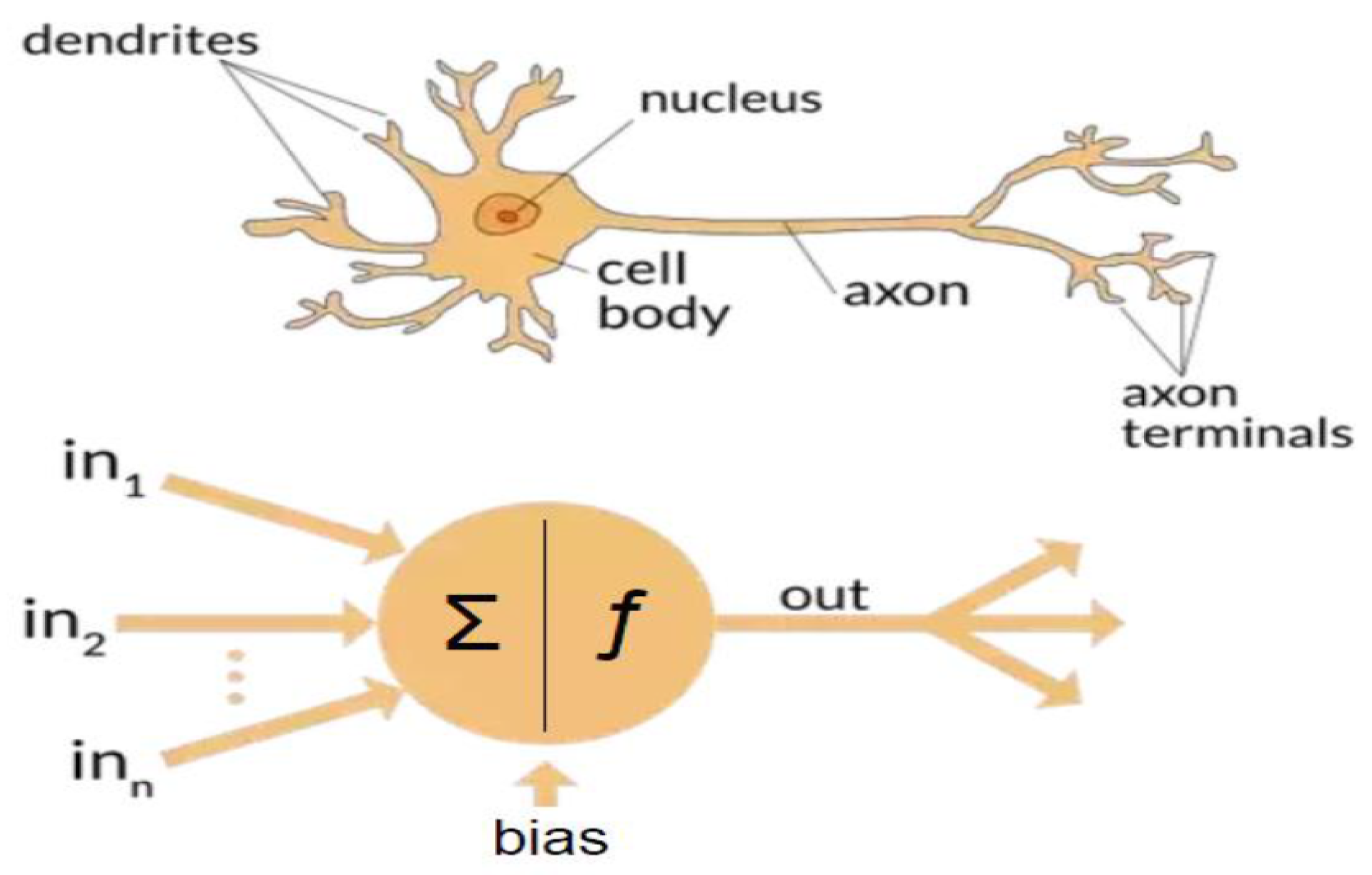
Traumatic brain injury (TBI) can produce temporary biochemical imbalance due to leaks through cell membranes or disruption of the axoplasmic flow due to the misalignment of intracellular neurofilaments. If untreated, TBI can lead to Alzheimer’s, Parkinson’s, or total disability. Mild TBI (mTBI) accounts for about about 90 percent of all TBI cases. The detection of TBI as soon as it happens is crucial for successful treatment management. Neuroimaging-based tests provide only a structural and functional mapping of the brain with poor temporal resolution. Such tests may not detect mTBI. On the other hand, the electroencephalogram (EEG) provides good spatial resolution and excellent temporal resolution of the brain activities beside its portability and low cost.

traumatic brain injury neuroimaging electroencephalography preprocessing

1. Introduction

Traumatic brain injury can lead to three types of injuries: a collection of the blood within the skull called intracranial hematoma (ICH) [1], an elevated intracranial pressure (ICP) [2], and a midline shift (MLS) [3]. Nonfatal injuries can result in a severe lifetime disability, which has significant impact on the injured and his/her family, as well as on healthcare cost. The estimated annual worldwide number of TBI cases is 69 millions [4]. In 2016, the annual total cost of nonfatal TBI was about 40.6 billion dollars in the United States [5]. For the armed forces in the US, it was found that the TBI cases were 91 percent higher for deployed combat forces than those who are not in a combat zone [6]. It is known that the sooner nonfatal TBI is detected and treated, the lesser the long term impact on the injured will be. The first hour following a brain injury is known as the golden hour [7][8][9]. The Glasgow Coma Scale (GCS) is used to classify the severity of TBI injury as mild, moderate and severe according to their level and severity [10]. Mild TBI (mTBI) accounts for 70 to 90 percent of all TBI cases [11]. In the case where mTBI is not diagnosed, its effect may lead to limited to impaired cognitive function, fatigue, depression, irritability, and headaches.

2. Conventional TBI Detection Using EEG

EEG signals are voltage signals collected with respect to a neutral reference electrode(s). Even though EEG contains valuable information about the brain waves that can be used for TBI detection, the brain waves amplitudes are very small, typically less than 100 µV. Furthermore, the recordings themselves are distorted by physical and non-physiological noises called artifacts. Non-physiological artifacts include electrode displacement, environment, and electrode-scalp impedance. Physiological artifacts include the effect of eye movement, blinking, muscle activity and cardiac activity. Such artifacts, if not removed, can lead to misleading TBI detection. Before any automated TBI detection, EEG signals must be first preprocessed to remove noise and artifacts.

2.1. EEG Signal Preprocessing

A significant amount of research work has been conducted to remove noise and artifacts form EEG signals. Artifacts removal uses linear regression, filtering/regression, independent component analysis (ICA), and principal component analysis (PCA) or a combination of different techniques [12][13][14][15][16][17][18][19]. Currently, for regression or blind source separation, it is assumed that the EEG model is linear, whereas the noise that models the artifacts is additive. The brain waves are also assumed to be stationary. The use of principal component analysis is based on the assumption that EEG signals and the artifacts are independent and linearly mixed with the true brain wave signals. The principal component analysis (PCA) uses orthogonal transformation under the assumption that neuronal activity waves are orthogonal to artifacts. Most of the studies focus on the removal of a particular artifact. There is no known solution that accommodates the different types of artifacts at once. Brain wave signals are non-stationary [20], non causal and nonlinear. Obtaining the average of the collected EEG signal over a relatively long period of time, in order to get around the non-stationarity of the signal, will make it less sensitive to the fast dynamics of the cortex that requires sampling in the order of milliseconds. The current state of the art in EEG preprocessing using analytical tools is expected to have limited performance with potential false alarm in detection.

2.2. TBI Detection Using EEG

The physiological method of evaluating the TBI level of injury is the Glasgow coma scale (GCS) obtained by adding the scores from eye opening, verbal response and motor response. The visual inspection of EEG may be successfully performed by a highly trained professional. Unfortunately, those professionals are in short supply. Furthermore, when it comes to quantifying the oscillatory activities of the brain waves, visual inspection sometimes fails to even differentiate between normal and abnormal brain waves. In an attempt to help the detection process, the concept of quantitative EEG (qEEG) has been introduced. Quantitative EEG represents a set of features extracted from the EEG signals to assess the functional state of the brain. The frequency bands of clinical interest in which brain waves oscillate are delta (0.5–4 Hz), theta (4–7 Hz), alpha (8–13 Hz), beta (14–30 Hz), and gamma (30–100 Hz) [21]. The features extraction uses signal-processing-derived tools, such as signal powers, power spectrum parameters, regularity measures, and coherence [22][23][24][25]. Several features can be extracted from the EEG signals. These include relative power, amplitude symmetry, coherence and phase difference [26]. Given the qEEG feature, multivariate analysis and discriminant functions are used to detect the existence of a TBI and its severity [24][26][27][28][29][30][31]. There is no universal index for TBI detection at this time. As pointed out earlier, due to the nonstationary nature of EEG signals and the fact that the signal features can vary for the same person and from person to person, the quantitative analysis using qEEG for TBI detection is helpful, but not reliable. There is no guarantee that the extracted features will have a unique behavior for TBI. At this time, there is no universal clinical index for TBI detection. It is very difficult to establish the one-to-one mapping between the TBI severity index and the qEEGs.
If one treats the TBI severity index as the output of an unknown complex map whose inputs are the EEG signals, one can think of the artificial intelligence to help establish such a map. Recent advances in artificial intelligence and especially machine learning and deep learning led to successfully obtaining the proper output, given the input information, without knowing the actual analytical map between inputs and outputs.

3. TBI Detection Using Artificial Intelligence

When it comes to learning and making complex decisions, even when based on partial information, the human brain does miraculous things. The human brain is equipped with approximately 100 billion neurons connected by about 1000 trillion synapses. Human learning takes place via adjustment of the synapses strength during training and generalization. An artificial neuron is a simplified model of a biological neuron; see Figure 1. Artificial neural systems are made up with interconnected artificial neurons.
Figure 1. Biological and artificial neuron.

3.1. Artificial Intelligence

Artificial intelligence (AI) is a technology that attempts to make a machine an “intelligent” device. Static AI systems, known as expert systems, deal with machines that perform specific tasks. It typically uses rule-based programming and does not require any training. The limitation of expert systems is in its inability to make decisions regarding situations that are not covered by the rule base. Instead of using an expert to generate the set of rules, in machine learning, a machine learns without being programmed. It is much easier to obtain an “intelligent” machine by showing it examples of desired input and corresponding outputs than to program it manually. It uses training data to acquire knowledge that can be used for decision making. Contrary to classical expert systems, a well-trained machine learning algorithm can make a decision based on new input data that have not been seen before. In machine learning, the learning uses features extracted from the data, i.e., it does not operate on raw data directly. However, in deep learning, which is a sub field of machine learning, learning and decision making uses only raw clean data without human interference. It uses multi-layer artificial neural networks and attempts to operate in a similar manner to the brain. A conventional neural network is made up of an input layer, one hidden layer, and one output layer. A deep learning neural network is a neural network with multiple hidden layers; see Figure 2 and Figure 3, respectively.
Figure 2. Conventional artificial neural network.
Figure 3. Deep learning neural network.

3.2. Machine Learning for TBI Detection

Machine learning is a data-driven algorithm that learns and update its learning as new information is provided. There are three major types of machine learning: supervised, unsupervised, and reinforcement learning machines. Supervised learning uses input and desired output data, also called labeled data, to develop a predictive model for classification and regression. The learning algorithms include regression analysis, support vector machines, naïve Bayes, and decision tree. Unsupervised training or learning without a teacher uses only input data for the purpose of identifying patterns/structures in the data or clustering. A well-known learning algorithm is the K-means algorithm. Reinforcement learning ML is not provided with any data. It is provided with only a response to tell whether the output is true or false. Reinforcement learning is typically used for the brain–computer interface (BCI) [32][33][34][35][36][37][38][39][40]. The recent growth in data in different application areas has led to the availability of a huge amount of data, known as big data. The best example of big data availability is in the healthcare industry. To take advantage of such data, machine learning was one of the artificial intelligence technologies used [39][41][42]. This section focuses on the application of machine learning to TBI detection.
At the high level, the ML TBI detection based approach uses features extracted from the EEG(qEEG) data, as is the case for the conventional approach, then uses a learning algorithm for detection/classification. Supervised learning ML is used for classification, such as the presence or absence of a TBI in EEG signals. Supervised ML using support vector machine for TBI detection was done in [43][44], among others. The performance of the decision tree (DT), random forest (RF), and K-nearest neighbors (KNN) supervised learning algorithms was compared in [45] using random sampling and independent validation, respectively. In the study of [46], carried out using a relatively small sample of patients, the DT and KNN performed the worst. The study showed several interesting facts. First, the performance depends on the feature selection used. The second and most surprising one is that the performance using raw data and "artifacts free" data was similar. This appears to be a good indicator that existing artifact removal techniques are not helpful. A ML survey paper was published in 2020 [47].

3.3. Artificial Neural Networks: Deep Learning Neural Networks

A deep learning neural network is an artificial neural network with more than two hidden layers. In the open literature, there seem to be confusion between ML and DL. DL is a subfield of ML that uses artificial neural networks and attempts to operate in a similar way as the brain in terms of learning and generalization. There are three types of deep learning neural networks: artificial neural networks (ANN), convolutional neural networks (CNN), and recurrent neural networks (RNN). They are shown in Figure 3, Figure 4 and Figure 5, respectively. Contrary to ML, a deep learning neural network can operate directly on raw data instead of needing human intervention to select features extracted from the data whenever needed. Furthermore, DL is used for establishing complex maps between the inputs and the outputs. Typically, DL requires a high performance computing platform and a large amount of data for learning. The computing platform needs graphical and tensor processing units to reduce the processing time. DL has been applied to big data [49], medical [50][51], computer vision [52][53], power systems [54], nuclear power [55], etc. (Figure 6). As an example, ANN has been applied to detect TBI using CT scans of children admitted to the emergency unit [56]. They used CT scans to train an ANN to decide whether a patient is a clinically relevant TBI (CRTBI). CRTBI features are defined by the multicenter Pediatric Emergency Care Applied Research Network (PECARN). According to [56], this study gave excellent results. However, as discussed before, CT scans can only detect structural brain damage. It will be interesting if the study focuses on the detection of mTBI.
Figure 4. Convolutional neural network (CNN).
Figure 5. Recurrent neural network (RNN).
Figure 6. Deep learning applications.

References

  1. Miller, J.D.; Murray, L.S.; Teasdale, G.M. Development of a traumatic intracranial hematoma after a “minor” head injury. Neurosurgery 1990, 27, 669–673.
  2. Kristiansson, H.; Nissborg, E.; Bartek, J., Jr.; Andresen, M.; Reinstrup, P.; Romner, B. Measuring elevated intracranial pressure through noninvasive methods: A review of the literature. J. Neurosurg. Anesthesiol. 2013, 25, 372–385.
  3. Puffer, R.C.; Yue, J.K.; Mesley, M.; Billigen, J.B.; Sharpless, J.; Fetzick, A.L.; Puccio, A.; Diaz-Arrastia, R.; Okonkwo, D.O. Long-term outcome in traumatic brain injury patients with midline shift: A secondary analysis of the Phase 3 COBRIT clinical trial. J. Neurosurg. 2018, 131, 596–603.
  4. Dewan, M.C.; Rattani, A.; Gupta, S.; Baticulon, R.E.; Hung, Y.C.; Punchak, M.; Agrawal, A.; Adeleye, A.O.; Shrime, M.G.; Rubiano, A.M.; et al. Estimating the global incidence of traumatic brain injury. J. Neurosurg. 2018, 130, 1080–1097.
  5. Miller, G.F.; DePadilla, L.; Xu, L. Costs of nonfatal traumatic brain injury in the United States, 2016. Med. Care 2021, 59, 451–455.
  6. Agimi, Y.; Regasa, L.E.; Stout, K.C. Incidence of traumatic brain injury in the US Military, 2010–2014. Mil. Med. 2019, 184, e233–e241.
  7. Dinh, M.M.; Bein, K.; Roncal, S.; Byrne, C.M.; Petchell, J.; Brennan, J. Redefining the golden hour for severe head injury in an urban setting: The effect of prehospital arrival times on patient outcomes. Injury 2013, 44, 606–610.
  8. Hu, W.; Freudenberg, V.; Gong, H.; Huang, B. The “Golden Hour” and field triage pattern for road trauma patients. J. Saf. Res. 2020, 75, 57–66.
  9. Polinder, S.; Cnossen, M.C.; Real, R.G.L.; Covic, A.; Gorbunova, A.; Voormolen, D.C.; Master, C.L.; Haagsma, J.A.; Diaz-Arrastia, R.; von Steinbuechel, N. A Multidimensional Approach to Post-concussion Symptoms in Mild Traumatic Brain Injury. Front. Neurol. 2018, 9, 1113.
  10. Reith, F.; Van den Brande, R.; Synnot, A.; Gruen, R.; Maas, A.I. The reliability of the Glasgow Coma Scale: A systematic review. Intensive Care Med. 2016, 42, 3–15.
  11. Shan, R.; Szmydynger-Chodobska, J.; Warren, O.U.; Mohammad, F.; Zink, B.J.; Chodobski, A. A new panel of blood biomarkers for the diagnosis of mild traumatic brain injury/concussion in adults. J. Neurotrauma 2016, 33, 49–57.
  12. Congedo, M.; Gouy-Pailler, C.; Jutten, C. On the blind source separation of human electroencephalogram by approximate joint diagonalization of second order statistics. Clin. Neurophysiol. 2008, 119, 2677–2686.
  13. Mammone, N.; La Foresta, F.; Morabito, F.C. Automatic Artifact Rejection From Multichannel Scalp EEG by Wavelet ICA. IEEE Sens. J. 2012, 12, 533–542.
  14. Zou, Y.; Nathan, V.; Jafari, R. Automatic Identification of Artifact-Related Independent Components for Artifact Removal in EEG Recordings. IEEE J. Biomed. Health Inform. 2016, 2, 73–81.
  15. Mannan, M.M.N.; Jeong, M.Y.; Kamran, M.A. Hybrid ICA—Regression: Automatic identification and removal of ocular artifacts from electroencephalographic signals. Front. Hum. Neurosci. 2016, 10, 193.
  16. Mannan, M.M.N.; Kamran, M.A.; Jeong, M.Y. Identification and Removal of Physiological Artifacts From Electroencephalogram Signals: A Review. IEEE Access 2018, 6, 30630–30652.
  17. Dai, C.; Wang, J.; Xie, J.; Li, W.; Gong, Y.; Li, Y. Removal of ECG Artifacts From EEG Using an Effective Recursive Least Square Notch Filter. IEEE Access 2019, 7, 158872–158880.
  18. Chang, C.Y.; Hsu, S.H.; Pion-Tonachini, L.; Jung, T.P. Evaluation of Artifact Subspace Reconstruction for Automatic Artifact Components Removal in Multi-Channel EEG Recordings. IEEE Trans. Biomed. Eng. 2020, 67, 1114–1121.
  19. Robbins, K.A.; Touryan, J.; Mullen, T.; Kothe, C.; Bigdely-Shamlo, N. How Sensitive Are EEG Results to Preprocessing Methods: A Benchmarking Study. IEEE Trans. Neural Syst. Rehabil. Eng. 2020, 28, 1081–1090.
  20. Blanco, S.; Garcia, H.; Quiroga, R.Q.; Romanelli, L.; Rosso, O.A. Stationarity of the EEG series. IEEE Eng. Med. Biol. Mag. 1995, 14, 395–399.
  21. Penttonen, M.; Buzsáki, G. Natural logarithmic relationship between brain oscillators. Thalamus Relat. Syst. 2003, 2, 145–152.
  22. Nuwer, M.R.; Hovda, D.A.; Schrader, L.M.; Vespa, P.M. Routine and quantitative EEG in mild traumatic brain injury. Clin. Neurophysiol. 2005, 116, 2001–2025.
  23. Islam, M.K.; Rastegarnia, A.; Yang, Z. Methods for artifact detection and removal from scalp EEG: A review. Neurophysiol. Clin. Neurophysiol. 2016, 46, 287–305.
  24. Albert, B.; Zhang, J.; Noyvirt, A.; Setchi, R.; Sjaaheim, H.; Velikova, S.; Strisland, F. Automatic EEG processing for the early diagnosis of traumatic brain injury. Procedia Comput. Sci. 2016, 96, 703–712.
  25. Lewine, J.D.; Plis, S.; Ulloa, A.; Williams, C.; Spitz, M.; Foley, J.; Paulson, K.; Davis, J.; Bangera, N.; Snyder, T.; et al. Quantitative EEG biomarkers for mild traumatic brain injury. J. Clin. Neurophysiol. 2019, 36, 298–305.
  26. Kostarelos, F.; MacNamee, C.; Mullane, B. A hardware implementation of a qEEG-based discriminant function for brain injury detection. In Proceedings of the 2021 IEEE Biomedical Circuits and Systems Conference (BioCAS), Berlin, Germany, 7–9 October 2021; pp. 1–6.
  27. Sjaaheim, H.; Albert, B.; Setchi, R.; Noyvirt, A.; Strisland, F. A portable medical system for the early diagnosis and treatment of traumatic brain injury. In Proceedings of the 2014 IEEE International Conference on Systems, Man, and Cybernetics (SMC), San Diego, CA, USA, 5–8 October 2014; pp. 2529–2534.
  28. Rapp, P.E.; Keyser, D.O.; Albano, A.; Hernandez, R.; Gibson, D.B.; Zambon, R.A.; Hairston, W.D.; Hughes, J.D.; Krystal, A.; Nichols, A.S. Traumatic Brain Injury Detection Using Electrophysiological Methods. Front. Hum. Neurosci. 2015, 9, 11.
  29. Dingle, A.A.; Jones, R.D.; Carroll, G.J.; Fright, W.R. A multistage system to detect epileptiform activity in the EEG. IEEE Trans. Biomed. Eng. 1993, 40, 1260–1268.
  30. Chamanzar, A.; George, S.; Venkatesh, P.; Chamanzar, M.; Shutter, L.; Elmer, J.; Grover, P. An algorithm for automated, noninvasive detection of cortical spreading depolarizations based on EEG simulations. IEEE Trans. Biomed. Eng. 2018, 66, 1115–1126.
  31. Fisher, J.A.; Huang, S.; Ye, M.; Nabili, M.; Wilent, W.B.; Krauthamer, V.; Myers, M.R.; Welle, C.G. Real-time detection and monitoring of acute brain injury utilizing evoked electroencephalographic potentials. IEEE Trans. Neural Syst. Rehabil. Eng. 2016, 24, 1003–1012.
  32. Jo, T. Machine Learning Foundations; Springer: Berlin/Heidelberg, Germany, 2021.
  33. Alzubi, J.; Nayyar, A.; Kumar, A. Machine learning from theory to algorithms: An overview. Proc. J. Phys. Conf. Ser. 2018, 142, 012012.
  34. Hastie, T.; Tibshirani, R.; Friedman, J.H.; Friedman, J.H. The Elements of Statistical Learning: Data Mining, Inference, and Prediction; Springer: Berlin/Heidelberg, Germany, 2009; Volume 2.
  35. Sutton, R.S.; Barto, A.G. Reinforcement Learning: An Introduction; MIT Press: Cambridge, MA, USA, 2018.
  36. Hosseini, M.P.; Hosseini, A.; Ahi, K. A review on machine learning for EEG signal processing in bioengineering. IEEE Rev. Biomed. Eng. 2020, 14, 204–218.
  37. Rothmann, M.; Porrmann, M. A Survey of Domain-Specific Architectures for Reinforcement Learning. IEEE Access 2022, 10, 13753–13767.
  38. Roh, Y.; Heo, G.; Whang, S.E. A survey on data collection for machine learning: A big data-ai integration perspective. IEEE Trans. Knowl. Data Eng. 2019, 33, 1328–1347.
  39. Jordan, M.I.; Mitchell, T.M. Machine learning: Trends, perspectives, and prospects. Science 2015, 349, 255–260.
  40. Bonaccorso, G. Machine Learning Algorithms; Packt Publishing Ltd.: Birmingham, UK, 2017.
  41. Zhou, L.; Pan, S.; Wang, J.; Vasilakos, A.V. Machine learning on big data: Opportunities and challenges. Neurocomputing 2017, 237, 350–361.
  42. L’heureux, A.; Grolinger, K.; Elyamany, H.F.; Capretz, M.A. Machine learning with big data: Challenges and approaches. IEEE Access 2017, 5, 7776–7797.
  43. Cao, C.; Tutwiler, R.L.; Slobounov, S. Automatic classification of athletes with residual functional deficits following concussion by means of EEG signal using support vector machine. IEEE Trans. Neural Syst. Rehabil. Eng. 2008, 16, 327–335.
  44. Lai, C.Q.; Abdullah, M.Z.; Abdullah, J.M.; Azman, A.; Ibrahim, H. Screening of moderate traumatic brain injury from power feature of resting state electroencephalography using support vector machine. In Proceedings of the 2019 2nd International Conference on Electronics and Electrical Engineering Technology, Penang, Malaysia, 25–27 September 2019; pp. 99–103.
  45. Schmid, E.; Fan, Y.; Chi, T.; Golanov, E.; Regnier-Golanov, A.S.; Austerman, R.J.; Podell, K.; Cherukuri, P.; Bentley, T.; Steele, C.T.; et al. Review of wearable technologies and machine learning methodologies for systematic detection of mild traumatic brain injuries. J. Neural Eng. IOP Publ. 2021, 18, 041006.
  46. Vivaldi, N.; Caiola, M.; Solarana, K.; Ye, M. Evaluating performance of eeg data-driven machine learning for traumatic brain injury classification. IEEE Trans. Biomed. Eng. 2021, 68, 3205–3216.
  47. Noor, N.S.E.M.; Ibrahim, H. Machine learning algorithms and quantitative electroencephalography predictors for outcome prediction in traumatic brain injury: A systematic review. IEEE Access 2020, 8, 102075–102092.
  48. Gravesteijn, B.Y.; Nieboer, D.; Ercole, A.; Lingsma, H.F.; Nelson, D.; Van Calster, B.; Steyerberg, E.W.; Åkerlund, C.; Amrein, K.; Andelic, N.; et al. Machine learning algorithms performed no better than regression models for prognostication in traumatic brain injury. J. Clin. Epidemiol. 2020, 122, 95–107.
  49. Nti, I.K.; Quarcoo, J.A.; Aning, J.; Fosu, G.K. A mini-review of machine learning in big data analytics: Applications, challenges, and prospects. Big Data Min. Anal. 2022, 5, 81–97.
  50. Hussein, S.; Kandel, P.; Bolan, C.W.; Wallace, M.B.; Bagci, U. Lung and Pancreatic Tumor Characterization in the Deep Learning Era: Novel Supervised and Unsupervised Learning Approaches. IEEE Trans. Med. Imaging 2019, 38, 1777–1787.
  51. Roy, S.; Menapace, W.; Oei, S.; Luijten, B.; Fini, E.; Saltori, C.; Huijben, I.; Chennakeshava, N.; Mento, F.; Sentelli, A.; et al. Deep Learning for Classification and Localization of COVID-19 Markers in Point-of-Care Lung Ultrasound. IEEE Trans. Med. Imaging 2020, 39, 2676–2687.
  52. White, J.; Kameneva, T.; McCarthy, C. Vision Processing for Assistive Vision: A Deep Reinforcement Learning Approach. IEEE Trans. Hum. Mach. Syst. 2022, 52, 123–133.
  53. Wei, X.S.; Song, Y.Z.; Mac Aodha, O.; Wu, J.; Peng, Y.; Tang, J.; Yang, J.; Belongie, S. Fine-Grained Image Analysis with Deep Learning: A Survey. IEEE Trans. Pattern Anal. Mach. Intell. 2021.
  54. Khodayar, M.; Liu, G.; Wang, J.; Khodayar, M.E. Deep learning in power systems research: A review. CSEE J. Power Energy Syst. 2021, 7, 209–220.
  55. Tang, C.; Yu, C.; Gao, Y.; Chen, J.; Yang, J.; Lang, J.; Liu, C.; Zhong, L.; He, Z.; Lv, J. Deep learning in nuclear industry: A survey. Big Data Min. Anal. 2022, 5, 140–160.
  56. Hale, A.T.; Stonko, D.P.; Lim, J.; Guillamondegui, O.D.; Shannon, C.N.; Patel, M.B. Using an artificial neural network to predict traumatic brain injury. J. Neurosurgery Pediatr. PED 2019, 23, 219–226.
More
Information
Contributors MDPI registered users' name will be linked to their SciProfiles pages. To register with us, please refer to https://encyclopedia.pub/register : ,
View Times: 382
Revisions: 3 times (View History)
Update Date: 01 Nov 2022
1000/1000
Video Production Service