Submitted Successfully!
To reward your contribution, here is a gift for you: A free trial for our video production service.
Thank you for your contribution! You can also upload a video entry or images related to this topic.
Version Summary Created by Modification Content Size Created at Operation
1 -- 2002 2022-10-27 20:43:20 |
2 format change + 15 word(s) 2017 2022-10-28 04:59:44 |

Video Upload Options

Do you have a full video?

Confirm

Are you sure to Delete?
Cite
If you have any further questions, please contact Encyclopedia Editorial Office.
Monroe, J.D.;  Basheer, F.;  Gibert, Y. Xmrk and Human Epidermal Growth Factor Receptor. Encyclopedia. Available online: https://encyclopedia.pub/entry/31668 (accessed on 02 July 2024).
Monroe JD,  Basheer F,  Gibert Y. Xmrk and Human Epidermal Growth Factor Receptor. Encyclopedia. Available at: https://encyclopedia.pub/entry/31668. Accessed July 02, 2024.
Monroe, Jerry D., Faiza Basheer, Yann Gibert. "Xmrk and Human Epidermal Growth Factor Receptor" Encyclopedia, https://encyclopedia.pub/entry/31668 (accessed July 02, 2024).
Monroe, J.D.,  Basheer, F., & Gibert, Y. (2022, October 27). Xmrk and Human Epidermal Growth Factor Receptor. In Encyclopedia. https://encyclopedia.pub/entry/31668
Monroe, Jerry D., et al. "Xmrk and Human Epidermal Growth Factor Receptor." Encyclopedia. Web. 27 October, 2022.
Xmrk and Human Epidermal Growth Factor Receptor
Edit

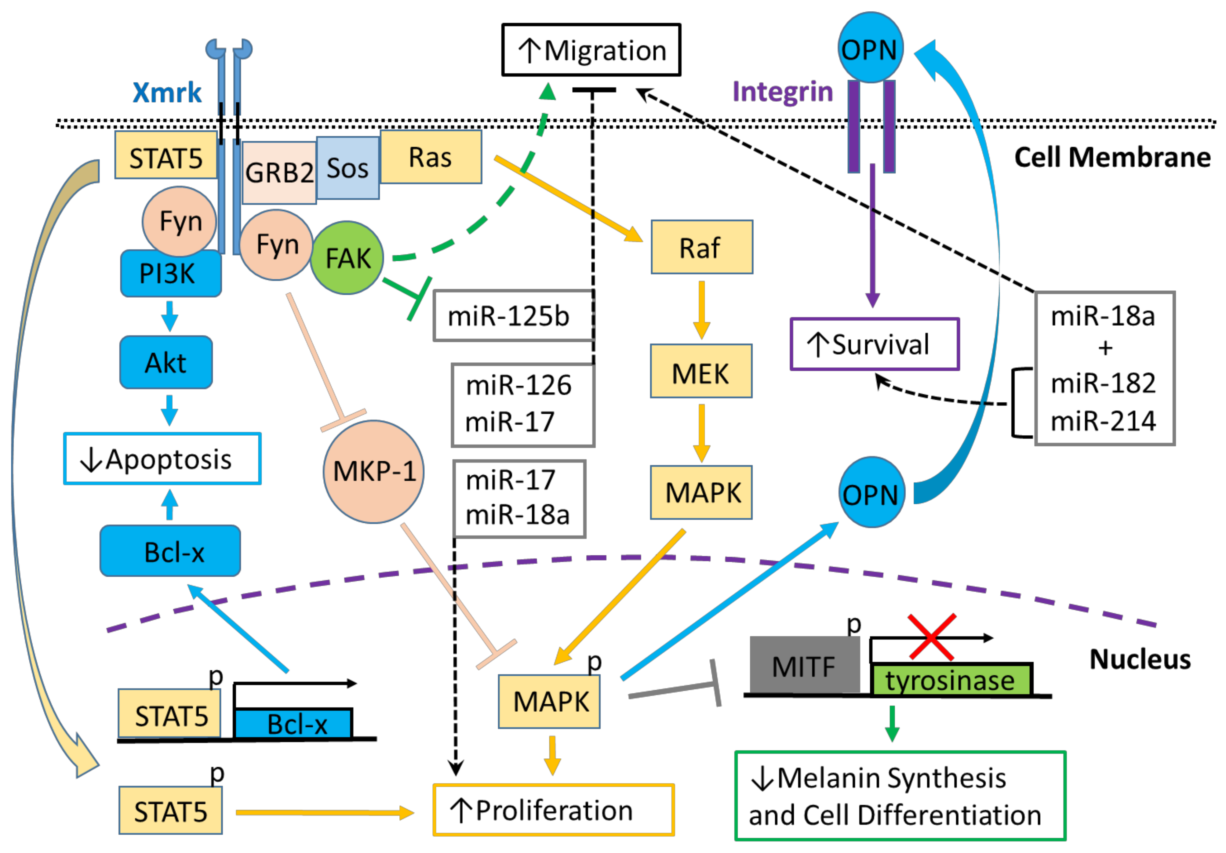
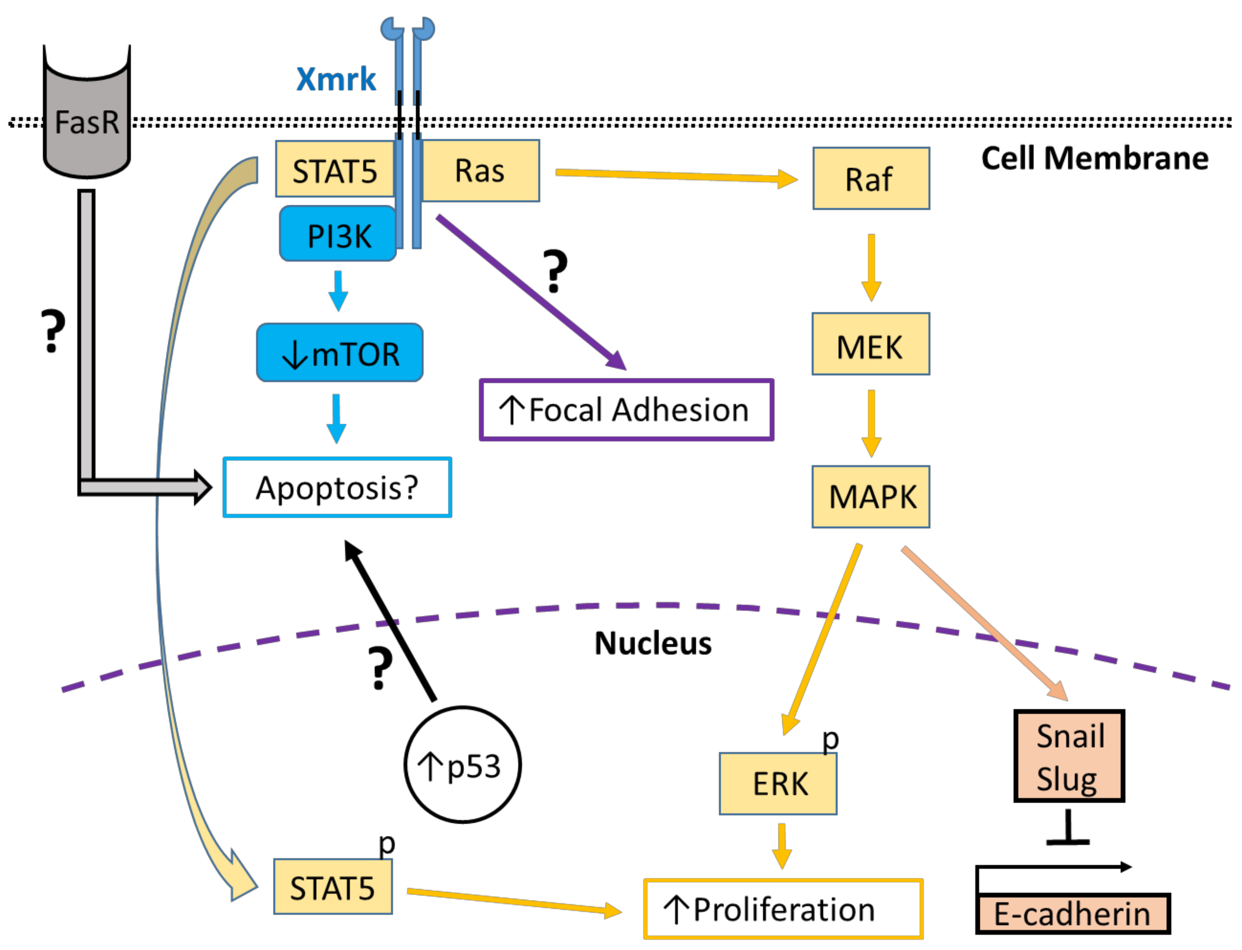
Xmrk is a gene product closely related to the human epidermal growth factor receptor (EGFR), which is associated with a wide variety of pathological conditions, including cancer. Comparative analyses of Xmrk and EGFR signal transduction in melanoma have shown that both utilize signal transducer and activator of transcription 5 (STAT5) signaling to regulate apoptosis and cell proliferation, phosphoinositide 3-kinase (PI3K) to modulate apoptosis, focal adhesion kinase (FAK) to control migration, and the Ras/Raf/MEK/mitogen-activated protein kinase (MAPK) pathway to regulate cell survival, proliferation, and differentiation. Further, Xmrk and EGFR may also modulate similar chemokine, extracellular matrix, oxidative stress, and microRNA signaling pathways in melanoma. In hepatocellular carcinoma (HCC), Xmrk and EGFR signaling utilize STAT5 to regulate cell proliferation, and Xmrk may signal through PI3K and FasR to modulate apoptosis. At the same time, both activate the Ras/Raf/MEK/MAPK pathway to regulate cell proliferation and E-cadherin signaling.

epidermal growth factor receptor Xmrk cancer

1. Introduction

The epidermal growth factor receptor (EGFR) is a member of the erythroblastic leukemia viral oncogene (ErbB) family of receptor tyrosine kinases (RTKs). Ligand-dependent EGFR activation triggers receptor autophosphorylation creating phosphotyrosine-binding sites for other proteins that then signal along various signal transduction pathways, e.g., mitogen-activated protein kinase (MAPK), phosphoinositide 3-kinase/protein kinase B (PI3K/Akt) and c-Jun N-terminal kinase (JNK), which control cellular behaviors, including adhesion, migration and proliferation [1][2] (Figure 1 and Figure 2). Mutations in EGFR have been linked to developing various diseases, including diverse cancers, including melanoma and hepatocarcinoma (HCC) [3][4]. Comparative genomic studies have shown that species of many other organisms, including fish, have genetic sequences homologous to EGFR [5].
Figure 1. Xmrk signaling in the Xiphophorus melanoma model. Xiphophorus melanoma receptor kinase (Xmrk) is a receptor tyrosine kinase that recruits and activates several kinases, transcription factors and adaptor proteins integrated into signaling networks to regulate pigment cell cancer transformation. Activation of the kinase, phosphoinositide 3-kinase (PI3K), prevents apoptosis in malignant cells, as does activation of the transcription factor, signal transducer and activator of transcription 5 (STAT5), via B-cell lymphoma (Bcl-x) signaling, and STAT5 also promotes proliferation. The Src kinase protein, Fyn, acts as a docking protein and, through focal adhesion kinase (FAK), can induce pigment cell migration. Further, Fyn inhibits MAPK phosphatase 1 (MKP-1), a repressor of MAPK, causing the promotion of MAPK’s function to increase melanoma cell proliferation. Xmrk can also signal via the adaptor proteins, GRB2 and Sos, to activate the Ras/Raf/MEK/MAPK pathway, thereby enhancing proliferation. Additionally, MAPK signaling causes degradation of the transcription factor, microphthalmia transcription factor (MITF), leading to reduced transcription of tyrosinase, which decreases melanin synthesis and inhibits pigment cell differentiation. Another function of activated MAPK is to induce the synthesis of osteopontin (OPN), which is secreted, binds to integrins on the cell membrane surface, and promotes pigment cell survival. Several miRNAs that are regulated in fish Xmrk models also have functions in human melanoma. The miRNAs miR-18a, -182, and -214, can act to promote migration in human melanoma, while miR-182 and -214 can also stimulate cell survival. MiR-17, 125b, and -126 act to inhibit melanoma cell migration, with -125b’s action inhibited by FAK. MiR-17, along with miR-18a, also function to increase melanoma proliferation.
Figure 2. Xmrk signaling in the zebrafish hepatocellular carcinoma model. In transgenic zebrafish models of HCC, Xmrk signals through kinases and transcription factors that modulate liver cancer progression. PI3K activation may modulate apoptosis through the mechanistic target of rapamycin (mTOR), although mTOR expression may be decreased in HCC, making its effect on apoptosis uncertain. STAT5 signaling can promote proliferation, as does MAPK via activation of extracellular signal-regulated kinases (ERK). MAPK also has a dual role in signaling through the transcription factors, Snail and Slug, to inhibit E-cadherin activity. The formation of focal adhesions is increased through a pathway that is currently uncharacterized. Xmrk is also associated with increased expression of the tumor suppressor, p53, and the Fas receptor (FasR), which regulates apoptosis during HCC, but the details of their associated signaling mechanisms are not currently understood.
Crosses between species of the fish genus Xiphophorus: X. maculatus (the southern platyfish) and X. hellerii (the green swordtail) produce fish that spontaneously develop malignant melanoma induced by a proto-oncogene encoding a receptor tyrosine kinase designated Xiphophorus melanoma receptor kinase (Xmrk), which is located on the sex chromosome [6]. The encoded Xmrk protein is structurally related to the human EGFR with an extracellular ligand-binding domain, a transmembrane domain and an intracellular catalytic domain [7]. Studies in human melanoma cell lines and the Xiphophorus cross-derived melanoma cell line (PSM-1) [8] using chimeric receptors combining the human epidermal growth factor receptor (HER) with Xmrk (HER-mrk) have shown that Xmrk displays constitutive autophosphorylation and is a functional receptor tyrosine kinase with activity in malignant melanoma [9]. Blockage of Xmrk’s kinase activity in Xiphophorus derived PSM cells demonstrates that receptor autophosphorylation is sufficient to trigger mitogenic signaling in melanoma cells [10]. Further, receptor stimulation induces autophosphorylation resulting in the production of the physiological ligand in an autocrine manner [11].
The role of Xmrk in melanoma also has been investigated in Oryzias latipes, the medaka fish. Ectopic expression of the Xmrk oncogene cloned from Xiphophorus and introduced into medaka embryos induces tumors, typically classifiable as cysts or hyperplasias, primarily in the epithelium, but also in the brain, retina, integument and pigment cells [12]. Expression of HER-mrk in medaka has demonstrated that one ligand, human transforming growth factor-alpha (hTGF alpha), can cause activation of this chimeric receptor [12]. Mutational studies of Xmrk in medaka embryos also have shown that point mutations can lead to constitutive, ligand-independent receptor kinase activity [13]. Medaka expressing HER-mrk, which exhibits invasive melanoma, has been used to show that activated Xmrk induces EGFR expression and ligand production leading to the formation of an autocrine loop that promotes pro-tumorigenic signaling [14].
RNA-Seq comparative analyses of Xiphophorus hellerii/maculatus hybrid fish and human melanoma tissues have shown shared pathways in these tissues governing: inflammation, cell migration, cell proliferation, pigmentation, cancer development, and metastasis [15]. Structural studies using the Xiphophorus and medaka models suggest that oncogenic signaling mechanisms and related translational studies can be studied using zebrafish transgenic Xmrk models. Recently, the tetracycline-controlled transcriptional activation Tet-on system [16][17] has been used in zebrafish to express Xmrk under the control of the liver-specific fatty acid-binding protein 10 (fabp10) promoter (also designated as liver fatty acid-binding protein (LFABP)) to study signaling in hepatocellular carcinoma (HCC) [18][19][20][21].

2. Genetic Characterization and Comparison of Xmrk with the Human EGFR

The Xmrk gene is hypothesized to have arisen from gene duplication and nonhomologous recombination during evolution [5][22]. There are three Xiphophorus orthologs of the EGFR gene: egfra, which encodes a tyrosine kinase receptor; egfrb, which is a proto-oncogene form termed INV-Xmrk; and ONC-Xmrk, a duplicated version of INV-Xmrk, which is highly expressed in melanoma where it is responsible for inducing oncogenic transformation [23][24][25][26][27]. Oncogenic activation of the ONC-Xmrk receptor occurs via two mutations, i.e., G336R and C555S, both located in the extracellular domain that is not found in INV-Xmrk; these mutations result in constitutive receptor dimerization leading to constitutive kinase signaling [27][28].
The Xmrk promoter is highly methylated in nontransformed tissues, but in the Xiphophorus cross-derived melanoma cell line (PSM-1) [8], it is unmethylated, suggesting that methylation status also regulates Xmrk expression [29]. Transcription of Xmrk does not stem from the action of cis-regulatory elements present in the promoter region of pigment cells, suggesting that epigenetic mechanisms also play a role in Xmrk’s oncogenic activation [30].
In comparison to Xmrk, the human EGFR is encoded by a single gene that has orthologs in all vertebrate species [5][31]; two transcriptions start sites have been identified, resulting in two major transcripts (10 and 5.6 kb) encoding the full-length receptor [31]. In addition to the two transcripts that encode EGFR Isoform A (the “full-length” or holo-receptor), three alternately processed transcripts encode alternate EGFR receptor isoforms (isoforms B, C, and D) [31], all of which are expressed in normal human tissues. While the full-length EGFR isoform (A) is known to signal through both ligand-dependent and independent signaling pathways [32], all three alternate isoforms lack the receptor kinase domain, rendering these isoforms unable to signal directly through protein phosphorylation. These isoforms have been hypothesized to function through signaling mechanisms similar to those used by the IGF-binding proteins, including regulating cell surface to extracellular matrix interactions, i.e., as matricellular proteins [33].
The transmembrane glycoprotein EGFR is derived from the 1210 residue precursor, which is cleaved at the N-terminus resulting in an 1186 residue mature EGFR protein [34]. The extracellular or ligand-binding domain (exons 1–16, amino acids 1–621) of the human EGFR receptor comprises four subdomains, two leucine-rich repeats (I and III) and two cysteine-rich repeats (II and IV) [35]. Subdomains I and III are involved in ligand-binding, domain II facilitates the formation of homo- or heterodimers with an analogous family member domain, and domain IV forms disulfide bonds to domain II. These subdomains are followed by a single hydrophobic transmembrane domain (exon 17, amino acids 645–668) and an intracellular cytoplasmic domain (exon 18–28, amino acids 669–1210). The cytoplasmic domain includes the tyrosine kinase (exons 18–24, amino acids 669–979) and C-terminal tail (exon 24–28, amino acids 980–1210) domains [35].
To date, several EGFR variants have been studied in humans [36][37][38]. In addition to the full-length isoform (isoform A), cells can also produce alternate EGFR isoforms that lack the intracellular domain [39]. The different roles of these alternative isoforms have not yet been established, but some have now been shown to play crucial roles [40]. Therefore, the researchers analyzed the different EGFR transcript variants reported in the genomic and protein databases for zebrafish and humans, as summarized in Table 1 and Table 2.
Table 1. List of zebrafish EGFR isoforms in the Ensembl database. ID, transcript name, transcript length, protein name and protein length for all zebrafish EGFR isoforms listed in the Ensembl database.

Ensembl ID

Transcript

Length (nt)

Protein (Uniprot)

Length (aa)

egfra-201

ENSDART00000108964.5

1868

F1RBY7

503

egfra-202

ENSDART00000128514.2

1881

F1RA48

389

egfra-203

ENSDART00000136906.3

2437

F1R671

760

egfra-205

ENSDART00000147261.3

3168

F1QU74

243

egfra-206

ENSDART00000150499.3

3007

F1Q7X2

625

egfra-207

ENSDART00000164152.3

6151

A0A0R4IFV9

1191

Table 2. List of human EGFR isoforms in the Ensembl and NCBI genomic databases. ID, transcript name, transcript length, protein name and protein length for all human EGFR isoforms listed in the Ensembl (top) and NCBI (bottom) databases. Isoforms highlighted in green are common to both databases.

Ensembl ID

Transcript

Length (nt)

Protein (Uniprot)

Length (aa)

EGFR-201

ENST00000275493.7

9905

P0053-1

1210

EGFR-202

ENST00000342916.7

2239

P00533-4

628

EGFR-203

ENST00000344576.6

2864

P00533-3

705

EGFR-204

ENST00000420316.6

1570

P00533-2

405

EGFR-205

ENST00000450046.1

691

C9JYS6

128

EGFR-206

ENST00000454757.6

5464

E9PFD7

1165

EGFR-207

ENST00000455089.5

3844

Q504U8

1091

NCBI ID

Transcript

Length (nt)

Protein (NCBI)

Length (aa)

Isoform A

NM_005228.5

9905

NP_005219.1

1210

Isoform G

NM_001346899.2

9770

NP_001346899.2

1165

Isoform I

NM_001346941.2

9104

NP_001333870.1

943

Isoform F

NM_001346898.2

3983

NP_001333827.1

1136

Isoform E

NM_001346897.2

3848

NP_001333826.1

1091

Isoform D

NM_201284.2

2872

NP_958441.1

705

Isoform B

NM_201282.2

2254

NP_958439.1

628

Isoform C

NM_201283.2

1575

NP_958440.1

405

Isoform H

NM_001346900.2

9676

NP_001333829.1

1157

Analysis of zebrafish genomic data version 11 on the Ensembl website, RGCz11; www.ensembl.org (accessed on 4 March 2021), revealed identifying six transcripts for egfra (egfra-201; 202; 203; 205; 206 and 207) (Table 1), and one transcript, which retained an intron and, therefore, is not listed in Table 1. Teleosts have experienced a fish-specific genome duplication [41], and, therefore, around one-third of the zebrafish genes are duplicated. In the zebrafish, a putative paralog named egfrb has been identified, but to date, no isoforms or transcript variants have been reported [42], and only one paper has reported the expression of the egfrb paralog in zebrafish [43]. The latest human genomic data on the Ensembl website (www.ensembl.org, accessed on 4 March 2021), human genome version (GRCh38.p13), revealed identifying 7 reported transcripts (EGFR-201 to 207) ranging from 1210 amino acids in length to 128 amino acids in length (Table 2). Ensembl human genomic data analysis also revealed the identification of two processed transcripts (561 and 452 bp, respectively) and one retained intron (665 bp), none of which are translated into a protein and, therefore, were excluded from Table 2.
NCBI analysis of the latest human EGFR transcript sequences (updated 7 March 2021) within the National Library of Medicine protein database revealed the existence of 9 EGFR transcripts encoding proteins from 1210 amino acids in length to 405 amino acids in length (Table 2). Comparison of the Ensembl and NCBI data sets revealed that 7 transcripts are common in both databases (highlighted in green in Table 2), while the reported transcript EGFR-205 is unique to the Ensembl database, and isoforms I, F and H are only present in the NCBI database (Table 2).

References

  1. Yarden, Y. The EGFR family and its ligands in human cancer: Signalling mechanisms and therapeutic opportunities. Eur. J. Cancer 2001, 37, S3–S8.
  2. Chen, J.; Zeng, F.; Forrester, S.J.; Eguchi, S.; Zhang, M.; Harris, R.C. Expression and Function of the Epidermal Growth Factor Receptor in Physiology and Disease. Physiol. Rev. 2016, 96, 1025–1069.
  3. Komposch, K.; Sibilia, M. EGFR Signaling in Liver Diseases. Int. J. Mol. Sci. 2015, 17, 30.
  4. Tímár, J.; Vizkeleti, L.; Doma, V.; Barbai, T.; Rásó, E. Genetic progression of malignant melanoma. Cancer Metastasis Rev. 2016, 35, 93–107.
  5. Barberán, S.; Martín-Durán, J.M.; Cebrià, F. Evolution of the EGFR pathway in Metazoa and its diversification in the planarian Schmidtea mediterranea. Sci. Rep. 2016, 6, 28071.
  6. Schartl, M. Homology of melanoma-inducing loci in the genus Xiphophorus. Genetics 1990, 126, 1083–1091.
  7. Schartl, M.; Adam, D. Molecular cloning, structural characterization, and analysis of transcription of the melanoma oncogene of Xiphophorus. Pigment Cell Res. 1992, 2, 173–180.
  8. Wakamatsu, Y. Establishment of a cell line from the platyfish-swordtail hybrid melanoma. Cancer Res. 1981, 41, 679–680.
  9. Wittbrodt, J.; Lammers, R.; Malitschek, B.; Ullrich, A.; Schartl, M. The Xmrk receptor tyrosine kinase is activated in Xiphophorus malignant melanoma. EMBO J. 1992, 11, 4239–4246.
  10. Wellbrock, C.; Fischer, P.; Schartl, M. Receptor tyrosine kinase Xmrk mediates proliferation in Xiphophorus melanoma cells. Int. J. Cancer 1998, 76, 437–442.
  11. Malitschek, B.; Wittbrodt, J.; Fischer, P.; Lammers, R.; Ullrich, A.; Schartl, M. Autocrine stimulation of the Xmrk receptor tyrosine kinase in Xiphophorus melanoma cells and identification of a source for the physiological ligand. J. Biol. Chem. 1994, 269, 10423–10430.
  12. Winkler, C.; Wittbrodt, J.; Lammers, R.; Ullrich, A.; Schartl, M. Ligand-dependent tumor induction in medakafish embryos by a Xmrk receptor tyrosine kinase transgene. Oncogene 1994, 9, 1517–1525.
  13. Winnemoeller, D.; Wellbrock, C.; Schartl, M. Activating mutations in the extracellular domain of the melanoma inducing receptor Xmrk are tumorigenic in vivo. Int. J. Cancer 2005, 117, 723–729.
  14. Laisney, J.A.; Mueller, T.D.; Schartl, M.; Meierjohann, S. Hyperactivation of constitutively dimerized oncogenic EGF receptors by autocrine loops. Oncogene 2013, 32, 2403–2411.
  15. Lu, Y.; Boswell, M.; Boswell, W.; Kneitz, S.; Hausmann, M.; Klotz, B.; Regneri, J.; Savage, M.; Amores, A.; Postlethwait, J.; et al. Comparison of Xiphophorus and human melanoma transcriptomes reveals conserved pathway interactions. Pigment Cell Melanoma Res. 2018, 31, 496–508.
  16. Gossen, M.; Freundlieb, S.; Bender, G.; Müller, G.; Hillen, W.; Bujard, H. Transcriptional activation by tetracyclines in mammalian cells. Science 1995, 268, 1766–1769.
  17. Knopf, F.; Schnabel, K.; Haase, C.; Pfeifer, K.; Anastassiadis, K.; Weidinger, G. Dually inducible TetON systems for tissue-specific conditional gene expression in zebrafish. Proc. Natl. Acad. Sci. USA 2010, 107, 19933–19938.
  18. Li, Z.; Huang, X.; Zhan, H.; Zeng, Z.; Li, C.; Spitsbergen, J.M.; Meierjohann, S.; Schartl, M.; Gong, Z. Inducible and repressable oncogene-addicted hepatocellular carcinoma in Tet-on xmrk transgenic zebrafish. J. Hepatol. 2012, 56, 419–425.
  19. Li, Z.; Luo, H.; Li, C.; Huo, X.; Yan, C.; Huang, X.; Al-Haddawi, M.; Mathavan, S.; Gong, Z. Transcriptomic analysis of a transgenic zebrafish hepatocellular carcinoma model reveals a prominent role of immune responses in tumour progression and regression. Int. J. Cancer 2014, 135, 1564–1573.
  20. Zheng, W.; Li, Z.; Nguyen, A.T.; Li, C.; Emelyanov, A.; Gong, Z. Xmrk, kras and myc transgenic zebrafish liver cancer models share molecular signatures with subsets of human hepatocellular carcinoma. PLoS ONE 2014, 9, e91179.
  21. Li, Z.; Zheng, W.; Li, H.; Li, C.; Gong, Z. Synergistic Induction of Potential Warburg Effect in Zebrafish Hepatocellular Carcinoma by Co-Transgenic Expression of Myc and xmrk Oncogenes. PLoS ONE 2015, 10, e0132319.
  22. Förnzler, D.; Altschmied, J.; Nanda, I.; Kolb, R.; Baudler, M.; Schmid, M.; Schartl, M. The Xmrk oncogene promoter is derived from a novel amplified locus of unusual organization. Genome Res. 1996, 6, 102–113.
  23. Dimitrijevic, N.; Winkler, C.; Wellbrock, C.; Gómez, A.; Duschl, J.; Altschmied, J.; Schartl, M. Activation of the Xmrk proto-oncogene of Xiphophorus by overexpression and mutational alterations. J. Biol. Chem. 1997, 272, 131–137.
  24. Weis, S.; Schartl, M. The macromelanophore locus and the melanoma oncogene Xmrk are separate genetic entities in the genome of Xiphophorus. Genetics 1998, 149, 1909–1920.
  25. Schartl, M.; Hornung, U.; Gutbrod, H.; Volff, J.N.; Wittbrodt, J. Melanoma loss-of-function mutants in Xiphophorus caused by Xmrk-oncogene deletion and gene disruption by a transposable element. Genetics 1999, 153, 1385–1394.
  26. Gómez, A.; Volff, J.N.; Hornung, U.; Schartl, M.; Wellbrock, C. Identification of a second egfr gene in Xiphophorus uncovers an expansion of the epidermal growth factor receptor family in fish. Mol. Biol. Evol. 2004, 21, 266–275.
  27. Meierjohann, S.; Mueller, T.; Schartl, M.; Buehner, M. A structural model of the extracellular domain of the oncogenic EGFR variant Xmrk. Zebrafish 2006, 3, 359–369.
  28. Gómez, A.; Wellbrock, C.; Gutbrod, H.; Dimitrijevic, N.; Schartl, M. Ligand-independent dimerization and activation of the oncogenic Xmrk receptor by two mutations in the extracellular domain. J. Biol. Chem. 2001, 276, 3333–3340.
  29. Altschmied, J.; Ditzel, L.; Schartl, M. Hypomethylation of the Xmrk oncogene promoter in melanoma cells of Xiphophorus. Biol. Chem. 1997, 378, 1457–1466.
  30. Regneri, J.; Volff, J.N.; Schartl, M. Transcriptional control analyses of the Xiphophorus melanoma oncogene. Comp. Biochem. Physiol. C Toxicol. Pharmacol. 2015, 178, 116–127.
  31. Reiter, J.L.; Threadgill, D.W.; Eley, G.D.; Strunk, K.E.; Danielsen, A.J.; Sinclair, C.S.; Pearsall, R.S.; Green, P.J.; Yee, D.; Lampland, A.L.; et al. Comparative genomic sequence analysis and isolation of human and mouse alternative EGFR transcripts encoding truncated receptor isoforms. Genomics 2001, 71, 1–20.
  32. Boerner, J.L.; Danielsen, A.; Maihle, N.J. Ligand-independent oncogenic signaling by the epidermal growth factor receptor: V-ErbB as a paradigm. Exp. Cell Res. 2003, 284, 111–121.
  33. Murphy-Ullrich, J.E. The de-adhesive activity of matricellular proteins: Is intermediate cell adhesion an adaptive state? J. Clin. Investig. 2001, 107, 785–790.
  34. Ullrich, A.; Coussens, L.; Hayflick, J.S.; Dull, T.J.; Gray, A.; Tam, A.W.; Lee, J.; Yarden, Y.; Libermann, T.A.; Schlessinger, J. Human epidermal growth factor receptor cDNA sequence and aberrant expression of the amplified gene in A431 epidermoid carcinoma cells. Nature 1984, 309, 418–425.
  35. Wee, P.; Wang, Z. Epidermal Growth Factor Receptor Cell Proliferation Signaling Pathways. Cancers 2017, 9, 52.
  36. Kim, H.M.; Lee, S.H.; Lim, J.; Yoo, J.; Hwang, D.Y. The epidermal growth factor receptor variant type III mutation frequently found in gliomas induces astrogenesis in human cerebral organoids. Cell Prolif. 2021, 54, e12965.
  37. Du, Z.; Brown, B.P.; Kim, S.; Ferguson, D.; Pavlick, D.C.; Jayakumaran, G.; Benayed, R.; Gallant, J.N.; Zhang, Y.K.; Yan, Y.; et al. Structure-function analysis of oncogenic EGFR Kinase Domain Duplication reveals insights into activation and a potential approach for therapeutic targeting. Nat. Commun. 2021, 12, 1382.
  38. Albitar, L.; Pickett, G.; Morgan, M.; Wilken, J.A.; Maihle, N.J.; Leslie, K.K. EGFR isoforms and gene regulation in human endometrial cancer cells. Mol. Cancer 2010, 9, 166.
  39. Guillaudeau, A.; Durand, K.; Bessette, B.; Chaunavel, A.; Pommepuy, I.; Projetti, F.; Robert, S.; Caire, F.; Rabinovitch-Chable, H.; Labrousse, F. EGFR soluble isoforms and their transcripts are expressed in meningiomas. PLoS ONE 2012, 7, e37204.
  40. Guillaudeau, A.; Durand, K.; Rabinovitch-Chable, H.; Pommepuy, I.; Mesturoux, L.; Robert, S.; Chaunavel, A.; Moreau, J.J.; Labrousse, F. Adult diffuse gliomas produce mRNA transcripts encoding EGFR isoforms lacking a tyrosine kinase domain. Int. J. Oncol. 2012, 40, 1142–1152.
  41. Jaillon, O.; Aury, J.M.; Brunet, F.; Petit, J.L.; Stange-Thomann, N.; Mauceli, E.; Bouneau, L.; Fischer, C.; Ozouf-Costaz, C.; Bernot, A.; et al. Genome duplication in the teleost fish Tetraodon nigroviridis reveals the early vertebrate proto-karyotype. Nature 2004, 431, 946–957.
  42. Laisney, J.A.; Braasch, I.; Walter, R.B.; Meierjohann, S.; Schartl, M. Lineage-specific co-evolution of the Egf receptor/ligand signaling system. BMC Evol. Biol. 2010, 10, 27.
  43. Ningappa, M.; So, J.; Glessner, J.; Ashokkumar, C.; Ranganathan, S.; Min, J.; Higgs, B.W.; Sun, Q.; Haberman, K.; Schmitt, L.; et al. The Role of ARF6 in Biliary Atresia. PLoS ONE 2015, 10, e0138381.
More
Information
Contributors MDPI registered users' name will be linked to their SciProfiles pages. To register with us, please refer to https://encyclopedia.pub/register : , ,
View Times: 435
Revisions: 2 times (View History)
Update Date: 28 Oct 2022
1000/1000
Video Production Service