Submitted Successfully!
To reward your contribution, here is a gift for you: A free trial for our video production service.
Thank you for your contribution! You can also upload a video entry or images related to this topic.
Version Summary Created by Modification Content Size Created at Operation
1 -- 1820 2022-10-25 08:34:08 |
2 Format correction Meta information modification 1820 2022-10-26 02:42:08 |

Video Upload Options

Do you have a full video?

Confirm

Are you sure to Delete?
Cite
If you have any further questions, please contact Encyclopedia Editorial Office.
Min, J.W.S.;  Saeed, N.;  Coene, A.;  Adriaens, M.;  Ceelen, W. Fundamental Principles in Electromotive Enhanced Drug Administration. Encyclopedia. Available online: https://encyclopedia.pub/entry/31099 (accessed on 14 August 2024).
Min JWS,  Saeed N,  Coene A,  Adriaens M,  Ceelen W. Fundamental Principles in Electromotive Enhanced Drug Administration. Encyclopedia. Available at: https://encyclopedia.pub/entry/31099. Accessed August 14, 2024.
Min, Jolene Wong Si, Nidda Saeed, Annelies Coene, Mieke Adriaens, Wim Ceelen. "Fundamental Principles in Electromotive Enhanced Drug Administration" Encyclopedia, https://encyclopedia.pub/entry/31099 (accessed August 14, 2024).
Min, J.W.S.,  Saeed, N.,  Coene, A.,  Adriaens, M., & Ceelen, W. (2022, October 25). Fundamental Principles in Electromotive Enhanced Drug Administration. In Encyclopedia. https://encyclopedia.pub/entry/31099
Min, Jolene Wong Si, et al. "Fundamental Principles in Electromotive Enhanced Drug Administration." Encyclopedia. Web. 25 October, 2022.
Fundamental Principles in Electromotive Enhanced Drug Administration
Edit

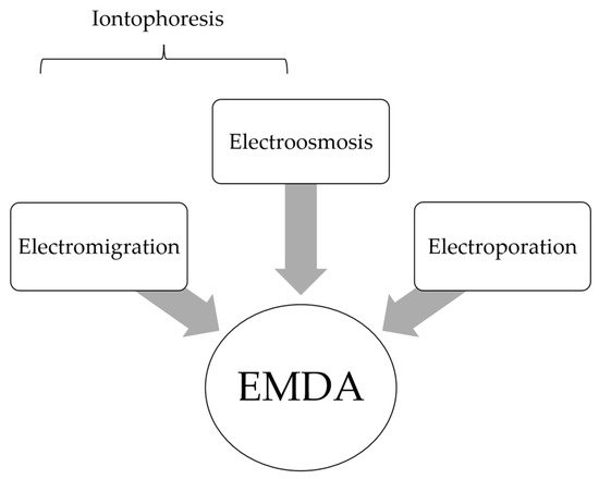
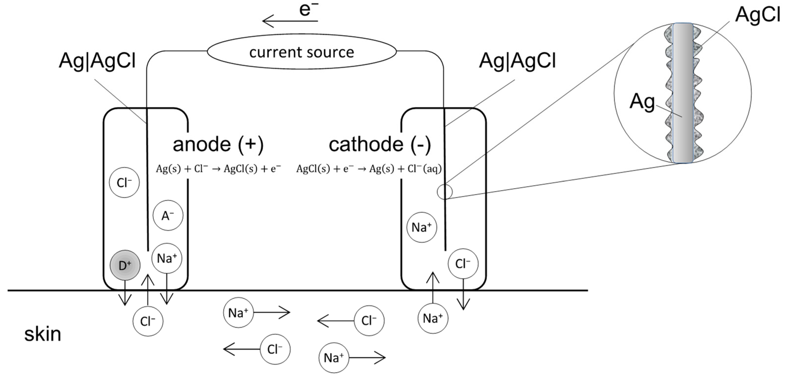
Local-regional administration of cytotoxic drugs is an important adjunct to systemic chemotherapy amongst cancer patients. It allows for targeted delivery of agents at high concentration to target sites while minimizing systemic side effects. Despite the pharmacokinetic advantages of the local–regional approach, drug transport into tumor nodules remains limited due to the biophysical properties of these tissues. Electromotive enhanced drug administration (EMDA) represents a potential solution to overcome challenges in local drug transport by applying electric currents. Through electrokinetic phenomena of electromigration, electroosmosis and electroporation, electric currents have been shown to improve drug penetration and distribution in a wide variety of clinical applications.

electromotive electric-driven iontophoresis electroporation drug transport

1. Introduction

The ideal drug delivery model is safe, convenient, site-specific and can maximize therapeutic efficacy while ensuring minimal toxicity to unintended sites [1]. As such, a local–regional approach is logical and has pharmacokinetic (PK) advantages when compared with systemic drug administration [2]. This approach has been most widely adopted in bladder and peritoneal cancers, where intra-vesical and intra-peritoneal (IP) delivery of cytotoxic chemotherapeutic agents have been shown to have superior anti-tumoral effects [3][4][5]. However, both delivery modes rely heavily on passive diffusion for the transport of drugs into target tissues—a process hindered by the relative impermeability of both urothelial and peritoneal membranes [6][7][8]. Furthermore, the penetration of drugs into tumor nodules with elevated interstitial fluid pressures (IFP) represents an additional barrier to efficient drug transport [9].
Recognizing the challenges in local–regional drug delivery, the use of electric forces to enhance drug penetration has gained increasing popularity in the recent decade [10][11]. First described by Veratti in 1747, the concept of electricity-enhanced drug transport is not new [12]. In the early 20th century, Leduc, through a classical experiment using two rabbits connected in an electrical series circuit with strychnine sulfate and potassium cyanide solutions, proved that ionized drugs could penetrate the skin and exert a systemic effect. He also demonstrated the importance of polarity with respect to an ionized drug and its counter ion [13]. This phenomenon was termed iontophoresis, describing an accelerated delivery of charged ions into tissues when an electric current was applied through a drug solution [14][15][16].
By the 1930s, iontophoresis was frequently adopted in ophthalmologic and skin conditions and was found to be highly effective in the treatment of hyperhidrosis [17][18]. It was also used to enhance the effects of local anesthesia to the tympanic membrane, oral mucosa, skin and the eye [19]. The term electromotive drug administration (EMDA) was subsequently coined in 1994, recognizing electroporation as a secondary force at play with iontophoresis that resulted in an increased penetration of both ionized and unionized drugs into surrounding tissues [20][21][22]. Since then, EMDA has been frequently used to describe electricity-enhanced intra-vesical administration of cytotoxic and anesthetic agents for urological conditions.

2. Fundamental Principles in EMDA

EMDA refers to the use of a low intensity electrical current to drive drugs across various tissues. There are three main electrokinetic phenomena that govern EMDA: electromigration, electroosmosis and electroporation (Figure 1) [21][22][23]. The summation of electro-migratory and electro-osmotic forces is termed iontophoresis and describes the mechanism in which the delivery of ionized drugs across biological membranes is enhanced through the application of a mild electric current in an electrolyte drug solution [23]. In electromigration, the repulsion of cations by the anode and anions by the cathode result in ionic fluxes across tissues to maintain electrical neutrality. This is coupled with electroosmosis, where solvent flows in an anode to cathode direction, providing a secondary driving force in the transport of cationic drugs [24]. This convective solvent flow also represents the main mechanism by which unionized drug compounds are transported across membranes [21]. The two main electrokinetic phenomena (electromigration and electroosmosis) in EMDA-mediated drug transport can be described by the modified Nernst–Planck equation [25][26]. Finally, electroporation describes the formation of aqueous pores by the application of an electric current, thereby increasing the permeability of membranes and facilitating drug delivery [22][27]. This should be differentiated from “high voltage electroporation” techniques where voltages of >100 volts over very short durations (microseconds to milliseconds) are used to permeabilize the skin [28].
Figure 1. Electromotive drug administration (EMDA) encompasses the electrokinetic phenomena of electromigration (EM), electroosmosis (EO) and electroporation (EP). Conventionally, iontophoresis alone was commonly used to describe electric-driven drug transport.

2.1. EMDA Devices

In transdermal applications, several Food and Drug Administration (FDA) approved EMDA devices exist; most use a current source and two electrode compartments, the latter consisting of an electrode immersed in an electrolyte (ionic conductor) solution or gel [29]. Both electrode compartments are placed at two distant sites on the skin while aiming for transdermal drug delivery [20]. The total set-up operates as an electrochemical cell (Figure 2):
Figure 2. Schematic diagram of an iontophoretic device consisting of a current source and two Ag|AgCl electrodes. During EMDA, D+ is placed inside the electrode compartment bearing the same charge (i.e., the anode). Cations, including D+, are transported from the anode into the skin. At the same time, anions from the skin move into the anode. In the cathode, anions leave the cathode towards the skin, while cations move into the cathode.
In intra-vesical EMDA, a catheter-electrode is connected to a current generator, the Physionizer®® Mini 30N2 (Medolla, MO, Italy) and a positive polarity applied (Figure 3). The bladder is filled with a drug containing electrolyte solution such that if a cationic drug ion is present, penetration across the bladder wall is enhanced predominantly through an electromigratory phenomenon [24]. This contrasts with unionized large drug molecules, whose transport is more electro-osmotically driven [21].
Figure 3. Schematic depiction of EMDA of a cationic drug molecule (+) within a filled bladder. The foley catheter contains a spiral Ag electrode at its tip and is connected to a current generator. During EMDA, grounding skin electrodes are placed over the anterior abdominal wall and connected to the cathode component of the generator.

2.2. Relationship between Current Intensity, Ion Valency and EMDA

Faraday’s law of electrochemical reaction states that [30]:
Q = t i I t Z i F
where Q is the mass of drug delivered by electromotive transport, ti the transference number of the ionic species i, I the current applied (in amperes) and t the time duration (in seconds).
As such, the amount of drug delivered during EMDA is directly proportional to the current intensity and treatment time but inversely proportional to the charge of the drug ion. The transference number of a specific drug ion refers to its ability to carry electric current and is defined as the ratio of the electric current carried by the drug ion i to the total current carried by all ionic species within the electrolyte solution.
The linear relationship between applied current and electromotive transport was illustrated by Harding (1987), who found increasing rates of angiotensin release from Ringer’s solution with increasing amplitudes of electric current applied [31]. Amongst drug compounds with similar molecular weight, monovalent sodium ions were found to have a drug delivery efficiency that was more than twice that of divalent magnesium ions, reflecting the slower migration of drug ions with high valency when a constant electric current is applied [16]. This is also reflected by the inverse relationship between valency and drug delivery in Faraday’s equation above.
However, while increasing current can increase drug delivery in experimental models, thermal damage to healthy tissues is a significant concern in clinical applications. For example, in transdermal EMDA treatments, a current density exceeding 0.5 mA/cm2 induces skin irritation while a maximum of 15 to 20 mA is used in intravesical EMDA to prevent discomfort and tissue damage [10]. Other adjuncts adopted to reduce the likelihood of skin injury during transdermal EMDA include the use of well-saturated absorbent pads and ensuring that there is no direct contact between metallic components and the skin [32]. In addition, based on early transdermal experiments, pulsed direct current (DC) is preferred over continuous DC, as the latter has been found to cause skin polarization, which in turn results in skin irritation and likely a reduced drug delivery efficiency [33].

2.3. Relationship between Drug Physicochemical Properties and EMDA

The physicochemical properties of drug molecules and their carrier solution are important factors influencing EMDA [15]. The charge and molecular size of drug molecules and the pH and presence of buffer ions within its carrier solution affect iontophoresis. As only ionized or charged drugs may be delivered via electromotive forces, unionized drugs rely solely on electroosmosis for transport. Since the degree of drug ionization is often pH dependent, altering the pH of carrier solutions can have a significant impact during EMDA. In an experiment using lidocaine (a local anesthetic agent), Gangarosa et al. demonstrated that when an alkaline solution was added, conductivity was reduced by 15%, as an increase in pH drove the conversion of positively charged lidocaine ions to unionized molecules [34]. The impact of pH on EMDA delivery was further evaluated by Murthy et al., who found that iontophoretic flux of salicylic acid (SA) was significantly increased at higher pH due to the increased ionization of SA (at a pH of 1.2, SA is present solely in the unionized form and the only driving force for transport is electroosmosis. At a pH of 7.1, SA is completely ionized and electromigration drives drug transport) [35].
When present in carrier solutions, buffer ions act as competitors to charged drug ions, resulting in reduced iontophoretic delivery of the latter. As such, if EMDA is desired, the inclusion of small mobile ions in drug diluent solutions should be avoided.

2.4. Membrane or Barrier Properties and EMDA

The skin, urothelium and sclera are examples of biological barriers that can affect EMDA. In general, the porosity of membranes is affected by their thickness as well as pore size and charge. In transdermal EMDA, this membrane barrier is composed of up to 15 layers of corneocytes embedded within the intracellular lamellar lipid membrane, which make up the stratum corneum [36]. At physiological pH, the skin carries a net negative charge; hence, EMDA enhances the transport of positively charged drug ions with an anode while retarding the movement of negatively charged drug ions with a cathode. Neutral drug molecules whose transport is mainly driven by electroosmosis will experience enhanced transport with an anode due to the negatively charged skin barrier [32]. With transscleral applications, since the sclera also carries a net negative charge under physiological conditions, a similar pattern of iontophoretic flux is seen [37]. As the sclera is relatively porous when compared with the skin due to larger pore sizes, it allows for the easy penetration of macromolecules. An example is the fact that the transport of bevacizumab (molecular weight 149 kDa), an antibody targeting the vascular endothelial growth factor (VEGF), is enhanced 32-fold when 2 mA of current is applied during EMDA [38].
In intravesical oncological applications, the ideal penetration depth is to the lamina propria, which is at 1193 ± 26.9 µm [39]. When EMDA with mitomycin-C (MMC) is applied in an in vitro human bladder model, drug penetration at a constant current of 20 mA was highest at a depth between 80 and 200 µm and lowest at a depth between 2000 and 4000 µm [39][40]. This illustrates the importance of barrier thickness and the desired ‘target’ depth in oncological EMDA applications.

References

  1. Petrak, K. Essential properties of drug-targeting delivery systems. Drug Discov. Today 2005, 10, 1667–1673.
  2. Collins, J.M. Pharmacologic rationale for regional drug delivery. J. Clin. Oncol. 1984, 2, 498–504.
  3. Joice, G.A.; Bivalacqua, T.J.; Kates, M. Optimizing pharmacokinetics of intravesical chemotherapy for bladder cancer. Nat. Rev. Urol. 2019, 16, 599–612.
  4. Ceelen, W.; Demuytere, J.; de Hingh, I. Hyperthermic Intraperitoneal Chemotherapy: A Critical Review. Cancers 2021, 13, 3114.
  5. Ceelen, W.P.; Flessner, M.F. Intraperitoneal therapy for peritoneal tumors: Biophysics and clinical evidence. Nat. Rev. Clin. Oncol. 2010, 7, 108–115.
  6. Mishina, T.; Watanabe, H.; Kobayashi, T.; Maegawa, M.; Nakao, M.; Nakagawa, S. Absorption of anticancer drugs through bladder epithelium. Urology 1986, 27, 148–157.
  7. Lasič, E.; Višnjar, T.; Kreft, M.E. Properties of the Urothelium that Establish the Blood-Urine Barrier and Their Implications for Drug Delivery. Rev. Physiol. Biochem. Pharmacol. 2015, 168, 1–29.
  8. Dedrick, R.L. Theoretical and experimental bases of intraperitoneal chemotherapy. Semin. Oncol. 1985, 12 (Suppl. S4), 1–6.
  9. Heldin, C.H.; Rubin, K.; Pietras, K.; Ostman, A. High interstitial fluid pressure—An obstacle in cancer therapy. Nat. Rev. Cancer 2004, 4, 806–813.
  10. Hashemi, S.; Sahai, A.; Malde, S. Applications of electromotive drug administration in urology. Urol. Ann. 2020, 12, 301–308.
  11. Charoo, N.A.; Rahman, Z.; Repka, M.A.; Murthy, S.N. Electroporation: An avenue for transdermal drug delivery. Curr. Drug Deliv. 2010, 7, 125–136.
  12. Turnell, W.J. Therapeutic action of constant current. Proc. R. Soc. Med. 1921, 14, 41–52.
  13. Leduc, S. Introduction of medicinal substances into the depth of tissues by electric current. Ann. D’Eleetrobiol. 1900, 3, 545–560.
  14. Leduc, S. Electric Ions and Their Use in Medicine; Rebman Ltd.: London, UK, 1908.
  15. Banga, A.K.; Chien, Y.W. Iontophoretic delivery of drugs: Fundamentals, developments and biomedical application. J. Control. Release 1988, 7, 1–24.
  16. Phipps, J.B.; Padmanabhan, R.B.; Lattin, G.A. Iontophoretic delivery of model inorganic and drug ions. J. Pharm. Sci. 1989, 48, 365.
  17. Hughes, L.; Maurice, D.M. A fresh look at iontophoresis. Arch. Ophthalmol. 1984, 102, 1825–1829.
  18. Shelley, W.B.; Horwath, P.; Weidrnan, F.; Pillsbury, D.M. Experimental milaria in man. Production of sweat retention anhidrosis and vesicles by means of iontophoresis. J. Investig. Dermatol. 1948, 11, 275–291.
  19. Harris, R. Treatment of post-therapeutic neuralgia. Lancet 1957, 269, 378–379.
  20. Stephen, R.; Miotti, D.; Bettaglio, R.; Rossi, C.; Bonezzi, C. Electromotive administration of a new morphine formulation: Morphine citrate. Artif. Organs 1994, 18, 461–465.
  21. Gangarosa, L.P.; Park, N.H.; Wiggins, C.A.; Hill, J.M. Increased penetration of nonelectrolytes into mouse skin during iontophoretic water transport (iontohydrokinesis). J. Pharmacol. Exp. Ther. 1980, 212, 377–381.
  22. Prausnitz, M.R.; Bose, V.G.; Langer, R.; Weaver, J.C. Electroporation of mammalian skin: A mechanism to enhance transdermal drug delivery. Proc. Natl. Acad. Sci. USA 1993, 90, 10504–10508.
  23. Guy, R.H.; Kalia, Y.N.; Delgado-Charro, M.B.; Merino, V.; López, A.; Marro, D. Iontophoresis: Electrorepulsion and electroosmosis. J. Control. Release 2000, 64, 129–132.
  24. Kalia, Y.N.; Naik, A.; Garrison, J.; Guy, R.H. Iontophoretic drug delivery. Adv. Drug Deliv. Rev. 2004, 56, 619–658.
  25. Bard, A.J.; Faulkner, L.R. Electrochemical Methods: Fundamentals and Applications, 1st ed.; Wiley: New York, NY, USA, 1980.
  26. Fick, A.V. On liquid diffusion. Lond. Edinb. Dublin Philos. Mag. J. Sci. 1855, 63, 30–39.
  27. Li, S.K.; Hao, J.; Liddell, M.R. Electrotransport across membranes in biological media: Electrokinetic theories and applications in drug delivery. In Transport in Biological Media; Becker, S., Kuznetsoy, A., Eds.; Elsevier: Philadelphia, PA, USA, 2013; pp. 417–454.
  28. Banga, A.K.; Bose, S.; Ghosh, T.K. Iontophoresis and electroporation: Comparisons and contrasts. Int. J. Pharm. 1999, 179, 1–19.
  29. Available online: https://www.federalregister.gov/documents/2016/07/26/2016-17609/physical-medicine-devices-reclassification-of-iontophoresis-device-intended-for-any-other-purposes (accessed on 7 October 2022).
  30. Stillwell, G.K. Electrical stimulation and iontophoresis. In Handbook of Physical Medicine and Rehabilitation, 2nd ed.; Krussen, F.H., Ed.; W.B. Saunders Company: St. Louis, MO, USA, 1971; Chapter 14.
  31. Harding, J.W.; Felix, D. Quantification of angiotensin iontophoresis. J. Neurosci. Methods 1987, 19, 209–215.
  32. Sloan, J.B.; Soltani, K. Iontophoresis in dermatology. A review. J. Am. Acad. Dermatol. 1986, 15 Pt 1, 671–684.
  33. Sun, Y.; Siddiqui, O.; Liu, J.C.; Chien, Y.W. Transdermal modulated delivery of polypeptides: Effect of DC pulse wave-form on enhancement. In Proceedings of the 13th International Symposium on Controlled Release of Bioactive Materials, Controlled Release Society, Lincolnshire, IL, USA, 23–25 July 1986.
  34. Gangarosa, L.P.; Park, N.H.; Fong, B.C.; Scott, D.F.; Hill, J.M. Conductivity of drugs used for iontophoresis. J. Pharm. Sci. 1978, 67, 1439–1443.
  35. Narasimha Murthy, S.; Wiskirchen, D.E.; Bowers, C.P. Iontophoretic drug delivery across human nail. J. Pharm. Sci. 2007, 96, 305–311.
  36. Hadgraft, J. Skin, the final frontier. Int. J. Pharm. 2001, 224, 1–18.
  37. Chopra, P.; Hao, J.; Li, S.K. Iontophoretic transport of charged macromolecules across human sclera. Int. J. Pharm. 2010, 388, 107–113.
  38. Li, S.K.; Liddell, M.R.; Wen, H. Effective electrophoretic mobilities and charges of anti-VEGF proteins determined by capillary zone electrophoresis. J. Pharm. Biomed. Anal. 2011, 55, 603–607.
  39. Di Stasi, S.M.; Giannantoni, A.; Massoud, R.; Dolci, S.; Navarra, P.; Vespasiani, G.; Stephen, R.L. Electromotive versus passive diffusion of mitomycin C into human bladder wall: Concentration-depth profiles studies. Cancer Res. 1999, 59, 4912–4918.
  40. Lugnani, F.; Mazza, G.; Cerulli, N.; Rossi, C.; Stephen, R. Iontophoresis of drugs in the bladder wall: Equipment and preliminary studies. Artif. Organs 1993, 17, 8–17.
More
Information
Subjects: Oncology
Contributors MDPI registered users' name will be linked to their SciProfiles pages. To register with us, please refer to https://encyclopedia.pub/register : , , , ,
View Times: 728
Revisions: 2 times (View History)
Update Date: 26 Oct 2022
1000/1000
Video Production Service