Submitted Successfully!
To reward your contribution, here is a gift for you: A free trial for our video production service.
Thank you for your contribution! You can also upload a video entry or images related to this topic.
Version Summary Created by Modification Content Size Created at Operation
1 -- 3449 2022-10-21 03:13:41 |
2 update references and layout -5 word(s) 3444 2022-10-24 03:09:36 | |
3 update layout Meta information modification 3444 2022-10-24 03:10:22 |

Video Upload Options

Do you have a full video?

Confirm

Are you sure to Delete?
Cite
If you have any further questions, please contact Encyclopedia Editorial Office.
Carr, B.P.;  Chen, Z.;  Chung, J.H.Y.;  Wallace, G.G. Collagen Alignment via Electro-Compaction for Biofabrication Applications. Encyclopedia. Available online: https://encyclopedia.pub/entry/30579 (accessed on 05 September 2024).
Carr BP,  Chen Z,  Chung JHY,  Wallace GG. Collagen Alignment via Electro-Compaction for Biofabrication Applications. Encyclopedia. Available at: https://encyclopedia.pub/entry/30579. Accessed September 05, 2024.
Carr, Benjamin P., Zhi Chen, Johnson H. Y. Chung, Gordon G. Wallace. "Collagen Alignment via Electro-Compaction for Biofabrication Applications" Encyclopedia, https://encyclopedia.pub/entry/30579 (accessed September 05, 2024).
Carr, B.P.,  Chen, Z.,  Chung, J.H.Y., & Wallace, G.G. (2022, October 21). Collagen Alignment via Electro-Compaction for Biofabrication Applications. In Encyclopedia. https://encyclopedia.pub/entry/30579
Carr, Benjamin P., et al. "Collagen Alignment via Electro-Compaction for Biofabrication Applications." Encyclopedia. Web. 21 October, 2022.
Collagen Alignment via Electro-Compaction for Biofabrication Applications
Edit

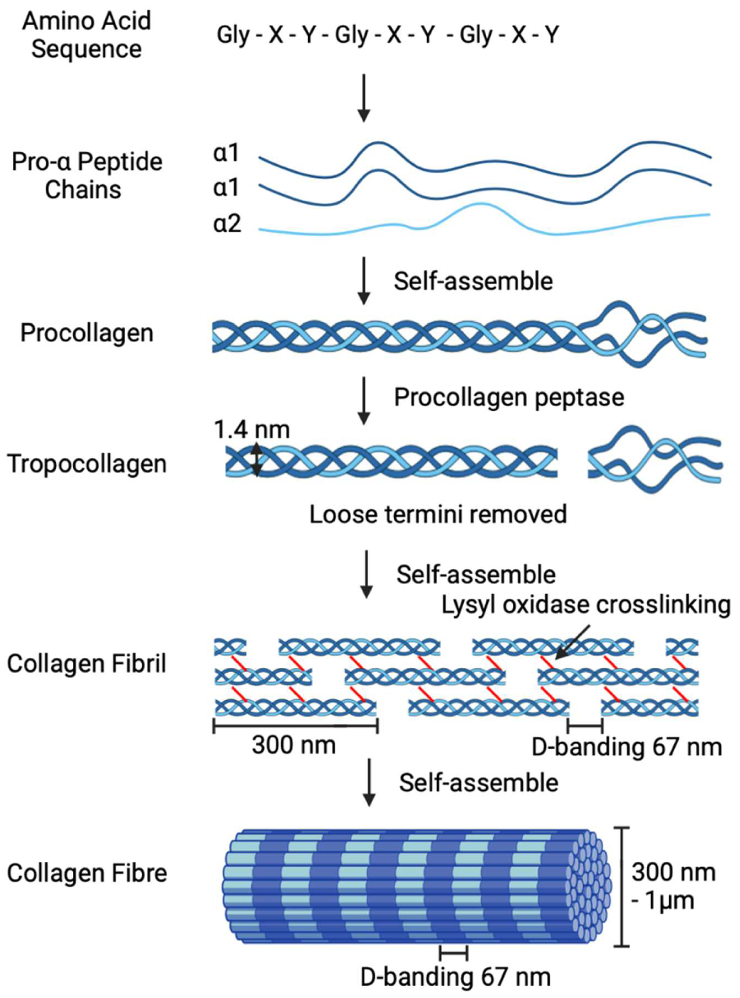
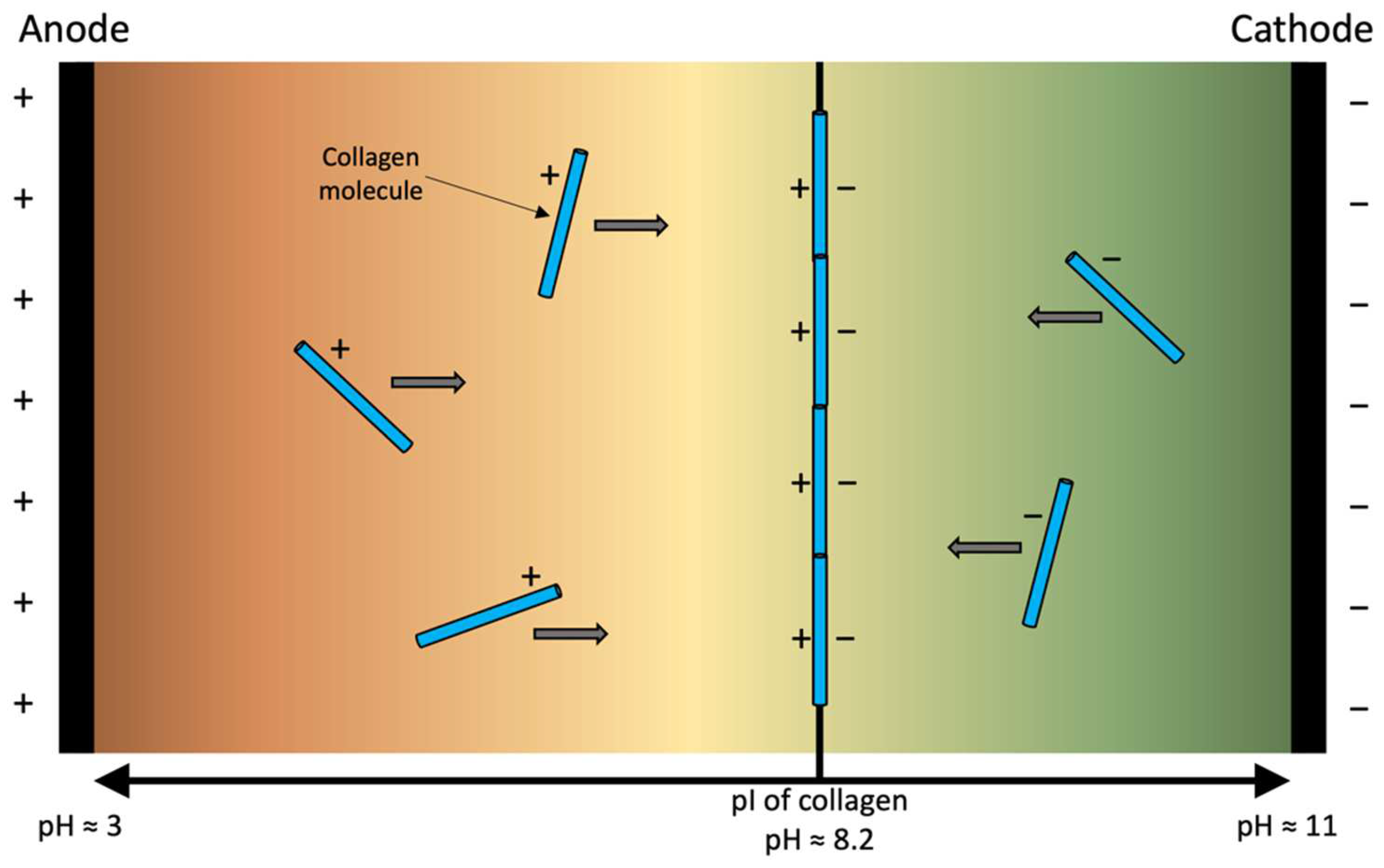
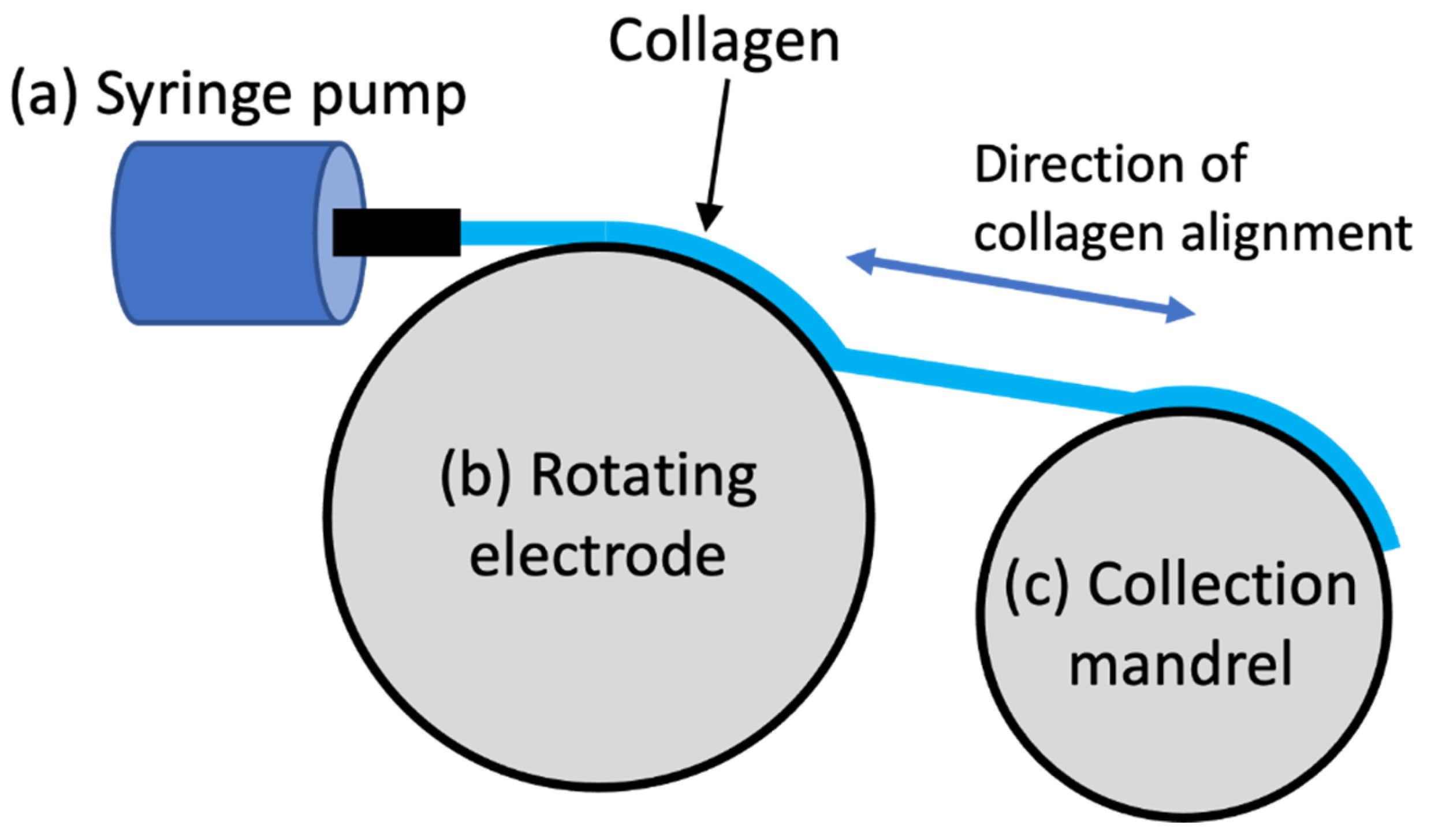
Collagen is the most prevalent structural protein in the extracellular matrix, resulting in the biopolymer being extensively investigated for biofabrication-based applications. However, its utilisation has been impeded due to a lack of sufficient mechanical toughness and the inability of the scaffolds to mimic complex natural tissues. The anisotropic alignment of collagen has been proven to be an effective method to enhance overall mechanical properties and produce biomimetic scaffolds. The alignment of collagen can be achieved by several methods, namely, gravity and extrusion-based fluidic alignment, static magnetic alignment, magnetic-flow alignment, cell-based stress-induced self-alignment, electrospinning, and electrophoretic-based electro-compaction (EC). Each existing approach of aligning collagen is described and compared with a sharper focus on electro-compaction.

collagen electro-compaction electro-chemical alignment

1. Overview of Collagen Alignment Techniques

The strength of collagen-based scaffolds can be enhanced through achieving anisotropic alignment. Anisotropically aligned collagen-based structures are observed throughout the body in tissues such as tendons, muscles, nerves, intervertebral discs [1], blood vessels [2][3], and corneas [4]. Ex vivo alignment aims to mimic the tissue’s natural fibre direction, contributing to the tissue’s ability to withstand physiological loads and mechanical stressors [5]. Another advantage of aligned-collagen-based scaffolds is that they provide biophysical cues to direct cell adherence, migration, and proliferation [6]. Collagen alignment can be achieved by several methods, namely, gravity and extrusion-based fluidic alignment [7], static magnetic alignment [8], magnetic-flow alignment [9], cell-based stress-induced self-alignment [10], electrospinning [11], and electrophoretic-based electro-compaction (EC; Figure 1) [12]. The principles and recent advances are introduced herein, with discussion on the advantages and disadvantages of each method, including a sharper focus on EC. This information can contribute to producing mechanically robust biomimetic scaffolds for various biofabrication applications.
Figure 1. Schematic of collagen alignment methods: (a) gravity-based fluidic, (b) extrusion-based, (c) static magnetic, (d) magnetic-flow, (e) cell-based stress induced, (f) electrospinning, and (g) electro-compaction.

2. Electro-Compaction

The method for EC consists of an electrical current being applied between two electrodes across a solution of collagen, generating a pH gradient (anode pH ≈ 3 and cathode pH ≈ 11) and charging the collagen molecules (Figure 2) [2][13][14]. The collagen nearer the anode (positive electrode) gains a positive charge, whilst those nearer the cathode are charged negatively [14]. The combination of the pH gradient and charged collagen produces a highly organised anisotropically-aligned aggregation at the isoelectric point. The isoelectric point (pI) of collagen varies depending on the source, but for bovine hide, it is approximately at a pH = 8.2, where the net charge is 0 (Equation 1) [14][15].
Figure 2. Schematic of collagen electro-compaction, illustrating the anode and cathode with associated charges and generated pH gradient, and demonstrating the charges gained by the collagen molecules and the aggregation at the isoelectric point (pI), where the net charge is 0.
Anode: 2H2O − 4e → 4H+ + O2; Cathode: 4H2O + 4e → 4OH +2H2   
Due to the specific pI of collagen, the aligned scaffold is formed between the electrodes, favouring the cathode with the collagen fibres parallel to the electrodes [12]. In addition to the alignment, the packing density of collagen increases from 5 mg.cm−3 non-EC to 50 mg.cm−3 post-treatment [14]. This also makes the concentration of EC collagen ~1030 mg.mL−1 [16][17][18][19] 17 times denser than non-EC conventional collagen gels. The optimal EC parameters, such as concentration, voltage, current density, and time, are generally specific to the application and desired outcome. However, higher voltage, closer electrodes, and collagen concentration have different optimal alignment times [20][21]. Specifically, 3 volts for 45 min [13] and 40 volts for 10 s [22] have both been successfully utilised.

2.1. Sources of Collagen for Electro-Compaction

Several different sources of collagen have been used for EC, with the most common being type I, extracted from bovine hide. Other collagen sources used for EC have included porcine, fish, and rats. However, additionally, collagen has been successfully extracted from other sources such as human [23], ovine [24], equine [25], avian [26], and various marine (mammals and fish) [27][28]. The extraction method alters the structure and consists of, namely, chemically produced procollagen/telocollagen and enzymatically digested tropocollagen/atelocollagen [29][30]. Furthermore, each source of collagen has numerous subtypes of collage that exist in different tissues and aid the specific functions within the body [15][31]. Different sources, types, and extraction methods possess various characteristics [15][32][33], such as viscosity [34][35], isoelectric point (pI) [36], and molecular weight [37][38], which ultimately allows for the ability to control the scaffold properties and EC processing.

2.2. Electro-Compacted Scaffold Types

Over recent years, progressively more complex EC collagen scaffolds have been utilized for biofabrication applications, commencing in 2008 with the generation of simple threads using linear electrodes [12] and membranes with planar electrodes [39]. Membranes have since been put into various shapes via EC, determined by the shape of the spacer or mould between the electrodes. Such examples have included rectangular, round [4], and irregular hexadecagon [40] shaped membranes. More recently, concentric tube electrodes have been used to fabricate tubular scaffolds [2], and curviplanar electrodes have been utilised to fabricate domes [14]. Initially, the length of the aligned collagen threads was restricted to the length of the linear electrodes. Younesi et al. addressed this limitation by developing a device for the continuous EC of collagen threads called REEAD (rotating electrode electro-chemical alignment device, Figure 3). The REEAD device utilises a syringe pump to extrude collagen onto the first of two rotating wheels [41]. The first wheel contains two parallel electrodes, as is used in a regular EC setup. Collagen is extruded between the electrodes whilst the wheel is in motion. Depending on the parameters, mainly speed and voltage, different thicknesses of collagen threads can be achieved (0.10–0.15 mm) [22][42]. At the same time, the collection mandrel rotates proportionally to the electrode to collect the aligned thread.
Figure 3. Schematic of rotating electrode electro-chemical alignment device (REEAD) for continuous electro-compaction of threads, (a) syringe pump, (b) rotating wheel with electrodes on the edge and collagen between, and (c) rotating collection mandrel.
The mechanical properties of EC collagen scaffolds vary depending on the scaffold shape and preparation process; in particular crosslinking and the use of filler materials. Additionally, there is no standardised method for the mechanical testing of EC collagen scaffolds, thus resulting in some samples being desiccated prior to testing whilst others remained hydrated, leading to the inconsistency of the tests performed and reported. It is well established that EC collagen has superior mechanical properties compared to conventional collagen gels. Specifically, the ultimate tensile strength of EC collagen is 6.2 MPa, compared to conventional collagen <10 kPa [14]. Young’s modulus has been shown to increase this from ~1 MPa (when unaligned) to ~50 MPa after EC [14].

2.3. Enhancing Electro-Compacted Collagen Strength

After the alignment of the collagen, further steps can be used to increase scaffold strength, such as phosphate-buffered saline (PBS) treatment or crosslinking. Uquillas et al. investigated the mechanical effects of immediate post-alignment incubation and PBS treatment to promote fibrillogenesis. The results demonstrated that 1 × PBS incubated for 12 h produced mechanically competent threads with D-banding similar to the native tendons. Specifically, the ultimate tensile stress was 0.4 MPa, the strain was 100%, and Young’s modulus was 0.4 MPa [16]. However, despite 12 h being the identified period for optimal mechanics, methodologies from subsequent studies have commonly used 4–6 h [2][6][13][40][41][43][44][45][46].
Additionally, non-fibre-forming structural molecules, mainly glycosaminoglycans (GAG) and proteoglycans, have been used to enhance the scaffold strength, functioning similarly to a crosslinker [47]. Paderi and Panitch synthesised a dermatan sulphate-peptide sequence (DS-SILY) which mimics the natural structure and function of decorin, a small leucine-rich proteoglycan (SLRP) [48]. The leucine-rich protein core binds to the D-bands on the collagen fibrils and the dermatan sulphate glycosaminoglycan chain, followed by the binding of the chains to adjacent molecules to form inter-fibrillar crosslinks. When incorporated into EC collagen, the ultimate tensile strength increased to 1.5 MPa (Col:DS-SILY 1:30) [49].

2.4. Co-Electro-Compaction of Collagen with Fillers

As previously established, collagen has excellent biocompatibility, and when it is aligned, it has better mechanical properties. However, the mechanical strength remains suboptimal for many biofabrication applications. The use of additional materials, termed fillers, can be utilised to reinforce collagen-based scaffolds. The methods of filler incorporation are grouped into two main techniques: (a) homogenous co-electro-compaction (Co-EC) and (b) post-EC fabrication methods. Co-EC involves the homogenous incorporation of fillers into the collagen and then applying a current. Examples of materials suited for Co-EC are biopolymers, such as elastin [2], or polysaccharides, such as nanocellulose [43]. The primary determining factor of this method is the isoelectric point of the filler in relation to collagen (pH ≈ 8.2). Both materials are required to have similar isoelectric points allowing for isoelectric focusing on the same point. On the contrary, if the isoelectric point between the materials is too great, the materials will separate during EC. Thus, forming two independent scaffolds, each aligned at their respective isoelectric points [50].
Similar to collagen, elastin is a protein that forms part of the extracellular matrix. However, it is responsible for the elastic properties in tissues, specifically, stretching and contraction [51]. Nguyen et al. initially investigated the effects of soluble versus insoluble elastin when incorporated into EC collagen threads for application in small-diameter blood vessels [46]. Briefly, solutions of elastin (soluble or insoluble, 200 mg.mL−1) were mixed with collagen (3.1 mg.mL−1) at a ratio of 40:60 (w/w%). The solutions were subjected to Co-EC using stainless steel wire electrodes at 3 volts for 30 min, and then incubated (37 °C) in PBS for 6 h. Mechanical testing showed a decrease in Young’s modulus, ultimate tensile stress, and strain with the incorporation of elastin. However, in vitro characterisation using rat aorta smooth muscle cells (rSMCs) and real-time polymerase chain reaction (PCR) determined a positive effect on the contractile phenotype of the cells when elastin was incorporated. Additionally, it was determined that the cells could sense the composition and topography of Co-EC fibres. A direct comparison between soluble and insoluble elastin determined that insoluble was better suited for biofabrication-based applications. This method was used again with insoluble elastin, with the ratio of collagen to elastin adjusted to 50:50 (w/w) [2].
Nanocellulose is a polymer sourced from the cellulose found in plants, bacteria, algae, and animals [52] and comes in two primary forms. First, nanostructured materials, including microcrystals and microfibrils, whilst the second are nanofibers such as nanofibrils, nanocrystals, and bacterial cellulose [53]. Nanocellulose has been used in various applications, including wound healing, blood vessel, corneal, heart valve, urethra, bone, and cartilage biofabrication [54]. The wide use of nanocellulose is due to its increased mechanical properties, biocompatibility, and low cytotoxicity [55]. As previously described, one primary consideration when choosing a material for Co-EC is the isoelectric point of the two materials. Cudjoe et al. modified the isoelectric point of TEMPO (2,2,6,6-Tetramethylpiperidine 1-oxyl)-oxidised cellulose nanocrystals (t-CNC), resulting in t-CNC−COOH, which favours the anode, whilst t-CNC−COOH27−NH273 favoured the cathode at a pH of 7 [43]. The modification of nanocellulose allowed for successful Co-EC with collagen. The Co-EC utilised two parallel wire electrodes, and 20 volts were applied for 30 s. It was found that t-CNC−COOH27−NH273 at 5% (w/w %) with collagen was optimal to increase strength when fabricating threads. There has not been any reported in vitro characterisation of Co-EC collagen and nanocellulose.

2.5. Post-Alignment Fabrication Methods

In contrast to homogenous Co-EC, the second method for incorporating fillers into collagen-based scaffolds uses post-alignment fabrication methods. Due to the ease of generating threads via EC, textile-based fabrication methods have been investigated as a promising post-EC fabrication process [56]. Comparatively, the simplest fabrication method involves forming yarn, made by twisting several threads together. The mechanical properties of EC collagen yarn are better than that of the threads. Specifically, there was a reported 30% and 20% increase in the ultimate tensile strength (65 MPa) and Young’s modulus (530 MPa), respectively [41]. Braiding is comprised of three or more threads being intertwined in an overlapping pattern [56]. Furthermore, three individual braids can been braided again, thus, using nine individual threads [57]. This twice braided technique is suited for tissues under high load and has been used with EC collagen for tendon applications [12][57][58]. There were increases in ultimate tensile strength (24–88 MPa), strain (7–14%), and tensile modulus (277–671 MPa) when braided once and crosslinked with genipin. Additionally, braiding increased cell attachment, as the cells were able to infiltrate the space between the bundles [12]. Weaving involves overlapping the two distinct directions of the threads, termed warp and weft. The warp is stationary, whilst the weft is perpendicular. The weft moves in a repeating under-over fashion, forming rows [56]. Younesi et al. combined these fabrication methods by firstly forming yarn with three EC collagen threads (3-ply), and then the yarn was subsequently woven [41]. Xie et al. used polylactic acid (PLA) threads twisted around a two-ply EC collagen core yarn, which were then woven into a scaffold [59]. Both methods were used to fabricate sheets for tendon applications. The resulting scaffolds have a reported porosity of 81%. High porosity has been noted as necessary for the diffusion of oxygen, nutrients, and waste [60]. Finally, knitting is the most complicated textile fabrication method. Individual threads or yarns that are interlaced in a highly ordered arrangement of connected loops brought through a previous loop forming new rows [56]. This method has been used by Xie et al. to fabricate myocardial patches. Specifically, two continuous EC collagen threads and a PLA were grouped into yarn. The yarn was subsequently knitted and crosslinked with EDC/NHS, resulting in a maximum scaffold load of 1.4 N, an extension of 3.1 mm, and 1.8 N.mm−1 stiffness [22].
The Layer-by-layer assembly provides a simple method for reinforcing scaffolds via stacking, increasing their robustness and providing more surface area. Chen et al. layered EC collagen membranes with human corneal stromal cells attached in alternating directions of alignment to mimic the structure of corneas [4]. Nguyen et al. investigated the reinforcing of scaffolds using tubes with threads for small-diameter blood vessels [2]; EC collagen tubes and threads were fabricated, with the reinforcing threads positioned around the collagen tubular lumen in either longitudinal or circumferential directions and crosslinked with EDC/NHS. The addition of the EC collagen threads increased the overall tube scaffold strength, with the circumferentially-directed threads demonstrating better performance when compared to the longitudinally orientated version.

2.6. Clinical Applications Using Electro-Compacted Collagen Scaffolds

EC collagen has been used to fabricate biomimetic scaffolds for various applications. Such applications have included the biofabrication of tendons [12], corneas [13], nerves [44], blood vessels [46], myocardium [22], and wound-healing dressings [45]. Due to the wide scope of clinical applications for EC collagen scaffolds, there has been a variety of cell types used and in vitro characterisations made; however, currently, there are limited in vivo studies available.
EC collagen has been investigated for corneal biofabrication due to the highly transparent and mechanically robust nature of EC collagen membranes. Initially, Kishore et al. assessed the in vitro response of human keratocytes (corneal fibroblasts) on EDC/NHS crosslinked EC membranes [13]. A live-dead assay showed the high keratocyte viability on the crosslinked membranes, and F-actin staining (at day 2) demonstrated well-spread morphology and attachment; by day 7, a highly confluent layer was observed. Additionally, due to the function of corneas, scaffold transparency was investigated where light transmission measurements determined that the crosslinking reduced the scaffold’s transparency (EDC/NHS 67–89%; genipin 33–78%). However, after 14 days of culture with keratocytes, the EC collagen scaffolds (EDC/NHS) had increased in transparency by 75–100%. Meanwhile, Chen et al. aimed to fabricate a biomimetic corneal stromal structure with orthogonally aligned layers [4]. Four EC membranes seeded with human corneal stromal cells (hCSCs) were layered onto each other in alternating alignment directions, forming an orthogonally arranged scaffold. Cell orientation was investigated by F-actin staining, showing the underlying scaffold topography affecting cell alignment. Specifically, cells on the EC membranes were clearly aligned with collagen fibrils whilst conventional collagen scaffolds were patently disordered, resulting in scaffold alignment directly affecting cell orientation. The multilayered scaffold was shown to upregulate keratocyte expression (ALDH3) whilst reducing fibroblast phenotypes (α-SMA and Thy-1), confirming keratocyte differentiation from hCSCs, mimicking the quintessential state of human corneal stroma. Furthermore, there was no change in glucose permeability or the mass of the cornea scaffolds over time, whilst a small decrease in the dehydrated mass was observed at days 7 and 14, and the presence of the cells marginally impaired light transmission (81–83%).
The effects of collagen alignment via EC were investigated for the application of nerve growth by Abu-Rub et al. [44]. Rat pheochromocytoma (PC12) cells were cultured on either EC threads or conventional unaligned collagen membranes, and embryonic rat dorsal root ganglion explants were subsequently placed on the collagen scaffolds (or adjacent to the aligned threads) to assess neurite extension after growth. The cells seeded onto the threads displayed outgrowth that continued in the direction of the fibre, and the unaligned scaffolds displayed no preferential neurite outgrowth, whilst the cells seeded away from the thread showed random outgrowth until contacting the thread, which then changed their trajectory to follow the orientation of the threads. It was also noted, for the first time, that cells were able to overcome myelin-associated glycoprotein-induced inhibition when on EC collagen threads, without surface modification or chemical functionalisation.
EC collagen has been used to fabricate small-diameter blood vessels. Nguyen et al. initially investigated the effects of incorporating elastin into collagen threads due to elastin’s natural prevalence in the wall of blood vessels [46]. Rat aortic smooth muscle cells (rSMCs) were seeded onto collagen-only and collagen and elastin scaffolds. The Alamar blue assay (at day 1) showed that the collagen-only scaffolds displayed preferential alignment, which was not seen in the elastin-containing threads; however, by day 14, a confluent and highly aligned layer of cells was observed on both fibre types. Contractile (α-SMA and calponin) and synthetic (thrombospondin) phenotype markers were examined by PCR, where elastin-containing scaffolds showed an increased expression of α-SMA and calponin from days 3–14 whilst remaining the same on the collagen-only scaffolds. Furthermore, thrombospondin expression increased in both thread types over time, confirming that the incorporation of elastin into EC collagen induces a contractile expression in rSMCs. Further work by Nguyen et al. fabricated tube scaffolds, seeded initially with rSMCs, as previously described [2]. Additionally, human umbilical vein endothelial cells (hUVECs) were seeded to a scaffold lumen cell cytoskeleton, and staining demonstrated that the cells could successfully attach and proliferate on the luminal surface. The immunostaining of hUVECs showed evidence of gap-junction (Cx43) expression around dense colonies, confirming the presence of intercellular interactions. Furthermore, the cells were positive for nitric oxide production (eNOS) and endothelial cell phenotypes (vWF), suggesting successful endothelial cell differentiation.
Xie et al. investigated continuous EC collagen threads as materials for textile-based fabrication methods for the application of myocardium [22]. The scaffolds were fabricated by grouping two collagen and one polylactic acid (PLA) or PLA-only threads together, forming yarn, and were subsequently knitted into scaffolds. The collagen-containing scaffolds, when seeded with human cardiosphere-derived cells (hCDCs), allowed for attachment, proliferation, and migration across the full surface as determined by Alamar blue assay. Whilst PLA had a limited initial biological response, the cells formed surface aggregates and attached between the adjacent yarns and were able to proliferate. On day 28, both groups were compatible, with a maintained confluence.
Yuan et al. used a bacterial nanocellulose (BNC) scaffold impregnated with collagen and lactoferrin (LF) via EC for wound healing applications [54]. Five groups of dressings were investigated (BNC, BNC-LF, BNC-Col, BNC-LF-Col, and cotton gauze) by making wounds (1 cm in diameter) on the dorsal flank of male Sprague-Dawley rats, with the dressings changed daily. The groups that had collagen incorporated into the dressing showed greater healing efficiency than those without. The BNC-LF-Col scaffold showed the highest reduction in wound size after nine days at 85% and had the highest presence of fibroblasts. However, it is noteworthy that it did not directly assess EC collagen in vivo, even more so, the effects of collagen and lactoferrin integration into nanocellulose scaffolds for healing.

References

  1. Dewle, A.; Pathak, N.; Rakshasmare, P.; Srivastava, A. Multifarious fabrication approaches of producing aligned collagen scaffolds for tissue engineering applications. ACS Biomater. Sci. Eng. 2020, 6, 779–797.
  2. Nguyen, T.U.; Shojaee, M.; Bashur, C.A.; Kishore, V. Electrochemical fabrication of a biomimetic elastin-containing bi-layered scaffold for vascular tissue engineering. Biofabrication 2019, 11, 015007.
  3. Caliari, S.R.; Harley, B.A.C. The effect of anisotropic collagen-GAG scaffolds and growth factor supplementation on tendon cell recruitment, alignment, and metabolic activity. Biomaterials 2011, 32, 5330–5340.
  4. Chen, Z.; Liu, X.; You, J.; Song, Y.; Tomaskovic-Crook, E.; Sutton, G.; Crook, J.M.; Wallace, G.G. Biomimetic corneal stroma using electro-compacted collagen. Acta Biomater. 2020, 113, 360–371.
  5. Wang, T.; Chen, P.; Zheng, M.; Wang, A.; Lloyd, D.; Leys, T.; Zheng, Q.; Zheng, M.H. In vitro loading models for tendon mechanobiology. J. Orthop. Res. 2018, 36, 566–575.
  6. Kishore, V.; Bullock, W.; Sun, X.; Van Dyke, W.S.; Akkus, O. Tenogenic differentiation of human MSCs induced by the topography of electrochemically aligned collagen threads. Biomaterials 2012, 33, 2137–2144.
  7. Kirkwood, J.E.; Fuller, G.G. Liquid crystalline collagen: A self-assembled morphology for the orientation of mammalian cells. Langmuir 2009, 25, 3200–3206.
  8. Debons, N.; Matsumoto, K.; Hirota, N.; Coradin, T.; Ikoma, T.; Aimé, C. Magnetic field alignment, a perspective in the engineering of collagen-silica composite biomaterials. Biomolecules 2021, 11, 749.
  9. Guo, C.; Kaufman, L.J. Flow and magnetic field induced collagen alignment. Biomaterials 2007, 28, 1105–1114.
  10. Wilks, B.T.; Evans, E.B.; Nakhla, M.N.; Morgan, J.R. Directing fibroblast self-assembly to fabricate highly-aligned, collagen-rich matrices. Acta Biomater. 2018, 81, 70–79.
  11. Blackstone, B.N.; Gallentine, S.C.; Powell, H.M. Collagen-based electrospun materials for tissue engineering: A systematic review. Bioengineering 2021, 8, 39.
  12. Cheng, X.; Gurkan, U.A.; Dehen, C.J.; Tate, M.P.; Hillhouse, H.W.; Simpson, G.J.; Akkus, O. An electrochemical fabrication process for the assembly of anisotropically oriented collagen bundles. Biomaterials 2008, 29, 3278–3288.
  13. Kishore, V.; Iyer, R.; Frandsen, A.; Nguyen, T.U. In vitro characterization of electrochemically compacted collagen matrices for corneal applications. Biomed. Mater. 2016, 11, 055008.
  14. Younesi, M.; Islam, A.; Kishore, V.; Panit, S.; Akkus, O. Fabrication of compositionally and topographically complex robust tissue forms by 3D-electrochemical compaction of collagen. Biofabrication 2015, 7, 035001.
  15. Oliveira, V.d.M.; Assis, C.R.D.; Costa, B.d.A.M.; Neri, R.C.d.A.; Monte, F.T.D.; Freitas, H.M.S.d.C.V.; França, R.C.P.; Santos, J.F.; Bezerra, R.d.S.; Porto, A.L.F. Physical, biochemical, densitometric and spectroscopic techniques for characterization collagen from alternative sources: A review based on the sustainable valorization of aquatic by-products. J. Mol. Struct. 2021, 1224, 129023.
  16. Uquillas, J.A.; Kishore, V.; Akkus, O. Effects of phosphate-buffered saline concentration and incubation time on the mechanical and structural properties of electrochemically aligned collagen threads. Biomed. Mater. 2011, 6, 035008.
  17. Cross, V.L.; Zheng, Y.; Choi, N.W.; Verbridge, S.S.; Sutermaster, B.A.; Bonassar, L.J.; Fischbach, C.; Stroock, A.D. Dense type I collagen matrices that support cellular remodeling and microfabrication for studies of tumor angiogenesis and vasculogenesis in vitro. Biomaterials 2010, 31, 8596–8607.
  18. Giraud Guille, M.M.; Helary, C.; Vigier, S.; Nassif, N. Dense fibrillar collagen matrices for tissue repair. Soft Matter 2010, 6, 4963–4967.
  19. Islam, A.; Chapin, K.; Younesi, M.; Akkus, O. Computer aided biomanufacturing of mechanically robust pure collagen meshes with controlled macroporosity. Biofabrication 2015, 7, 035005.
  20. Kumar, M.R.; Merschrod, S.E.F.; Poduska, K.M. Correlating mechanical properties with aggregation processes in electrochemically fabricated collagen membranes. Biomacromolecules 2009, 10, 1970–1975.
  21. Sun, W.; Paulovich, J.; Webster-Wood, V. Tuning the mechanical and geometric properties of electrochemically aligned collagen threads toward applications in biohybrid robotics. J. Biomech. Eng. 2021, 143, 051005.
  22. Xie, Y.; Chen, J.; Celik, H.; Akkus, O.; King, M.W. Evaluation of an electrochemically aligned collagen yarn for textile scaffold fabrication. Biomed. Mater. 2021, 16, 025001.
  23. Karami, A.; Tebyanian, H.; Soufdoost, R.S.; Motavallian, E.; Barkhordari, A.; Nourani, M.R. Extraction and characterization of collagen with cost-effective method from human placenta for biomedical applications. World J. Plast. Surg. 2019, 8, 352–358.
  24. Gao, L.; Wang, Z.; Li, Z.; Zhang, C.; Zhang, D. The characterization of acid and pepsin soluble collagen from ovine bones (Ujumuqin sheep). J. Integr. Agric. 2018, 17, 704–711.
  25. Salvatore, L.; Gallo, N.; Aiello, D.; Lunetti, P.; Barca, A.; Blasi, L.; Madaghiele, M.; Bettini, S.; Giancane, G.; Hasan, M.; et al. An insight on type I collagen from horse tendon for the manufacture of implantable devices. Int. J. Biol. Macromol. 2020, 154, 291–306.
  26. Akram, A.N.; Zhang, C. Extraction of collagen-II with pepsin and ultrasound treatment from chicken sternal cartilage; physicochemical and functional properties. Ultrason. Sonochem. 2020, 64, 105053.
  27. Felician, F.F.; Xia, C.; Qi, W.; Xu, H. Collagen from marine biological sources and medical applications. Chem. Biodivers. 2018, 15, e1700557.
  28. Liu, S.; Lau, C.-S.; Liang, K.; Wen, F.; Teoh, S.H. Marine collagen scaffolds in tissue engineering. Curr. Opin. Biotechnol. 2022, 74, 92–103.
  29. Sarrigiannidis, S.O.; Rey, J.M.; Dobre, O.; González-García, C.; Dalby, M.J.; Salmeron-Sanchez, M. A tough act to follow: Collagen hydrogel modifications to improve mechanical and growth factor loading capabilities. Mater 2021, 10, 100098.
  30. Maher, M.K.; White, J.F.; Glattauer, V.; Yue, Z.; Hughes, T.C.; Ramshaw, J.A.M.; Wallace, G.G. Variation in hydrogel formation and network structure for Telo-, Atelo- and Methacrylated collagens. Polymers 2022, 14, 1775.
  31. Blidi, O.E.; Omari, N.E.; Balahbib, A.; Ghchime, R.; Menyiy, N.E.; Ibrahimi, A.; Kaddour, K.B.; Bouyahya, A.; Chokairi, O.; Barkiyou, M. Extraction methods, characterization and biomedical applications of collagen: A review. Biointerface Res. Appl. Chem. 2021, 11, 13587–13613.
  32. León-López, A.; Morales-Peñaloza, A.; Martínez-Juárez, V.M.; Vargas-Torres, A.; Zeugolis, D.I.; Aguirre-Álvarez, G. Hydrolyzed collagen-sources and applications. Molecules 2019, 24, 4031.
  33. Matinong, A.M.E.; Chisti, Y.; Pickering, K.L.; Haverkamp, R.G. Collagen extraction from animal skin. Biology 2022, 11, 905.
  34. León-López, A.; Fuentes-Jiménez, L.; Hernández-Fuentes, A.D.; Campos-Montiel, R.G.; Aguirre-Álvarez, G. Hydrolysed collagen from sheepskins as a source of functional peptides with antioxidant activity. Int. J. Mol. Sci. 2019, 20, 3931.
  35. Pan, B.S.; En Chen, H.O.A.; Sung, W.C. Molecular and thermal characteristics of acid-soluble collagen from orbicular batfish: Effects of deep-sea water culturing. Int. J. Food Prop. 2018, 21, 1080–1090.
  36. Kezwoń, A.; Chromińska, I.; Frączyk, T.; Wojciechowski, K. Effect of enzymatic hydrolysis on surface activity and surface rheology of type I collagen. Colloids Surf. B 2016, 137, 60–69.
  37. Zhang, G.; Sun, A.; Li, W.; Liu, T.; Su, Z. Mass spectrometric analysis of enzymatic digestion of denatured collagen for identification of collagen type. J. Chromatogr. A 2006, 1114, 274–277.
  38. Li, Z.; Wang, B.; Chi, C.; Gong, Y.; Luo, H.; Ding, G. Influence of average molecular weight on antioxidant and functional properties of cartilage collagen hydrolysates from Sphyrna lewini, Dasyatis akjei and Raja porosa. Food Res. Int. 2013, 51, 283–293.
  39. Baker, H.R.; Merschrod, S.E.F.; Poduska, K.M. Electrochemically controlled growth and positioning of suspended collagen membranes. Langmuir 2008, 24, 2970–2972.
  40. Webster, V.A.; Hawley, E.L.; Akkus, O.; Chiel, H.J.; Quinn, R.D. Effect of actuating cell source on locomotion of organic living machines with electrocompacted collagen skeleton. Bioinspir. Biomim. 2016, 11, 036012.
  41. Younesi, M.; Islam, A.; Kishore, V.; Anderson, J.M.; Akkus, O. Tenogenic induction of human MSCs by anisotropically aligned collagen biotextiles. Adv. Funct. Mater. 2014, 24, 5762–5770.
  42. Zhang, F.; Bambharoliya, T.; Xie, Y.; Liu, L.; Celik, H.; Wang, L.; Akkus, O.; King, M.W. A hybrid vascular graft harnessing the superior mechanical properties of synthetic fibers and the biological performance of collagen filaments. Mater. Sci. Eng. C 2021, 118, 111418.
  43. Cudjoe, E.; Younesi, M.; Cudjoe, E.; Akkus, O.; Rowan, S.J. Synthesis and fabrication of nanocomposite fibers of collagen-cellulose nanocrystals by coelectrocompaction. Biomacromolecules 2017, 18, 1259–1267.
  44. Abu-Rub, M.T.; Billiar, K.L.; van Es, M.H.; Knight, A.; Rodriguez, B.J.; Zeugolis, D.I.; McMahon, S.; Windebank, A.J.; Pandit, A. Nano-textured self-assembled aligned collagen hydrogels promote directional neurite guidance and overcome inhibition by myelin associated glycoprotein. Soft Matter 2011, 7, 2770–2781.
  45. Kang, L.; Liu, X.; Yue, Z.; Chen, Z.; Baker, C.; Winberg, P.C.; Wallace, G.G. Fabrication and in vitro characterization of electrochemically compacted collagen/sulfated xylorhamnoglycuronan matrix for wound healing applications. Polymers 2018, 10, 415.
  46. Nguyen, T.U.; Bashur, C.A.; Kishore, V. Impact of elastin incorporation into electrochemically aligned collagen fibers on mechanical properties and smooth muscle cell phenotype. Biomed. Mater. 2016, 11, 025008.
  47. Okamoto, O.; Fujiwara, S. Dermatopontin, a novel player in the biology of the extracellular matrix. Connect Tissue Res. 2006, 47, 177–189.
  48. Paderi, J.E.; Panitch, A. Design of a synthetic collagen-binding peptidoglycan that modulates collagen fibrillogenesis. Biomacromolecules 2008, 9, 2562–2566.
  49. Kishore, V.; Paderi, J.E.; Akkus, A.; Smith, K.M.; Balachandran, D.; Beaudoin, S.; Panitch, A.; Akkus, O. Incorporation of a decorin biomimetic enhances the mechanical properties of electrochemically aligned collagen threads. Acta Biomater. 2011, 7, 2428–2436.
  50. Pergande, M.R.; Cologna, S.M. Isoelectric point separations of peptides and proteins. Proteomes 2017, 5, 4.
  51. Vindin, H.; Mithieux, S.M.; Weiss, A.S. Elastin architecture. Matrix Biol. 2019, 84, 4–16.
  52. Omran, A.A.B.; Mohammed, A.A.B.A.; Sapuan, S.M.; Ilyas, R.A.; Asyraf, M.R.M.; Rahimian Koloor, S.S.; Petrů, M. Micro- and nanocellulose in polymer composite materials: A Review. Polymers 2021, 13, 231.
  53. Trache, D.; Tarchoun, A.F.; Derradji, M.; Hamidon, T.S.; Masruchin, N.; Brosse, N.; Hussin, M.H. Nanocellulose: From fundamentals to advanced applications. Front. Chem. 2020, 8, 392.
  54. Yuan, H.; Chen, L.; Hong, F.F. Homogeneous and efficient production of a bacterial nanocellulose-lactoferrin-collagen composite under an electric field as a matrix to promote wound healing. Biomater. Sci. 2021, 9, 930–941.
  55. Xu, C.; Zhang Molino, B.; Wang, X.; Cheng, F.; Xu, W.; Molino, P.; Bacher, M.; Su, D.; Rosenau, T.; Willför, S.; et al. 3D printing of nanocellulose hydrogel scaffolds with tunable mechanical strength towards wound healing application. J. Mater. Chem. B 2018, 6, 7066–7075.
  56. Akbari, M.; Tamayol, A.; Bagherifard, S.; Serex, L.; Mostafalu, P.; Faramarzi, N.; Mohammadi, M.H.; Khademhosseini, A. Textile technologies and tissue engineering: A path toward organ weaving. Adv. Healthc. Mater. 2016, 5, 751–766.
  57. Kishore, V.; Uquillas, J.A.; Dubikovsky, A.; Alshehabat, M.A.; Snyder, P.W.; Breur, G.J.; Akkus, O. In vivo response to electrochemically aligned collagen bioscaffolds. J. Biomed. Mater. Res. Part B Appl. Biomater. 2012, 100B, 400–408.
  58. Gurkan, U.A.; Cheng, X.; Kishore, V.; Uquillas, J.A.; Akkus, O. Comparison of morphology, orientation, and migration of tendon derived fibroblasts and bone marrow stromal cells on electrochemically aligned collagen constructs. J. Biomed. Mater. Res. A 2010, 94A, 1070–1079.
  59. Xie, Y.; Zhang, F.; Akkus, O.; King, M.W. A collagen/PLA hybrid scaffold supports tendon-derived cell growth for tendon repair and regeneration. J. Biomed. Mater. Res. Part B Appl. Biomater. 2022.
  60. Loh, Q.L.; Choong, C. Three-dimensional scaffolds for tissue engineering applications: Role of porosity and pore size. Tissue Eng. Part B Rev. 2013, 19, 485–502.
  61. Park, H.; Nazhat, S.N.; Rosenzweig, D.H. Mechanical activation drives tenogenic differentiation of human mesenchymal stem cells in aligned dense collagen hydrogels. Biomaterials 2022, 286, 121606.
  62. McClellan, P.; Ina, J.G.; Knapik, D.M.; Isali, I.; Learn, G.; Valente, A.; Wen, Y.; Wen, R.; Anderson, J.M.; Gillespie, R.J.; et al. Mesenchymal stem cell delivery via topographically tenoinductive collagen biotextile enhances regeneration of segmental tendon defects. Am. J. Sports Med. 2022, 50, 2281–2291.
More
Information
Contributors MDPI registered users' name will be linked to their SciProfiles pages. To register with us, please refer to https://encyclopedia.pub/register : , , ,
View Times: 489
Revisions: 3 times (View History)
Update Date: 24 Oct 2022
1000/1000
ScholarVision Creations