 +1 credit
| Version | Summary | Created by | Modification | Content Size | Created at | Operation |
|---|---|---|---|---|---|---|
| 1 | Teresa Kowalska | -- | 5848 | 2022-10-19 04:05:10 | | | |
| 2 | Camila Xu | Meta information modification | 5848 | 2022-10-19 04:24:00 | | |
Video Upload Options
Thin-layer chromatography both in its standard (TLC) and high-performance (HPTLC) format is known as a versatile and high-throughput liquid chromatography technique, with a wide range of important applications. These applications can roughly be divided into those in direct service of life sciences (such as botany, phytochemistry and medicine, and handling rather fundamental issues such as contributing to chemotaxonomy of plants, or searching for enzyme inhibitor templates) and the more practical goals.
1. Introduction
Thin-layer chromatography both in its standard (TLC) and high-performance (HPTLC) format is known as a versatile and high-throughput liquid chromatography technique, with a wide range of important applications. These applications can roughly be divided into those in direct service of life sciences (such as botany, phytochemistry and medicine, and handling rather fundamental issues such as contributing to chemotaxonomy of plants, or searching for enzyme inhibitor templates) and the more practical goals. Practical applications are usually made on the demand of different regulatory bodies closely related to authentication and quality control of herbal medicines, alimentary herbs and spices, and they include forensic control of these ethnobotanicals, which as illicit highs, are subject to criminal law.
Among the most often employed screening TLC procedures, researchers find those which focus on drug control of synthetic pharmaceuticals, with special emphasis laid on substandard and fake drugs which are illegally traded in developing countries [1][2][3][4][5], yet this purely pharmaceutical issue is out of scope of researchers' present overview. Construction of a chemistry-based taxonomy of plants is a meaningful help in plant systematics. Contribution of TLC is significant in this sense that the thin-layer chromatographic screening of plant extracts facilitates identification of chemotaxonomic markers and the entire chemotaxonomic profiles, and hence, it facilitates determination of botanical taxa [6][7][8][9]. The TLC-based screening methods often target medicinal plants in the search for various different physiological properties of botanical material (e.g., the free radical scavenging, antimicrobial, and enzyme-inhibiting activity [10][11][12]). Hyphenation of TLC with other analytical techniques which allow for an in situ (i.e., directly on the chromatographic plate surface) recognition of biological properties of an individual compound or compound fraction has evolved into the so-called TLC-EDA strategy (with EDA held for the effect-directed analysis) [13][14], which in particularly favourable cases, might suggest novel structural motifs for synthetic medicines. Two more areas of application of the TLC-based screening approach are quality control of plant medicines, botanical alimentary and cosmetic products (e.g., [15][16][17]) and psychoactive ethnobotanicals, which in many cases are illicit and subject to criminal law. In the latter case however, the TLC methods of screening psychoactive botanicals sometimes tend to remain unpublished, or purposely provide incomplete analytical details (e.g., [18]).
It is not an aim of this research to provide a traditional and exhaustive review on applications of TLC to screening botanicals in their various different roles and functions but instead, to provide a comprehensive blueprint equipped with a selection of experimental examples, which clearly define distinct competences of this versatile, efficient and high-throughput analytical technique. It is an intentional and perhaps a slightly provocative mosaic of the diverse TLC possibilities dispensed in a single paper and intended to make the readers feel a bit dizzy and, in that way, to capture their attention. It will also be an added value of this entry and an undoubted joy for its researchers if it manages to light a spark of inspiration with some of those who are interested in plant analysis and who search for simple, reliable and cost-effective approaches in this field.
Last not least, researchers would like to share with the readers a reflection on the distinction made by many researchers who divide their thin-layer chromatographic methods into two categories, i.e., belonging to the TLC in its standard version, or to the high-performance TLC (HPTLC). Citation of all working examples presented in the forthcoming sections of this entry preserves original classification of the techniques (either TLC, or HPTLC), as declared in respective publications by their researchers. However, no normative guidance is available thus far which might allow for an official distinction between TLC and HPTLC, and the most compelling argument is that the IUPAC Gold Book of chemical terminology does not mention HPTLC, but TLC only (see Orange Book, 2nd Edition). Basically, thin-layer chromatography is a separation technique, and the two major factors which rule separation are chemical and physical structure of stationary phases, so that the lower is the average diameter of the stationary phase particles of a given type, and the more regular their shape, the more efficient separation can be expected. To this effect, manufacturers of the TLC plates which are intended for analytical (and not for preparative) tasks denote them either as the TLC- or the HPTLC-type plates, and this differentiation should unequivocally define the technique itself. However, numerous practitioners of thin-layer chromatography firmly believe that using an advanced auxiliary instrumentation (e.g., an automatic sample applicator, densitometric scanner of the developed chromatograms, or video acquisition of the separation results) enhances the separation result obtained on the TLC-type chromatographic plates such that their method can justifiably be regarded as the HPTLC method.
2. Thin-Layer Chromatography in Chemotaxonomy of Plants
Although slightly more than one third of a million species of plants are known to humans today, taxonomy was recognized as a formal subject in the early 19th century only, and then, it was understood as the science of identifying, naming, and classifying plants. The earliest systems of plant classification, certainly used even in prehistorical times, were based on one or a few easily observable characters of plants, such as their habit (trees, shrubs, herbs, etc.) or floral characteristics (particularly the number of stamens and carpels). These classification systems were based on arbitrarily selected and easily observable features, and they are therefore viewed as artificial. Natural classification systems are based upon overall resemblances, mostly in gross morphology, thus, utilizing as many taxonomic characters as possible to group taxa. The most advanced classification systems also use as many taxonomic characters as possible yet in addition to phylogenetic (i.e., evolutionary) interpretations. Chemotaxonomy is an advanced approach to plant classification, which is based on plant biochemistry and chemistry, as it supplements morphological evidence at another (mostly molecular) level of structural organization. Within the framework of this approach, relations are investigated between the classes of plants and the occurrence of specific substances or substance groups in plant tissues [19][20]. Thin-layer chromatography has proved an excellent tool for fast screening of plant material for chemotaxonomic purposes by providing easy and reliable access to the plant fingerprints, which is a very important step in the chemotaxonomic procedure exerted with the aid of TLC. Relatively simple, inexpensive and fast TLC methods of fractionating complex plant extracts and obtaining respective TLC fingerprints permit for easy perceiving similarities among different plant species and make a solid base for the consecutive chemotaxonomic steps [21]. In the following paragraphs, selected examples are given of TLC contributions to the chemotaxonomy of plants.
The researchers of [8] provided an example of the TLC application to solving a taxonomic problem with certain plants from the sandstone region of southern KwaZulu-Natal and Pondoland areas, identified as a distinct centre of endemism in South Africa and therefore called the Pondoland Centre (PC). This region abounds in species from the Maytenus genus (family of Celastraceae), out of which at least four species seem to be endemic to the region. Thus, an idea arose about a possible split of representatives of the endemic Maytenus genus into more natural and closely related complexes of species.
Knowing that a vast number of the secondary plant metabolites (such as sesquiterpenoids, triterpenoids, alkaloids and flavonoids) have earlier been isolated from the Maytenus genus specimens from around the globe, the researchers decided to explore chemotaxonomic potential of the secondary metabolites fractions extracted from the leaves of the Maytenus genus representatives characteristic of the PC region and to fingerprint them by means of TLC. Thus, the leaves of fourteen Maytenus genus species originating from the PC region underwent an extraction with methanol, and then, the fingerprint analysis of the obtained extracts was carried out on the silica gel pre-coated chromatographic plates using four solvent systems of an increasing polarity (light petroleum–ethyl acetate (8:3, v/v), light petroleum–ethyl acetate chloroform–formic acid (8:7:5:1, v/v/v/v), chloroform–ethyl acetate–formic acid (5:4:1, v/v/v), and chloroform–methanol–water (12:3:1, v/v/v)), to obtain fingerprints targeting different groups of secondary metabolites. The chromatograms were acquired photographically. Based on these fingerprints, the researchers succeeded in splitting certain Maytenus genus species into smaller and closely related complexes (e.g., M. oleosa and M. undata make one such cluster), or to the contrary, they attributed separate status of non-clustering species to M. peduncularis, M. acuminata, and M. cordata.
In the reported experiment, bark samples were collected from all plants and extracted with methanol to obtain secondary metabolites, and they were also hydrodistilled for essential oils. The TLC analyses of the methanol extracts were carried out on the silica gel pre-coated chromatographic plates, using separate solvent systems for different groups of secondary metabolites. For quantification of anthraquinones, ethyl acetate–methanol–water (100:13.5:10, v/v/v) was used, and visualization of chromatograms was carried out at 365 nm in UV light. For quantification of flavonoids and phenolics, ethyl acetate–formic acid–acetic acid–water (100:11:11:27, v/v/v/v) was employed, and visualization was carried out at 365 nm in UV light (for flavonoids) and in visible light (for phenolics). Moreover, chromatograms of flavonoids and phenolics underwent the DPPH• test for antioxidant potential of individual fractions. Last but not least, TLC was also used to fingerprint essential oils, and then visualization of chromatograms was carried out by spraying the plates with anisaldehyde followed by heating, while detection was carried out in visible light. Based on multiple test repetitions and high amounts of the collected fingerprint results, cluster analysis was eventually applied to analyse the obtained data, which confirmed chemotaxonomic correlation of the two genera (Cinnamomum and Litsea) of the Lauraceae family, and the Cinnamonum genus proved distinctly separate from the Litsea genus. It was also shown that C. camphora makes an independent entity, while a close relationship is observed among C. tamala, C. verum and C. bejolghota. In the case of Litsea, four genera were divided into two dendrogram branches, one branch representing L. laeta and L. glutinosa, and the other branch standing for L. monopetala and L. assamica.
The TLC method for chemotaxonomic differentiation between two medicinal plants listed in Chinese Pharmacopoeia, i.e., field thistle (Cirsium setosum) and Japanese field thistle (Cirsium japonicum) was introduced in [28]. Both plants belong to the family of Compositae, and they are important components of traditional Chinese medicines, internally and externally used to treat diverse kinds of bleeding and inflammation. They are herbaceous perennials, and the aerial parts of both plants, which are used medicinally, are difficult to distinguish morphologically, while differentiation of the dried and cut crude plants is even more challenging. A simple TLC method proposed in [28] permits for an unambiguous differentiation, however, of C. japonicum and C. setosum by their flavonoid fingerprints. To this effect, plant material was extracted with methanol, and then, the chromatograms of the extracts were developed with use of the silica gel pre-coated aluminium sheets and ethyl acetate–formic acid–acetic acid–water (12:1.5:1.5:4, v/v/v/v) as the mobile phase. Two characteristic flavonoids, i.e., pectolinarin and linarin, were used as chemotaxonomic markers and external standards for analysis. Visualization was carried out by spraying the plates with the natural products spray reagent (a 1% solution of 2-aminoethyldiphenylborinate in methanol), and after gentle heating, the plates were inspected in UV light at 366 nm for fluorescent spots of linarin (yellow) and pectolinarin (brown). This procedure allowed for distinguishing between C. setosum (containing in its extract linarin only) and C. japonicum (containing both pectolinarin and linarin).
3. Thin-Layer Chromatography Coupled with Bioassays
Thin-layer chromatography hyphenated with a number of bioassays is an excellent strategy for rapid screening of various different botanicals (and in the first instance, medicinal plants and culinary herbs and spices), mainly for their free radical scavenging activity and antimicrobial- and enzyme-inhibiting properties [14][29][30]. This trend of coupling TLC with a variety of other analytical tools is effective and very promising for the future; hence, it is on a rising tide now and is far from having said its last word. According to newly coined terminology, the discussed strategy is often referred to as TLC-EDA, where EDA holds for the effect-directed analysis. Certain bottlenecks in development of new couplings (or hyphenations) are due to technical demands caused by increasingly more sophisticated analytical tools, but inventiveness of researchers is hard to overestimate, and numbers of new technical solutions are steadily growing. In this section, researchers present a selection of illustrative and practical enough TLC-EDA examples in the three main application fields, focused on (i) the free radical scavenging properties, (ii) the antimicrobial properties, and (iii) the enzyme-inhibiting properties of selected fractions of secondary plant metabolites.
3.1. Thin-Layer Chromatography in Screening of Botanicals for Their Free Radical Scavenging Activity
Polyphenolic compounds are commonly found in many herbs (but also in selected fruits, vegetables and even in grain products), and they have been reported to have multiple biological effects, including strong antioxidant activity. There are also other classes of chemical compounds with well-pronounced antioxidant activity, e.g., carotenoids, tocopherols and ascorbates. Currently, there is a growing interest in correlating phytochemical constituents of indigenous plants originating from all different regions of the world with their pro-health antioxidant activity. Methods to determine total antioxidant activity (TAA) of plants are generally based on inhibition of certain reactions by antioxidants present in plant samples. The most widely used methods are those which involve generation of radical compounds which then disappear in the presence of antioxidants. The two most common reference systems employ either 2,20-azino-bis-3-ethylbenzthiazoline-6-sulphonic acid (ABTS) (in the presence of sodium persulfate giving the free radical cation), or the stable 1,1-diphenyl-2-picrylhydrazyl (DPPH•) radical as the reference free radical models, and trolox, gallic acid (GA), or ascorbic acid (AA) as the reference free radical scavenger models, against which set the antioxidant potential of the plant extracts to be calibrated. These (and some other) free radical vs. free radical scavenger interactions produce colour effects, so that quantification of the antioxidant potential is usually performed by means of the UV–VIS spectrophotometric method. Measurement of the antioxidant potential with use of DPPH• as a free radical and trolox, GA, or AA as a free radical scavenger can also be performed with use of the electron paramagnetic resonance (EPR) spectroscopic approach. Although the results originating from the UV–VIS spectrophotometry or the EPR spectroscopy are quantitative and therefore considerably more accurate than the qualitative or semi-quantitative assessments originating from the TLC-EDA tests, they need instrumental equipment (which in the case of the EPR spectroscopy is particularly expensive) and moreover not always available in laboratories engaged in rapid screening of botanicals. Faced with a huge number of plants which have not yet been tested for their antioxidant potential, rapid qualitative or semi-qualitative screening of their extracts by means of TLC hyphenated with the DPPH• test seems to be a very convenient step in the initial diagnosis of plant material for its antioxidant capacity. This is the simplest method, wherein the plant extract is brought into contact with the DPPH• solution and the result is recorded after a certain time. In [31], the researchers presented the genesis and development of the DPPH• method, including the basic mechanism which stands behind it. This mechanism can be given in the following manner: If researchers represent the DPPH• radical by Z• and the free radical scavenging molecule (e.g., phenolic acid) by AH, then the primary reaction is given below:
Z• + AH ⇔ ZH + A• (1)
As a result of this reaction, the deep violet colour of the DPPH• radical disappears and instead, the pale yellow colour of the reduced ZH form is observed. The DPPH• method was first introduced to TLC in 1967 [32] for qualitative assessment of chromatographic plates with the plant extracts developed and fractionated on them. Upon spraying the developed and dried plates with the DPPH• solution, the pale yellow zones appeared on the deep violet background of these plant extract fractions which were endowed with the free radical scavenging potential. In 2005, the reversed phase TLC method combined with the video scanning detection was first developed for quantitative evaluation of the free radical scavenging activity of antioxidative fractions from rapeseed meal by the DPPH• method [33]. Comparison of the results obtained by this approach showed good correlation between the activities measured by TLC-DPPH• and the conventional spectrophotometric assay.
An important paper on optimization of the TLC conditions when studying the free radical scavenging properties of medicinal and culinary herbs was published in 2012 [34]. The researchers admitted that although the TLC-DPPH• test belongs to the arsenal of popular and frequently used TLC-EDA strategies with an aim to reveal radical scavenging properties of plant extracts, it has to be employed following certain rules which might allow for a comparison of the data derived from different laboratories. To this effect, the researchers focused on selection of a stationary phase most suitable for the TLC-DPPH• tests, and for this purpose, they performed a pilot dot-blot comparison among plain silica gel stationary phase and the chemically bonded stationary phases (NP-CN and RP-18), using for the experiment a wide spectrum of the test compounds abundantly present in botanicals and known for their considerable free radical scavenging potential (e.g., rosmarinic acid, cinnamic acid, caffeic acid etc.). The most active adsorbent (silica gel) unnecessarily strengthens the result of the radical–antioxidant reaction, and the polar bonded stationary phase (NP-CN) unnecessarily weakens it. Based on this observation, it was concluded that (i) the TLC-DPPH• assay should preferably be performed on the surface of a non-specific adsorbent (e.g., RP-18), (ii) the DPPH• reagent should be dissolved in n-hexane, and (iii) documentation of results should be made every 5 minutes after staining with the DPPH• solution, as visual effects perceptibly change in the course of time.
Application of TLC to monitor the free radical scavenging activity of nineteen Salvia species which were cultivated in Poland and to develop respective free radical scavenging fingerprints of these plants was presented in [35]. Chromatography was performed on the silica gel layers with use of two eluents, one for resolution of the less polar compounds (toluene-ethyl acetate-formic acid (60:40:1, v/v/v)), and the other one for resolution of the medium and highly polar ones (ethyl acetate–water–formic acid–acetic acid (100:26:11:11, v/v/v/v)). As reference compounds, the researchers used the gallic acid, hiperoside, rutin, caffeic acid, chlorogenic acid and rosmarinic acid standards. Developed plates were sprayed with the vanillin–sulphuric acid reagent (to produce chemical fingerprints) and with DPPH• solution (to generate the free radical scavenging fingerprints). With four Salvia species, it was revealed that their strong free radical scavenging properties were not only due to polar flavonoids and phenolic acids present in the extracts, but also due to the other free radical scavengers in the less polar fractions. It was also established that due to similarities in chromatographic and free radical scavenging fingerprints of S. triloba and S. officinalis, the former one can be regarded as a pharmacopoeial species candidate. The developed method was validated for its specificity, precision (repeatability and intermediate precision), stability and robustness, according to the recognised AOAC guidelines for the qualitative TLC procedures [36].
3.2. Thin-Layer Chromatography in Screening of Botanicals for Their Antimicrobial Properties
In the 1960s, when the “golden era” in drug discovery has come to its end and almost all groups of important antibiotics (tetracyclines, cephalosporins, aminoglycosides and macrolides) were already discovered, it seemed to many that all the main problems of chemotherapy were once and for all solved. Currently, an exciting potential of antibiotics from that “golden era” is in a serious danger of losing its efficacy and importance due to growing microbial multidrug resistance. For this reason, the discovery of new antibiotics is an important objective for public health, and in this context, plants which can provide a huge range of complex and structurally diverse compounds still remain one of major sources of the new drug molecules or molecular concepts for tomorrow. To this effect, many researchers have currently focused on the investigation of plant extracts for their antibacterial activity. A variety of laboratory methods can be used to the preliminary in vitro screening of antimicrobial activity with plant extracts, and the leading traditional approaches are the agar disk-diffusion and the broth or agar dilution methods. All of them are tedious, time-consuming, relatively complicated and needing adequate facilities, and moreover, they demand from the experimenters considerable skills in carrying out these tests. To the contrary, thin-layer chromatography in screening anti-microbial capacity of plant extracts is a far simpler and easier to perform alternative approach, and it can be put into practice in one of the three variants, known as (i) agar diffusion, (ii) direct bioautography, and (iii) agar overlay bioassay. Direct bioautography (DB) is the most often applied method among these three, and it consists in dipping into or spraying with a microbial suspension the developed TLC plate. Then, the bioautogram is incubated at a fixed temperature and for a fixed time under humid condition. To visualize the microbial growth directly on the chromatographic plate, tetrazolium salts are frequently used as a spray reagent. These salts undergo conversion by dehydrogenases of the living cells to the corresponding and intensely coloured formazan, as schematically shown in the below Scheme 1:
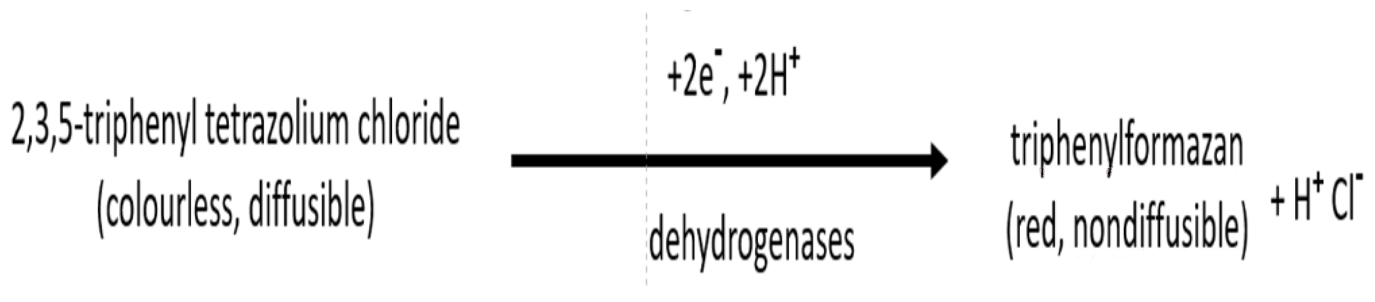
Scheme 1. Conversion of colourless tetrazolium chloride to red formazan in the reaction with dehydrogenases of living cells.
Review [40] contains a thorough and inspiring spectrum of different TLC approaches to characterize plants for their biological properties. It focuses on antimicrobial and antifungal assays, enzyme inhibition, antioxidant testing, and free radical scavenging activity, and it comes with 66 references of the original research papers. A similar review taking on a vast range of the TLC approaches to screening botanicals for their different biological properties is also given in [41], which dispenses a solid number of 99 references to the original research papers. Apart from general reviews which attempt to cover an entire spectrum of the TLC applications to screening different biological properties of botanicals, researchers also have reviews which cover selected kinds of TLC applications, and a good example is paper [42], which focuses on the screening of antimicrobial properties and is implemented with 75 references. In an interesting paper [11] (published in the Journal of Visual Experiments), a step-by-step explanation (implemented with a nice selection of instructive figures) is provided on how to practically perform the TLC-direct bioautography (TLC-DB) test for plant extracts to identify antimicrobial compounds (with direct bioautography, also known as the dot-blot test). As a working example given in [41], the TLC separation of phenolics extracted from the red clover (Trifolium pratense cv. Kenland) plant is presented, followed by screening of the separated fractions for their activity against Clostridium sticklandii, a hyper ammonia-producing bacterium (HAB) that is native to bovine rumen.
A kind of precursor to the TLC-DB approach is the dot-blot test alone, performed with aid of the thin-layer chromatographic adsorbent, yet without preliminary chromatographic fractionation of the sample considered. In that way, information is derived on an overall antibacterial potential of the plant extract, but without pointing out any specific fraction or individual compound derived from the scrutinized sample. A good practical example of such an approach is given in [43]. The researchers applied this test to compare antibacterial activity of 18 thyme (Thymus) specimens and species (originating from the same gardening plot and harvested in the same period). To this effect, polar fractions of the secondary metabolites were derived from each thyme plant, which were then drop-wise deposited on the silica gel pre-coated chromatographic plates, yet without developing the chromatograms. Then, the well-described dot-blot procedure was performed for antibacterial activity against the Gram-positive Bacillus subtilis strain. It was established that all investigated extracts exhibited antibacterial activity, yet distinct differences in the size of the bacterial growth inhibition zones were observed among the compared thyme species. Based on the results obtained, T. citriodorus “golden dwarf” and T. marschallianus were selected as prominent targets for further investigations and possible inclusion in herbal pharmacopeia, which was an essential scientific novelty of this research.
Practical illustration of the TLC-DB assay is provided in [44], and it focuses on two medicinal plants belonging to the European ethnopharmacy, i.e., on Matricaria recutita L. (chamomile) and Achillea millefolium L. (yarrow). To this effect, tinctures from aerial flowering parts of these plants were prepared by seven days of maceration in 70% ethanol (according to Polish Pharmacopoeia VI) and then chromatographically developed, and the chromatograms with separated fractions were tested against eight bacterial strains, i.e., Staphylococcus epidermidis, S. aureus, the methicillin-resistant S. aureus, Escherichia coli, Pseudomonas syringae pv. maculicola, Xanthomonas campestis pv. vesicatoria, Aliivibrio fischeri, and Bacillus subtilis. As a result, considerable antibacterial properties were for the first time confirmed with two compounds found in the examined tinctures, i.e., with apigenin and α-linolenic acid, and their identity was additionally confirmed by means of LC/MS.
Rapid screening of botanicals for their antimicrobial properties is advantageous, especially with plants originating from sub-tropical and tropical regions which are more abundant in flora and which are relatively less researched than flora originating from the temperate climatic zones. In that way, a shortcut verification can be assured of healing potential with traditional local medicines, which in terms of accessibility and use are ahead of Western medicines. A working example is provided in [45], which focuses on antibacterial properties of the leaf extract derived from the Philippine Piper betle L. plant belonging to the family of Piperaceae, which is recognized in India, Sri Lanka, Malaysia, Philippines and the other subtropical countries for its antibacterial, cytotoxic, hepatoprotective and many other advantageous pro-health properties. From the research data obtained from instrumental techniques (more advanced than the TLC-DB assay), it has already been known that the methanol, ethanol and supercritical CO2 leaf extracts from P. betle are exceptionally active against a number of the multidrug resistant (MDR) bacteria. In the discussed research, the ethanol leaf extract of P. betle was separated with use of TLC into eight fractions, which then underwent the dot-blot test. To this effect, the TLC system used consisted of the silica gel stationary phase and ethyl acetate–n-hexane (7:3, v/v) mobile phase. Two spots with RF values of 0.86 and 0.13 showed inhibitory activities against two Gram-positive MDR bacteria, i.e., the methicillin-resistant Staphylococcus aureus and the vancomycin-resistant Enterococcus. The spot with RF = 0.86 also showed inhibitory activity against two Gram-negative MDR bacteria, i.e., the carbapenem-resistant Enterobacteriaceae, Klebsiella pneumoniae and the metallo-𝛽-lactamase-producing Acinetobacter baumannii. With aid of the GC/MS technique, six compounds contained in the spots showing antibacterial activity were identified, with four of them never before having been mentioned in the medical literature.
3.3. Thin-Layer Chromatography in Screening of Botanicals for Their Enzyme-Inhibiting Potential
Many drugs are inhibitors of enzymes involved in mediating disease processes, and the same can be said about numerous plant constituents. Understanding the mechanism of action (MOA) of the target enzyme is critical in early discovery and development of drug candidates through extensive structure–activity relationship (SAR) studies. The purpose of a mechanism of action (MOA) study is to characterize the interaction of a compound with its target enzyme to understand how the compound interacts with this target and how natural substrates at physiologic concentrations can modulate this activity. These compounds which prove as inhibitors of enzymes rarely become proper drugs, however, due to strict requirements for a drug not only to inhibit the target but to have acceptable solubility, permeability, protein binding, and the selectivity, metabolism and toxicity profiles. This potential for the compound to become a proper drug is slowly revealed through tracking these characteristics in the specially designed structure–activity relationship (SAR) studies.
Similar to what has been stated in the preceding section, plants can offer a huge range of complex and structurally diverse compounds, and for this reason, they still remain one of the major reservoirs of the new drug molecules or molecular concepts for tomorrow, also in their capacity as enzyme inhibitors. Rapidity and cost-effectiveness of screening plant extracts with an aid of TLC-direct bioautography and human or mammal enzymes for testing their enzyme inhibiting capacity is a very attractive option for medicinal chemists in their search for novel structural motifs. To this effect, plant extract undergoes thin-layer chromatographic separation to individual constituents and/or constituent fractions, and then the dried chromatographic plate is sprayed with enzyme solution followed by a strict incubation protocol under humid conditions, and finally, it is visualized by spraying the plate with a visualizing reagent (which in the case of tracing cholinesterase or glucosidase inhibitors, is often the Fast Blue B salt).
Currently, one of the most acute health conditions among ageing populations worldwide is Alzheimer’s disease, and for this reason, the inhibitors of acetylcholinesterase (AChE) and butyrylcholinesterase (BuChE) currently form the basis of the newest drugs available for management of this disease. In view of a considerable potential of plants, the TLC bioautographic assays have been performed for screening plant extracts in the search for natural cholinesterase inhibitors. Research on these inhibitors which are available from natural botanical sources [47], implemented with 76 references to the original research papers, introduces a long list of plants recognized for their cholinesterase-inhibiting activity. These plants have long been used in ethnomedicines, particularly for memory-related disorders, and their efficiency is attributed to specific alkaloids, terpenes, sterols, flavanoids and glycosides. The most potent cholinesterase inhibitors are observed with the components of plants from the families of Buxaceae, Amaryllidaceae and Lycopodiaceae, although certain activity has also been established with the extracts from plants belonging to the families of Lamiaceae, Chenopodiaceae, Papaveraceae, Apocynaceae, and Labiatae. A number of papers have been published on this subject matter (e.g., [48][49][50][51][52]), and the basic principle of all these methods is that the enzyme (cholinesterase) converts 1-naphthyl acetate into naphthol, which reacts with Fast Blue B salt to make a purple-coloured background on TLC plates. Thus, upon the thin-layer chromatographic fractionation of the plant extract, the plate is dried for complete removal of liquid mobile phase, and then, it is sprayed with the enzyme stock solution. Then, it is again dried and kept in humid atmosphere for incubation at 37 °C for 20 min, and eventually, it is sprayed again with solution of α-naphthyl acetate and Fast Blue B salt, to give purple colouration after 1–2 min. The AChE and BuChE enzyme-inhibiting zones (i.e., fractions derived from the plant extracts) produce white spots on the purple background.
Among the first reports on a possibility of screening plant extracts for the detection of the cholinesterase-inhibition zones was that issued by Verpoorte et al. [48], who used acetylcholinesterase inhibitors from the Amaryllidaceae extracts in the TLC-DB test instead of synthetic galanthamine. Working conditions of the TLC-DB assay (which follows a very detailed protocol) were tested upon two alkaloids (galanthamine and physostigmine), and the results were reported in [49]. Although galanthamine and physostigmine are currently produced synthetically and are used for the treatment of cognitive decline in mild to moderate Alzheimer’s disease, both alkaloids have originally been isolated from plants and, more specifically, from the bulbs and plants of Galanthus nivalis (known as common snowdrop) and Physostigma venenosum, a leguminous plant endemic to tropical Africa. Over time, working conditions of the original TLC-BD method were modified, and in [50], an improved methodology was proposed focusing on the concentration of enzymes, the reagents, and the reaction time. As a consequence, the consumption of enzymes was reduced by 85%, and the detection limits were remarkably decreased. The researchers of [51] provided a comparison between the performance of the TLC-DB assay for inhibitors of cholinesterase and the 96-well plate assay based on Ellman’s method. For the majority (83%) of the 138 test compounds of natural and synthetic origin, the results obtained with the two assays have converged, and both screening assays were considered as suitable for the generation of new hits. The TLC-DB methodology for the screening of plant extracts in the search for new botanical inhibitors of cholinesterase is still in use, and in [52], a report is given on the examination of the never-before-tested single components from the ginger (Zingiber officinale) extract. The experiment led to recognition of three active inhibitors among volatile constituents of this plant, i.e., ar-curcumene, α-sesquiphellandrene, and α-zingiberene. Identification was possible owing to the TLC-HPLC-MS interface analysis of active zones and the GC-MS analysis of tested samples. The researchers of [53] provided another report on the TLC-DB test of the methanol extract of Schisandra chinensis for the cholinesterase inhibition effect, and the dibenzocyclooctadiene lignans contained in this plant were proven as responsible for the inhibition.
Along with Alzheimer’s disease, obesity (i.e., an abnormal or excessive fat accumulation) is also considered as one of great threats to human health on a global scale. It tends to aggravate the chances of acquiring diseases such as type 2 diabetes, hypertension, fatty liver, and cancer, which in turn reduce both life expectancy and the quality of life. Pancreatic lipase is a key enzyme for digestion of triacylglycerols, and inhibition of lipase has until now been the most explored strategy for treatment of obesity. In recent years, increasingly more medicinal herbs have been reported to show inhibitory activities against pancreatic lipase, and discovery of lipase inhibitors among herbal medicines may provide potential alternatives for the treatment of obesity. The researchers of [54] provided a report on the TLC-DB screening of methanolic extracts from the Camellia sinensis (L.) Kuntz, Rosmarinus officinalis L. and Morus alba leaves, for their ability to inhibit pancreatic lipase. Upon the thin-layer chromatographic fractionation of the plant extracts in the TLC system composed of silica gel as stationary phase and ethyl acetate–methanol–water (60:30:10, v/v/v) mobile phase, chromatographic plates were sprayed with α-naphtyl acetate, and the enzyme solutions before incubation at 37 °C for 20 min. Finally, solution of Fast Blue B salt was sprayed onto the TLC plates, giving a purple background colouration. Orlistat (a synthetic lipase inhibitor drug) was used as a reference analyte. The pancreatic lipase inhibiting zones of the plant extracts and the reference drug zone appeared as white spots on the purple background. In that way, it was demonstrated that the extracts from C. sinensis and R. officinalis exert an inhibitory effect upon pancreatic lipase, whereas Morus alba lacks analogical activity.
An alternative TLC-DB procedure was also proposed for screening pancreatic lipase [55], and to this effect, the p-nitrophenyl butyrate (PNPB) and bromothymol blue system were used to detect the lipase inhibition fractions derived from three unexplored species of Streptomyces (S. tendae, S. aurantiacus and S. albaduncus), with orlistat used as a positive control of procedure performance. Upon developing orlistat and supernatants derived from the Streptomyces samples on the silica gel pre-coated chromatographic plates (with chloroform–methanol (90:10, v/v) for orlistat and benzene–methanol (60:40, v/v) for Streptomyces), the plates were air dried until the mobile phase was evaporated completely. Then, a complex yet well-described procedure involving the porcine pancreatic lipase enzyme was implemented, and finally, the inhibitory zones were visualized as blue spots against the greenish-yellow background.
References
- Kenyon A.S., Flinn P.E., Layloff T.P., 1995. Rapid screening of pharmaceuticals by thin-layer chromatography: Analysis of essential drugs by visual methods. J. AOAC Int. 78:41-49.
- Kaale E., Risha P., Layloff T., 2011. TLC for pharmaceutical analysis in resource limited countries. J. Chromatogr. A, 1218:2732-2736.
- Shewiyo D.H., “Development and validation of HPTLC methods to assay pharmaceutical formulations”, the Ph.D. Thesis, Department of Pharmaceutical Biotechnology and Molecular Biology, Center for Neurosciences, Free University of Brussels, Brussels, Belgium, 2012.
- Kaale E., Risha P., Layloff T., Sherma J., Chapter 14:“Screening of substandard and fake drugs in underdeveloped countries by TLC”, in: (Eds, Komsta Ł., Waksmundzka-Hajnos M., Sherma J.) “Thin-Layer Chromatography in Drug Analysis”, CRC Press, Taylor & Francis Group, Boca Raton, Fl, USA, 2014; pp. 247-266.
- O’Sullivan C, Sherma J., 2012. A model procedure for the transfer of TLC pharmaceutical product screening methods designed for use in developing countries to quantitative HPTLC-densitometry methods. Acta Chromatogr. 24:241-252.
- Kaltsikes P.J., Dedio W., 1970. A thin-layer chromatographic study of the phenolics of the genus Aegilops. I. Numerical chemotaxonomy of the diploid species. Can. J. Bot., https://doi.org/10.1139/b70-260
- Kaltsikes P.J., Dedio W., 1970. A thin-layer chromatographic study of the phenolics of the genus Aegilops. II. Numerical chemotaxonomy of the polyploid species. Can. J. Bot., https://doi.org/10.1139/b70-261
- Rogers C.B., Abbot A.T.D., van Wyk A.E., 1999. A convenient thin layer chromatographic technique for chemotaxonomic application in Maytenus (Celastraceae). S. Afr. J. Bot. 66:174-176.
- Mandal P., Choudhury D., Ghosal M., Das A.P., 2016. TLC based chemotaxonomic approach of some laurels present in sub-Himalayan Terai and Duars region of West Bengal, India. Int. J. Pharm. Sci. Rev. Res. 41:193-196.
- Cieśla Ł., Waksmundzka-Hajnos M., 2010. Application of thin-layer chromatography for the quality control and screening the free radical scavenging activity of selected pharmacuetical preparations containing Salvia officinalis L. extract. Acta Pol. Pharm.-Drug Res. 67:481-485.
- Kagan I.A., Flythe M.D., 2014. Thin-layer chromatographic (TLC) separations and bioassays of plant extracts to identify antimicrobial compounds. J. Vis. Exp. 85 | e51411 | Page 1 of 8.
- Wang M., Zhang Y., Wang R., Wang Z., Yang B., Kuang H., 2021. An evolving technology that integrates classical methods with continuous technological developments: Thin-layer chromatography bioautography. Molecules 26(15), 4647;
- Schönborn A., Grimmmer A., 2013. Coupling sample preparation with effect-directed analysis of estrogenic activity – proposal for a new rapid screening concept for water samples. J. Planar Chromatogr. – Modern TLC 26:402-408.
- [Agatonovic-Kustrin S., Morton D.W., 2020. Hyphenated TLC as a tool in the effect-directed discovery of bioactive natural products. Appl. Sci. 2020, 10, 1123; doi:10.3390/app10031123.
- Sherma, J., 2000. Thin-layer chromatography in food and agricultural analysis. Review. J. Chromatogr. A 880:129-147.
- De Mey E., De Maere H., Dewulf L., Paelinck H., Sajewicz M., Fraeye I., KowalskaT., 2014. Application of accelerated solvent extraction (ASE) and thin layer chromatography (TLC) to determination of piperine in commercial samples of pepper (Piper nigrum L.). J. Liq. Chromatogr. Relat. Technol. 37:2980-2988.
- [Łata E., Fulczyk A., Kowalska T., Sajewicz M., 2017. Thin-layer chromatographic method of screening the anthocyanes containing alimentary products and precautions taken at the method development step. J. Chromatogr. A 1530:211-218.
- Kowalczuk A.P., Raman V., Galal A.M., Khan I.A., Siebert D.J., Zjawiony J.K., 2014. Vegetative anatomy and micromorphology of Salvia divinorum (Lamiaceae) from Mexico, combined with chromatographic analysis of salvinorin A. J. Nat. Med. 68:63–73.
- Waksmundzka-Hajnos M., Sherma J., Kowalska T., 2008. Overview of the field of TLC in phytochemistry and the structure of the book, Chapter 1, in: Thin-Layer Chromatography in Phytochemistry, CRC Press, FL, USA, pp. 3-14.
- Singh R., 2016. Chemotaxonomy: A tool for plant classification. J. Med. Plants Stud. 4:90-93.
- Schibli A., Reich E., 2005. Modern TLC: A key technique for identification and quality control of botanicals and dietary supplements. J. Planar Chromatogr. – Modern TLC 18:34-38.
- Staszek D., Orłowska M., Waksmundzka-Hajnos M., Sajewicz M., Kowalska T., 2013. Marker fingerprints originating from TLC and HPLC for selected plants from the Lamiaceae family. J. Liq. Chromatogr. Relat. Technol. 36:2463-2475.
- Jerzmanowska Z., 1967, Plant Material. The Isolation Methods, PWN, Warszawa, (in Polish).
- Świątek L., 1977. The iridoid phenolic acids and glycosides in certain Polish medicinal herbs from the Plantago genus. Herba Polon. 23:201–210.
- Ibrahim R.K., Towers G.H., 1960. The identification by chromatography of plant phenolic acids. Arch. Biochem. Biophys. 87:125–127.
- Świątek L., Dombrowicz E., 1984. Phenolic acids in the bitter raw materials. Part I. Analysis of the Artemisia absinthium herb and the Gentian root. Farm. Pol. 40:729–732.
- Schmidtlein, H., Hermann K., 1975. Quantitative analysis for phenolic acids by thin layer chromatography. J. Chromatogr. 115:123-128.
- Ganzera M., Pöcher A., Stuppner H., 2005. Differentiation of Cirsium japonicum and C. setosum by TLC and HPLC-MS. Phytochem. Anal. 16:205-209
- Sherma J., 2021. Thin-layer chromatography in the determination of synthetic and natural colorants in foods, Chapter 4, in: (Eds Grinberg N., Carr P.W.), Advances in Chromatography Vol. 56 CRC Press, Taylor & Francis Group, Boca Raton, FL, SA; pp. 109-136.
- Choma I., 2013. Thin-layer chromatography hyphenated with bioassays. J. AOAC Int. 96:1165-1166.
- Kedare S.B., Singh R.P., 2011. Genesis and development of DPPH method of antioxidant assay. J. Food Sci. Technol. 48:412-422.
- Glavind J., Holmer G., 1967. Thin-layer chromatographic determination of antioxidants by the stable free radical α,α’-diphenyl--picrylhydrazyl. J. Am. Oil Chem. Soc. 44:539–542.
- Li P., Anu H., Jari S., Yrjönen T., Vuorela H., 2005. TLC method for evaluation of free radical scavenging activity of rapeseed meal by video scanning technology. http://www.regional.org.au (accessed in July, 2022)
- Cieśla Ł., Kryszeń, Stochmal A., Oleszek W., Waksmundzka-Hajnos M., 2012. Approach to develop a standardized TLC-DPPH test for assessing free radical scavenging properties of selected phenolic compounds. J. Pharm. Biomed. Anal. 70:126-135.
- Cieśla Ł., Staszek D., Hajnos M., Kowalska T., Waksmundzka-Hajnos M., 2011. Development of chromatographic and free-radical scavenging activity fingerprints by thin-layer chromatography for selected Salvia species. Phytochem. Anal. 22:59-65.
- Reich E., Schibli A., DeBatt A., 2008. Validation of high-performance thin-layer chromatographic methods for the identification of botanicals in a cGMP environment. J. AOAC Int. 91:13–20.
- Cieśla Ł., Staszek D., Kowalska T., Waksmundzka-Hajnos M., 2013. The use of TLC-DPPH test with image processing to study direct antioxidant activity of phenolic acid fractions of selected Lamiaceae species. J. AOAC Int. 96:1228-1232.
- Agatonovic-Kustrin S., Morton D.W., 2017. High-performance thin-layer chromatography-direct bioautography as a method of choice for alpha-amylase and antioxidant activity evaluation in marine algae. J. Chromatogr. A 1530:197-203.
- Sherma J., 2018. Review of the determination of the antioxidant activity of foods, food ingredients, and dietary supplements by thin layer chromatography-direct bioautography, spectrometry, and the dot-blot procedure. J. AOAC Int. 101:1285-1294.
- Marston A., 2011. Thin layer chromatography with biological detection in phytochemistry. J. Chromatogr. A 1218:2676-2683.
- Dewanjee S., Gangopadhayay M., Bhattacharya N., Khanra R., Dua T.K., 2015. Bioautography and its scope in the field of natural product chemistry. J. Pharm. Anal. 5:75-84.
- Choma I.M., Jesionek W., 2015. TLC-direct bioautography as a high throughput method for detection of antimicrobials in plants. Chromatography 2:225-238.
- Kagan I.A., Flythe M.D., 2014. Thin-layer chromatographic (TLC) separations and bioassays of plant extracts to identify antimicrobial compounds. J. Vis. Exp. (85):51411; doi: 10.3791/51411.
- Orłowska M., Kowalska T., Sajewicz M., Jesionek W., Choma I.M., Majer-Dziedzic B., Szymczak G., Waksmundzka-Hajnos M., 2015. A comparison of antibacterial activity of selected thyme (Thymus) species by means of the dot blot test with direct bioautographic detection. J. AOAC Int. 98:871-875.
- Jesionek W., Móricz Á.M., Ott P.G., Kocsis B., Horváth B., Choma I.M., 2015. TLC-direct bioautography and LC/MS as complementary methods in identification of antibacterial agents in plant tinctures from the Asteraceae family. J. AOAC Int. 98:857-861.
- Valle D.M. Jr., Puzon J.J.M., Cabrera E.C., Rivera W.L., 2016. Thin layer chromatography-bioautography and gas chromatography-mass spectrometry of antimicrobial leaf extracts from Philippine Piper betle L. against multidrug-resistant bacteria. Evidence Based Complementary and Alternative Medicine, Vol. 2016, Article ID 4976791, 7 pages; http://dx.doi.org/10.1155/2016/497679
- Abiri R., Abdul-Hamid H., Sytar O., Abiri R., de Almeida E.B.. Sharma S., Bulgakov V., Arroo R., Malik S., 2021. A brief overview of potential treatments for viral diseases using natural plant compounds: The case of SARS-CoV. Molecules 2021, 26, 3868.
- Ahmed F., Ghalib R.M., Sasikala P., Mueen Ahmed K.K., 2013. Cholinesterase inhibitors from botanicals. Pharmacogn. Rev. 7:121-130.
- Rhee I.K., van de Meent M., Ingkaninan K., Verpoorte R., 2001. Screening for acetylcholinesterase inhibitors from Amaryllidaceae using silica gel thin-layer chromatography in combination with bioactivity staining. J. Chromatogr. A 915:217-223.
- Marston A., Kissling J., Hostettmann K., 2002. A rapid TLC bioautographic method for the detection of acetylcholinesterase and butyrylcholinesterase inhibitors in plants. Phytochem. Anal. 13:51-54.
- [51] Yang Z., Zhang X., Duan D., Song Z., Yang M., Li S., 2009. Modified TLC bioautographic method for screening acetylcholinesterase inhibitors from plant extracts. J. Sep. Sci. 32:3257-3259.
- Di Giovanni S., Borloz A., Urbain A., Marston A., Hostettmann K., Carrupt P.-A., Reist M., 2008. In vitro screening assays to identify natural or synthetic acetylcholinesterase inhibitors: thin layer chromatography versus microplate methods. Eur. J. Pharm. Sci. 32:109-119.
- Czernicka L., Ludwiczuk A., Rój E., Marzec Z., Jarzab A., Kukula-Koch W., 2020. Acetylcholinesterase inhibitors among Zingiber officinale terpenes – extraction conditions and thin layer chromatography-based bioautography studies. Molecules, 2020, 25(7), 1643.1680
- Sobstyl E., Szopa A., Ekiert H., Gnat S., Typek R., Choma I.M., 2020. Effect directed analysis and TLC screening of Schisandra chinensis fruits. J. Chromatogr. A 1618 (2020) 460942
- Hassan A.M.S., 2012. TLC bioautographic method for detecting lipase inhibitors. Phytochem. Anal. 23:405-407.

