Submitted Successfully!
To reward your contribution, here is a gift for you: A free trial for our video production service.
Thank you for your contribution! You can also upload a video entry or images related to this topic.
Version Summary Created by Modification Content Size Created at Operation
1 -- 1669 2022-09-29 06:03:46 |
2 format correct Meta information modification 1669 2022-09-29 10:46:58 |

Video Upload Options

Do you have a full video?

Confirm

Are you sure to Delete?
Cite
If you have any further questions, please contact Encyclopedia Editorial Office.
Ayana, G.;  Ryu, J.;  Choe, S. Ultrasound-Responsive Nanocarriers for Breast Cancer Chemotherapy. Encyclopedia. Available online: https://encyclopedia.pub/entry/27928 (accessed on 24 April 2024).
Ayana G,  Ryu J,  Choe S. Ultrasound-Responsive Nanocarriers for Breast Cancer Chemotherapy. Encyclopedia. Available at: https://encyclopedia.pub/entry/27928. Accessed April 24, 2024.
Ayana, Gelan, Jaemyung Ryu, Se-Woon Choe. "Ultrasound-Responsive Nanocarriers for Breast Cancer Chemotherapy" Encyclopedia, https://encyclopedia.pub/entry/27928 (accessed April 24, 2024).
Ayana, G.,  Ryu, J., & Choe, S. (2022, September 29). Ultrasound-Responsive Nanocarriers for Breast Cancer Chemotherapy. In Encyclopedia. https://encyclopedia.pub/entry/27928
Ayana, Gelan, et al. "Ultrasound-Responsive Nanocarriers for Breast Cancer Chemotherapy." Encyclopedia. Web. 29 September, 2022.
Ultrasound-Responsive Nanocarriers for Breast Cancer Chemotherapy
Edit

Breast cancer is the most common type of cancer and is treated with surgical intervention, radiotherapy, chemotherapy, or a combination of these regimens. Despite its ample use, chemotherapy has limitations such as bioavailability, adverse side effects, high-dose requirements, low therapeutic indices, multiple drug resistance development, and non-specific targeting. Drug delivery vehicles or carriers, of which nanocarriers are prominent, have been introduced to overcome chemotherapy limitations. Nanocarriers have been preferentially used in breast cancer chemotherapy because of their role in protecting therapeutic agents from degradation, enabling efficient drug concentration in target cells or tissues, overcoming drug resistance, and their relatively small size. However, nanocarriers are affected by physiological barriers, bioavailability of transported drugs, and other factors. To resolve these issues, the use of external stimuli has been introduced, such as ultrasound, infrared light, thermal stimulation, microwaves, and X-rays. Recently, ultrasound-responsive nanocarriers have become popular because they are cost-effective, non-invasive, specific, tissue-penetrating, and deliver high drug concentrations to their target.

ultrasound nanocarriers micro-/nano-bubbles breast cancer chemotherapy

1. Nanocarriers for Breast Cancer Chemotherapy

Nanoparticles designed for either targeted or non-targeted drug delivery have a small diameter (1–100 nm) and possess a large surface area to volume ratio [1]. These properties allow them to bind, absorb, and carry therapeutic agents with high efficiency 
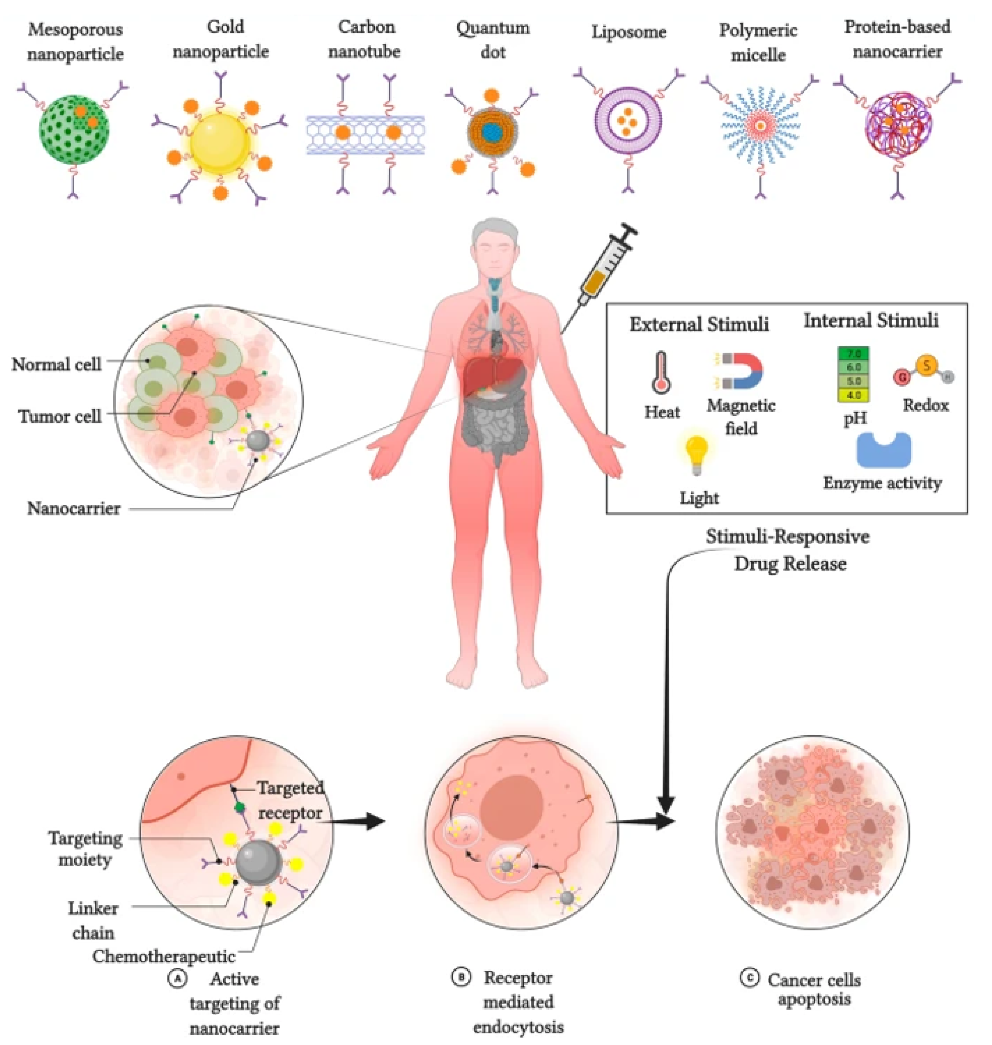
[2]. Nanocarriers for breast cancer chemotherapy are broadly divided into two types: organic and inorganic (Figure 1 [3]. Inorganic nanocarriers include quantum dots (QD), mesoporous silica nanoparticles (MSN), layered double hydroxide (LDH) nanoparticles, carbon nanotubes, and magnetic nanoparticles. Inorganic nanocarriers are preferred for their better anti-cancer agent-loading capacity, large surface area, reduced side effects, bioavailability, well-regulated drug release, and—most importantly—for their organic solvent tolerance. Organic nanocarriers, on the other hand, include polymeric nanoparticles, liposomes, micelles, protein nanoparticles, and dendrites. Organic nanocarriers are preferred for their easy synthesis and modification, enabling improved drug-loading efficacy, biodistribution, and therapeutic efficacy. Moreover, organic nanoparticles allow sustained drug release over a period of time and the use of organic solvents [4].
Figure 1. Types of nanocarriers used in breast cancer chemotherapy.
The most commonly used nanocarriers in breast cancer chemotherapy include liposomes, dendrimers, micelles, carbon nanotubes, polymeric nanoparticles, solid lipid nanoparticles (SLNs), and nanostructured lipid carriers (NLCs) [5]. Liposomes are used for various purposes to increase drug-loading capacity while suppressing unnecessary drug effects. In contrast, lipids cause toxicity, and nanocarriers are quickly destroyed by phagocytes. Dendrimers have been commended for their higher loading capacity and bioavailability. However, dendrimers suffer from rapid clearance, organ accumulation, and synthesis variability. Micelles reduce toxicity and other side effects, but are used only for limited drugs and exhibit low drug-loading capacity [6]. Carbon nanotubes are capable of penetrating and localizing at the cellular level, but as a material, they can be potentially toxic. Polymeric nanoparticles are biocompatible, degradable, non-toxic; however, they are less effective and susceptible to carrier degradation. SLNs have the advantage of being soluble and better controlled for drug release, despite their low drug-loading capacity and containing other complex structures [7]. NLCs have multiple advantages compared to others, and their limitations include gelation of lipid dispersion and polymorphic transition. In general, nanocarriers for breast chemotherapy have their advantages and shortcomings. To improve their shortcomings while increasing treatment efficacy, different stimuli are utilized, which are designed to make the nanocarriers responsive.

2. Stimuli-Responsive Nanocarriers for Breast Cancer Chemotherapy

The application of stimuli to improve the efficacy of therapeutic agents delivered by nanocarriers has received considerable attention in recent years. Stimuli-responsive nanocarriers have been developed to compensate for the shortcomings of conventional nanocarrier-based chemotherapy [8]. The delivery of therapeutic agents responsive to stimuli is based on both internal (endogenous) and external (exogenous) stimuli (Figure 2).
Figure 2. Stimulus responsive nanocarriers. Adapted from Kaushik et al. [9] with permission under the terms of the CC BY 4.0 License, Copyright 2022.

2.1. Internal Stimuli

Various internal stimuli are used for nanocarrier-based anti-cancer agent delivery to increase therapeutic efficacy and suppress adverse effects. Internal stimuli used with nanocarriers in breast cancer chemotherapy include pH, redox, and enzymatic stimuli [10]. pH-responsive nanocarriers are internalized and dissociate, causing protonation and extracellular drug release. Subsequently, the nanoparticle is detached, which promotes endocytosis of nanocarriers and release of the drug [11]. The redox-responsive nanocarrier system is the S–S bond that is chemically cross-linked as a gating or capping molecule on the surface of the nanoparticle and is cleaved upon the addition of agents, causing rapid drug release to the tumor cells [12]. Drug release from NPs in an enzyme-responsive manner originates from specific enzyme-catalyzed chemical reactions that lead to the degradation, dissociation, or morphological transitions of the parent NPs [13].

2.2. External Stimuli

External stimuli originate from outside of the body to initiate anti-cancer agent delivery. External stimuli used in breast cancer chemotherapy include magnetic fields, ultrasound, and light [14]. In contrast to the internal stimuli, the external stimuli would introduce contrast agents to the image—that of nanoparticles located in the target tissues, cells, or organelles. This further triggers nanocarriers from outside the body through particular stimuli at a specific time. Magnetic systems are widely utilized for targeting and imaging [15]. As magnetic-responsive nanotherapeutics are non-invasive signals, an externally applied magnetic field can damage the moving particles and increase the accumulation of therapeutic agents in tumors. A magnetic field could be employed for in vivo applications, and could have greater advantages for targeted cancer therapy as compared with intrinsic stimuli-responsive nanotherapeutics. Ultrasound is one of the most commonly used exogenous stimuli in cancer therapy [16]. The unique advantages of ultrasound responsiveness include safety, non-invasiveness, and deeper penetration into the tissue. Many exogenous stimuli are used for drug delivery systems, among which temperature-responsive drug delivery systems offer potential advantages compared to other counterparts. This is due to their flexible design, regulation of phase transition temperatures, and passive targeting capability. The localized hyperthermia from 42.5 to 43.5 °C helps to evade cancer cells by inducing high temperatures in tumor tissues. However, these hyperthermic stimuli would enlarge the blood vessels and modify the perforation of tumor cell membranes, thereby enhancing anti-tumor drug delivery [17].

2.3. Internal vs. External Stimuli

Both internal and external stimuli have their own advantages and disadvantages, as presented in Table 1. Internal stimuli are safe and provide efficient and controllable drug release without compromising cell and site specificity. Internal stimuli have the disadvantage of not being controlled manually. External stimuli have the advantage of being manually controlled and modulated based on individual requirement making it vital in personalized treatment. They also provide upgraded site-specific drug delivery and enable regulated and payload release. However, external stimuli need more sophisticated equipment and normal cell injury may happen. Nevertheless, compared to internal stimuli, external stimuli are preferred for nanocarrier based chemotherapy.
Table 1. Advantages and disadvantages of internal and external stimuli.

3. Therapeutic Agents in Ultrasound-Responsive Breast Cancer Treatment

Therapeutic agents are chemical substances that are delivered to the body for the treatment or mitigation of disease conditions or ailments. These substances can be drugs, proteins, genes, compounds, or other pharmaceutically active ingredients. As the human genome has been sequenced and genetic technology has advanced, there is a growing body of knowledge on genetic changes, initiation and proliferation, therapeutic mechanisms, and novel treatment targets for cancer therapy. Understanding the pathophysiology of the disease, human gene sequences, and discovery of novel molecular targets is the core of modern medicine to conquer cancer therapy. Numerous noteworthy advances have been made in the development of targeted therapy. These targeted therapies are designed to attack cancer cells while causing less damage to normal healthy cells. Targeted therapies are drugs or other substances that block the growth and spread of cancer by interfering with specific molecules or targets that are involved in the growth, spread, and progression of cancer. Targeted therapies are currently at the center of anti-cancer drug development; hence, they are the cornerstone of precision medicine. Similarly, in breast cancer, many drugs are being developed and integrated with nanocarriers. Table 2 lists some of the drugs used in breast cancer treatment along with nanocarriers responsive to ultrasound.
Table 2. Drugs used in ultrasound-responsive nanocarrier breast cancer treatment.
Drug Product/Platform Type of Nanocarrier Reference
Doxorubicin Perfluoropropane Liposome [18][19]
Doxorubicin Polyethylene glycol Liposome [20]
Doxorubicin Polyethylene glycol Liposome [21]
Cisplatin Soy phosphatidyl choline (SPC-3), cholesterol, dipalmitoyl phos-phatidyl glycerol (DPPG), and methoxy-polyethylene glycol-distearoyl phosphatidylethanolamine (mPEG 2000-DSPE) Liposome [22]
EndoTAG-1 and paclitaxel Cationic Liposome [23]
Paclitaxel 1,2-dioleoyl-sn-glycero-3-phosphocholine Liposome [24]
Resveratrol Chloroform solutions of cadmium oxide and sucrose laurate Liposome [25]
Cisplatin Distearoyl phosphoethanolamine-polyethylene glycol and phosphatidylcholine Liposome [26]
Paclitaxel Polyethyleneglycol (PEG)-phosphatidylethanolamine (PE) (PEG-PE) Liposome [27]
Doxorubicin and silymarin 3-(4,5-dimethylthiazol-2-yl)-2,5-diphenyltetrazolium bromide (MTT) Liposome [28]
Epirubicin-hydrochloride Phosphatidylcholines with thin film hydration using egg yolk Liposome [29]
Curcumin Polyethylene glycol (PEG) Liposome [30]
A7R-cysteine peptide Distearoylphosphosphatidyl-ethanolamine
(DSPE-PEG2000)
Liposome [31]
Raloxifene Methanol-ethyl acetate Liposome [32]
Artemisinin Polyethylene glycol 2000 (PEG 2000) Liposome [33]
Thymoquinone Thymoquinone (2-isopropyl-5-methyl-1,4-benzoquinone) and Triton X-100; 1,2-dipalmitoyl-sn-glycero-3-phospho-choline (DPPC) Liposome [34]
Doxorubicin Lipoic acid, hyaluronic acid, L-lysine methyl ester Polymer nanoparticles [35]
Doxorubicin Chitosan and pluronic F127 Polymer nanoparticles [36]
Cisplatin Luteinizing hormone-releasing hormone (LHRH)-modified dextran Polymer nanoparticles [37]
Tamoxifen citrate Polylactide-co-glycolide Polymer nanoparticles [38]
Paclitaxel Albumin nanoparticle Polymer nanoparticles [39]
Paclitaxel Folic acid Polylactic-co-glycolic acid, polyethylene glycol succinate Polymer nanoparticles [40]
Paclitaxel Montmorillonite and Poly(D, L-lactide-co-glycolide) Polymer nanoparticles [41]
Paclitaxel and ceramide Poly(beta-amino ester) and poly(D,L-lactide-co-glycolide) Polymer nanoparticles [42]
Docetaxel Albumin nanoparticle Polymer nanoparticles [43]
Quercetin Polylactic-co-glycolic acid, polyethylene glycol 1000 succinate Polymer nanoparticles [44]
Doxorubicin and Salinomycin Polyacrylic acid and Polyethylene glycol Micellar nanoparticle [45]
Paclitaxel Polyethylene glycol succinimidyl succinate Micellar nanoparticle [12]
Doxorubicin and Paclitaxel Lauryl carbamate derivative of plant-based polymer inulin Micellar nanoparticle [46]
Paclitaxel Polyethylene glycol-b-polylactide Micellar nanoparticle [11]
Fisetin Pluronic127 folic acid Micellar nanoparticle [47]
Paclitaxel Dextran-g-indomethacin Micellar nanoparticle [48]
Aminoflavone Anti-epidermal growth factor receptor Micellar nanoparticle [49]
Paclitaxel PEG-block-poly[(1,4-butanediol)-diacrylate-β-5-amino-1-pentanol] polyethyleneimine-block-PDHA Micellar nanoparticle [50]
Aminoflavone Poly(amidoamine) dendrimer, polyethylene glycol derivatives Micellar nanoparticle [51]
Doxorubicin Pluronic copolymer P123 polyethylene glycol-block-poly (di-isopropanolamino ethyl methacrylate) diblock copolymer Micellar nanoparticle [52]
Paclitaxel Methoxy polyethylene glycol-polylactide (mPEG-PLA) Micellar nanoparticle [53]
Paclitaxel polyethylene glycol (PEG)-polyacrylic acid (PAA) (PEG-PAA) Micellar nanoparticle [54]

References

  1. Yao, Y.; Zhou, Y.; Liu, L.; Xu, Y.; Chen, Q.; Wang, Y.; Wu, S.; Deng, Y.; Zhang, J.; Shao, A. Nanoparticle-Based Drug Delivery in Cancer Therapy and Its Role in Overcoming Drug Resistance. Front. Mol. Biosci. 2020, 7, 193.
  2. Liu, G.; Yang, L.; Chen, G.; Xu, F.; Yang, F.; Yu, H.; Li, L.; Dong, X.; Han, J.; Cao, C.; et al. A Review on Drug Delivery System for Tumor Therapy. Front. Pharmacol. 2021, 12, 735446.
  3. Yap, K.M.; Sekar, M.; Fuloria, S.; Wu, Y.S.; Gan, S.H.; Mat Rani, N.N.I.; Subramaniyan, V.; Kokare, C.; Lum, P.T.; Begum, M.Y.; et al. Drug Delivery of Natural Products Through Nanocarriers for Effective Breast Cancer Therapy: A Comprehensive Review of Literature. Int. J. Nanomed. 2021, 16, 7891–7941.
  4. Kandekar, U. Nanocarriers For Breast Cancer: Advanced Perspective Review Article. Hacet. Univ. J. Fac. Pharm. 2021, 41, 179–195.
  5. Tagde, P.; Najda, A.; Nagpal, K.; Kulkarni, G.T.; Shah, M.; Ullah, O.; Balant, S.; Rahman, M.H. Nanomedicine-Based Delivery Strategies for Breast Cancer Treatment and Management. Int. J. Mol. Sci. 2022, 23, 2856.
  6. Mu, W.; Chu, Q.; Liu, Y.; Zhang, N. A Review on Nano-Based Drug Delivery System for Cancer Chemoimmunotherapy. Nano-Micro Lett. 2020, 12, 142.
  7. Panikar, S.S.; Banu, N.; Haramati, J.; del Toro-Arreola, S.; Riera Leal, A.; Salas, P. Nanobodies as Efficient Drug-Carriers: Progress and Trends in Chemotherapy. J. Control. Release 2021, 334, 389–412.
  8. Senapati, S.; Mahanta, A.K.; Kumar, S.; Maiti, P. Controlled Drug Delivery Vehicles for Cancer Treatment and Their Performance. Signal Transduct. Target. Ther. 2018, 3, 7.
  9. Kaushik, N.; Borkar, S.B.; Nandanwar, S.K.; Panda, P.K.; Choi, E.H.; Kaushik, N.K. Nanocarrier Cancer Therapeutics with Functional Stimuli-Responsive Mechanisms. J. Nanobiotechnol. 2022, 20, 152.
  10. Zou, T.; Lu, W.; Mezhuev, Y.; Lan, M.; Li, L.; Liu, F.; Cai, T.; Wu, X.; Cai, Y. A Review of Nanoparticle Drug Delivery Systems Responsive to Endogenous Breast Cancer Microenvironment. Eur. J. Pharm. Biopharm. 2021, 166, 30–43.
  11. Wang, J.; De, G.; Yue, Q.; Ma, H.; Cheng, J.; Zhu, G.; Du, M.; Yi, H.; Zhao, Q.; Chen, Y. PH Responsive Polymer Micelles Enhances Inhibitory Efficacy on Metastasis of Murine Breast Cancer Cells. Front. Pharmacol. 2018, 9, 543.
  12. Zhang, Y.; Zhu, X.; Chen, X.; Chen, Q.; Zhou, W.; Guo, Q.; Lu, Y.; Li, C.; Zhang, Y.; Liang, D.; et al. Activated Platelets-Targeting Micelles with Controlled Drug Release for Effective Treatment of Primary and Metastatic Triple Negative Breast Cancer. Adv. Funct. Mater. 2019, 29, 1806620.
  13. Li, M.; Zhao, G.; Su, W.K.; Shuai, Q. Enzyme-Responsive Nanoparticles for Anti-Tumor Drug Delivery. Front. Chem. 2020, 8, 647.
  14. Oshiro-Júnior, J.A.; Rodero, C.; Hanck-Silva, G.; Sato, M.R.; Alves, R.C.; Eloy, J.O.; Chorilli, M. Stimuli-Responsive Drug Delivery Nanocarriers in the Treatment of Breast Cancer. Curr. Med. Chem. 2018, 27, 2494–2513.
  15. Dwivedi, P.; Kiran, S.; Han, S.; Dwivedi, M.; Khatik, R.; Fan, R.; Mangrio, F.A.; Du, K.; Zhu, Z.; Yang, C.; et al. Magnetic Targeting and Ultrasound Activation of Liposome-Microbubble Conjugate for Enhanced Delivery of Anticancer Therapies. ACS Appl. Mater. Interfaces 2020, 12, 23737–23751.
  16. Wei, P.; Cornel, E.J.; Du, J. Ultrasound-Responsive Polymer-Based Drug Delivery Systems. Drug Deliv. Transl. Res. 2021, 11, 1323–1339.
  17. Wu, P.; Jia, Y.; Qu, F.; Sun, Y.; Wang, P.; Zhang, K.; Xu, C.; Liu, Q.; Wang, X. Ultrasound-Responsive Polymeric Micelles for Sonoporation-Assisted Site-Specific Therapeutic Action. ACS Appl. Mater. Interfaces 2017, 9, 25706–25716.
  18. Ueno, Y.; Sonoda, S.; Suzuki, R.; Yokouchi, M.; Kawasoe, Y.; Tachibana, K.; Maruyama, K.; Sakamoto, T.; Komiya, S. Combination of Ultrasound and Bubble Liposome Enhance the Effect of Doxorubicin and Inhibit Murine Osteosarcoma Growth. Cancer Biol. Ther. 2011, 12, 270–277.
  19. Lybaek, D.; Iversen, L. Pegylated Liposomal Doxorubicin in the Treatment of Mycosis Fungoides. Acta Derm. Venereol. 2006, 86, 545–547.
  20. Rau, K.M.; Lin, Y.C.; Chen, Y.Y.; Chen, J.S.; Lee, K.D.; Wang, C.H.; Chang, H.K. Pegylated Liposomal Doxorubicin (Lipo-Dox®) Combined with Cyclophosphamide and 5-Fluorouracil Is Effective and Safe as Salvage Chemotherapy in Taxane-Treated Metastatic Breast Cancer: An Open-Label, Multi-Center, Non-Comparative Phase II Study. BMC Cancer 2015, 15, 423.
  21. Batist, B.G.; Ramakrishnan, G.; Rao, C.S.; Chandrasekharan, A.; Gutheil, J.; Guthrie, T.; Shah, P.; Khojasteh, A.; Nair, M.K.; Hoelzer, K.; et al. Reduced Cardiotoxicity and Preserved Antitumor Efficacy of Liposome-Encapsulated Doxorubicin and Cyclophosphamide Compared with Conventional Doxorubicin and Cyclophosphamide in a Randomized, Multicenter Trial of Metastatic Breast Cancer. J. Clin. Oncol. 2001, 19, 1444–1454.
  22. Stathopoulos, G.P.; Boulikas, T. Lipoplatin Formulation Review Article. J. Drug Deliv. 2012, 2012, 581363.
  23. Awada, A.; Bondarenko, I.N.; Bonneterre, J.; Nowara, E.; Ferrero, J.M.; Bakshi, A.V.; Wilke, C.; Piccart, M. A Randomized Controlled Phase Ii Trial of a Novel Composition of Paclitaxel Embedded into Neutral and Cationic Lipids Targeting Tumor Endothelial Cells in Advanced Triple-Negative Breast Cancer (Tnbc). Ann. Oncol. 2014, 25, 824–831.
  24. Slingerland, M.; Guchelaar, H.J.; Rosing, H.; Scheulen, M.E.; van Warmerdam, L.J.C.; Beijnen, J.H.; Gelderblom, H. Bioequivalence of Liposome-Entrapped Paclitaxel Easy-To-Use (LEP-ETU) Formulation and Paclitaxel in Polyethoxylated Castor Oil: A Randomized, Two-Period Crossover Study in Patients With Advanced Cancer. Clin. Ther. 2013, 35, 1946–1954.
  25. Zhao, Y.N.; Cao, Y.N.; Sun, J.; Wu, Q.; Cui, S.H.; Zhi, D.F.; Zhang, S.B.; Liang, Z.; Zhen, Y.H.; Guo, S.T. Anti-Breast Cancer Activity of Resveratrol Encapsulated in Liposomes. J. Mater. Chem. B 2019, 8, 27–37.
  26. Ghafari, M.; Haghiralsadat, F.; Khanamani Falahati-Pour, S.; Zavar Reza, J. Development of a Novel Liposomal Nanoparticle Formulation of Cisplatin to Breast Cancer Therapy. J. Cell. Biochem. 2020, 121, 3584–3592.
  27. Okamoto, Y.; Taguchi, K.; Imoto, S.; Giam Chuang, V.T.; Yamasaki, K.; Otagiri, M. Cell Uptake and Anti-Tumor Effect of Liposomes Containing Encapsulated Paclitaxel-Bound Albumin against Breast Cancer Cells in 2D and 3D Cultured Models. J. Drug Deliv. Sci. Technol. 2020, 55, 101381.
  28. Gheybi, F.; Alavizadeh, S.H.; Rezayat, S.M.; Zendehdel, E.; Jaafari, M.R. Chemotherapeutic Activity of Silymarin Combined with Doxorubicin Liposomes in 4T1 Breast Cancer Cells. Nanomed. Res. J. 2019, 4, 29–34.
  29. Alexander, P.; Jainamboo, M.; Praseetha, P.K.; Gopukumar, S.T. Silica Coated Liposomes for Drug Delivery towards Breast Cancer Cells. Rasayan J. Chem. 2016, 9, 300–308.
  30. Jadia, R.; Kydd, J.; Piel, B.; Rai, P. Liposomes Aid Curcumin’s Combat with Cancer in a Breast Tumor Model. Oncomedicine 2018, 3, 94–109.
  31. Cao, J.; Wang, R.; Gao, N.; Li, M.; Tian, X.; Yang, W.; Ruan, Y.; Zhou, C.; Wang, G.; Liu, X.; et al. A7RC Peptide Modified Paclitaxel Liposomes Dually Target Breast Cancer. Biomater. Sci. 2015, 3, 1545–1554.
  32. Ağardan, N.B.M.; Değim, Z.; Yılmaz, Ş.; Altıntaş, L.; Topal, T. The Effectiveness of Raloxifene-Loaded Liposomes and Cochleates in Breast Cancer Therapy. AAPS PharmSciTech 2016, 17, 968–977.
  33. Dadgar, N.; Koohi Moftakhari Esfahani, M.; Torabi, S.; Alavi, S.E.; Akbarzadeh, A. Effects of Nanoliposomal and Pegylated Nanoliposomal Artemisinin in Treatment of Breast Cancer. Indian J. Clin. Biochem. 2014, 29, 501–504.
  34. Odeh, F.; Ismail, S.I.; Abu-Dahab, R.; Mahmoud, I.S.; Al Bawab, A. Thymoquinone in Liposomes: A Study of Loading Efficiency and Biological Activity towards Breast Cancer. Drug Deliv. 2012, 19, 371–377.
  35. Zhong, Y.; Zhang, J.; Cheng, R.; Deng, C.; Meng, F.; Xie, F.; Zhong, Z. Reversibly Crosslinked Hyaluronic Acid Nanoparticles for Active Targeting and Intelligent Delivery of Doxorubicin to Drug Resistant CD44 + Human Breast Tumor Xenografts. J. Control. Release 2015, 205, 144–154.
  36. Manaspon, C.; Viravaidya-Pasuwat, K.; Pimpha, N. Preparation of Folate-Conjugated Pluronic F127/Chitosan Core-Shell Nanoparticles Encapsulating Doxorubicin for Breast Cancer Treatment. J. Nanomater. 2012, 2012, 593878.
  37. Li, M.; Tang, Z.; Zhang, Y.; Lv, S.; Li, Q.; Chen, X. Targeted Delivery of Cisplatin by LHRH-Peptide Conjugated Dextran Nanoparticles Suppresses Breast Cancer Growth and Metastasis. Acta Biomater. 2015, 18, 132–143.
  38. Maji, R.; Dey, N.S.; Satapathy, B.S.; Mukherjee, B.; Mondal, S. Preparation and Characterization of Tamoxifen Citrate Loaded Nanoparticles for Breast Cancer Therapy. Int. J. Nanomed. 2015, 9, 3107–3118.
  39. Miele, E.; Spinelli, G.P.; Miele, E.; Tomao, F.; Tomao, S. Albumin-Bound Formulation of Paclitaxel (Abraxane® ABI-007) in the Treatment of Breast Cancer. Int. J. Nanomed. 2009, 4, 99–105.
  40. Chen, S.H.; Liu, T.I.; Chuang, C.L.; Chen, H.H.; Chiang, W.H.; Chiu, H.C. Alendronate/Folic Acid-Decorated Polymeric Nanoparticles for Hierarchically Targetable Chemotherapy against Bone Metastatic Breast Cancer. J. Mater. Chem. B 2020, 8, 3789–3800.
  41. Sun, B.; Ranganathan, B.; Feng, S.S. Multifunctional Poly(d,l-Lactide-Co-Glycolide)/Montmorillonite (PLGA/MMT) Nanoparticles Decorated by Trastuzumab for Targeted Chemotherapy of Breast Cancer. Biomaterials 2008, 29, 475–486.
  42. van Vlerken, L.E.; Duan, Z.; Little, S.R.; Seiden, M.V.; Amiji, M.M. Biodistribution and Pharmacokinetic Analysis of Paclitaxel and Ceramide Administered in Multifunctional Polymer-Blend Nanoparticles in Drug Resistant Breast Cancer Model. Mol. Pharm. 2008, 5, 516–526.
  43. Zheng, Y.R.; Suntharalingam, K.; Johnstone, T.C.; Yoo, H.; Lin, W.; Brooks, J.G.; Lippard, S.J. Pt(IV) Prodrugs Designed to Bind Non-Covalently to Human Serum Albumin for Drug Delivery. J. Am. Chem. Soc. 2014, 136, 8790–8798.
  44. Zhou, Y.; Chen, D.; Xue, G.; Yu, S.; Yuan, C.; Huang, M.; Jiang, L. Improved Therapeutic Efficacy of Quercetin-Loaded Polymeric Nanoparticles on Triple-Negative Breast Cancer by Inhibiting UPA. RSC Adv. 2020, 10, 34517–34526.
  45. Cui, Y.; Yang, Y.; Ma, M.; Xu, Y.; Sui, J.; Li, H.; Liang, J.; Sun, Y.; Fan, Y.; Zhang, X. Reductive Responsive Micelle Overcoming Multidrug Resistance of Breast Cancer by Co-Delivery of DOX and Specific Antibiotic. J. Mater. Chem. B 2019, 7, 6075–6086.
  46. Kesharwani, S.S.; Dachineni, R.; Bhat, G.J.; Tummala, H. Hydrophobically Modified Inulin-Based Micelles: Transport Mechanisms and Drug Delivery Applications for Breast Cancer. J. Drug Deliv. Sci. Technol. 2019, 54, 101254.
  47. Pawar, A.; Singh, S.; Rajalakshmi, S.; Shaikh, K.; Bothiraja, C. Development of Fisetin-Loaded Folate Functionalized Pluronic Micelles for Breast Cancer Targeting. Artif. Cells Nanomed. Biotechnol. 2018, 46, 347–361.
  48. Ji, W.; Wang, B.; Fan, Q.; Xu, C.; He, Y.; Chen, Y. Chemosensitizing Indomethacin-Conjugated Dextran-Based Micelles for Effective Delivery of Paclitaxel in Resistant Breast Cancer Therapy. PLoS ONE 2017, 12, e0180037.
  49. Wang, Y.; Wang, Y.; Chen, G.; Li, Y.; Xu, W.; Gong, S. Quantum-Dot-Based Theranostic Micelles Conjugated with an Anti-EGFR Nanobody for Triple-Negative Breast Cancer Therapy. ACS Appl. Mater. Interfaces 2017, 9, 30297–30305.
  50. Qiu, Y.; Ren, K.; Zhao, W.; Yu, Q.; Guo, R.; He, J.; Mei, L.; Liu, Y.; Tang, J.; Xu, S.; et al. A “Dual-Guide” Bioinspired Drug Delivery Strategy of a Macrophage-Based Carrier against Postoperative Triple-Negative Breast Cancer Recurrence. J. Control. Release 2021, 329, 191–204.
  51. Brinkman, A.M.; Chen, G.; Wang, Y.; Hedman, C.J.; Sherer, N.M.; Havighurst, T.C.; Gong, S.; Xu, W. Aminoflavone-Loaded EGFR-Targeted Unimolecular Micelle Nanoparticles Exhibit Anti-Cancer Effects in Triple Negative Breast Cancer. Biomaterials 2016, 101, 20–31.
  52. Yu, H.; Cui, Z.; Yu, P.; Guo, C.; Feng, B.; Jiang, T.; Wang, S.; Yin, Q.; Zhong, D.; Yang, X.; et al. PH- and NIR Light-Responsive Micelles with Hyperthermia-Triggered Tumor Penetration and Cytoplasm Drug Release to Reverse Doxorubicin Resistance in Breast Cancer. Adv. Funct. Mater. 2015, 25, 2489–2500.
  53. Ahn, H.K.; Jung, M.; Sym, S.J.; Shin, D.B.; Kang, S.M.; Kyung, S.Y.; Park, J.W.; Jeong, S.H.; Cho, E.K. A Phase II Trial of Cremorphor EL-Free Paclitaxel (Genexol-PM) and Gemcitabine in Patients with Advanced Non-Small Cell Lung Cancer. Cancer Chemother. Pharmacol. 2014, 74, 277–282.
  54. Kato, K.; Chin, K.; Yoshikawa, T.; Yamaguchi, K.; Tsuji, Y.; Esaki, T.; Sakai, K.; Kimura, M.; Hamaguchi, T.; Shimada, Y.; et al. Phase II Study of NK105, a Paclitaxel-Incorporating Micellar Nanoparticle, for Previously Treated Advanced or Recurrent Gastric Cancer. Investig. New Drugs 2012, 30, 1621–1627.
More
Information
Contributors MDPI registered users' name will be linked to their SciProfiles pages. To register with us, please refer to https://encyclopedia.pub/register : , ,
View Times: 428
Entry Collection: Biopharmaceuticals Technology
Revisions: 2 times (View History)
Update Date: 29 Sep 2022
1000/1000