Submitted Successfully!
To reward your contribution, here is a gift for you: A free trial for our video production service.
Thank you for your contribution! You can also upload a video entry or images related to this topic.
Version Summary Created by Modification Content Size Created at Operation
1 -- 4113 2022-09-27 09:35:10 |
2 format corrected. Meta information modification 4113 2022-09-28 03:08:51 |

Video Upload Options

Do you have a full video?

Confirm

Are you sure to Delete?
Cite
If you have any further questions, please contact Encyclopedia Editorial Office.
Shaw, D.S.;  Honeychurch, K.C. Applications of Nanosensor Technology for the Plant Sciences. Encyclopedia. Available online: https://encyclopedia.pub/entry/27643 (accessed on 29 April 2024).
Shaw DS,  Honeychurch KC. Applications of Nanosensor Technology for the Plant Sciences. Encyclopedia. Available at: https://encyclopedia.pub/entry/27643. Accessed April 29, 2024.
Shaw, Daniel S., Kevin C. Honeychurch. "Applications of Nanosensor Technology for the Plant Sciences" Encyclopedia, https://encyclopedia.pub/entry/27643 (accessed April 29, 2024).
Shaw, D.S., & Honeychurch, K.C. (2022, September 27). Applications of Nanosensor Technology for the Plant Sciences. In Encyclopedia. https://encyclopedia.pub/entry/27643
Shaw, Daniel S. and Kevin C. Honeychurch. "Applications of Nanosensor Technology for the Plant Sciences." Encyclopedia. Web. 27 September, 2022.
Applications of Nanosensor Technology for the Plant Sciences
Edit

The detection of analytes is optically difficult in planta due to tissue thickness and the presence of photosynthetic pigments in plant tissues. Nanosensors are well-suited for the detection of analytes as they are easily embedded in plant tissues. They are thus well-suited for in vivo studies of cellular signalling and metabolism.

nanosensors plants botany nanobiotechnology agriculture

1. Detection of Molecular Oxygen

Molecular oxygen is the terminal electron acceptor in the electron transport chain during aerobic respiration. Plants (as well as algae and photosynthetic bacteria) are able to produce oxygen via photolysis, which is part of the light-dependent reaction of photosynthesis. However, the availability of oxygen in the atmosphere is still an essential substrate for plant metabolism as photosynthetic activity varies in tissue and there are times when plants are not photosynthetically active [1]. Many stresses, for example flooding, can also result in hypoxia in plant tissues. In addition, differential patterns of the abundance of oxygen occur in organs and meristems and the regulation of oxygen status is mechanistically related to plant development [2].
Extensive work on oxygen sensing has utilised Clark-type polarographic electrode sensors to detect a current flow caused by the chemical reduction of oxygen to water [3]. These microelectrodes have been used to determine the rates of photosynthesis and respiration by potato leaf protoplasts [4], measure the respiration rate of mitochondria extracted from pea shoots [5] and the leaves of Arabidopsis thaliana [6], as well as to measure alternative oxidase activity in soybean cotyledons and roots [7]. However, these electrodes have practical limitations when compared to optical sensors; they are invasive and can require extensive sample preparation, and they consume oxygen causing experimental errors when used in a living cell. Some of these problems can be overcome by using nanosensors. At present, two categories of nanosensor are being utilised to assess oxygen distribution inside tissues, namely electrochemical and optical systems.
Electrochemical nanosensors (carbon-filled quartz micropipettes with platinum-coated tips) have been used to detect a considerable drop in oxygen concentration at the surface of Chara corallina internodes in response to micro-perforation of the cell wall [8]. The decline in oxygen concentration at the wounding site could be due to several causes, such as the stimulation of the plasma membrane NADPH oxidase, and modulation of antioxidant systems.
Optical nanosensors for O2 also have features that make them an attractive alternative to the Clark-type polarographic electrode sensors, whilst enabling oxygen to be sensed on a nanoscale and to be imaged over large areas. Probes encapsulated by biologically localised embedding (PEBBLEs) are a prominent class of this type of nanosensor. The sensing elements, i.e., fluorescent dyes of PEBBLEs are encapsulated within an inert matrix which reduces dye leakage [9]. In addition, the protective shell retains stability and prevents interference with other proteins [10]. However, PEBBLEs commonly emitted a red phosphorescence signal that interfered with the autofluorescence of the plant chlorophyll when applied in plant cells. To circumvent the interference of the plant autofluorescence, a microbead-based probe was developed for application in plant- and algae-based systems that utilised the two-frequency phase modulation technique [11]. Nanoparticle oxygen sensors typically have excellent brightness and photostability, and are relatively simple to produce, with the added benefit that long-term storage is possible [1]. However, the size of the probes—ranging from 20 to 600 nm in diameter [9]—may lead to cell damage, and hence limit their application in living plants [11].

2. Water and Humidity Nanosensors

Utilising an aluminium oxide nano-porous ceramic plate (mean pore size: 30 ± 15 nm), an optical-based sensor for the direct, continuous monitoring of soil water has been reported [12]. The nano-porous ceramic disc was used in conjunction with a silicon diaphragm and a miniature optical displacement detection unit, composed of an integrated light source and photodetector. When the sensor is buried into unsaturated soil water, a negative pressure inside the reservoir is established, inducing diaphragm bending. The resulting displacement caused by the diaphragm could then be used to measure the dry soil saturation.
Subsequently, Leone et al. [13] presented a compact innovative optical, low-cost platform for soil water content measurement based on a nano-porous ceramic disc, however, in this case, in connection with an engineered optical fibre with near-infrared-based detection. The sensor consisted of a Y-shaped bifurcated cable housing two fibres in a single body. These fibres were placed side-by-side with one connected to the light source and the other to the detector. The common leg allowed for illumination of the fibres and served to collect the light from the disc. For a soil testing, the sensor was placed in a protective PVC tube buried in a soil tank.
Lan et al. [14] have reported on the fabrication of a capacitive wearable graphene-based plant humidity sensor. The capacitive-type humidity nanosensor was fabricated using laser direct writing technology on a polyimide film to give a graphene interdigital electrode (LIG-IDE). An aqueous solution of graphene oxide (GO) was then drop-cast onto the surface of LIG-IDE to act as the sensing element of the humidity sensor. The flexible GO humidity nanosensor could be readily attached to the surface of plant leaves without reportedly adversely affecting the growth of the plant. The nanosensor could be combined with wireless devices to give an integrated system.

3. Detection of Adenosine Triphosphate

Adenosine triphosphate (ATP) is an organic chemical that provides energy to drive many processes in living cells. ATP is dephosphorylated either to adenosine diphosphate or to adenosine monophosphate when consumed in metabolic processes. In plants, ATP is synthesised in chloroplasts and mitochondria. A decrease in cytoplasmic ATP levels following the addition of oligomycin A (a mitochondrial ATP synthase inhibitor) was detected using a chimera of enhanced Renilla luciferase and a fluorescent protein with high bioluminescence resonance energy transfer efficiency (Venus) [15]—an optical nanosensor based on FRET. In addition, ATP production in chloroplasts during photosynthesis has been visualised in transgenic Arabidopsis plants by targeting this optical nanosensor to the chloroplast stroma by using a transit peptide fusion [15].

4. Detection of Calcium Ions

Calcium plays an important role in signal transduction pathways. Calcium ions are involved in multiple plant processes such as stomatal closing, cellular division, and cell signalling. For example, in the process of stomatal closing, free Ca2+ ions enter the cytosol from both outside the cell and internal stores following abscisic acid signals to the guard cells. This has the effect of reversing the concentration gradient and K+ ions begin exiting the cell. The loss of solutes makes the cell flaccid and closes the stomatal pores. FRET-based genetically encoded sensors allow high-resolution live cell imaging of Ca2+ dynamics [16]. Analysis of Ca2+ dynamics in Lotus japonicus revealed distinct Nod factor-induced Ca2+ spiking patterns in the nucleus and the cytosol.

5. Detection of Reactive Oxygen Species

Nanosensors are also capable of detecting reactive oxygen species. Superoxide anion (O2•−), singlet oxygen (1O2), hydroxyl radical (•OH), and hydrogen peroxide (H2O2) are the major ROS in plants. ROS can be produced during normal cellular metabolism at cellular sites such as the chloroplast, mitochondria, peroxisomes, and apoplast [17][18][19]. ROS are involved in numerous signalling pathways in plants, including those involved in plant development, cell death, and responses to various types of stress [20][21]. ROS and redox potentials can be measured using genetically encoded ratiometric single-fluorescent protein sensors, such as roGFPs [22][23], to monitor the glutathione redox state [24][25] and extrinsic sensors, such as HyPer—a single fluorescent ROS sensor that directly reports H2O2. In plants, roGFPs have been extensively used to determine glutathione redox potential [25][26][27][28]. HyPer has been used to detect H2O2 changes in Arabidopsis guard cells and roots [29][30][31]. In plants, oxidative bursts can play significant roles in plant disease defences and signal transduction. The real-time monitoring of oxidative burst from single plant protoplasts has been achieved using electrochemical sensors modified with platinum nanoparticles [32].

6. Detection of Nitric Oxide

Nitric oxide is a gaseous reactive nitrogen species that acts as a signalling molecule throughout the plant life cycle. Nitric oxide is involved in a range of physiological activities in plants, ranging from seed germination to senescence and programmed cell death [33]. Furthermore, nitric oxide also acts as a signal in response to biotic and abiotic stresses [34]. However, the precise role of nitric oxide in signalling pathways and ideal methods of measurement remain an active area of research. One method of measurement employs semiconducting single-walled carbon nanotubes as signal transducers for nanosensors. Semiconducting single-walled carbon nanotubes have been exploited for near-infrared fluorescence monitoring of nitric oxide in A. thaliana [35]. This technique employed corona phase molecular recognition, which uses the specific adsorption of a compositionally designed polymer at a nanoparticle interface to enable recognition.

7. Detection of Plant Hormones

It is possible to detect a range of plant hormones, such as strigolactones [36], ethylene [37][38], and auxin [39][40], using a variety of nanosensors. Plant hormones (also known as phytohormones) are signal molecules produced within plants and are involved throughout a plant’s growth and development from embryogenesis [41] to reproductive development [42], as well as in biotic [43][44] and abiotic stress tolerance [45][46]. Nanosensors offer opportunities to study plant hormones and signalling mechanisms in vivo.
Strigolactones are a group of chemical compounds produced by a plant’s roots [47] and represent a class of plant hormones that regulate developmental processes and play a role in the response of plants to various biotic and abiotic stresses [48]. They have been identified as being involved in three different processes: the promotion of the germination of parasitic organisms that grow in the host plant’s roots [47][49][50], in the recognition of the plant by symbiotic fungi [47][51], and the inhibition of plant shoot branching [47][52][53]. One such parasitic organism that grows in the host plant’s roots is Striga [47][49][50]. The use of an optical nanosensor (a fluorescence turn-on probe called Yoshimulactone Green) allowed for the spatiotemporal monitoring of strigolactone levels in germinating Striga seeds [36]. The recognition of Yoshimulactone Green by strigolactone receptors and its subsequent hydrolysis generates detectable fluorescent products. In addition to the spatiotemporal monitoring of strigolactone levels, Yoshimulactone Green was used to determine specific strigolactone receptors [36].
Ethylene regulates many aspects of the plant life cycle, including seed germination, root initiation, flower development, fruit ripening, senescence, and responses to biotic and abiotic stresses [54]. Ethylene is widely used in agriculture to force the ripening of fruits [55]. Chemiresistive sensors have been used to sense the gaseous plant hormone ethylene [56][57][58][59] and have shown a reliable ethylene response toward different fruit types such as banana, avocado, apple, pear, and orange.
Auxins are plant hormones that influence multiple aspects of plant development such as cell enlargement, bud formation, and root initiation. A sensor employing platinum black and carbon nanotube surface modifications characterised auxin flux in 3- to 5-day roots non-invasively [39]. Moreover, a sensor utilising a porous graphene bionanocomposite of porous graphene, gold nanoparticles, and anti-indole-3-acetic acid antibody for sensitive and label-free amperometric immunoassay of indole-3-acetic acid (IAA; an auxin-class hormone) was reported to have a low detection limit and can been applied to the detection of IAA in plant sample extracts [60].
Gibberellins are plant hormones that promote organ growth and regulate a variety of developmental processes. Mutants defective in GA biosynthesis are characterized by reduced elongation of roots, stems, and floral organs [61]. A FRET-based nanosensor has been developed for the high-resolution quantification of spatiotemporal gibberellin distribution [62]. To develop the FRET-based nanosensor for gibberellin, plant hormone receptors were used as sensory domains. The GIBBERELLIN INSENSITIVE DWARF 1 (GID1) protein is a soluble receptor protein that interacts with gibberellins in an internal binding pocket [63]. Gibberellin binding promotes GID1 interaction with members of the DELLA family of growth regulators in plants. The GID1–gibberellin complex leads to the degradation of the DELLA protein after binding to the N-termini of the DELLA protein [64]. The Arabidopsis gibberellin perception machinery was adapted into a conformationally dynamic gibberellins binding domain within a FRET nanosensor by fusing GID1 variants to DELLA N-termini. This fusion converts the gibberellin-dependent intermolecular interactions into gibberellin-dependent intramolecular structural rearrangements. In this way, the nanosensor responds to nanomolar concentrations of bioactive gibberellins with an increase in the emission ratio and has been used to report gibberellin distribution and gradients in vivo in multiple tissues [62].
Salicylic acid (SA) is an important plant hormone that is best known for mediating host responses upon pathogen infection [65]. Derivatives of salicylic acid can be found in food products, medicines, cosmetics, and preservatives. A structure-switching aptamer-based nanopore thin film sensor has been developed for the detection of salicylic acid in plant extracts [66]. Due to its small size and scarcity of reactive groups for immobilization, salicylic acid is reportedly a challenging target for aptamer selection using conventional systemic evolution of ligands. However, the authors Chen et al. reported the development of a nanopore thin film sensor platform capable of determining levels as low as 0.1 μM salicylic acid, which showed good selectivity towards salicylic acid and its metabolites. It was shown possible to determine salicylic acid in Arabidopsis and rice using only about 1 μL plant extracts, with an assay time of less than 30 min.

8. Determination of Fruit Ripening

The perishability of fruits is a long-standing supply chain issue, causing a sizeable proportion of harvested fruits to be discarded before distribution to consumers [67]. As discussed above, ethylene is a major plant hormone that dictates fruit ripening [54]. It is possible to regulate the ripening dynamics of climacteric fruits through the manipulation of ethylene concentration—a technique widely used to extend shelf-life and ensure shelf-maturity. The mechanisms of fruit ripening and spoilage have been well studied [55][68][69][70]. Ethylene concentrations at 1 parts-per-million (ppm; 10−6) have been shown to initiate the ripening of climacteric fruits [70], while ethylene-sensitive fruits such as bananas and kiwis were found to be affected by sustained exposure to 10 parts-per-billion (ppb; 10−9) ethylene [71][72]. For this reason, ethylene concentration has been used to identify an optimal harvest period [73], define ideal storage conditions [74][75], and control the speed of ripening. Chemiresistive sensors have demonstrated ethylene detection as low as 0.5 ppm [56] as well as their utility in the determination of fruit ripeness [56][57][58][59].
Equally, the deliberate or accidental adulteration of plant oils can have notable effects on the supply chain. Spaniolas et al. [76] have developed lab-on-a-chip based technology for the determination of the adulteration of plant oils. The methodology was based on the combinatorial use of a polymerase chain reaction (PCR) assay with a capillary electrophoresis lab-on-a-chip based assay. The variability in the length of chloroplast trnL intron among different plant species was used for the authentication of oils. The application of the assay on DNA extracted from different plant-derived oils was undertaken and determined to be capable of detecting the adulteration of olive oil with various other plant oils.

9. Plant Pathogen Detection

Crop losses to plant pathogens represent a significant cost to farmers. Nanosensors offer the opportunity to detect pathogens so that containment is possible. Diagnosis is currently performed using microbiological or PCR-based techniques [77][78][79][80]. While these techniques are often sophisticated and accurate, they can also be time-consuming. Nanosensors offer an alternative method of detection as they allow for the rapid detection of fungi, bacteria, and viruses in plants [81][82].
Sensors encompassing fluorescent silica nanoparticles combined with antibody molecules have been used to detect Xanthomonas axonopodis pv. vesicatoria, which causes bacterial spot disease in Solanaceae plants [83]. In addition, a nanosensor based on fluorescently labelled-DNA oligonucleotide conjugated to 2-nm gold nanoparticles detected phytoplasma associated with the Flavescence dorée disease of grapevine [84]. Moreover, an electrochemical sensor utilising gold nanoparticles was shown to be capable of detecting Pseudomonas syringae in A. thaliana by differential pulse voltammetry [85]. Nanosensors are also available for mycotoxin detection. The 4mycosensor is a competitive antibody-based assay capable of detecting ZEA, T-2/HT-2, DON, and FB1/FB2 mycotoxin residues in corn, wheat, oat, and barley [86][87]. QD-based biosensors have been used to detect Cowpea mosaic virus [88], Cauliflower mosaic virus [89], Citrus tristeza virus [90][91][92], Grapevine virus A [93], Tomato ringspot virus [94], Bean pod mottle virus [94], and Arabis mosaic virus [94].
The synthesis of gold nanoparticle glycoconjugates based on functionalised sugars was recently reported [95]. The gold nanoparticle glycoconjugates were subsequently employed in the development of a sensor for the detection of the spores and hyphae of the blue-green mould Penicillium italicum in fruit [96]. This was based on the recognition of lectin. Lateral tests using standalone poly(amic) acid (PAA) membranes on glass and 96-well polystyrene plates utilising paper electrodes were investigated. Both substrates were functionalised with derivatised sugar-based ligands and stained with gold nanoparticles. The authors reported strong signals for 104 spores/mL of P. italicum isolated from infected lemons. The 96-well plate approach was found to be the most sensitive approach with a detection limit of 4 × 10² spores/mL, with a linear range from 2.9 × 10³ to 6.02 × 10⁴ spores/mL. A standard deviation of less than 5% for five replicate measurements was reported. The fungi P. italicum was successfully identified over related fungi species Trichaptum biforme, Glomerulla cingulata (Colletotrichum gloeosporioides), and Aspergillus nidulans. The authors concluded that this specificity resulted from the sugar ligands employed in the synthesis of the gold nanoparticles and was unaffected by their size and shapes [97].
The pathogen, Xylella fastidiosa subsp. pauca strain CoDiRO, is responsible for olive quick decline syndrome (OQDS). This represents a great threat to agricultural-based economies such as that of South Italy. The bacteria can also infect other plant species. As a result, quarantine programs have been put in place in parts of Italy. Symptoms of OQDS include leaf scorching and wilting of the canopy, and can appear months after the initial infection with some hosts also being asymptomatic. Consequently, sensors for the rapid and early screening of plants are highly desirable. Determination of X. fastidiosa is normally undertaken by ELISA and PCR. Chiriacò et al. [98] have compared these two standard methods with a lab-on-a-chip assay for the determination of X. fastidiosa detection in leaf samples. The developed lab-on-a-chip includes a microfluidic module, and its performance is competitive with conventional diagnostic methods in terms of reliability, but with further advantages of portability, low costs, and ease of use. Thus, the proposed technology has the potential to be a useful assay method for large-scale monitoring programs.
The lab-on-a-chip system used for X. fastidiosa detection was based on a polydimethylsiloxane (PDMS) microfluidic module with microchannels and 20 μL microchambers fabricated by replica moulding. A system of inlet and outlet holes was incorporated to allow for the delivery of test samples directly on the surface of an interdigitated metallic microelectrode array, fabricated via optical lithography on a glass substrate. The device layout has a central inlet aperture and four peripheral outlets per side, allowing for the contemporaneous testing of different samples (Figure 1). The central inlet was used to perform functionalisation steps and to insert the sample to be measured and delivered to the four chambers, allowing for measurements to be made either in quadruplicate or for separate samples. The interdigitated electrodes were further functionalised with X. fastidiosa specific antibodies. Quantification was obtained by impedance spectroscopy, following the addition of a 1:1 solution of hexacyanoferrate (II/III).
Figure 1. Description of the lab-on-a-chip device for the detection of Xylella fastidiosa made up of a sensing and a microfluidic module [98] with permissions.
The application of microfluidic chip for the high-throughput phenotyping of A. thaliana [99]—a commonly used as a model organism in plant biology and genetics—has been reported. Multiple Arabidopsis seeds were germinated and propagated hydroponically in the chip, making it possible to continuously investigate phenotypic changes in plants at the whole organismal level and at the cellular level. Reportedly, the Arabidopsis plants grown in the device maintained normal morphological and physiological behaviour, and phenotypic variations between wildtype and mutant plants were measurable. The timeline for the plant’s different developmental stages in the chip was reported as being highly comparable to growth recorded on a conventional agar plate. Using the microfluidic device, it was shown possible to identify changes occurring during plant–pathogen interactions. The authors postulate their prototype plant chip technology could be used for the basis of a high-throughput and precise plant phenotyping device.
Julich et al. [100] have developed a lab-on-a-chip approach for the rapid nucleic acid-based diagnosis Phytophthora—a genus of plant-damaging oomycetes. PCR and hybridisation steps were performed consecutively within a single chip consisting of two layers; an inflexible and a flexible one, with integrated microchannels and microchambers containing a polymeric component, with integrated half channels placed on the inflexible component containing the DNA microarray. The 32 measurement points on the chip allow the incorporation of five different capture sequences in quadruplicates plus negative and positive controls and untreated electrode gaps to monitor the background signal. This allows for at least five different DNA fragments to be tested in parallel on the chip in the current setup. Data from the microarray was collected electrochemically, based on the deposition of elementary silver by enzymatical catalysation. After an initial 5-min period of silver deposition, increased conductivity values were recorded at the positive control. After a period of 8–10 min of total silver deposition, conductivities of 10−4 to 10−2 Siemens were reported only for fully complementary capture sequences. Incomplete complementary sequences and negative controls showed no increase in conductivity within 10 min at all measurement points. The electrical readout was reported to be simpler and faster than PCR technology generally used for such investigations. Deposited silver spots were reported to show long term stability compared to fluorescent signals that are affected by bleaching. The specificity of the lab-on-a-chip system was investigated for the determination of five species of Phytophthora. However, two of these species were reported to give signals below the threshold.

10. Fertiliser and Pesticide Management

The application of fertilisers plays an important role in increasing agricultural production. However, excessive use of fertilisers can alter the chemical ecology of soil and reduce the amount of land available for crop production [101]. Non-destructive nanosensors capable of transducing plant signals into digital signals permit the establishment of direct communication between plants and growers, facilitating controlled fertiliser release while minimising their use. In addition, electrochemical nanosensors can determine the concentration of various ions in the soil and so can be used to inform on appropriate levels of fertiliser applications. Ion-selective electrodes have been used to monitor the sap of potatoes [102][103] and broccoli [104]. Electrochemical nanosensors can detect heavy metal ions [105][106], as well as ions used for plant growth, such as H+, K+, and Na+ [102][107]. It is possible to incorporate these ion-selective electrodes into greenhouse industry systems to manage liquid fertilisation strategies [107][108][109].
Pesticides are widely used in modern agriculture. The adverse effects of pesticides on the agricultural ecosystem have been a matter of concern in recent decades, and have established the need for monitoring programmes to determine the fate and accumulation of pesticides in the soil [110]. Understanding the behaviour of pesticide translocation is significant for effectively applying pesticides and reducing pesticide overexposure. SERS utilising gold nanoparticles has been used in the real-time monitoring of pesticide translocation in tomato plant tissues, including in the leaves and flowers [111]. In addition, flame aerosol technology has been used to rapidly self-assemble uniform SERS sensing films to detect pesticides [112]. This technology combines particle synthesis and facile film fabrication in a cost-effective and single process step. To synthesise nanoparticles, solution containing Ag and Si precursors was fed through a capillary, atomised using pure oxygen into fine droplets, and ignited. The nanoparticles were generated through droplet evaporation and combustion, particle nucleation, growth by coalescence and sintering, aggregation, and agglomeration [113]. At the same time, the films are generated by the depositions of nanoparticles on a temperature-controlled glass substrate by thermophoresis to produce highly uniform and reproducible SERS sensing surfaces. Pesticide residues collected from the surface of an apple and dissolved in an ethanol solution were applied to the SERS substrate for SERS measurements to be taken. The presence of the pesticide parathion-ethyl was verified, demonstrating an application in food-safety diagnostics for pesticide detection on fruit surfaces [112].
Insect pheromones are used in pest management programs, typically for pest detection and monitoring, and deciding the timing of pesticide spray programs. Recently, a cantilever-based gas nanosensor coated with a polyaniline and sodium polystyrene sulfonate nanocomposite and a polyaniline-silver nanohybrid was reported for the monitoring of a pheromone released by the neotropical brown stink bug, Euschistus heros (F.) [114]. Rubber septa insect pheromone dispensers were impregnated with 2,6,10-methyl trimethyltridecanoate, which is the main component of the sexual pheromone of E. heros. Over a period of two months, the cantilever nanosensors showed a daily reduction in resonance frequency when exposed to the pheromone, which was not observed in the control cantilever. The authors reported that relative humidity did not influence the nanosensors resonance frequency, and the cantilever nanosensors were stable for twelve months.

11. Summary

The application of nanosensors has been shown to meet some of the greatest challenges presently facing us, allowing for insights that can be developed to support plant growth and food security. The ability to monitor and determine plant characteristics is essential for plant breeding programmes and incorporation of desirable traits in plants. The application of nanosensors in the plant science offers opportunities to investigate distribution and transport of analytes, nutrients and pathogens in vivo, as well as plant signalling, and plant responses to environmental conditions.

References

  1. Ast, C.; Schmälzlin, E.; Löhmannsröben, H.-G.; van Dongen, J.T. Optical Oxygen Micro- and Nanosensors for Plant Applications. Sensors 2012, 12, 7015–7032.
  2. Considine, M.J.; Diaz-Vivancos, P.; Kerchev, P.; Signorelli, S.; Agudelo-Romero, P.; Gibbs, D.J.; Foyer, C.H. Learning to Breathe: Developmental Phase Transitions in Oxygen Status. Trends Plant Sci. 2017, 22, 140–153.
  3. Clark, L.C.; Wolf, R.; Granger, D.; Taylor, Z. Continuous Recording of Blood Oxygen Tensions by Polarography. J. Appl. Physiol. 1953, 6, 189–193.
  4. Bykova, N.V.; Keerberg, O.; Pärnik, T.; Bauwe, H.; Gardeström, P. Interaction between Photorespiration and Respiration in Transgenic Potato Plants with Antisense Reduction in Glycine Decarboxylase. Planta 2005, 222, 130–140.
  5. Day, D.; Neuburger, M.; Douce, R. Biochemical Characterization of Chlorophyll-Free Mitochondria from Pea Leaves. Funct. Plant Biol. 1985, 12, 219.
  6. Shaw, D.S.; Meitha, K.; Considine, M.J.; Foyer, C.H. Mitochondrial Respiration and Oxygen Tension. Methods Mol. Biol. 2017, 1670, 97–113.
  7. Kearns, A.; Whelan, J.; Young, S.; Elthon, T.E.; Day, D.A. Tissue-Specific Expression of the Alternative Oxidase in Soybean and Siratro. Plant Physiol. 1992, 99, 712–717.
  8. Alova, A.; Erofeev, A.; Gorelkin, P.; Bibikova, T.; Korchev, Y.; Majouga, A.; Bulychev, A. Prolonged Oxygen Depletion in Microwounded Cells of Chara Corallina Detected with Novel Oxygen Nanosensors. J. Exp. Bot. 2020, 71, 386–398.
  9. Buck, S.M.; Xu, H.; Brasuel, M.; Philbert, M.A.; Kopelman, R. Nanoscale Probes Encapsulated by Biologically Localized Embedding (PEBBLEs) for Ion Sensing and Imaging in Live Cells. Talanta 2004, 63, 41–59.
  10. Lakowicz, J.R. (Ed.) Principles of Fluorescence Spectroscopy; Springer: Boston, MA, USA, 2006; ISBN 0387312781.
  11. Schmälzlin, E.; Van Dongen, J.T.; Klimant, I.; Marmodée, B.; Steup, M.; Fisahn, J.; Geigenberger, P.; Löhmannsröben, H.G. An Optical Multifrequency Phase-Modulation Method Using Microbeads for Measuring Intracellular Oxygen Concentrations in Plants. Biophys. J. 2005, 89, 1339–1345.
  12. Chen, Y.; Tian, Y.; Wang, X.; Dong, L. Miniaturized Soil Sensor for Continuous, In-Situ Monitoring of Soil Water Potential. In Proceedings of the 2019 20th International Conference on Solid-State Sensors, Actuators and Microsystems & Eurosensors XXXIII (Transducers & Eurosensors XXXIII), Berlin, Germany, 23–27 June 2019; IEEE: Piscataway, NJ, USA, 2019; pp. 2025–2028.
  13. Leone, M.; Consales, M.; Passeggio, G.; Buontempo, S.; Zaraket, H.; Youssef, A.; Persiano, G.V.; Cutolo, A.; Cusano, A. Fiber Optic Soil Water Content Sensor for Precision Farming. Opt. Laser Technol. 2022, 149, 107816.
  14. Lan, L.; Le, X.; Dong, H.; Xie, J.; Ying, Y.; Ping, J. One-Step and Large-Scale Fabrication of Flexible and Wearable Humidity Sensor Based on Laser-Induced Graphene for Real-Time Tracking of Plant Transpiration at Bio-Interface. Biosens. Bioelectron. 2020, 165, 112360.
  15. Saito, K.; Chang, Y.F.; Horikawa, K.; Hatsugai, N.; Higuchi, Y.; Hashida, M.; Yoshida, Y.; Matsuda, T.; Arai, Y.; Nagai, T. Luminescent Proteins for High-Speed Single-Cell and Whole-Body Imaging. Nat. Commun. 2012, 3, 1262.
  16. Krebs, M.; Held, K.; Binder, A.; Hashimoto, K.; den Herder, G.; Parniske, M.; Kudla, J.; Schumacher, K. FRET-Based Genetically Encoded Sensors Allow High-Resolution Live Cell Imaging of Ca2+ Dynamics. Plant J. 2012, 69, 181–192.
  17. Apel, K.; Hirt, H. REACTIVE OXYGEN SPECIES: Metabolism, Oxidative Stress, and Signal Transduction. Annu. Rev. Plant Biol. 2004, 55, 373–399.
  18. Foyer, C.H. Reactive Oxygen Species, Oxidative Signaling and the Regulation of Photosynthesis. Environ. Exp. Bot. 2018, 154, 134–142.
  19. Mattila, H.; Khorobrykh, S.; Havurinne, V.; Tyystjärvi, E. Reactive Oxygen Species: Reactions and Detection from Photosynthetic Tissues. J. Photochem. Photobiol. B Biol. 2015, 152, 176–214.
  20. Neill, S.J.; Desikan, R.; Clarke, A.; Hurst, R.D.; Hancock, J.T. Hydrogen Peroxide and Nitric Oxide as Signalling Molecules in Plants. J. Exp. Bot. 2002, 53, 1237–1247.
  21. Quan, L.-J.; Zhang, B.; Shi, W.-W.; Li, H.-Y. Hydrogen Peroxide in Plants: A Versatile Molecule of the Reactive Oxygen Species Network. J. Integr. Plant Biol. 2008, 50, 2–18.
  22. Hanson, G.T.; Aggeler, R.; Oglesbee, D.; Cannon, M.; Capaldi, R.A.; Tsien, R.Y.; Remington, S.J. Investigating Mitochondrial Redox Potential with Redox-Sensitive Green Fluorescent Protein Indicators. J. Biol. Chem. 2004, 279, 13044–13053.
  23. Wierer, S.; Peter, S.; Elgass, K.; Mack, H.-G.; Bieker, S.; Meixner, A.J.; Zentgraf, U.; Schleifenbaum, F. Determination of the in vivo Redox Potential by One-Wavelength Spectro-Microscopy of RoGFP. Anal. Bioanal. Chem. 2012, 403, 737–744.
  24. Schnaubelt, D.; Queval, G.; Dong, Y.; Diaz-Vivancos, P.; Makgopa, M.E.; Howell, G.; de Simone, A.; Bai, J.; Hannah, M.A.; Foyer, C.H. Low Glutathione Regulates Gene Expression and the Redox Potentials of the Nucleus and Cytosol in Arabidopsis thaliana. Plant Cell Environ. 2015, 38, 266–279.
  25. García-Quirós, E.; Alché, J.d.D.; Karpinska, B.; Foyer, C.H. Glutathione Redox State Plays a Key Role in Flower Development and Pollen Vigour. J. Exp. Bot. 2020, 71, 730–741.
  26. Jiang, K.; Schwarzer, C.; Lally, E.; Zhang, S.; Ruzin, S.; Machen, T.; Remington, S.J.; Feldman, L. Expression and Characterization of a Redox-Sensing Green Fluorescent Protein (Reduction-Oxidation-Sensitive Green Fluorescent Protein) in Arabidopsis. Plant Physiol. 2006, 141, 397–403.
  27. Rosenwasser, S.; Rot, I.; Meyer, A.J.; Feldman, L.; Jiang, K.; Friedman, H. A Fluorometer-Based Method for Monitoring Oxidation of Redox-Sensitive GFP (RoGFP) during Development and Extended Dark Stress. Physiol. Plant. 2010, 138, 493–502.
  28. Schwarzländer, M.; Fricker, M.D.; Müller, C.; Marty, L.; Brach, T.; Novak, J.; Sweetlove, L.J.; Hell, R.; Meyer, A.J. Confocal Imaging of Glutathione Redox Potential in Living Plant Cells. J. Microsc. 2008, 231, 299–316.
  29. Belousov, V.V.; Fradkov, A.F.; Lukyanov, K.A.; Staroverov, D.B.; Shakhbazov, K.S.; Terskikh, A.V.; Lukyanov, S. Genetically Encoded Fluorescent Indicator for Intracellular Hydrogen Peroxide. Nat. Methods 2006, 3, 281–286.
  30. Costa, A.; Drago, I.; Behera, S.; Zottini, M.; Pizzo, P.; Schroeder, J.I.; Pozzan, T.; Schiavo, F. lo H2O2 in Plant Peroxisomes: An in Vivo Analysis Uncovers a Ca2+-Dependent Scavenging System. Plant J. 2010, 62, 760–772.
  31. Hernández-Barrera, A.; Velarde-Buendía, A.; Zepeda, I.; Sanchez, F.; Quinto, C.; Sánchez-Lopez, R.; Cheung, A.Y.; Wu, H.M.; Cardenas, L. Hyper, a Hydrogen Peroxide Sensor, Indicates the Sensitivity of the Arabidopsis Root Elongation Zone to Aluminum Treatment. Sensors 2015, 15, 855–867.
  32. Ai, F.; Chen, H.; Zhang, S.-H.; Liu, S.-Y.; Wei, F.; Dong, X.-Y.; Cheng, J.-K.; Huang, W.-H. Real-Time Monitoring of Oxidative Burst from Single Plant Protoplasts Using Microelectrochemical Sensors Modified by Platinum Nanoparticles. Anal. Chem. 2009, 81, 8453–8458.
  33. Kolbert, Z.; Barroso, J.B.; Brouquisse, R.; Corpas, F.J.; Gupta, K.J.; Lindermayr, C.; Loake, G.J.; Palma, J.M.; Petřivalský, M.; Wendehenne, D.; et al. A Forty Year Journey: The Generation and Roles of NO in Plants. Nitric Oxide 2019, 93, 53–70.
  34. Hu, J.; Yang, H.; Mu, J.; Lu, T.; Peng, J.; Deng, X.; Kong, Z.; Bao, S.; Cao, X.; Zuo, J. Nitric Oxide Regulates Protein Methylation during Stress Responses in Plants. Mol. Cell 2017, 67, 702–710.e4.
  35. Giraldo, J.P.; Landry, M.P.; Faltermeier, S.M.; McNicholas, T.P.; Iverson, N.M.; Boghossian, A.A.; Reuel, N.F.; Hilmer, A.J.; Sen, F.; Brew, J.A.; et al. Plant Nanobionics Approach to Augment Photosynthesis and Biochemical Sensing. Nat. Mater. 2014, 13, 400–408.
  36. Tsuchiya, Y.; Yoshimura, M.; Sato, Y.; Kuwata, K.; Toh, S.; Holbrook-Smith, D.; Zhang, H.; McCourt, P.; Itami, K.; Kinoshita, T.; et al. Probing Strigolactone Receptors in Striga Hermonthica with Fluorescence. Science 2015, 349, 864–868.
  37. Janssen, S.; Schmitt, K.; Blanke, M.; Bauersfeld, M.L.; Wöllenstein, J.; Lang, W. Ethylene Detection in Fruit Supply Chains. Philos. Trans. R. Soc. A Math. Phys. Eng. Sci. 2014, 372, 20130311.
  38. Cristescu, S.M.; Mandon, J.; Arslanov, D.; De Pessemier, J.; Hermans, C.; Harren, F.J.M. Current Methods for Detecting Ethylene in Plants. Ann. Bot. 2013, 111, 347–360.
  39. McLamore, E.S.; Diggs, A.; Calvo Marzal, P.; Shi, J.; Blakeslee, J.J.; Peer, W.A.; Murphy, A.S.; Porterfield, D.M. Non-Invasive Quantification of Endogenous Root Auxin Transport Using an Integrated Flux Microsensor Technique. Plant J. 2010, 63, 1004–1016.
  40. Liu, J.T.; Hu, L.S.; Liu, Y.L.; Chen, R.S.; Cheng, Z.; Chen, S.J.; Amatore, C.; Huang, W.H.; Huo, K.F. Real-Time Monitoring of Auxin Vesicular Exocytotic Efflux from Single Plant Protoplasts by Amperometry at Microelectrodes Decorated with Nanowires. Angew. Chem.-Int. Ed. 2014, 53, 2643–2647.
  41. Méndez-Hernández, H.A.; Ledezma-Rodríguez, M.; Avilez-Montalvo, R.N.; Juárez-Gómez, Y.L.; Skeete, A.; Avilez-Montalvo, J.; De-la-Peña, C.; Loyola-Vargas, V.M. Signaling Overview of Plant Somatic Embryogenesis. Front. Plant Sci. 2019, 10, 77.
  42. Pierre-Jerome, E.; Drapek, C.; Benfey, P.N. Regulation of Division and Differentiation of Plant Stem Cells. Annu. Rev. Cell Dev. Biol. 2018, 34, 289–310.
  43. Shigenaga, A.M.; Argueso, C.T. No Hormone to Rule Them All: Interactions of Plant Hormones during the Responses of Plants to Pathogens. Semin. Cell Dev. Biol. 2016, 56, 174–189.
  44. Bürger, M.; Chory, J. Stressed Out About Hormones: How Plants Orchestrate Immunity. Cell Host Microbe 2019, 26, 163–172.
  45. Ku, Y.-S.; Sintaha, M.; Cheung, M.-Y.; Lam, H.-M. Plant Hormone Signaling Crosstalks between Biotic and Abiotic Stress Responses. Int. J. Mol. Sci. 2018, 19, 3206.
  46. Ullah, A.; Manghwar, H.; Shaban, M.; Khan, A.H.; Akbar, A.; Ali, U.; Ali, E.; Fahad, S. Phytohormones Enhanced Drought Tolerance in Plants: A Coping Strategy. Environ. Sci. Pollut. Res. 2018, 25, 33103–33118.
  47. Umehara, M.; Cao, M.; Akiyama, K.; Akatsu, T.; Seto, Y.; Hanada, A.; Li, W.; Takeda-Kamiya, N.; Morimoto, Y.; Yamaguchi, S. Structural Requirements of Strigolactones for Shoot Branching Inhibition in Rice and Arabidopsis. Plant Cell Physiol. 2015, 56, 1059–1072.
  48. Aliche, E.B.; Screpanti, C.; De Mesmaeker, A.; Munnik, T.; Bouwmeester, H.J. Science and Application of Strigolactones. New Phytol. 2020, 227, 1001–1011.
  49. Cook, C.E.; Whichard, L.P.; Turner, B.; Wall, M.E.; Egley, G.H. Germination of Witchweed (Striga Lutea Lour.): Isolation and Properties of a Potent Stimulant. Science 1966, 154, 1189–1190.
  50. Cook, C.E.; Whichard, L.P.; Wall, M.; Egley, G.H.; Coggon, P.; Luhan, P.A.; McPhail, A.T. Germination Stimulants. II. Structure of Strigol, a Potent Seed Germination Stimulant for Witchweed (Striga Lutea). J. Am. Chem. Soc. 1972, 94, 6198–6199.
  51. Akiyama, K.; Matsuzaki, K.; Hayashi, H. Plant Sesquiterpenes Induce Hyphal Branching in Arbuscular Mycorrhizal Fungi. Nature 2005, 435, 824–827.
  52. Gomez-Roldan, V.; Fermas, S.; Brewer, P.B.; Puech-Pagès, V.; Dun, E.A.; Pillot, J.-P.; Letisse, F.; Matusova, R.; Danoun, S.; Portais, J.-C.; et al. Strigolactone Inhibition of Shoot Branching. Nature 2008, 455, 189–194.
  53. Umehara, M.; Hanada, A.; Yoshida, S.; Akiyama, K.; Arite, T.; Takeda-Kamiya, N.; Magome, H.; Kamiya, Y.; Shirasu, K.; Yoneyama, K.; et al. Inhibition of Shoot Branching by New Terpenoid Plant Hormones. Nature 2008, 455, 195–200.
  54. Lin, Z.; Zhong, S.; Grierson, D. Recent Advances in Ethylene Research. J. Exp. Bot. 2009, 60, 3311–3336.
  55. Wang, K.L.C.; Li, H.; Ecker, J.R. Ethylene Biosynthesis and Signaling Networks. Plant Cell 2002, 14, S131–S151.
  56. Esser, B.; Schnorr, J.M.; Swager, T.M. Selective Detection of Ethylene Gas Using Carbon Nanotube-Based Devices: Utility in Determination of Fruit Ripeness. Angew. Chem. Int. Ed. 2012, 51, 5752–5756.
  57. Chauhan, R.; Moreno, M.; Banda, D.M.; Zamborini, F.P.; Grapperhaus, C.A. Chemiresistive Metal-Stabilized Thiyl Radical Films as Highly Selective Ethylene Sensors. RSC Adv. 2014, 4, 46787–46790.
  58. Krivec, M.; Mc Gunnigle, G.; Abram, A.; Maier, D.; Waldner, R.; Gostner, J.; Überall, F.; Leitner, R. Quantitative Ethylene Measurements with MOx Chemiresistive Sensors at Different Relative Air Humidities. Sensors 2015, 15, 28088–28098.
  59. Mirica, K.A.; Azzarelli, J.M.; Weis, J.G.; Schnorr, J.M.; Swager, T.M. Rapid Prototyping of Carbon-Based Chemiresistive Gas Sensors on Paper. Proc. Natl. Acad. Sci. USA 2013, 110, E3265–E3270.
  60. Su, Z.; Xu, X.; Cheng, Y.; Tan, Y.; Xiao, L.; Tang, D.; Jiang, H.; Qin, X.; Wang, H. Chemical Pre-Reduction and Electro-Reduction Guided Preparation of a Porous Graphene Bionanocomposite for Indole-3-Acetic Acid Detection. Nanoscale 2019, 11, 962–967.
  61. Sun, T. Gibberellin Metabolism, Perception and Signaling Pathways in Arabidopsis. Arab. Book 2008, 6, e0103.
  62. Rizza, A.; Walia, A.; Lanquar, V.; Frommer, W.B.; Jones, A.M. In Vivo Gibberellin Gradients Visualized in Rapidly Elongating Tissues. Nat. Plants 2017, 3, 803–813.
  63. Ueguchi-Tanaka, M.; Nakajima, M.; Katoh, E.; Ohmiya, H.; Asano, K.; Saji, S.; Hongyu, X.; Ashikari, M.; Kitano, H.; Yamaguchi, I.; et al. Molecular Interactions of a Soluble Gibberellin Receptor, GID1, with a Rice DELLA Protein, SLR1, and Gibberellin. Plant Cell 2007, 19, 2140–2155.
  64. Sun, T. Gibberellin-GID1-DELLA: A Pivotal Regulatory Module for Plant Growth and Development. Plant Physiol. 2010, 154, 567–570.
  65. Lefevere, H.; Bauters, L.; Gheysen, G. Salicylic Acid Biosynthesis in Plants. Front. Plant Sci. 2020, 11, 338.
  66. Chen, C.; Feng, S.; Zhou, M.; Ji, C.; Que, L.; Wang, W. Development of a Structure-Switching Aptamer-Based Nanosensor for Salicylic Acid Detection. Biosens. Bioelectron. 2019, 140, 111342.
  67. Plazzotta, S.; Manzocco, L.; Nicoli, M.C. Fruit and Vegetable Waste Management and the Challenge of Fresh-Cut Salad. Trends Food Sci. Technol. 2017, 63, 51–59.
  68. Dubois, M.; van den Broeck, L.; Inzé, D. The Pivotal Role of Ethylene in Plant Growth. Trends Plant Sci. 2018, 23, 311–323.
  69. Light, K.M.; Wisniewski, J.A.; Vinyard, W.A.; Kieber-Emmons, M.T. Perception of the Plant Hormone Ethylene: Known-Knowns and Known-Unknowns. JBIC J. Biol. Inorg. Chem. 2016, 21, 715–728.
  70. Saltveit, M.E. Effect of Ethylene on Quality of Fresh Fruits and Vegetables. Postharvest Biol. Technol. 1999, 15, 279–292.
  71. Pranamornkith, T.; East, A.; Heyes, J. Influence of Exogenous Ethylene during Refrigerated Storage on Storability and Quality of Actinidia Chinensis (Cv. Hort16A). Postharvest Biol. Technol. 2012, 64, 1–8.
  72. Li, Y.; Golding, J.B.; Arcot, J.; Wills, R.B.H. Continuous Exposure to Ethylene in the Storage Environment Adversely Affects ‘Afourer’ Mandarin Fruit Quality. Food Chem. 2018, 242, 585–590.
  73. Sun, M.; Yang, X.; Zhang, Y.; Wang, S.; Wong, M.W.; Ni, R.; Huang, D. Rapid and Visual Detection and Quantitation of Ethylene Released from Ripening Fruits: The New Use of Grubbs Catalyst. J. Agric. Food Chem. 2019, 67, 507–513.
  74. Zhang, J.; Cheng, D.; Wang, B.; Khan, I.; Ni, Y. Ethylene Control Technologies in Extending Postharvest Shelf Life of Climacteric Fruit. J. Agric. Food Chem. 2017, 65, 7308–7319.
  75. Jedermann, R.; Praeger, U.; Geyer, M.; Lang, W. Remote Quality Monitoring in the Banana Chain. Philos. Trans. R. Soc. A Math. Phys. Eng. Sci. 2014, 372, 20130303.
  76. Spaniolas, S.; Bazakos, C.; Awad, M.; Kalaitzis, P. Exploitation of the Chloroplast Trn L (UAA) Intron Polymorphisms for the Authentication of Plant Oils by Means of a Lab-on-a-Chip Capillary Electrophoresis System. J. Agric. Food Chem. 2008, 56, 6886–6891.
  77. Bilodeau, G.J.; Lévesque, C.A.; De Cock, A.W.A.M.; Duchaine, C.; Brière, S.; Uribe, P.; Martin, F.N.; Hamelin, R.C. Molecular Detection of Phytophthora Ramorum by Real-Time Polymerase Chain Reaction Using TaqMan, SYBR Green, and Molecular Beacons. Phytopathology 2007, 97, 632–642.
  78. Sanzani, S.M.; Li Destri Nicosia, M.G.; Faedda, R.; Cacciola, S.O.; Schena, L. Use of Quantitative PCR Detection Methods to Study Biocontrol Agents and Phytopathogenic Fungi and Oomycetes in Environmental Samples. J. Phytopathol. 2014, 162, 1–13.
  79. Weller, S.A.; Elphinstone, J.G.; Smith, N.C.; Boonham, N.; Stead, D.E. Detection of Ralstonia Solanacearum Strains with a Quantitative, Multiplex, Real-Time, Fluorogenic PCR (TaqMan) Assay. Appl. Environ. Microbiol. 2000, 66, 2853–2858.
  80. Jongman, M.; Carmichael, P.C.; Bill, M. Technological Advances in Phytopathogen Detection and Metagenome Profiling Techniques. Curr. Microbiol. 2020, 77, 675–681.
  81. Duhan, J.S.; Kumar, R.; Kumar, N.; Kaur, P.; Nehra, K.; Duhan, S. Nanotechnology: The New Perspective in Precision Agriculture. Biotechnol. Rep. 2017, 15, 11–23.
  82. Hong, S.; Lee, C. The Current Status and Future Outlook of Quantum Dot-Based Biosensors for Plant Virus Detection. Plant Pathol. J. 2018, 34, 85–92.
  83. Yao, K.S.; Li, S.J.; Tzeng, K.C.; Cheng, T.C.; Chang, C.Y.; Chiu, C.Y.; Liao, C.Y.; Hsu, J.J.; Lin, Z.P. Fluorescence Silica Nanoprobe as a Biomarker for Rapid Detection of Plant Pathogens. Adv. Mater. Res. 2009, 79–82, 513–516.
  84. Firrao, G.; Moretti, M.; Ruiz Rosquete, M.; Gobbi, E.; Locci, R. Nanobiotransducer for Detecting Flavescence Dorée Phytoplasma. J. Plant Pathol. 2005, 87, 101–107.
  85. Lau, H.Y.; Wu, H.; Wee, E.J.H.; Trau, M.; Wang, Y.; Botella, J.R. Specific and Sensitive Isothermal Electrochemical Biosensor for Plant Pathogen DNA Detection with Colloidal Gold Nanoparticles as Probes. Sci. Rep. 2017, 7, 38896.
  86. Lattanzio, V.M.T.; Nivarlet, N. Multiplex Dipstick Immunoassay for Semiquantitative Determination of Fusarium Mycotoxins in Oat. In Methods in Molecular Biology; Elsevier: Amsterdam, The Netherlands, 2017; pp. 137–142.
  87. Lattanzio, V.M.; Nivarlet, N.; Lippolis, V.; Della Gatta, S.; Huet, A.C.; Delahaut, P.; Granier, B.; Visconti, A. Multiplex Dipstick Immunoassay for Semi-Quantitative Determination of Fusarium Mycotoxins in Cereals. Anal. Chim. Acta 2012, 718, 99–108.
  88. Medintz, I.L.; Sapsford, K.E.; Konnert, J.H.; Chatterji, A.; Lin, T.; Johnson, J.E.; Mattoussi, H. Decoration of Discretely Immobilized Cowpea Mosaic Virus with Luminescent Quantum Dots. Langmuir 2005, 21, 5501–5510.
  89. Sun, W.; Zhong, J.; Qin, P.; Jiao, K. Electrochemical Biosensor for the Detection of Cauliflower Mosaic Virus 35 S Gene Sequences Using Lead Sulfide Nanoparticles as Oligonucleotide Labels. Anal. Biochem. 2008, 377, 115–119.
  90. Shojaei, T.R.; Salleh, M.A.M.; Sijam, K.; Rahim, R.A.; Mohsenifar, A.; Safarnejad, R.; Tabatabaei, M. Fluorometric Immunoassay for Detecting the Plant Virus Citrus Tristeza Using Carbon Nanoparticles Acting as Quenchers and Antibodies Labeled with CdTe Quantum Dots. Microchim. Acta 2016, 183, 2277–2287.
  91. Moreau, A.L.D.; Janissen, R.; Santos, C.A.; Peroni, L.A.; Stach-Machado, D.R.; de Souza, A.A.; de Souza, A.P.; Cotta, M.A. Highly-Sensitive and Label-Free Indium Phosphide Biosensor for Early Phytopathogen Diagnosis. Biosens. Bioelectron. 2012, 36, 62–68.
  92. Shojaei, T.R.; Salleh, M.A.M.; Sijam, K.; Rahim, R.A.; Mohsenifar, A.; Safarnejad, R.; Tabatabaei, M. Detection of Citrus Tristeza Virus by Using Fluorescence Resonance Energy Transfer-Based Biosensor. Spectrochim. Acta-Part A Mol. Biomol. Spectrosc. 2016, 169, 216–222.
  93. Tereshchenko, A.; Fedorenko, V.; Smyntyna, V.; Konup, I.; Konup, A.; Eriksson, M.; Yakimova, R.; Ramanavicius, A.; Balme, S.; Bechelany, M. ZnO Films Formed by Atomic Layer Deposition as an Optical Biosensor Platform for the Detection of Grapevine Virus A-Type Proteins. Biosens. Bioelectron. 2017, 92, 763–769.
  94. Zhang, M.; Chen, W.; Chen, X.; Zhang, Y.; Lin, X.; Wu, Z.; Li, M. Multiplex Immunoassays of Plant Viruses Based on Functionalized Upconversion Nanoparticles Coupled with Immunomagnetic Separation. J. Nanomater. 2013, 1–8.
  95. Yazgan, I.; Osonga, F.J.; Miller, R.M.; Kariuki, V.M.; Zhang, J.; Feng, J.; Skeete, Z.; Crapo, H.; Schulte, J.; Sadik, O.A. Greener One-Pot Synthesis of Gold Nanoparticle Glycoconjugates Using Functionalized Sugars. ACS Agric. Sci. Technol. 2021, 1, 379–389.
  96. Yazgan, I.; Zhang, J.; Kariuki, V.; Akgul, A.; Cronmiller, L.E.; Akgul, A.; Osonga, F.; McMahon, A.; Gao, Y.; Eshun, G.; et al. Selective Sensing and Imaging of Penicillium Italicum Spores and Hyphae Using Carbohydrate–Lectin Interactions. ACS Sens. 2018, 3, 648–654.
  97. Yazgan, I.; Gümüş, A.; Gökkuş, K.; Demir, M.A.; Evecen, S.; Sönmez, H.A.; Miller, R.M.; Bakar, F.; Oral, A.; Popov, S.; et al. On the Effect of Modified Carbohydrates on the Size and Shape of Gold and Silver Nanostructures. Nanomaterials 2020, 10, 1417.
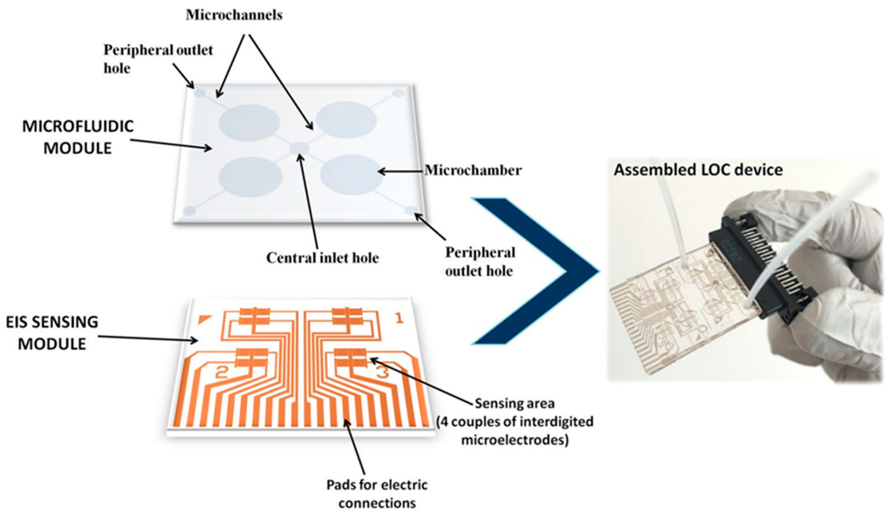
  98. Chiriacò, M.S.; Luvisi, A.; Primiceri, E.; Sabella, E.; de Bellis, L.; Maruccio, G. Development of a Lab-on-a-Chip Method for Rapid Assay of Xylella Fastidiosa Subsp. Pauca Strain CoDiRO. Sci. Rep. 2018, 8, 7376.
  99. Jiang, H.; Xu, Z.; Aluru, M.R.; Dong, L. Plant Chip for High-Throughput Phenotyping of Arabidopsis. Lab Chip 2014, 14, 1281–1293.
  100. Julich, S.; Riedel, M.; Kielpinski, M.; Urban, M.; Kretschmer, R.; Wagner, S.; Fritzsche, W.; Henkel, T.; Möller, R.; Werres, S. Development of a Lab-on-a-Chip Device for Diagnosis of Plant Pathogens. Biosens. Bioelectron. 2011, 26, 4070–4075.
  101. Shang, Y.; Hasan, M.K.; Ahammed, G.J.; Li, M.; Yin, H.; Zhou, J. Applications of Nanotechnology in Plant Growth and Crop Protection: A Review. Molecules 2019, 24, 2558.
  102. Vitosh, M.L.; Silva, G.H. A Rapid Petiole Sap Nitrate-Nitrogen Test for Potatoes. Commun. Soil Sci. Plant Anal. 1994, 25, 183–190.
  103. Errebhi, M.; Rosen, C.J.; Birong, D.E. Calibration of a Petiole Sap Nitrate Test for Irrigated “Russet Burbank” Potato. Commun. Soil Sci. Plant Anal. 1998, 29, 23–35.
  104. Kubota, A.; Thompson, T.L.; Doerge, T.A.; Godin, R.E. A Petiole Sap Nitrate Test for Broccoli. J. Plant Nutr. 1997, 20, 669–682.
  105. Zhang, H.; Zhang, G.; Xu, J.; Wen, Y.; Lu, B.; Zhang, J.; Ding, W. Novel Highly Selective Fluorescent Sensor Based on Electrosynthesized Poly(9-Fluorenecarboxylic Acid) for Efficient and Practical Detection of Iron(III) and Its Agricultural Application. Sens. Actuators B Chem. 2016, 230, 123–129.
  106. Roy, E.; Patra, S.; Madhuri, R.; Sharma, P.K. Simultaneous Determination of Heavy Metals in Biological Samples by a Multiple-Template Imprinting Technique: An Electrochemical Study. RSC Adv. 2014, 4, 56690–56700.
  107. Li, S.; Simonian, A.; Chin, B.A. Sensors for Agriculture and the Food Industry. Electrochem. Soc. Interface 2010, 19, 41–46.
  108. Gieling, T.H.; Van Straten, G.; Janssen, H.J.J.; Wouters, H. ISE and Chemfet Sensors in Greenhouse Cultivation. Sens. Actuators B Chem. 2005, 105, 74–80.
  109. Gutiérrez, M.; Alegret, S.; Cáceres, R.; Casadesús, J.; Marfà, O.; del Valle, M. Application of a Potentiometric Electronic Tongue to Fertigation Strategy in Greenhouse Cultivation. Comput. Electron. Agric. 2007, 57, 12–22.
  110. Sabzevari, S.; Hofman, J. A Worldwide Review of Currently Used Pesticides’ Monitoring in Agricultural Soils. Sci. Total Environ. 2022, 812, 152344.
  111. Yang, T.; Doherty, J.; Guo, H.; Zhao, B.; Clark, J.M.; Xing, B.; Hou, R.; He, L. Real-Time Monitoring of Pesticide Translocation in Tomato Plants by Surface-Enhanced Raman Spectroscopy. Anal. Chem. 2019, 91, 2093–2099.
  112. Li, H.; Merkl, P.; Sommertune, J.; Thersleff, T.; Sotiriou, G.A. SERS Hotspot Engineering by Aerosol Self-Assembly of Plasmonic Ag Nanoaggregates with Tunable Interparticle Distance. Adv. Sci. 2022, 2201133.
  113. Strobel, R.; Pratsinis, S.E. Flame Aerosol Synthesis of Smart Nanostructured Materials. J. Mater. Chem. 2007, 17, 4743.
  114. Martinazzo, J.; Ballen, S.C.; Steffens, J.; Steffens, C. Long Term Stability of Cantilever Gas Nanosensors to Detect Euschistus Heros (F.) Pheromone Release by Rubber Septa. Sens. Actuators B Chem. 2022, 359, 131566.
More
Information
Subjects: Plant Sciences
Contributors MDPI registered users' name will be linked to their SciProfiles pages. To register with us, please refer to https://encyclopedia.pub/register : ,
View Times: 423
Revisions: 2 times (View History)
Update Date: 11 Oct 2022
1000/1000