Your browser does not fully support modern features. Please upgrade for a smoother experience.
Submitted Successfully!
 +1 credit
Thank you for your contribution! You can also upload a video entry or images related to this topic.
For video creation, please contact our Academic Video Service.
| Version | Summary | Created by | Modification | Content Size | Created at | Operation |
|---|---|---|---|---|---|---|
| 1 | Ziyuan Wang | -- | 3178 | 2022-09-26 05:48:37 | | | |
| 2 | Conner Chen | + 7 word(s) | 3185 | 2022-09-27 04:05:40 | | | | |
| 3 | Conner Chen | + 5 word(s) | 3190 | 2022-09-29 05:58:27 | | |
Video Upload Options
We provide professional Academic Video Service to translate complex research into visually appealing presentations. Would you like to try it?
Cite
If you have any further questions, please contact Encyclopedia Editorial Office.
Li, Q.; Yang, H.; Song, S.; Liu, J.; Wang, Z.; Wang, J. Whole Grain Bioactive Compounds and Skeletal Muscle Function. Encyclopedia. Available online: https://encyclopedia.pub/entry/27572 (accessed on 21 May 2026).
Li Q, Yang H, Song S, Liu J, Wang Z, Wang J. Whole Grain Bioactive Compounds and Skeletal Muscle Function. Encyclopedia. Available at: https://encyclopedia.pub/entry/27572. Accessed May 21, 2026.
Li, Qing, Haihong Yang, Shuimiao Song, Jie Liu, Ziyuan Wang, Jing Wang. "Whole Grain Bioactive Compounds and Skeletal Muscle Function" Encyclopedia, https://encyclopedia.pub/entry/27572 (accessed May 21, 2026).
Li, Q., Yang, H., Song, S., Liu, J., Wang, Z., & Wang, J. (2022, September 26). Whole Grain Bioactive Compounds and Skeletal Muscle Function. In Encyclopedia. https://encyclopedia.pub/entry/27572
Li, Qing, et al. "Whole Grain Bioactive Compounds and Skeletal Muscle Function." Encyclopedia. Web. 26 September, 2022.
Copy Citation
Skeletal muscle plays a primary role in metabolic health and physical performance. Conversely, skeletal muscle dysfunctions such as muscular dystrophy, atrophy and aging-related sarcopenia could lead to frailty, decreased independence and increased risk of hospitalization. Dietary intervention has become an effective approach to improving muscle health and function. Evidence shows that whole grains possess multiple health benefits compared with refined grains. Importantly, there is growing evidence demonstrating that bioactive substances derived from whole grains such as polyphenols, γ-oryzanol, β-sitosterol, betaine, octacosanol, alkylresorcinols and β-glucan could contribute to enhancing myogenesis, muscle mass and metabolic function.
whole grains
bioactive components
skeletal muscle function
1. Whole Grain Bioactive Compounds in the Regulation of Skeletal Muscle Function
Whole grain consumption has been demonstrated to have an impressive effect on chronic disease prevention, such as type 2 diabetes, cardiovascular disease, cancer and muscle disease associated with obesity and aging [1][2]. Herein, the following contents introduce the evidence which supports the role of whole grain functional ingredients in regulating muscle health through their ability to promote myogenesis, affect muscle-fiber-type conversion, reduce muscle protein loss and enhance muscle metabolic function (Table 1 and Figure 1).
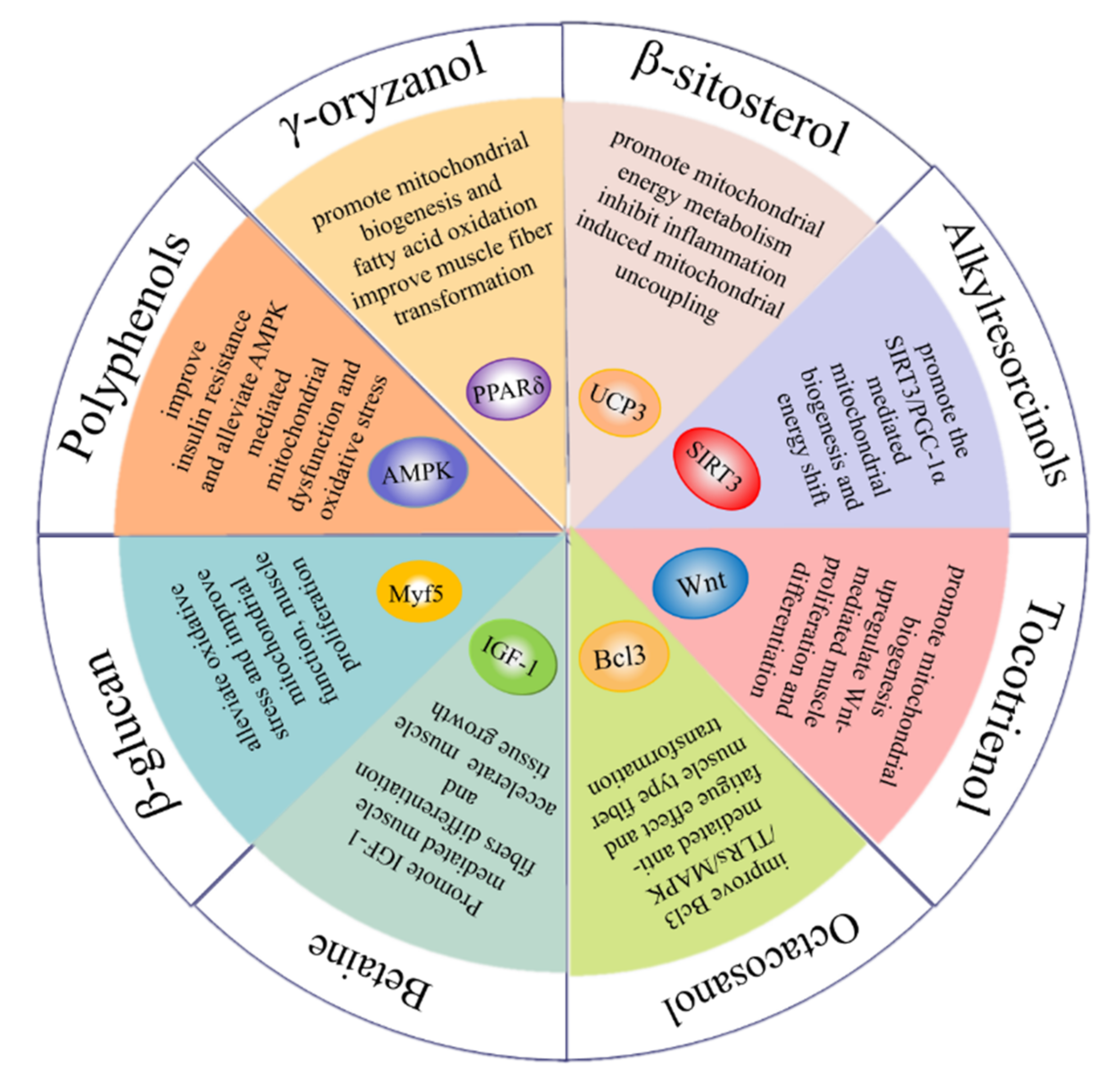
Figure 1. Bioactive components of whole grains for improving skeletal muscle function.
2. Phenolic Compounds
2.1. Phenolic Acids
Ferulic acid is a phenolic acid widespread in whole grains such as oat [3], wheat [4], rice [5], quinoa [6] and corn [7], which is evidenced to possess an impressive effect on modulating muscle function. It has been reported to prevent insulin resistance in skeletal muscle [8]. It can also promote glucose uptake and regulate fatty acid oxidative decomposition in a dose-dependent manner, promoting redox balance in rat muscle [9]. By triggering the PI3K/CpkC pathway, ferulic acid is proved to promote glucose uptake and glycogen synthesis, protecting against insulin resistance in L6 myotubes [10]. After 30 days of dietary ferulic acid supplementation, the number and cross-sectional area (CSA) of muscle fiber are enhanced in a zebrafish model. Ferulic acid also accelerated the muscle tissue growth of zebrafish by upregulating the expression of MyoD and myogenin [11]. In addition, ferulic acid is proven to modulate PGC-1α-activated mitogenesis and energy metabolism via upregulating the expression of NRF1, TFAM and TFB2M [12]. In C2C12 cells, ferulic acid exerts an anti-fatigue function via increasing succinate dehydrogenase (SDH) activity and decreasing lactate dehydrogenase (LDH) activity. Moreover, ferulic acid could stimulate the transformation from fast-twitch muscle fibers to slow-twitch muscle fibers via the SIRT1/AMPK signaling pathway [13]. This effect is further verified in a weaned piglets model, demonstrating that SIRT1/AMPK/PGC-1α signaling was involved in ferulic-acid-mediated muscle transformation and anti-fatigue function [14]. This evidence demonstrates the positive effect of ferulic acid on muscle metabolism and function.
p-coumaric acid is widely distributed in whole grains including rice [15], quinoa [6], barley [16] and wheat [4]. It has been found that p-coumaric acids possess the ability to regulate myogenic differentiation and lipid metabolism of skeletal muscle. For instance, p-coumaric acid could increase the expression of myogenic regulatory factors myogenin and MyoD via activating AMPK signaling, improving myogenic differentiation of C2C12 cells [17]. In the L6 cell model, p-coumaric acid could increase fatty acid β-oxidation through stimulating ACC phosphorylation and the expression of PPARγ, preventing skeletal muscle against oxidative stress and inflammation caused by fatty acid accumulation [18].
2.2. Resveratrol
Resveratrol is a natural source of antioxidant and mitochondrial nutrients which can be found in whole grains, such as buckwheat [19]. Resveratrol shows a positive effect on mitochondrial-dysfunction-induced muscle atrophy. For instance, in a dexamethasone-induced mouse model, resveratrol alleviates dexamethasone-induced mitochondrial dysfunction and muscle atrophy by blocking AMPK/FOXO3 signaling [20]. In high-fat-diet-induced aged mice, muscle loss and myofiber size decrease are reversed by resveratrol supplementation. Resveratrol could improve the mitochondrial dysfunction and oxidative-stress-induced muscle atrophy via activation of the PKA/LKB1/AMPK pathway [21]. Moreover, resveratrol has been reported to enhance muscle proliferation and differentiation in obese mouse muscle. The level of muscle regeneration proteins including MyoG, Myf5 and Pax7 is upregulated by resveratrol administration [22]. In response to skeletal-muscle glucose metabolism disorders caused by obesity, resveratrol can reduce insulin resistance by increasing muscle glycogen synthesis and reducing ROS levels in high-fat-diet-fed mice [23]. In the C2C12 model, resveratrol promotes AKT activation and glucose absorption, as well as decreased glutathione (GSH) level. These results suggest that resveratrol could alleviate muscle insulin resistance by modulating redox levels [24]. Resveratrol is evidenced to alleviate TNF-α-induced muscle hypertrophy and muscle atrophy in C2C12 cells via inhibiting the atrophy-related ubiquitin ligase through upregulating AKT/mTOR/FOXO1 signaling [25]. Furthermore, resveratrol displays impressive activity in promoting the transformation of muscle fiber types. The expression of myosin heavy chain (MyHC) 1, MyHC2a and MyHC2x in mouse muscle is increased with resveratrol supplementation, indicating the transformation from fast- to slow-twitch muscle fibers. This conversion is achieved by activating the AdiopR1-AMPK-PGC-1α signaling pathway [26]. In C2C12 myotubes, resveratrol supplementation increased the activities of lactate dehydrogenase (LDH) and malate dehydrogenase (MDH) while it reversed the elevated level of miR-22-3p, suggesting its anti-fatigue effect via stimulating the transformation of muscle fiber from fast-twitch to slow-twitch type [27]. In addition, resveratrol exerts an anti-fatigue effect in contusion-induced-injury mice, shown as the increased activity of lactate dehydrogenase (LDH) and creatine kinase (CK), displaying an important role in muscle metabolic regulation [28].
2.3. Flavonoids
As a special subclass of flavonoids, quercetin is a natural bioactive compound abundantly present in common cereal grains, especially in buckwheat [29], followed by rice [15], quinoa [6], corn [7] and oat [3]. Quercetin has displayed notable antioxidative and anti-inflammatory effects and shows potential benefits for muscle function [30]. By increasing the expression of adiponectin, quercetin stimulates muscle-fiber-type transformation from fast-twitch to slow-twitch myofibers [31]. It has been also demonstrated that quercetin increases slow-twitch myofibers by regulating AMPK/SIRT1 signaling [32]. Heme oxygenase-1 (HO-1) serves as an essential factor to decrease the inflammatory response and oxidative stress. Mediated by an HO-1/NRF2-dependent mechanism, quercetin is found to alleviate obesity-induced muscle atrophy [33]. In relation to the mitochondrial network, quercetin has been shown to significantly improve mitogenesis in denervated mice, through which muscle atrophy is improved [34]. Moreover, it is shown that quercetin ameliorates TNF-α-induced skeletal muscle insulin resistance in C2C12 cells [35]. The anti-fatigue function of quercetin might be related to its ability to scavenge free radicals [36]. Overloaded exercise causes excessive ROS generation, which further leads to mitochondrial dysfunction and muscle protein loss [37]. In a BALB/c mouse model, quercetin displayed obvious fatigue resistance activity, shown as enhanced muscle glycogen content, elevated mitochondrial fatty acid β-oxidation and decreased oxidative stress [38]. These findings demonstrate the impressive effect of quercetin on improving muscle mitochondrial function and alleviating the inflammatory reaction in the muscle.
Oligomeric procyanidins (OPCs) are mainly found in dark-colored grains, such as black rice and red rice [39]. As reported, OPCs could effectively inhibit glucose metabolism disorder in muscle tissue caused by diabetes. OPCs are shown to improve lipid metabolism by inhibiting mTOR signaling and enhancing glucose homeostasis and insulin sensitivity in the skeletal muscle of diabetic mice [40]. In obese mice, OPC supplementation increases glycogen synthesis and glucose uptake, exerting an antidiabetic effect in an insulin-independent manner [41]. Moreover, oral OPC administration alleviates acute hyperglycemia and improves insulin sensitivity via promoting AMPK-signaling-mediated GLUT4 translocation in ICR mice [42].
Cyanidin-3-glucoside (Cy3G) widely exists in pigmented whole grains, such as black rice [15], purple corn [7], oat, wheat and rye [43]. In vivo and in vitro studies have shown that Cy3G possesses anti-obesity and antidiabetic effects [44][45]. It has been demonstrated that as effective radical scavengers, Cy3G exerts an anti-diabetes effect via reducing oxidative stress in human skeletal muscle cells [46]. In an ICR mouse model, oral supplementation with Cy3G shows improved exercise endurance, performed as weight-loaded swimming time. Moreover, the increased level of lactic acid accumulated after excessive exercise is reversed by Cy3G via activating PGC-1α signaling, indicating that Cy3G exerts its anti-fatigue effect through modulating the PGC-1α-mediated lactic acid metabolism [47]. Consistent with the in vivo data, Cy3G could also upregulate PGC-1α expression in C2C12 myotubes, improving muscle metabolic function [47].
Catechins are flavanols widely found in whole grains such as wheat [48], barley and buckwheat [49]. The isomers of catechins including catechin, epicatechin (EC) and epigallocatechin gallate (EGCG) are proved to increase muscle mass and muscle strength, enhancing the exercise endurance of rats [50]. Catechins could upregulate the expression of Myf5 in muscle stem cells via activating Akt phosphorylation and thus improve muscle regeneration [51]. In the C2C12 cell model, catechin supplementation was found to promote myotube differentiation, shown as an increased level of MyoD, MyoG and MyHC, suggesting its improvement effect on skeletal muscle regeneration and repair [52]. Moreover, catechins have shown impressive modulation of the promotion of mitochondrial function in skeletal muscle. In an LCR (low running capacity) rat model, EC treatment improved mitochondrial biogenesis and respiratory capacity via activating p38 MAPK signaling in the skeletal muscle, enhancing muscle resistance to fatigue [53]. Using a diabetic rat model, EGCG supplementation could alleviate diabetes-induced mitochondrial dysfunction in skeletal muscle, characterized by increased expression of PGC-1α, MFN2 and COXⅠ. The improvement effect of catechins is possibly achieved by promoting mitochondrial autophagy via activating the ROS-ERK/JNK-p53 pathway [54]. Moreover, EGCG could target Wnt signaling and increase the level of downstream MyoD, promoting muscle regeneration [55]. In a rat model of sarcopenia, EGCG administration could increase skeletal muscle mass which is presumably attributed to the downregulated muscle protein degradation mediated by the ubiquitin–proteasome system (UPS) and the upregulated muscle protein synthesis mediated by IGF-1 [56]. Moreover, EGCG inhibits the expression of oxidatively modified protein induced by electrical stimulation in rat skeletal muscle, preventing the muscle from oxidative damage [57]. These data suggest that catechin could facilitate skeletal muscle function through promoting myotube differentiation, enhancing mitochondrial function and defending oxidative stress.
Rutin is a flavonol glycoside and is widely distributed in whole grains such as buckwheat [49], wheat [58] and quinoa [59]. The antioxidant and anti-inflammatory properties of rutin are demonstrated from both in vivo and in vitro studies [60]. The decreased mitochondrial number and mitochondrial biogenesis in skeletal muscle are associated with obesity [61]. In an obese rat model, rutin supplementation significantly enhances mitochondrial DNA (mtDNA) content and mitochondrial biogenesis in skeletal muscle by activating AMPK signaling. The expression levels of PGC-1α, NRF1, TFAM and SIRT1 are also increased, indicating that rutin could restore obesity-induced skeletal muscle mitochondrial dysfunction [62]. Using a forced swimming mouse model, rutin administration was proved to upregulate PGC-1α-mediated mitochondrial biogenesis and decrease the level of lactic acid in skeletal muscle, improving the fatigue-resistance capacity of mouse muscle [63]. In addition, rutin was found to inhibit oxidative stress and inflammation-induced muscle injury. Specifically, rutin intervention improves the inflammatory state by inhibiting NF-κB signaling, manifested in the decreased expression of IL-6 and iNOS, preventing muscle injury of C2C12 cells [64].
3. Carotenoids
Carotenoids are lipid-soluble phytochemicals and the inactive precursor of vitamin A [65], which are abundantly distributed in corn, wheat and barley [66]. As the main component of carotenoids, lutein is reported to improve muscle function via alleviating oxidative stress [67]. In a rat model of ischemia-reperfusion (IR) muscle injury, lutein supplementation decreases the production of ROS and the expression of COX-2 by inhibiting NF-κB signaling, protecting against skeletal muscle IR injury [68]. These observations imply the muscle improvement benefit of lutein presumably via its antioxidant and anti-inflammatory effect.
β-carotenes are the most ubiquitous and stable natural pigments in nature [69]. β-carotene supplementation could enhance muscle mass by promoting IGF-1-mediated muscle protein synthesis and reducing ubiquitin–protease-mediated muscle protein degradation in an atrophy mouse model [70]. Atrogin-1 and MuRF1 are identified as muscle-specific ubiquitin ligases that are upregulated in skeletal muscle under atrophy-inducing conditions [71]. β-carotene treatment is reported to increase muscle mass and decrease the level of Atrogin-1 and MuRF1 in denervated mice, exhibiting a promotion effect on oxidative-stress-induced muscle atrophy [72].
4. Tocotrienol
Tocotrienol, the unsaturated form of vitamin E, mainly exists in buckwheat [73], rice, rye and oat [3]. Tocotrienol receives attention due to its unique biological properties such as antioxidant and anti-inflammatory activity [74]. Increasing evidence has shown that tocopherol exhibits a positive effect on maintaining muscle function. It has been proven that dietary supplementation of tocotrienol improves muscle atrophy and insulin resistance in type 2 diabetic mice. This effect is achieved by activating AMPK/SIRT1/PGC-1α signaling and increasing mitochondrial biogenesis in the skeletal muscle, indicating that tocotrienol improves muscle disorder via upregulating mitochondrial function [75]. In addition, muscle differentiation and regeneration are essential for protecting against aging-related loss of muscle mass and strength. Tocotrienol treatment is found to increase the expression of myoblast-differentiation-related proteins such as MyoD, MyoG and MRF4. Moreover, tocotrienol increased the miR-206 level in human skeletal muscle myoblasts (HSMMs), which resulted in an increased expression of IGF1R and decreased expression of Pax7, promoting muscle cell differentiation [76]. Through transcriptomic analysis, tocotrienol was found to promote muscle cell regeneration via Wnt signaling and alleviate aging-related muscle loss through the FOXO pathway [77]. Additionally, Chung et al. reported that tocotrienol administration improved insulin intolerance and increased soleus muscle weight of obese mice, indicating its potential to ameliorate muscle dysfunction [78].
5. β-Glucan
β-glucan is a soluble dietary fiber which exists abundantly in oat [3], rice [79], wheat, barley and rye [73]. β-glucan has been evidenced to have a positive effect on muscle health [80]. For example, dietary supplementation with oat β-glucan is found to decrease the activity of lactate dehydrogenase and creatine kinase in serum, while it increases the glycogen content in the gastrocnemius muscle of rats. These results indicate that oat β-glucan facilitates the recovery of trained rats from fatigue [81]. In addition, β-glucan is found to promote the transformation of fast-twitch muscle fibers to slow-twitch type in the C2C12 model. Moreover, the levels of myoblast proliferation markers such as Myf5 and Mox2 are increased by β-glucan treatment, suggesting the anti-fatigue effect via improving muscle fiber proliferation and transformation [82]. Mitochondria are responsible for providing energy during immune action and the enhancement of mitochondrial function is conducive to preventing oxidative stress. In the Duchenne muscular dystrophy (DMD) zebrafish model, β-glucan intervention improves mitochondrial respiration and zebrafish exercise capacity, suggesting that β-glucan could alleviate the symptoms of DMD through regulating mitochondria function [83].
6. γ-Oryzanol
γ-oryzanol is a mixture of feruloyl esters with triterpenol as the main component and is mainly found in rice, corn and wheat [84][85]. γ-oryzanol supplementation has been evidenced to improve exercise performance such as grip strength and running time in aged mice. Additionally, γ-oryzanol has been found to promote the formation of slow-twitch muscle fiber, which is a fatigue-resistant myofiber type and conductive to the prevention of muscle aging. By binding to PPARδ and ERRγ directly, γ-oryzanol could upregulate mitochondrial biogenesis and alleviate inflammation, reducing the occurrence of muscle weakness of the aged mice [86]. Moreover, γ-oryzanol supplementation is demonstrated to promote GLUT4 translocation, alleviating insulin resistance in obese mice [87]. In addition, the antioxidant function of γ-oryzanol in muscle could be enhanced under an exercise condition [88], suggesting the physical exercise combined with γ-oryzanol supplementation might be a beneficial strategy to prevent muscle dysregulation.
7. β-Sitosterol
As the most abundant phytosterol, β-sitosterol exists widely in plant products such as barley [16], wheat, oat and quinoa [3][89]. It is considered to be a mild free-radical scavenger and has positive effects on maintaining cell membrane stability, as well as inhibiting inflammation and Alzheimer’s disease [90]. In ICR mice, β-sitosterol promotes mitochondrial electron transport and mitochondrial oxidative phosphorylation, enhancing muscle strength. In addition, β-sitosterol promotes the energy metabolism of C2C12 cells by activating UCP3 and inducing mitochondrial uncoupling [91]. Meanwhile, β-sitosterol dramatically enhanced mitochondrial biogenesis in chicken skeletal muscle via activating the PGC-1α/TFAM (transcription factor A) pathway [92]. In an L6 cell model, β-sitosterol could promote glucose uptake and lipid metabolism by activating ACC phosphorylation. A decreased level of triglycerides and cholesterol is achieved with LKB1-mediated AMPK activation [93]. Likewise, β-sitosterol supplementation is proved to facilitate GLUT4 translocation and upregulate insulin sensitivity in HFD-induced diabetic rats, manifested in the increased activity of glycolytic and gluconeogenesis enzymes [94]. Taken together, these data suggest the promoting effect of β-sitosterol on mitochondrial function and glucose metabolism in the skeletal muscle.
8. Alkylresorcinols
Alkylresorcinols (ARs) are phenolic lipids and exist abundantly in the bran part of cereal grains such as wheat, barley and rye [73]. They consists of various homologues distinguished by the length of the alkyl chain (C17:0−C25:0) attached to the phenyl ring [95]. ARs derived from wheat bran extracts have been indicated to prevent muscle atrophy in denervated mice via regulating the disruption of fatty acid metabolism induced by lipid autophagy [96]. In addition, dietary supplementation containing ARs was found to protect against isoproterenol-induced myofibrillar degeneration in rats via inhibiting oxidative damage caused by lipid accumulation [97]. 5-heptadecylresorcinol (AR-C17) as the main homologue of ARs has been proven to have the best anti-inflammatory effect among the identified ARs homologues in a previous study [95]. It can be found that AR-C17 alleviated muscle dysfunction via upregulating SIRT3/PGC-1α-mediated mitochondrial function. The mitochondria content and mitochondrial-biogenesis-related proteins such TFAM and NRF1 were significantly increased, contributing to enhanced exercise performance [98].
9. Betaine
Betaine is a natural antioxidant widely present in common cereals including whole grain wheat, barley, rye and oat [99]. Betaine supplementation is reported to improve muscle strength and power [100]. IGF-1 stimulates myotube growth and differentiation by activating the expression of downstream MyoD and myogenin. Betaine is reported to increase the expression of IGF-1 in murine myoblasts [101]. AKT is considered as a key regulator of muscle signaling and protein synthesis. Additionally, betaine supplementation could increase anabolism and weaken catabolism in muscle tissue by stimulating IGF-1/AKT signaling [102]. In an in vitro C2C12 model, betaine supplementation promoted myotube differentiation by upregulating the secretion of miR-29b-3p. In addition, through the activation of NFATc1/MyoD signaling, betaine promotes the transformation from fast-twitch to slow-twitch myofibers [103]. Moreover, betaine could promote muscle energy metabolism by upregulating mitochondrial biogenesis, accompanied by increased glucose consumption and ATP production in C2C12 cells [104].
10. Octacosanol
Octacosanol is a primary aliphatic alcohol that is abundant in rice [105] and wheat [106]. According to literature data, octacosanol has been identified as an anti-fatigue agent which could be stored in skeletal muscle after serial doses administration [107]. In a trained rats model, dietary supplementation with octacosanol also showed a better physical recovery after exhaustion and there was a shift from fast-twitch to slow-twitch myofibers in skeletal muscle [108]. In addition, orally administrated octacosanol might be converted into fatty acids and related to energy utilization, indicating its potential to improve exercise capacity through upregulating the energy supply [109]. The underlying mechanism of the anti-fatigue effect by octacosanol might be through increasing the expression of Prx and decreasing the expression of Trim63 in trained mice. Based on the gene ontology analysis, this improvement might be mediated by the Bcl3/TLRs/MAPK signaling pathway [110]. Overall, octacosanol resists fatigue possibly via modulating muscle-fiber-type transition and muscular energy metabolism.
Table 1. The structure and content of bioactive components in whole grains.
| Components | Structure | Derived Grains | Content (mg/kg) | References |
|---|---|---|---|---|
| γ-oryzanol | 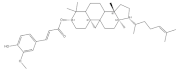 |
Rice | 1550.0–8400.0 | [84] |
| Wheat | 297.0–584.0 | [85] | ||
| Corn | 200.0–250.0 | [85] | ||
| β-sitosterol | 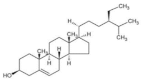 |
Wheat | 365.6–673.1 | [89] |
| Barley | 510.0–676.0 | [16] | ||
| Quinoa | 76.0–556.0 | [3] | ||
| Oat | 210.0–409.2 | [3] | ||
| Alkylresorcinols | 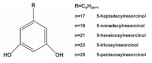 |
|||
| Rye | 360.0–3200.0 | [73] | ||
| Wheat | 317.0–1732.0 | [73] | ||
| Ferulic acid | 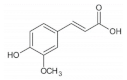 |
Barley | 41.0–210.0 | [73] |
| Corn | 55.2 | [7] | ||
| Wheat | 472.0–813.8 | [4] | ||
| Barley | 41.0–210.0 | [73] | ||
| Quinoa | 126.8–281.7 | [6] | ||
| Brown rice | 7.1–52.7 | [15] | ||
| Rice | 155.6–271.1 | [5] | ||
| Oat | 1493.6 | [3] | ||
| p-coumaric acid | 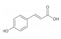 |
Black rice | 84.8 ± 1.4 | [15] |
| Red rice | 176.9 ± 2.5 | [15] | ||
| Brown rice | 76.1–152.0 | [15] | ||
| Wheat | 23.8–35.6 | [4] | ||
| Barley | 0.8–58.4 | [16] | ||
| Quinoa | 31.3–42.0 | [6] | ||
| Resveratrol | 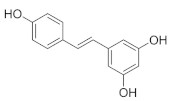 |
Buckwheat | 5.7–7.9 | [19] |
| Quercetin | 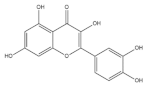 |
Buckwheat | 3700.0 | [29] |
| Rice | 22.0–28.0 | [15] | ||
| Barley | 1.4–8.7 | [16] | ||
| Quinoa | 11.3–42.8 | [6] | ||
| Corn | 15.8 | [7] | ||
| Oat | 89.0 | [3] | ||
| Oligomeric procyanidins | 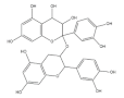 |
Black rice | 3500.0 | [39] |
| Red rice | 200.0 | [39] | ||
| Cyanidin-3-glucoside | 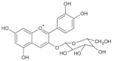 |
Wheat | 1590.0 | [43] |
| Rice | 2682.0–4700.0 | [15] | ||
| Purple corn | 1430.0 | [7] | ||
| Rye | 2270.0 | [43] | ||
| Barley | 1020.0 | [43] | ||
| Oat | 430.0 | [43] | ||
| Catechins | 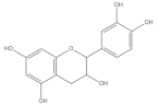 |
Barley | 1312.47 ± 7.11 | [49] |
| Wheat | 355.87 | [48] | ||
| Buckwheat | 46.47 ± 0.17 | [49] | ||
| Rutin | 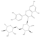 |
Wheat | 236.2 | [58] |
| Quinoa | 609.1 | [59] | ||
| Buckwheat | 69.95 ± 2.25 | [49] | ||
| Carotenoids | 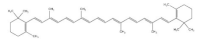 |
Corn | 9.69–13.0 | [66] |
| Wheat | 32.1–39.7 | [66] | ||
| Barley | 0.15–10.5 | [66] | ||
| Betaine | 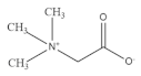 |
Oat | 200.0–1000.0 | [99] |
| Wheat | 270.0–1110.0 | [99] | ||
| Rye | 444.0–2213.0 | [99] | ||
| Barley | 460.0–980.0 | [99] | ||
| Octacosanol | 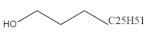 |
Rice | 95.7 | [105] |
| Wheat | 0.4–8.9 | [106] | ||
| β-glucan | 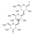 |
Rice | 140.0–570.0 | [79] |
| Barley | 4000.0–7000.0 | [73] | ||
| Oat | 51,800.0–282,000.0 | [3] | ||
| Rye | 1200.0–2900.0 | [73] | ||
| Wheat | 400.0–1400.0 | [73] | ||
| Tocotrienol | 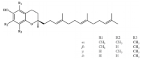 |
Black rice | 31.9 ± 0.4 | [15] |
| Red rice | 36.9 ± 1.6 | [15] | ||
| Brown rice | 19.6 ± 0.4 | [15] | ||
| Wheat | 27.81 | [73] | ||
| Rye | 27.78 | [73] | ||
| Barley | 18.73 | [73] | ||
| Oat | 11.59 | [73] | ||
| Buckwheat | 54.6–552.2 | [3] |
References
- Liu, J.; Yu, L.L.; Wu, Y. Bioactive Components and Health Beneficial Properties of Whole Wheat Foods. J. Agric. Food Chem. 2020, 68, 12904–12915.
- Wang, Z.; Yang, Z.; Liu, J.; Hao, Y.; Sun, B.; Wang, J. Potential Health Benefits of Whole Grains: Modulation of Mitochondrial Biogenesis and Energy Metabolism. J. Agric. Food Chem. 2021, 69, 14065–14074.
- Raguindin, P.F.; Itodo, O.A.; Stoyanov, J.; Dejanovic, G.M.; Gamba, M.; Asllanaj, E.; Minder, B.; Bussler, W.; Metzger, B.; Muka, T.; et al. A systematic review of phytochemicals in oat and buckwheat. Food Chemistry 2021, 338, 127982.
- Saini, P.; Kumar, N.; Kumar, S.; Mwaurah, P.W.; Panghal, A.; Attkan, A.K.; Singh, V.K.; Garg, M.K.; Singh, V. Bioactive compounds, nutritional benefits and food applications of colored wheat: A comprehensive review. Crit. Rev. Food Sci. Nutr. 2021, 61, 3197–3210.
- Yu, L.; Li, G.; Li, M.; Xu, F.; Beta, T.; Bao, J. Genotypic variation in phenolic acids, vitamin E and fatty acids in whole grain rice. Food Chemistry 2016, 197, 776–782.
- Han, Y.; Chi, J.; Zhang, M.; Zhang, R.; Fan, S.; Huang, F.; Xue, K.; Liu, L. Characterization of saponins and phenolic compounds: Antioxidant activity and inhibitory effects on alpha-glucosidase in different varieties of colored quinoa (Chenopodium quinoa Willd). Biosci. Biotechnol. Biochem. 2019, 83, 2128–2139.
- Cristianini, M.; Sanchez, J.S. Extraction of bioactive compounds from purple corn using emerging technologies: A review. J. Food Sci. 2020, 85, 862–869.
- Gogoi, B.; Chatterjee, P.; Mukherjee, S.; Buragohain, A.K.; Bhattacharya, S.; Dasgupta, S. A polyphenol rescues lipid induced insulin resistance in skeletal muscle cells and adipocytes. Biochem. Biophys. Res. Commun. 2014, 452, 382–388.
- Salau, V.F.; Erukainure, O.L.; Koorbanally, N.A.; Islam, M.S. Ferulic acid promotes muscle glucose uptake and modulate dysregulated redox balance and metabolic pathways in ferric-induced pancreatic oxidative injury. J. Food Biochem. 2022, 46, e13641.
- Kang, B.-B.; Chiang, B.-H. Amelioration of insulin resistance using the additive effect of ferulic acid and resveratrol on vesicle trafficking for skeletal muscle glucose metabolism. Phytother. Res. 2020, 34, 808–816.
- Wen, Y.; Ushio, H. Ferulic Acid Promotes Hypertrophic Growth of Fast Skeletal Muscle in Zebrafish Model. Nutrients 2017, 9, 66.
- Anis, E.; Zafeer, M.F.; Firdaus, F.; Islam, S.N.; Khan, A.A.; Ali, A.; Hossain, M.M. Ferulic acid reinstates mitochondrial dynamics through PGC1 alpha expression modulation in 6-hydroxydopamine lesioned rats. Phytother. Res. 2020, 34, 214–226.
- Chen, X.; Guo, Y.; Jia, G.; Zhao, H.; Liu, G.; Huang, Z. Ferulic acid regulates muscle fiber type formation through the Sirt1/AMPK signaling pathway. Food Funct. 2019, 10, 259–265.
- Wang, Y.; Chen, X.; Huang, Z.; Chen, D.; Yu, B.; Chen, H.; Yu, J.; Luo, Y.; Zheng, P.; He, J. Effects of dietary ferulic acid supplementation on growth performance and skeletal muscle fiber type conversion in weaned piglets. J. Sci. Food Agric. 2021, 101, 5116–5123.
- Gong, E.S.; Liu, C.; Li, B.; Zhou, W.; Chen, H.; Li, T.; Wu, J.; Zeng, Z.; Wang, Y.; Si, X.; et al. Phytochemical profiles of rice and their cellular antioxidant activity against ABAP induced oxidative stress in human hepatocellular carcinoma HepG2 cells. Food Chem. 2020, 318, 126484.
- Li, Y.; Li, T.; Liu, R.H. Bioactive compounds of highland barley and their health benefits. J. Cereal Sci. 2022, 103, 103366.
- Ilavenil, S.; Kim, D.H.; Srigopalram, S.; Arasu, M.V.; Lee, K.D.; Lee, J.C.; Lee, J.S.; Renganathan, S.; Choi, K.C. Potential Application of p-Coumaric Acid on Differentiation of C2C12 Skeletal Muscle and 3T3-L1 Preadipocytes-An In Vitro and In Silico Approach. Molecules 2016, 21, 997.
- Yoon, S.-A.; Kang, S.-I.; Shin, H.-S.; Kang, S.-W.; Kim, J.-H.; Ko, H.-C.; Kim, S.-J. p-Coumaric acid modulates glucose and lipid metabolism via AMP-activated protein kinase in L6 skeletal muscle cells. Biochem. Biophys. Res. Commun. 2013, 432, 553–557.
- Nemcova, L.; Zima, J.; Barek, J.; Janovska, D. Determination of resveratrol in grains, hulls and leaves of common and tartary buckwheat by HPLC with electrochemical detection at carbon paste electrode. Food Chem. 2011, 126, 374–378.
- Liu, J.; Peng, Y.; Wang, X.; Fan, Y.; Qin, C.; Shi, L.; Tang, Y.; Cao, K.; Li, H.; Long, J.; et al. Mitochondrial Dysfunction Launches Dexamethasone-Induced Skeletal Muscle Atrophy via AMPK/FOXO3 Signaling. Mol. Pharm. 2016, 13, 73–84.
- Huang, Y.; Zhu, X.; Chen, K.; Lang, H.; Zhang, Y.; Hou, P.; Ran, L.; Zhou, M.; Zheng, J.; Yi, L.; et al. Resveratrol prevents sarcopenic obesity by reversing mitochondrial dysfunction and oxidative stress via the PKA/LKB1/AMPK pathway. Aging-Us 2019, 11, 2217–2240.
- Niu, W.; Wang, H.; Wang, B.; Mao, X.; Du, M. Resveratrol improves muscle regeneration in obese mice through enhancing mitochondrial biogenesis. J. Nutr. Biochem. 2021, 98, 108804.
- Gong, L.; Guo, S.; Zou, Z. Resveratrol ameliorates metabolic disorders and insulin resistance in high-fat diet-fed mice. Life Sci. 2020, 242, 117212.
- Quan, Y.; Hua, S.; Li, W.; Zhan, M.; Li, Y.; Lu, L. Resveratrol bidirectionally regulates insulin effects in skeletal muscle through alternation of intracellular redox homeostasis. Life Sci. 2020, 242, 117188.
- Wang, D.-T.; Yin, Y.; Yang, Y.-J.; Lv, P.-J.; Shi, Y.; Lu, L.; Wei, L.-B. Resveratrol prevents TNF-alpha-induced muscle atrophy via regulation of Akt/mTOR/FoxO1 signaling in C2C12 myotubes. Int. Immunopharmacol. 2014, 19, 206–213.
- Jiang, Q.; Cheng, X.; Cui, Y.; Xia, Q.; Yan, X.; Zhang, M.; Lan, G.; Liu, J.; Shan, T.; Huang, Y. Resveratrol regulates skeletal muscle fibers switching through the AdipoR1-AMPK-PGC-1 alpha pathway. Food Funct. 2019, 10, 3334–3343.
- Wen, W.; Chen, X.; Huang, Z.; Chen, D.; Chen, H.; Luo, Y.; He, J.; Zheng, P.; Yu, J.; Yu, B. Resveratrol regulates muscle fiber type conversion via miR-22-3p and AMPK/SIRT1/PGC-1 alpha pathway. J. Nutr. Biochem. 2020, 77, 108297.
- Hsu, Y.-J.; Ho, C.-S.; Lee, M.-C.; Ho, C.-S.; Huang, C.-C.; Kan, N.-W. Protective Effects of Resveratrol Supplementation on Contusion Induced Muscle Injury. Int. J. Med. Sci. 2020, 17, 53–62.
- Gabr, A.M.M.; Fayek, N.M.; Mahmoud, H.M.; El-Bahr, M.K.; Ebrahim, H.S.; Sytar, O.; El-Halawany, A.M. Effect of Light Quality and Media Components on Shoot Growth, Rutin, and Quercetin Production from Common Buckwheat. ACS Omega 2022, 7, 26566–26572.
- Paulina Luna-Castillo, K.; Lin, S.; Francisco Munoz-Valle, J.; Vizmanos, B.; Lopez-Quintero, A.; Marquez-Sandoval, F. Functional Food and Bioactive Compounds on the Modulation of the Functionality of HDL-C: A Narrative Review. Nutrients 2021, 13, 1165.
- Chen, X.; Liang, D.; Huang, Z.; Jia, G.; Zhao, H.; Liu, G. Quercetin regulates skeletal muscle fiber type switching via adiponectin signaling. Food Funct. 2021, 12, 2693–2702.
- Iwabu, M.; Yamauchi, T.; Okada-Iwabu, M.; Sato, K.; Nakagawa, T.; Funata, M.; Yamaguchi, M.; Namiki, S.; Nakayama, R.; Tabata, M.; et al. Adiponectin and AdipoR1 regulate PGC-1 alpha and mitochondria by Ca2+ and AMPK/SIRT1. Nature 2010, 464, 1313–1319.
- Kim, Y.; Kim, C.-S.; Joe, Y.; Chung, H.T.; Ha, T.Y.; Yu, R. Quercetin Reduces Tumor Necrosis Factor Alpha-Induced Muscle Atrophy by Upregulation of Heme Oxygenase-1. J. Med. Food 2018, 21, 551–559.
- Mukai, R.; Matsui, N.; Fujikura, Y.; Matsumoto, N.; Hou, D.-X.; Kanzaki, N.; Shibata, H.; Horikawa, M.; Iwasa, K.; Hirasaka, K.; et al. Preventive effect of dietary quercetin on disuse muscle atrophy by targeting mitochondria in denervated mice. J. Nutr. Biochem. 2016, 31, 67–76.
- Dai, X.; Ding, Y.; Zhang, Z.; Cai, X.; Bao, L.; Li, Y. Quercetin But Not Quercitrin Ameliorates Tumor Necrosis Factor-Alpha-Induced Insulin Resistance in C2C12 Skeletal Muscle Cells. Biol. Pharm. Bull. 2013, 36, 788–795.
- Chen, C.; Yang, J.-S.; Lu, C.-C.; Chiu, Y.-J.; Chen, H.-C.; Chung, M.-I.; Wu, Y.-T.; Chen, F.-A. Effect of Quercetin on Dexamethasone-Induced C2C12 Skeletal Muscle Cell Injury. Molecules 2020, 25, 3267.
- Liang, Q.; Chen, Y.; Li, C.; Lu, L. Quercetin attenuates Ox-LDL-induced calcification in vascular smooth muscle cells by regulating ROS-TLR4 signaling pathway. Nan Fang Yi Ke Da Xue Xue Bao J. South. Med. Univ. 2018, 38, 980–985.
- Chen, X.; Liang, D.; Huang, Z.; Jia, G.; Zhao, H.; Liu, G. Anti-fatigue effect of quercetin on enhancing muscle function and antioxidant capacity. J. Food Biochem. 2021, 45, e13968.
- Finocchiaro, F.; Ferrari, B.; Gianinetti, A.; Dall’Asta, C.; Galaverna, G.; Scazzina, F.; Pellegrini, N. Characterization of antioxidant compounds of red and white rice and changes in total antioxidant capacity during processing. Mol. Nutr. Food Res. 2007, 51, 1006–1019.
- Li, X.; Sui, Y.; Wu, Q.; Xie, B.; Sun, Z. Attenuated mTOR Signaling and Enhanced Glucose Homeostasis by Dietary Supplementation with Lotus Seedpod Oligomeric Procyanidins in Streptozotocin (STZ)-Induced Diabetic Mice. J. Agric. Food Chem. 2017, 65, 3801–3810.
- Bowser, S.M.; Moore, W.T.; McMillan, R.P.; Dorenkott, M.R.; Goodrich, K.M.; Ye, L.; O’Keefe, S.F.; Hulver, M.W.; Neilson, A.P. High-molecular-weight cocoa procyanidins possess enhanced insulin-enhancing and insulin mimetic activities in human primary skeletal muscle cells compared to smaller procyanidins. J. Nutr. Biochem. 2017, 39, 48–58.
- Yamashita, Y.; Wang, L.; Nanba, F.; Ito, C.; Toda, T.; Ashida, H. Procyanidin Promotes Translocation of Glucose Transporter 4 in Muscle of Mice through Activation of Insulin and AMPK Signaling Pathways. PLoS ONE 2016, 11, e0161704.
- Corol, D.-I.; Ravel, C.; Raksegi, M.; Bedo, Z.; Charmet, G.; Beale, M.H.; Shewry, P.R.; Ward, J.L. Effects of Genotype and Environment on the Contents of Betaine, Choline, and Trigonelline in Cereal Grains. J. Agric. Food Chem. 2012, 60, 5471–5481.
- Guo, H.; Guo, J.; Jiang, X.; Li, Z.; Ling, W. Cyanidin-3-O-beta-glucoside, a typical anthocyanin, exhibits antilipolytic effects in 3T3-L1 adipocytes during hyperglycemia: Involvement of FoxO1-mediated transcription of adipose triglyceride lipase. Food Chem. Toxicol. 2012, 50, 3040–3047.
- Matsukawa, T.; Inaguma, T.; Han, J.; Villareal, M.O.; Isoda, H. Cyanidin-3-glucoside derived from black soybeans ameliorate type 2 diabetes through the induction of differentiation of preadipocytes into smaller and insulin-sensitive adipocytes. J. Nutr. Biochem. 2015, 26, 860–867.
- Giang Thanh Thi, H.; Kase, E.T.; Wangensteen, H.; Barsett, H. Phenolic Elderberry Extracts, Anthocyanins, Procyanidins, and Metabolites Influence Glucose and Fatty Acid Uptake in Human Skeletal Muscle Cells. J. Agric. Food Chem. 2017, 65, 2677–2685.
- Matsukawa, T.; Motojima, H.; Sato, Y.; Takahashi, S.; Villareal, M.O.; Isoda, H. Upregulation of skeletal muscle PGC-1 alpha through the elevation of cyclic AMP levels by Cyanidin-3-glucoside enhances exercise performance. Sci. Rep. 2017, 7, 44799.
- Dinelli, G.; Segura Carretero, A.; Di Silvestro, R.; Marotti, I.; Fu, S.; Benedettelli, S.; Ghiselli, L.; Fernandez Gutierrez, A. Determination of phenolic compounds in modern and old varieties of durum wheat using liquid chromatography coupled with time-of-flight mass spectrometry. J. Chromatogr. A 2009, 1216, 7229–7240.
- Hes, M.; Dziedzic, K.; Gorecka, D.; Drozdzynska, A.; Gujska, E. Effect of Boiling in Water of Barley and Buckwheat Groats on the Antioxidant Properties and Dietary Fiber Composition. Plant Foods Hum. Nutr. 2014, 69, 276–282.
- Li, P.; Liu, A.; Xiong, W.; Lin, H.; Xiao, W.; Huang, J.; Zhang, S.; Liu, Z. Catechins enhance skeletal muscle performance. Crit. Rev. Food Sci. Nutr. 2020, 60, 515–528.
- Kim, A.R.; Kim, K.M.; Byun, M.R.; Hwang, J.-H.; Park, J.I.; Oh, H.T.; Kim, H.K.; Jeong, M.G.; Hwang, E.S.; Hong, J.-H. Catechins activate muscle stem cells by Myf5 induction and stimulate muscle regeneration. Biochem. Biophys. Res. Commun. 2017, 489, 142–148.
- Li, P.; Liu, A.; Liu, C.; Qu, Z.; Xiao, W.; Huang, J.; Liu, Z.; Zhang, S. Role and mechanism of catechin in skeletal muscle cell differentiation. J. Nutr. Biochem. 2019, 74, 108225–108260.
- Huettemann, M.; Lee, I.; Perkins, G.A.; Britton, S.L.; Koch, L.G.; Malek, M.H. (-)-Epicatechin is associated with increased angiogenic and mitochondrial signalling in the hindlimb of rats selectively bred for innate low running capacity. Clin. Sci. 2013, 124, 663–674.
- Yan, J.; Feng, Z.; Liu, J.; Shen, W.; Wang, Y.; Wertz, K.; Weber, P.; Long, J.; Liu, J. Enhanced autophagy plays a cardinal role in mitochondrial dysfunction in type 2 diabetic Goto-Kakizaki (GK) rats: Ameliorating effects of (-)-epigallocatechin-3-gallate. J. Nutr. Biochem. 2012, 23, 716–724.
- Kim, A.R.; Kim, K.M.; Byun, M.R.; Hwang, J.-H.; Park, J.I.; Oh, H.T.; Jeong, M.G.; Hwang, E.S.; Hong, J.-H. (-)-Epigallocatechin-3-gallate stimulates myogenic differentiation through TAZ activation. Biochem. Biophys. Res. Commun. 2017, 486, 378–384.
- Meador, B.M.; Mirza, K.A.; Tian, M.; Skelding, M.B.; Reaves, L.A.; Edens, N.K.; Tisdale, M.J.; Pereira, S.L. The Green Tea Polyphenol Epigallocatechin-3-Gallate (EGCg) Attenuates Skeletal Muscle Atrophy in a Rat Model of Sarcopenia. J. Frailty Aging 2015, 4, 209–215.
- Nagasawa, T.; Hayashi, H.; Fujimaki, N.; Nishizawa, N.; Kitts, D.D. Induction of oxidatively modified proteins in skeletal muscle by electrical stimulation and its suppression by dietary supplementation of (-)-epigallocatechin gallate. Biosci. Biotechnol. Biochem. 2000, 64, 1004–1010.
- Li, Y.; Ma, D.; Sun, D.; Wang, C.; Zhang, J.; Xie, Y.; Guo, T. Total phenolic, flavonoid content, and antioxidant activity of flour, noodles, and steamed bread made from different colored wheat grains by three milling methods. Crop J. 2015, 3, 328–334.
- Maria Gomez-Caravaca, A.; Segura-Carretero, A.; Fernandez-Gutierrez, A.; Caboni, M.F. Simultaneous Determination of Phenolic Compounds and Saponins in Quinoa (Chenopodium quinoa Willd) by a Liquid Chromatography-Diode Array Detection-Electrospray Ionization-Time-of-Flight Mass Spectrometry Methodology. J. Agric. Food Chem. 2011, 59, 10815–10825.
- Sheu, J.R.; Hsiao, G.; Chou, P.H.; Shen, M.Y.; Chou, D.S. Mechanisms involved in the antiplatelet activity of rutin, a glycoside of the flavonol quercetin, in human platelets. J. Agric. Food Chem. 2004, 52, 4414–4418.
- Kim, J.Y.; Hickner, R.C.; Cortright, R.L.; Dohm, G.L.; Houmard, J.A. Lipid oxidation is reduced in obese human skeletal muscle. Am. J. Physiol.-Endocrinol. Metab. 2000, 279, E1039–E1044.
- Seo, S.; Lee, M.-S.; Chang, E.; Shin, Y.; Oh, S.; Kim, I.-H.; Kim, Y. Rutin Increases Muscle Mitochondrial Biogenesis with AMPK Activation in High-Fat Diet-Induced Obese Rats. Nutrients 2015, 7, 8152–8169.
- Su, K.-Y.; Yu, C.Y.; Chen, Y.-W.; Huang, Y.-T.; Chen, C.-T.; Wu, H.-F.; Chen, Y.-L.S. Rutin, a Flavonoid and Principal Component of Saussurea Involucrata, Attenuates Physical Fatigue in a Forced Swimming Mouse Model. Int. J. Med. Sci. 2014, 11, 528–537.
- Liu, S.; Adewole, D.; Yu, L.; Sid, V.; Wang, B.; Karmin, O.; Yang, C. Rutin attenuates inflammatory responses induced by lipopolysaccharide in an in vitro mouse muscle cell (C2C12) model. Poult. Sci. 2019, 98, 2756–2764.
- Weber, D.; Grune, T. The contribution of beta-carotene to vitamin A supply of humans. Mol. Nutr. Food Res. 2012, 56, 251–258.
- Banwo, K.; Olojede, A.O.; Adesulu-Dahunsi, A.T.; Verma, D.K.; Thakur, M.; Tripathy, S.; Singh, S.; Patel, A.R.; Gupta, A.K.; Aguilar, C.N.; et al. Functional importance of bioactive compounds of foods with Potential Health Benefits: A review on recent trends. Food Biosci. 2021, 43, 101320.
- Loskutov, I.G.; Khlestkina, E.K. Wheat, Barley, and Oat Breeding for Health Benefit Components in Grain. Plants 2021, 10, 86.
- Cheng, F.; Zhang, Q.; Yan, F.-F.; Wan, J.-F.; Lin, C.-S. Lutein protects against ischemia/reperfusion injury in rat skeletal muscle by modulating oxidative stress and inflammation. Immunopharmacol. Immunotoxicol. 2015, 37, 329–334.
- Nagao, A. Metabolism of Carotenoids in Mammals. In Carotenoids: Biosynthetic and Biofunctional Approaches; Misawa, N., Ed.; Advances in Experimental Medicine and Biology; Springer: Singapore, 2021; Volume 1261, pp. 67–78.
- Kitakaze, T.; Harada, N.; Imagita, H.; Yamaji, R. beta-Carotene Increases Muscle Mass and Hypertrophy in the Soleus Muscle in Mice. J. Nutr. Sci. Vitaminol. 2015, 61, 481–487.
- Foletta, V.C.; White, L.J.; Larsen, A.E.; Leger, B.; Russell, A.P. The role and regulation of MAFbx/atrogin-1 and MuRF1 in skeletal muscle atrophy. Pflug. Arch. -Eur. J. Physiol. 2011, 461, 325–335.
- Ogawa, M.; Kariya, Y.; Kitakaze, T.; Yamaji, R.; Harada, N.; Sakamoto, T.; Hosotani, K.; Nakano, Y.; Inui, H. The preventive effect of beta-carotene on denervation-induced soleus muscle atrophy in mice. Br. J. Nutr. 2013, 109, 1349–1358.
- Siurek, B.; Rosicka-Kaczmarek, J.; Nebesny, E. Bioactive compounds in cereal grains—Occurrence, structure, technological significance and nutritional benefits-a review. Food Sci. Technol. Int. 2012, 18, 559–568.
- Borneo, R.; Leon, A.E. Whole grain cereals: Functional components and health benefits. Food Funct. 2012, 3, 110–119.
- Lee, H.; Lim, Y. Tocotrienol-rich fraction supplementation reduces hyperglycemia-induced skeletal muscle damage through regulation of insulin signaling and oxidative stress in type 2 diabetic mice. J. Nutr. Biochem. 2018, 57, 77–85.
- Razak, A.M.; Khor, S.C.; Jaafar, F.; Karim, N.A.; Makpol, S. Targeting myomiRs by tocotrienol-rich fraction to promote myoblast differentiation. Genes Nutr. 2018, 13, 31.
- Lim, J.J.; Zurinah, W.N.W.; Moul, V.; Norwahidah, A.K. Tocotrienol-Rich Fraction (TRF) Treatment Promotes Proliferation Capacity of Stress-Induced Premature Senescence Myoblasts and Modulates the Renewal of Satellite Cells: Microarray Analysis. Oxidative Med. Cell. Longev. 2019, 2019, 9141343.
- Chung, E.; Campise, S.N.; Joiner, H.E.; Tomison, M.D.; Kaur, G.; Dufour, J.M.; Cole, L.; Ramalingam, L.; Moustaid-Moussa, N.; Shen, C.-L. Effect of annatto-extracted tocotrienols and green tea polyphenols on glucose homeostasis and skeletal muscle metabolism in obese male mice. J. Nutr. Biochem. 2019, 67, 36–43.
- Jung, T.-D.; Shin, G.-H.; Kim, J.-M.; Choi, S.-I.; Lee, J.-H.; Lee, S.J.; Park, S.J.; Woo, K.S.; Oh, S.K.; Lee, O.-H. Comparative Analysis of gamma-Oryzanol, beta-Glucan, Total Phenolic Content and Antioxidant Activity in Fermented Rice Bran of Different Varieties. Nutrients 2017, 9, 571.
- Prasadi, V.P.N.; Joye, I.J. Dietary Fibre from Whole Grains and Their Benefits on Metabolic Health. Nutrients 2020, 12, 3045.
- Xu, C.; Lv, J.; Lo, Y.M.; Cui, S.W.; Hu, X.; Fan, M. Effects of oat beta-glucan on endurance exercise and its anti-fatigue properties in trained rats. Carbohydr. Polym. 2013, 92, 1159–1165.
- Li, Y.; Fan, Y.; Pan, H.; Qian, H.; Qi, X.; Wu, G.; Zhang, H.; Xu, M.; Rao, Z.; Wang, L.; et al. Effects of functional beta-glucan on proliferation, differentiation, metabolism and its anti-fibrosis properties in muscle cells. Int. J. Biol. Macromol. 2018, 117, 287–293.
- Brogi, L.; Marchese, M.; Cellerino, A.; Licitra, R.; Naef, V.; Mero, S.; Bibbiani, C.; Fronte, B. beta-Glucans as Dietary Supplement to Improve Locomotion and Mitochondrial Respiration in a Model of Duchenne Muscular Dystrophy. Nutrients 2021, 13, 1619.
- Ratseewo, J.; Meeso, N.; Siriamornpun, S. Changes in amino acids and bioactive compounds of pigmented rice as affected by far-infrared radiation and hot air drying. Food Chem. 2020, 306, 125644.
- Zhu, D.; Sanchez-Ferrer, A.; Nystroem, L. Antioxidant Activity of Individual Steryl Ferulates from Various Cereal Grain Sources. J. Nat. Prod. 2016, 79, 308–316.
- Ahn, J.; Son, H.J.; Seo, H.D.; Ha, T.Y.; Ahn, J.; Lee, H.; Shin, S.H.; Jung, C.H.; Jang, Y.J. gamma-Oryzanol Improves Exercise Endurance and Muscle Strength by Upregulating PPAR delta and ERR gamma Activity in Aged Mice. Mol. Nutr. Food Res. 2021, 65, 2000652.
- Mattei, L.; Francisqueti-Ferron, F.V.; Garcia, J.L.; Ferron, A.J.T.; Silva, C.C.V.d.A.; Gregolin, C.S.; Nakandakare-Maia, E.T.; Silva, J.d.C.P.; Moreto, F.; Minatel, I.O.; et al. Antioxidant and anti-inflammatory properties of gamma- oryzanol attenuates insulin resistance by increasing GLUT-4 expression in skeletal muscle of obese animals. Mol. Cell. Endocrinol. 2021, 537, 111423.
- Dahleh, M.M.M.; Araujo, S.M.; Bortolotto, V.C.; Pinheiro, F.C.; Poetini, M.R.; Musachio, E.A.S.; Meichtry, L.B.; Couto, S.d.F.; Prigol, M. Exercise associated with gamma-oryzanol supplementation suppresses oxidative stress and prevents changes in locomotion in Drosophila melanogaster. Free. Radic. Res. 2021, 55, 198–209.
- Chen, C.Y.O.; Kamil, A.; Blumberg, J.B. Phytochemical composition and antioxidant capacity of whole wheat products. Int. J. Food Sci. Nutr. 2015, 66, 63–70.
- Ayaz, M.; Junaid, M.; Ullah, F.; Subhan, F.; Sadiq, A.; Ali, G.; Ovais, M.; Shahid, M.; Ahmad, A.; Wadood, A.; et al. Anti-Alzheimer’s Studies on beta-Sitosterol Isolated from Polygonum hydropiper L. Front. Pharmacol. 2017, 8, 697.
- Wong, H.S.; Leong, P.K.; Chen, J.; Leung, H.Y.; Chan, W.M.; Ko, K.M. beta-Sitosterol increases mitochondrial electron transport by fluidizing mitochondrial membranes and enhances mitochondrial responsiveness to increasing energy demand by the induction of uncoupling in C2C12 myotubes. J. Funct. Foods 2016, 23, 253–260.
- Cheng, Y.; Chen, Y.; Li, J.; Qu, H.; Zhao, Y.; Wen, C.; Zhou, Y. Dietary beta-Sitosterol Improves Growth Performance, Meat Quality, Antioxidant Status, and Mitochondrial Biogenesis of Breast Muscle in Broilers. Animals 2019, 9, 71.
- Hwang, S.-L.; Kim, H.-N.; Jung, H.-H.; Kim, J.-E.; Choi, D.-K.; Hur, J.-M.; Lee, J.-Y.; Song, H.; Song, K.-S.; Huh, T.-L. Beneficial effects of beta-sitosterol on glucose and lipid metabolism in L6 myotube cells are mediated by AMP-activated protein kinase. Biochem. Biophys. Res. Commun. 2008, 377, 1253–1258.
- Pei, J.; Prasad, M.; Mohamed Helal, G.; El-Sherbiny, M.; Abdelmonem Elsherbini, D.M.; Rajagopal, P.; Palanisamy, C.P.; Veeraraghavan, V.P.; Jayaraman, S.; Surapaneni, K.M. Beta-Sitosterol Facilitates GLUT4 Vesicle Fusion on the Plasma Membrane via the Activation of Rab/IRAP/Munc 18 Signaling Pathways in Diabetic Gastrocnemius Muscle of Adult Male Rats. Bioinorg. Chem. Appl. 2022, 2022, 7772305.
- Liu, J.; Hao, Y.; Wang, Z.; Ni, F.; Wang, Y.; Gong, L.; Sun, B.; Wang, J. Identification, Quantification, and Anti-inflammatory Activity of 5-n-Alkylresorcinols from 21 Different Wheat Varieties. J. Agric. Food Chem. 2018, 66, 9241–9247.
- Hiramoto, S.; Yahata, N.; Saitoh, K.; Yoshimura, T.; Wang, Y.; Taniyama, S.; Nikawa, T.; Tachibana, K.; Hirasaka, K. Dietary supplementation with alkylresorcinols prevents muscle atrophy through a shift of energy supply. J. Nutr. Biochem. 2018, 61, 147–154.
- Dianita, R.; Jantan, I.; Amran, A.Z.; Jalil, J. Protective Effects of Labisia pumila var. alata on Biochemical and Histopathological Alterations of Cardiac Muscle Cells in Isoproterenol-Induced Myocardial Infarction Rats. Molecules 2015, 20, 4746–4763.
- Wang, Z.; Li, Q.; Hao, Y.; Wang, Z.; Yang, H.; Liu, J.; Wang, J. Protective effect of 5-heptadecylresorcinol against obesity-associated skeletal muscle dysfunction by modulating mitochondrial biogenesis via the activation of SIRT3/PGC-1α signaling pathway. J. Funct. Foods 2022, 95, 105178.
- Filipcev, B.; Kojic, J.; Krulj, J.; Bodroza-Solarov, M.; Ilic, N. Betaine in Cereal Grains and Grain-Based Products. Foods 2018, 7, 49.
- Ismaeel, A. Effects of betaine supplementation on muscle strength and power: A systematic review. J. Strength Cond. Res. 2017, 31, 2338–2346.
- Senesi, P.; Luzi, L.; Montesano, A.; Mazzocchi, N.; Terruzzi, I. Betaine supplement enhances skeletal muscle differentiation in murine myoblasts via IGF-1 signaling activation. J. Transl. Med. 2013, 11, 174.
- Apicella, J.M.; Lee, E.C.; Bailey, B.L.; Saenz, C.; Anderson, J.M.; Craig, S.A.S.; Kraemer, W.J.; Volek, J.S.; Maresh, C.M. Betaine supplementation enhances anabolic endocrine and Akt signaling in response to acute bouts of exercise. Eur. J. Appl. Physiol. 2013, 113, 793–802.
- Du, J.; Shen, L.; Zhang, P.; Tan, Z.; Cheng, X.; Luo, J.; Zhao, X.; Yang, Q.; Gu, H.; Jiang, A.a.; et al. The regulation of skeletal muscle fiber-type composition by betaine is associated with NFATc1/MyoD. J. Mol. Med. 2018, 96, 685–700.
- Ma, J.; Meng, X.; Kang, S.Y.; Zhang, J.; Jung, H.W.; Park, Y.-K. Regulatory effects of the fruit extract of Lycium chinense and its active compound, betaine, on muscle differentiation and mitochondrial biogenesis in C2C12 cells. Biomed. Pharmacother. 2019, 118, 109297.
- Jung, D.M.; Lee, M.J.; Yoon, S.H.; Jung, M.Y. A Gas Chromatography-Tandem Quadrupole Mass Spectrometric Analysis of Policosanols in Commercial Vegetable Oils. J. Food Sci. 2011, 76, C891–C899.
- Irmak, S.; Dunford, N.T. Policosanol contents and compositions of wheat varieties. J. Agric. Food Chem. 2005, 53, 5583–5586.
- Kabir, Y.; Kimura, S. Tissue distribution of (8-14C)-octacosanol in liver and muscle of rats after serial administration. Ann. Nutr. Metab. 1995, 39, 279–284.
- Kim, H.; Park, S.; Han, D.S.; Park, T. Octacosanol supplementation increases running endurance time and improves biochemical parameters after exhaustion in trained rats. J. Med. Food 2003, 6, 345–351.
- Kabir, Y.; Kimura, S. Biodistribution and metabolism of orally administered octacosanol in rats. Ann. Nutr. Metab. 1993, 37, 33–38.
- Zhou, Y.; Cao, F.; Wu, Q.; Luo, Y.; Guo, T.; Han, S.; Huang, M.; Hu, Z.; Bai, J.; Luo, F.; et al. Dietary Supplementation of Octacosanol Improves Exercise-Induced Fatigue and Its Molecular Mechanism. J. Agric. Food Chem. 2021, 69, 7603–7618.
More
Information
Subjects:
Food Science & Technology
Contributors
MDPI registered users' name will be linked to their SciProfiles pages. To register with us, please refer to https://encyclopedia.pub/register
:
View Times:
1.1K
Revisions:
3 times
(View History)
Update Date:
29 Sep 2022
Table of Contents


Notice
You are not a member of the advisory board for this topic. If you want to update advisory board member profile, please contact office@encyclopedia.pub.
OK
Confirm
Only members of the Encyclopedia advisory board for this topic are allowed to note entries. Would you like to become an advisory board member of the Encyclopedia?
Yes
No
${ textCharacter }/${ maxCharacter }
Submit
Cancel
Back
Comments
${ item }
|
${ item.createdUser.fullName }
${ item.createdAt }
${ item.vote }
${ item.reply }
Delete
${ reply.createdUser.fullName }
${ reply.createdAt }
${ reply.vote }
Delete
There is no reply to this comment~
${ item.replyTextCharacter }/${ item.replyMaxCharacter }
Submit
Cancel
More
No more~
There is no comment~
${ textCharacter }/${ maxCharacter }
Submit
Cancel
${ selectedItem.replyTextCharacter }/${ selectedItem.replyMaxCharacter }
Submit
Cancel
Confirm
Are you sure to Delete?
Yes
No