Submitted Successfully!
To reward your contribution, here is a gift for you: A free trial for our video production service.
Thank you for your contribution! You can also upload a video entry or images related to this topic.
Version Summary Created by Modification Content Size Created at Operation
1 -- 1992 2022-09-22 09:36:45 |
2 Reference format revised. Meta information modification 1992 2022-09-22 11:30:13 | |
3 Add Abbreviations + 1 word(s) 1993 2022-10-14 02:54:49 |

Video Upload Options

Do you have a full video?

Confirm

Are you sure to Delete?
Cite
If you have any further questions, please contact Encyclopedia Editorial Office.
Park, B.K. Renal Artery Ultrasound. Encyclopedia. Available online: https://encyclopedia.pub/entry/27463 (accessed on 06 May 2024).
Park BK. Renal Artery Ultrasound. Encyclopedia. Available at: https://encyclopedia.pub/entry/27463. Accessed May 06, 2024.
Park, Byung Kwan. "Renal Artery Ultrasound" Encyclopedia, https://encyclopedia.pub/entry/27463 (accessed May 06, 2024).
Park, B.K. (2022, September 22). Renal Artery Ultrasound. In Encyclopedia. https://encyclopedia.pub/entry/27463
Park, Byung Kwan. "Renal Artery Ultrasound." Encyclopedia. Web. 22 September, 2022.
Renal Artery Ultrasound
Edit

Renal artery stenosis (RAS) is one of the major causes of secondary hypertension and renal impairment. Ultrasound (US) is a noninvasive, real-time examination method for detecting RAS. The available US scanners enable the depiction of small vessels or organs. Gray-scale US can assess the morphology of the renal artery and kidney. Hemodynamic changes in the renal artery and kidney are evaluated with color and spectral Doppler US. Contrast-enhanced US may directly show the diameter change in the renal artery with intravascular contrast material that is not harmful to patients with poor renal function. Therefore, US is a useful examination method for detecting RAS, regardless of patient renal function. 

renal artery stenosis kidney gray-scale ultrasound doppler ultrasound contrast-enhanced ultrasound

1. Introduction

There are many imaging studies on detecting RAS with ultrasound (US) [1][2][3], computed tomography (CT) [4][5], magnetic resonance imaging (MRI) [4][5][6], digital subtraction angiography (DSA) [7][8][9], and angiotensin-converting enzyme inhibitor scintigraphy [10][11]. CT or MRI is preferred because radiologists are familiar with CT and MR angiography. However, these examinations require the use of intravascular contrast material for evaluating the diameter of the renal artery. Given that these patients frequently have decreased renal function, serious complications can be induced by the intravascular administration of iodine [12][13][14] or gadolinium contrast material [6][15][16].

Renal artery US is a more-skilled technique than renal US because the renal artery is a small vessel that is deep-seated in the retroperitoneal space (Table 1). For this reason, many radiologists or sonographers rely on renal US to identify RAS. Understanding the anatomical characteristics and differences between the right and left renal arteries is essential for properly conducting renal artery US. Breath-holding is not necessary during renal artery US (Table 1). Accordingly, renal artery US appears to be better for detecting RAS in patients who cannot easily control respiration on their own. However, this US examination has several limitations: First, even though the main renal artery can be assessed with renal artery US, the assessment of segmental or subsegmental renal arteries is limited. Second, assessing renal arteries can be technically difficult because of poor sonic windows that result from bowel gas, poor image resolution, or weak frequent shift. Left RAS is more difficult to detect than right RAS. The left renal artery is farther from the transducer, more frequently obscured by bowel loops, and travels straighter without angulation. Third, multiple renal arteries are harder to detect with renal artery US because each is smaller than a single renal artery. Finally, renal artery US is more influenced by patient body mass index than renal US.
Table 1. Renal artery US versus renal US in detecting for RAS.
US Techniques and Accuracy Renal Artery US Renal US
Imaging techniques More difficult Less difficult
Scan time Longer Shorter
Breath hold Unnecessary Necessary
Bowel artifact Frequent Infrequent
Diagnostic performance Higher Lower
Note: RAS, renal artery stenosis.
Renal US requires less difficult techniques than renal artery US. However, breath-holding is essential to acquire an optimal Doppler spectrum which is important when calculating acceleration time, rate, and resistive index. This quantitative measurement is a key to precisely identify delayed and weak pulse in patients with RAS. Therefore, renal US has a limitation in patients who have shortness of breath or respiratory distress.

2. Renal Artery US: Imaging Techniques

Renal artery US is not established terminology on PubMed; there are no researchs defining it, even though many investigations have demonstrated the utility of US in assessing the velocity of bilateral renal arteries. It can be defined as an US technique used to directly assess the renal artery. The renal artery is not easy to detect with US because it is a deeply situated small vessel [17]. The right and left renal arteries are sited posterior to the left renal vein. Therefore, to assess RAS, the first step is to find the left renal vein [18]. The right renal artery arises from 9–12 o’clock of the aorta and passes behind the inferior vena cava (Figure 1). These anatomical characteristics result in the focal angulation of the right renal artery, in which blood flow is clearly visible because of the good frequency shift. In contrast, the left renal artery arises from 2–5 o’clock of the aorta and travels away from the transducer (Figure 1).
Figure 1. Normal anatomy of renal artery. (a) Gray-scale US axial image shows right renal artery (arrows) arising 10 o’clock from the aorta. It shows a short segmental angulation behind the inferior vena cava (IVC); (b) gray-scale US axial image shows left renal artery arising 4 o’clock from the aorta. It is located below the left renal vein (LRV) and is traveling away from the transducer without angulation.

3. Renal Artery US: Imaging Features
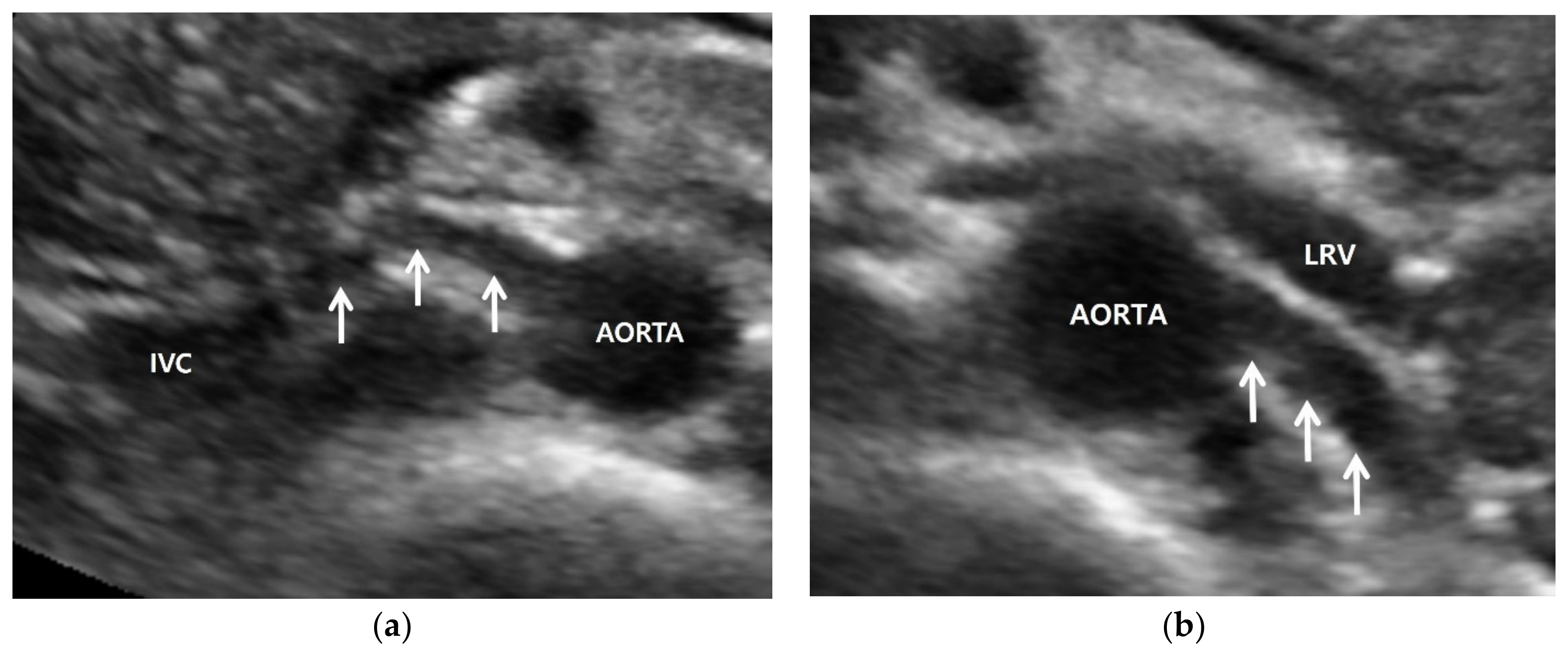
3.1. Gray-Scale US

Gray-scale US is not well-known to be particularly useful in detecting RAS. Previously published studies did not clearly describe the imaging features of gray-scale US, but rather those of Doppler US. Renal artery occlusion was not directly assessed even in an animal study [19]. However, current gray-scale US has the potential to assess renal arteries directly because the ongoing development of US scanners is providing higher resolution imaging than before. Transabdominal US can be used to evaluate proximal and middle segments of renal arteries. Transrenal US can be used to assess the distal segment of renal arteries in the flank. The location, number, and length can be shown depending on the patient’s obesity or bowel gas (Figure 2).
Figure 2. Renal artery and renal US examinations of a 50-year-old man. (a) Gray-scale US axial image shows focal stenosis (arrows) in the proximal right renal artery (RRA) and poststenotic dilatation (arrowhead). His RAS was incidentally detected in the routine check-up because his clinical or laboratory findings were unclear. (b) Gray-scale US sagittal image that shows a small (9 cm) right kidney in which the cortex (RC) is more hyperechoic compared with the liver parenchyma. Arrows indicate clear cortico-medullary differentiation in the right kidney. (c) Color Doppler US shows a focal stenosis (arrowhead) in the proximal renal artery. Bright red and blue signals are seen in the poststenotic dilatation (arrow). (d) Spectral Doppler US shows a high PSV (357 cm/s) in the poststenotic dilatation (arrow). However, a low PSV (108 cm/s) was measured in the stenotic artery because the frequency shift from the RAS was not sufficient.

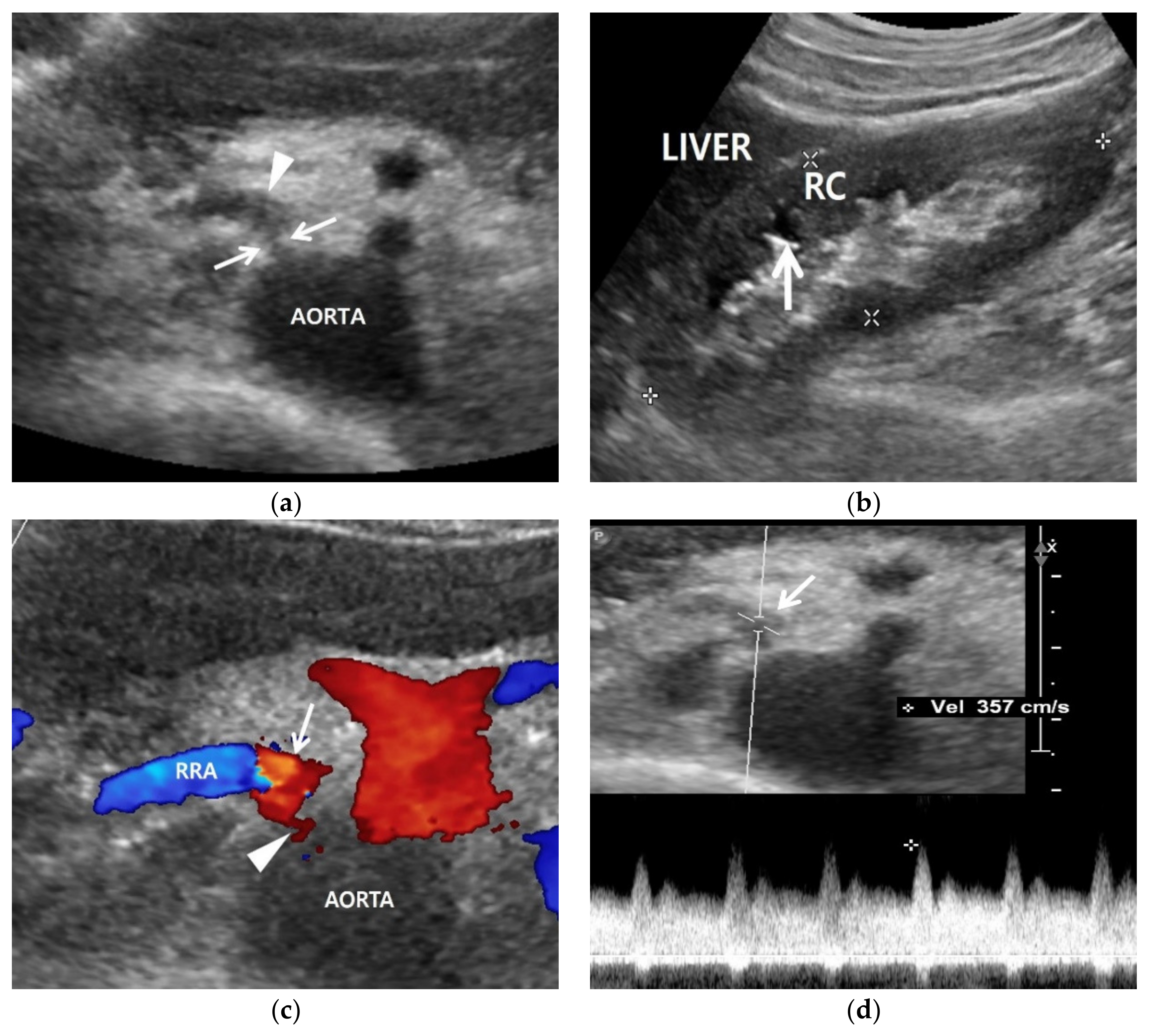
3.2. Color Doppler US

Color Doppler US shows mainly blue or red signals in the normal renal artery. The brightness of these signals is increased in the renal artery. The speed of blood flow increases as RAS becomes severe (Figure 3). These Doppler signals show mixed bright red and bright blue colors in the poststenotic dilatation because turbulence is created from the back-and-forth high-speed blood flows out of the stenosis by means of colliding with the lumen of renal artery (Figure 2 and Figure 3).
Figure 3. RAS-positive renal artery US in a 20-year-old man with negative renal US. (a) Spectral Doppler US does not show pulsus tardus and parvus pattern in the right kidney even though the size (11 cm) and echogenicity appear normal. (b) Color Doppler US shows a focal stenosis (arrow) in the proximal right renal artery (RRA), suggesting RAS. (c) Spectral Doppler US shows a high peak systolic velocity (PSV) (212 cm/s) in the stenotic right renal artery.
Color Doppler US of the kidneys is not an ideal approach for detecting RAS. Renal perfusion can be normal in early stage RAS (Figure 4) and decreases in intermediate or late-stage RAS. Renal perfusion is an indirect finding suggesting RAS. Accordingly, it is not adequately sensitive for detecting early stage RAS.
Figure 4. A high reno-aortic PSV ratio in a 78-year-old man. (a) Spectral Doppler US shows that a PSV is measured 83 cm/s 1–2 cm below the origin of superior mesenteric artery. (b) Spectral Doppler US shows that a PSV is measured 318 cm/s in the poststenotic area, showing turbulence flow. The frequency shift in the proximal left renal artery (arrow) is not sufficient to precisely quantify. The high-PSV RAR is more than 3.8 (318/83) because the PSV in the RAS should be higher than that in the poststenotic area.

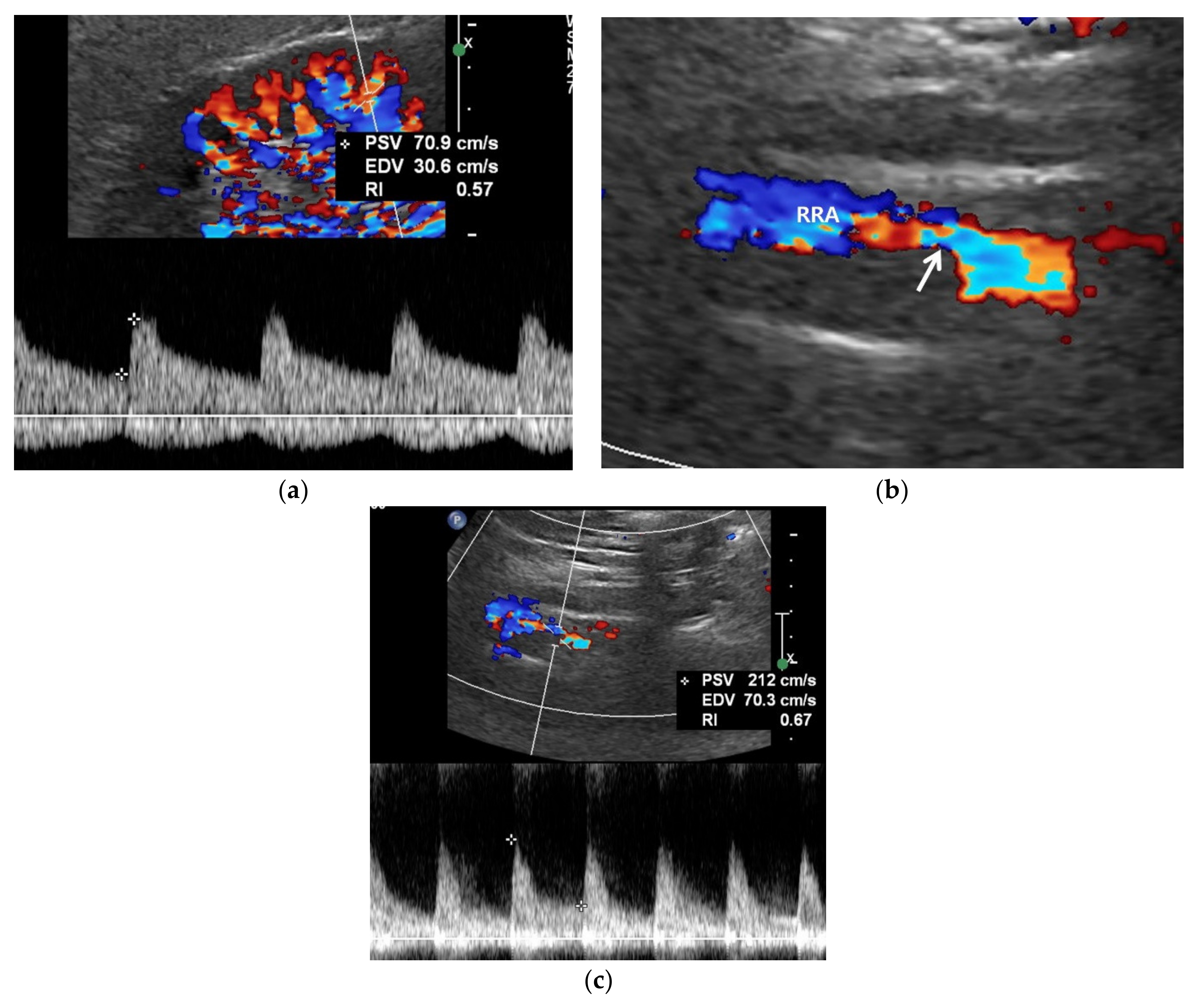
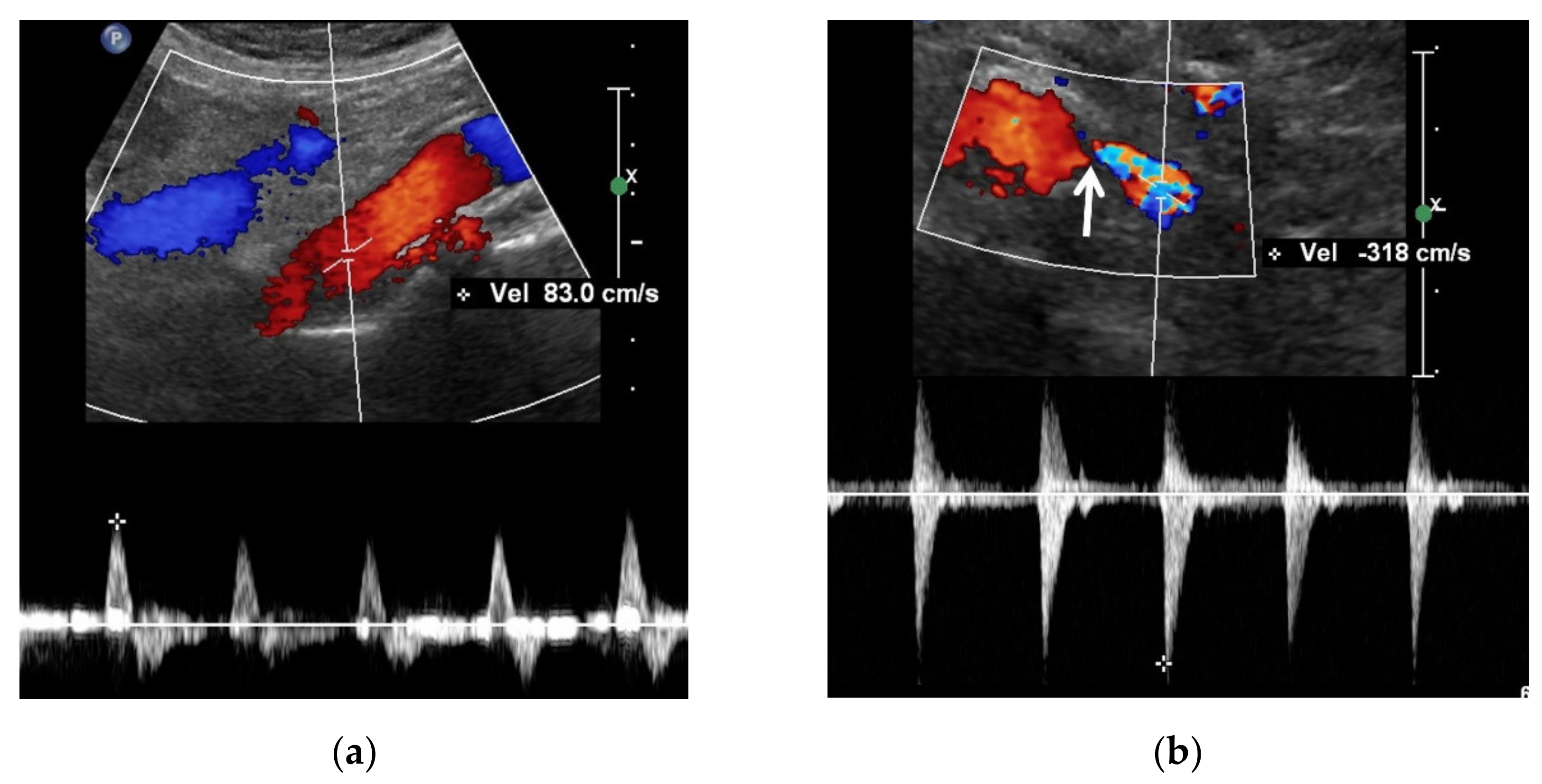
3.3. Spectral Doppler US

Spectral Doppler US quantitatively measures the velocity of blood flow in the stenotic renal artery. The peak systolic velocity (PSV) within the stenotic renal artery is frequently more than 180–200 cm/s [2][17][20][21][22][23] (Figure 2 and Figure 3). If PSV is more than 180 cm/s, the sensitivity and specificity for RAS range from 85–97% and 72–98%, respectively [21][22][24][25]. In case of unilateral RAS, PSV is significantly different between the two renal arteries. At this point, two simple concepts should be kept in mind: first, the normal PSV values in normal renal arteries without stenosis (about 70–100 cm/s), and, second, angle correction is essential to obtain reproducible and accurate measurements of PSVs. If the PSV of the renal artery is ≥3.5-fold that of the aorta (renal-to-aorta ratio (RAR)), it can suggest RAS [26][27][28][29] (Figure 4). The PSV RAR is another good indicator for identifying RAS. If RAR is 3.5 or greater, the sensitivity and specificity ranges are 91–92% and 71–95%, respectively [26][27][28].

3.4. Contrast-Enhanced US

Compared with CT or MRI contrast material, US contrast material does not harm patients with poor renal function because it does not influence renal function or induce nephrogenic systemic fibrosis. US contrast material is composed of microbubbles, which are destroyed with US and excreted from the pulmonary circulation [30]. Therefore, it does not deteriorate renal function in patients with chronic kidney disease. Initially, this US technique was frequently used for differentiating renal masses [31][32][33][34][35]. The use of US contrast is expanding to assess renal microcirculation for the detection of chronic ischemia [36][37][38][39]. RAS induces decreased blood flow to the renal cortex, which is more susceptible to ischemia than the renal medulla. Moreover, high-frame-rate, contrast-enhanced US can show changes in perfusion parameters, and the shape of the time–intensity curve is useful for assessing cortical perfusion after angio-intervention [40]. Finally, cortex thinning occurs after RAS is persistent. When it is intravenously injected, the renal artery can be imaged as if it were shown on DSA. Contrast-enhanced US can be called “US angiography” if it is used for vascular imaging. Accordingly, the renal artery can be hemodynamically assessed with contrast-enhanced US.

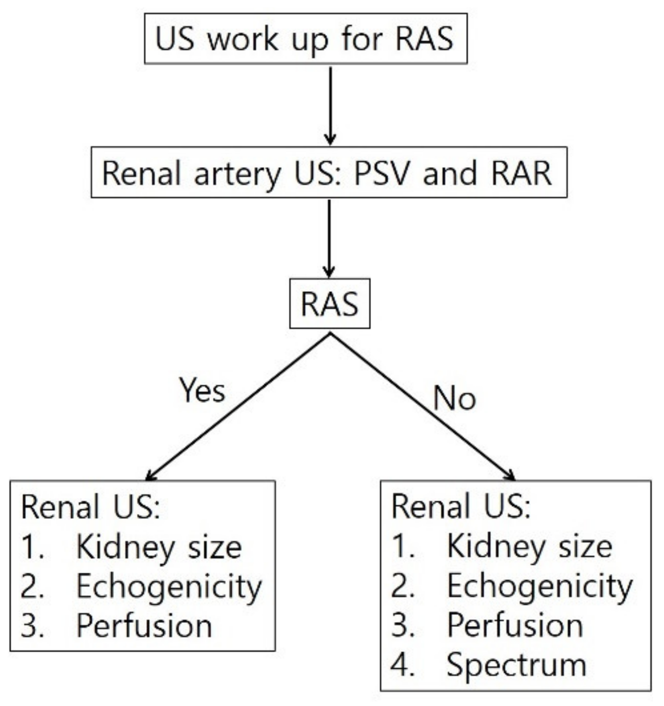
4. Diagnostic Steps for RAS

Radiologists or sonographers should be familiar with the following steps for detecting RAS: First, they should identify the left renal vein as the first step in detecting bilateral renal arteries with US (Figure 5). If the right or left renal artery is detected on the US, it should be carefully evaluated with gray-scale US. When RAS is detected with gray-scale US, color and spectral Doppler US also should be performed to depict the imaging features of RAS. However, even though RAS is not identified with gray-scale US, renal arteries must be assessed with color Doppler US to detect RAS. RAS may be staged earlier when it is negative on gray-scale US, but positive on color or spectral Doppler US. Angioplasty or stenting is more effective in gray-scale US-negative RAS than in gray-scale US-positive RAS.
Figure 5. Diagnostic steps for detecting RAS. The flow diagram shows that assessing the renal artery is the first step for diagnosing RAS with gray-scale US, color or spectral Doppler US, or contrast-enhanced US. Next, renal US should be performed to assess kidney size and cortical echogenicity. When RAS is indeterminate on renal artery US, kidneys should be assessed with gray-scale US, color or spectral Doppler US, or contrast-enhanced US. RAS, renal artery stenosis; PSV, peak systolic velocity; RAR, reno-aortic ratio.

5. Conclusions

CT, MRI, and DSA using contrast material are rarely recommended for patients who have poor renal function due to RAS. Hence, renal artery US is useful as a primary examination for RAS scanning. Radiologists or sonographers can assess renal arteries once they find the left renal vein behind which the right and left arteries travel. Therefore, RAS can be determined with various imaging features on gray-scale, color Doppler, spectral Doppler, and contrast-enhanced US. The direct assessment using renal artery US is more sensitive to detecting RAS compared with assessment by renal US. In addition, the ongoing development of US scanners has and will provide better hemodynamic information on RAS in patients who cannot undergo contrast-enhanced CT or MR angiography.

References

  1. Zierler, R.E. Is duplex scanning the best screening test for renal artery stenosis? Semin. Vasc. Surg. 2001, 14, 177–185.
  2. Olin, J.W.; Piedmonte, M.R.; Young, J.R.; DeAnna, S.; Grubb, M.; Childs, M.B. The utility of duplex ultrasound scanning of the renal arteries for diagnosing significant renal artery stenosis. Ann. Intern. Med. 1995, 122, 833–838.
  3. Riehl, J.; Schmitt, H.; Bongartz, D.; Bergmann, D.; Sieberth, H.G. Renal artery stenosis: Evaluation with colour duplex ultrasonography. Nephrol. Dial. Transpl. 1997, 12, 1608–1614.
  4. Vasbinder, G.B.; Nelemans, P.J.; Kessels, A.G.; Kroon, A.A.; de Leeuw, P.W.; van Engelshoven, J.M. Diagnostic tests for renal artery stenosis in patients suspected of having renovascular hypertension: A meta-analysis. Ann. Intern. Med. 2001, 135, 401–411.
  5. Vasbinder, G.B.; Nelemans, P.J.; Kessels, A.G.; Kroon, A.A.; Maki, J.H.; Leiner, T.; Beek, F.J.; Korst, M.B.; Flobbe, K.; de Haan, M.W.; et al. Accuracy of computed tomographic angiography and magnetic resonance angiography for diagnosing renal artery stenosis. Ann. Intern. Med. 2004, 141, 674–682.
  6. Roditi, G.; Maki, J.H.; Oliveira, G.; Michaely, H.J. Renovascular imaging in the NSF Era. J. Magn. Reson. Imaging 2009, 30, 1323–1334.
  7. Kim, D.; Porter, D.H.; Brown, R.; Crivello, M.S.; Silva, P.; Leeming, B.W. Renal artery imaging: A prospective comparison of intra-arterial digital subtraction angiography with conventional angiography. Angiology 1991, 42, 345–357.
  8. Hawkins, I.F., Jr.; Wilcox, C.S.; Kerns, S.R.; Sabatelli, F.W. CO2 digital angiography: A safer contrast agent for renal vascular imaging? Am. J. Kidney Dis. 1994, 24, 685–694.
  9. Liss, P.; Eklof, H.; Hellberg, O.; Hagg, A.; Bostrom-Ardin, A.; Lofberg, A.M.; Olsson, U.; Orndahl, P.; Nilsson, H.; Hansell, P.; et al. Renal effects of CO2 and iodinated contrast media in patients undergoing renovascular intervention: A prospective, randomized study. J. Vasc. Interv. Radiol. 2005, 16, 57–65.
  10. Stratigis, S.; Stylianou, K.; Kyriazis, P.P.; Dermitzaki, E.K.; Lygerou, D.; Syngelaki, P.; Stratakis, S.; Koukouraki, S.; Parthenakis, F.; Tsetis, D.; et al. Renal artery stenting for atherosclerotic renal artery stenosis identified in patients with coronary artery disease: Does captopril renal scintigraphy predict outcomes? J. Clin. Hypertens. 2018, 20, 373–381.
  11. Qanadli, S.D.; Soulez, G.; Therasse, E.; Nicolet, V.; Turpin, S.; Froment, D.; Courteau, M.; Guertin, M.C.; Oliva, V.L. Detection of renal artery stenosis: Prospective comparison of captopril-enhanced Doppler sonography, captopril-enhanced scintigraphy, and MR angiography. Am. J. Roentgenol. 2001, 177, 1123–1129.
  12. Mehran, R.; Nikolsky, E. Contrast-induced nephropathy: Definition, epidemiology, and patients at risk. Kidney Int. 2006, 69, S11–S15.
  13. Morcos, S.K.; Thomsen, H.S.; Webb, J.A.; Contrast Media Safety Committee, European Society of Urogenital Radiology (ESUR). Contrast-media-induced nephrotoxicity: A consensus report. Eur. Radiol. 1999, 9, 1602–1613.
  14. Gleeson, T.G.; Bulugahapitiya, S. Contrast-Induced Nephropathy. Am. J. Roentgenol. 2004, 183, 1673–1689.
  15. Grobner, T.; Prischl, F.C. Gadolinium and nephrogenic systemic fibrosis. Kidney Int. 2007, 72, 260–264.
  16. Grobner, T. Gadolinium—A specific trigger for the development of nephrogenic fibrosing dermopathy and nephrogenic systemic fibrosis? Nephrol. Dial. Transpl. 2006, 21, 1104–1108.
  17. Granata, A.; Fiorini, F.; Andrulli, S.; Logias, F.; Gallieni, M.; Romano, G.; Sicurezza, E.; Fiore, C.E. Doppler ultrasound and renal artery stenosis: An overview. J. Ultrasound 2009, 12, 133–143.
  18. Schäberle, W.; Leyerer, L.; Schierling, W.; Pfister, K. Ultrasound diagnostics of renal artery stenosis: Stenosis criteria, CEUS and recurrent in-stent stenosis. Gefasschirurgie 2016, 21, 4–13.
  19. Park, B.K.; Kim, S.H.; Moon, M.H.; Jung, S.I. Imaging features of gray-scale and contrast-enhanced color Doppler US for the differentiation of transient renal arterial ischemia and arterial infarction. Korean J. Radiol. 2005, 6, 179–184.
  20. Bokhari, S.W.; Faxon, D.P. Current advances in the diagnosis and treatment of renal artery stenosis. Rev. Cardiovasc. Med. 2004, 5, 204–215.
  21. Radermacher, J.; Chavan, A.; Schäffer, J.; Stoess, B.; Vitzthum, A.; Kliem, V.; Rademaker, J.; Bleck, J.; Gebel, M.J.; Galanski, M.; et al. Detection of significant renal artery stenosis with color Doppler sonography: Combining extrarenal and intrarenal approaches to minimize technical failure. Clin. Nephrol. 2000, 53, 333–343.
  22. Hua, H.T.; Hood, D.B.; Jensen, C.C.; Hanks, S.E.; Weaver, F.A. The use of colorflow duplex scanning to detect significant renal artery stenosis. Ann. Vasc. Surg. 2000, 14, 118–124.
  23. Revzin, M.V.; Imanzadeh, A.; Menias, C.; Pourjabbar, S.; Mustafa, A.; Nezami, N.; Spektor, M.; Pellerito, J.S. Optimizing Image Quality When Evaluating Blood Flow at Doppler US: A Tutorial. RadioGraphics 2019, 39, 1501–1523.
  24. Hoffmann, U.; Edwards, J.M.; Carter, S.; Goldman, M.L.; Harley, J.D.; Zaccardi, M.J.; Strandness, D.E., Jr. Role of duplex scanning for the detection of atherosclerotic renal artery disease. Kidney Int. 1991, 39, 1232–1239.
  25. Williams, G.J.; Macaskill, P.; Chan, S.F.; Karplus, T.E.; Yung, W.; Hodson, E.M.; Craig, J.C. Comparative accuracy of renal duplex sonographic parameters in the diagnosis of renal artery stenosis: Paired and unpaired analysis. Am. J. Roentgenol. 2007, 188, 798–811.
  26. Aytac, S.K.; Yigit, H.; Sancak, T.; Ozcan, H. Correlation between the diameter of the main renal artery and the presence of an accessory renal artery: Sonographic and angiographic evaluation. J. Ultrasound Med. 2003, 22, 433–439.
  27. Bude, R.O.; Forauer, A.R.; Caoili, E.M.; Nghiem, H.V. Is it necessary to study accessory arteries when screening the renal arteries for renovascular hypertension? Radiology 2003, 226, 411–416.
  28. Labropoulos, N.; Ayuste, B.; Leon, L.R., Jr. Renovascular disease among patients referred for renal duplex ultrasonography. J. Vasc. Surg. 2007, 46, 731–737.
  29. Zierler, R.E.; Bergelin, R.O.; Davidson, R.C.; Cantwell-Gab, K.; Polissar, N.L.; Strandness, D.E., Jr. A prospective study of disease progression in patients with atherosclerotic renal artery stenosis. Am. J. Hypertens. 1996, 9, 1055–1061.
  30. Morel, D.R.; Schwieger, I.; Hohn, L.; Terrettaz, J.; Llull, J.B.; Cornioley, Y.A.; Schneider, M. Human Pharmacokinetics and Safety Evaluation of SonoVue™, a New Contrast Agent for Ultrasound Imaging. Investig. Radiol. 2000, 35, 80.
  31. Park, B.K.; Kim, S.H.; Choi, H.J. Characterization of renal cell carcinoma using agent detection imaging: Comparison with gray-scale US. Korean J. Radiol. 2005, 6, 173–178.
  32. Park, B.K.; Kim, B.; Kim, S.H.; Ko, K.; Lee, H.M.; Choi, H.Y. Assessment of cystic renal masses based on Bosniak classification: Comparison of CT and contrast-enhanced US. Eur. J. Radiol. 2007, 61, 310–314.
  33. Gerst, S.; Hann, L.E.; Li, D.; Gonen, M.; Tickoo, S.; Sohn, M.J.; Russo, P. Evaluation of renal masses with contrast-enhanced ultrasound: Initial experience. Am. J. Roentgenol. 2011, 197, 897–906.
  34. Ignee, A.; Straub, B.; Schuessler, G.; Dietrich, C.F. Contrast enhanced ultrasound of renal masses. World J. Radiol. 2010, 2, 15–31.
  35. Barr, R.G. Use of lumason/sonovue in contrast-enhanced ultrasound of the kidney for characterization of renal masses-a meta-analysis. Abdom. Radiol. 2021, 47, 272–287.
  36. Tenant, S.C.; Gutteridge, C.M. The clinical use of contrast-enhanced ultrasound in the kidney. Ultrasound 2016, 24, 94–103.
  37. Dong, Y.; Wang, W.P.; Cao, J.; Fan, P.; Lin, X. Early assessment of chronic kidney dysfunction using contrast-enhanced ultrasound: A pilot study. Br. J. Radiol. 2014, 87, 20140350.
  38. Ma, F.; Cang, Y.; Zhao, B.; Liu, Y.; Wang, C.; Liu, B.; Wu, T.; Song, Y.; Peng, A. Contrast-enhanced ultrasound with SonoVue could accurately assess the renal microvascular perfusion in diabetic kidney damage. Nephrol. Dial. Transplant. 2012, 27, 2891–2898.
  39. Xu, Y.; Li, H.; Wang, C.; Zhang, M.; Wang, Q.; Xie, Y.; Shao, X.; Tian, L.; Yuan, Y.; Yan, W.; et al. Improving Prognostic and Chronicity Evaluation of Chronic Kidney Disease with Contrast-Enhanced Ultrasound Index-Derived Peak Intensity. Ultrasound Med. Biol. 2020, 46, 2945–2955.
  40. Ran, X.; Lin, L.; Yang, M.; Niu, G.; Chen, L.; Shao, Y.; Zou, Y.; Wang, B. Contrast-Enhanced Ultrasound Evaluation of Renal Blood Perfusion Changes after Percutaneous Transluminal Renal Angioplasty and Stenting for Severe Atherosclerotic Renal Artery Stenosis. Ultrasound Med. Biol. 2020, 46, 1872–1879.
More
Information
Contributor MDPI registered users' name will be linked to their SciProfiles pages. To register with us, please refer to https://encyclopedia.pub/register :
View Times: 888
Revisions: 3 times (View History)
Update Date: 14 Oct 2022
1000/1000