Submitted Successfully!
To reward your contribution, here is a gift for you: A free trial for our video production service.
Thank you for your contribution! You can also upload a video entry or images related to this topic.
Version Summary Created by Modification Content Size Created at Operation
1 -- 2256 2022-09-09 10:55:26 |
2 format corrected. + 4 word(s) 2260 2022-09-13 04:50:37 |

Video Upload Options

Do you have a full video?

Confirm

Are you sure to Delete?
Cite
If you have any further questions, please contact Encyclopedia Editorial Office.
Jarocki, M.;  Karska, J.;  Kowalski, S.;  Kiełb, P.;  Nowak, �.;  Krajewski, W.;  Saczko, J.;  Kulbacka, J.;  Szydełko, T.;  Małkiewicz, B. Involvement of Interleukin 17 in Renal Cell Carcinoma. Encyclopedia. Available online: https://encyclopedia.pub/entry/27055 (accessed on 27 July 2024).
Jarocki M,  Karska J,  Kowalski S,  Kiełb P,  Nowak �,  Krajewski W, et al. Involvement of Interleukin 17 in Renal Cell Carcinoma. Encyclopedia. Available at: https://encyclopedia.pub/entry/27055. Accessed July 27, 2024.
Jarocki, Michał, Julia Karska, Szymon Kowalski, Paweł Kiełb, Łukasz Nowak, Wojciech Krajewski, Jolanta Saczko, Julita Kulbacka, Tomasz Szydełko, Bartosz Małkiewicz. "Involvement of Interleukin 17 in Renal Cell Carcinoma" Encyclopedia, https://encyclopedia.pub/entry/27055 (accessed July 27, 2024).
Jarocki, M.,  Karska, J.,  Kowalski, S.,  Kiełb, P.,  Nowak, �.,  Krajewski, W.,  Saczko, J.,  Kulbacka, J.,  Szydełko, T., & Małkiewicz, B. (2022, September 09). Involvement of Interleukin 17 in Renal Cell Carcinoma. In Encyclopedia. https://encyclopedia.pub/entry/27055
Jarocki, Michał, et al. "Involvement of Interleukin 17 in Renal Cell Carcinoma." Encyclopedia. Web. 09 September, 2022.
Involvement of Interleukin 17 in Renal Cell Carcinoma
Edit

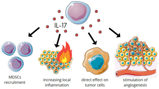
IL-17A (traditionally known as IL-17, in the past also termed as CTLA-8) is the first and best characterized member in its family composed of IL-17A, B, C, D, E (also known as Il-25), and F. IL-17 was first discovered in 1993, although it became well-known in 2005 with the finding of a new population of CD4+ Th cells—Th17 that expressed this cytokine. Naive CD4+ T-cells are triggered to differentiate into Th17 in the presence of both TGF-β and Il-6. The differentiation is characterized by the production of IL-17, Il-21, and ROR-yt (transcription factor). The differentiation of Th17 cells also depends on dendritic cells which produce IL-1, IL-6, and IL-23. These molecules preferentially activate STAT-3 which induces transcription factor ROR-yt. ROR-yt is also expressed in the presence of STAT-3 that is activated by Il-6, Il-21, and Il-23. There is also an autocrine generation of Th17 by Il-21 that is derived from these T-cells. Moreover, Il-21 leads to Il-23 receptor expression of Th17 and their susceptibility to Il-23 (produced by dendritic cells) stimulation. This cytokine gives Th17 phenotype stability and helps them with acquiring effector functions.

interleukin-17 renal cell carcinoma immunotherapy inflammation tumor microenvironment tumor development Th17 lymphocytes

1. IL-17 in Kidney Diseases

While the direct mechanism of IL-17 interaction in renal cell carcinoma (RCC) remains elusive, the researchers can safely extrapolate available data regarding IL-17 role regarding cancer in general. IL-17 is associated with chronic inflammation in many diseases and kidney diseases are no different. Increased levels of IL-17 are found in autoimmune kidney diseases and the serum level of IL-17 strictly correlates with disease activity [1]. The IL-17 receptors are expressed in many kidney cells (podocytes, mesangial, and renal endothelial cells) and are responsible for the stimulation of the inflammatory environment which leads to the damage and disturbance of nephron function [2]. Additionally, IL-17 receptors stimulate profibrotic pathways causing fibrosis and eventually the loss of organ function [3]. The result of these interactions is that autoimmune kidney diseases, when treated poorly or insufficiently, may lead to the onset of chronic kidney disease (CKD) which, in turn via various pathways, increases the risk of cancer development [4].

2. IL-17 in Tumors

IL-17 plays a dual role in tumor development. It is worth noting that IL-17 may have both stimulating and inhibiting effects on the growth of tumor cells. The potential role of IL-17 includes tumorigenesis, proliferation, angiogenesis, and metastasis [5][6]. IL-17 stimulates the secretion of cytokines such as G-CSF which recruit myeloid-derived suppressor cells (MDSC). MDSCs are immature myeloid cells with the ability to decrease the adaptive immunity [7][8]. These cells are recruited into the neoplastic tissue via IL-17-induced chemokines (CXCL1/CXCL5). IL-17 also induces pro-inflammatory NF-kB and Il-6, which acts as paracrine signalers and increases tumor growth and survival. Recent studies have shown that IL-17, independently of its effect on the tumor microenvironment, directly induces the proliferation of precancerous cells. An association has also been demonstrated between IL-17 signaling and tumor formation within wounds during healing. This is due to the induction of Lrig1 + cells with oncogenic mutations such as KrasG12D [7].
In summary, in the early stages of tumor development, IL-17 acts in a multidirectional way: (1) by activating MDSC, which results in decreasing systemic immunity, (2) increasing local inflammation, and (3) directly affecting tumor cells. Research has shown both pro- and anti-angiogenic effects. The angiogenic and lymphangiogenic effects are observed via the activation of VEGF. As a result, it promotes the formation of metastases [6][7][8]. IL-17s influence on early tumor development is summarized in Figure 1.
Figure 1. Multidirectional action of IL-17 in the early stages of tumorigenesis.
However, studies have indicated that the IL-17 knockout contributes to a more aggressive metastatic picture, which may be related to the loss of the potent antitumor cytokine IFN-gamma [6]. It has been shown that IL-17A directly inhibits the growth of P815 mastocytoma and J558L plasmacytoma [9][10]. There is also known evidence that IL-17A may exert an indirect pro-apoptotic effect. It was found that IL-17A, by increasing the production of inducible nitric oxide synthase (iNOS), leads to an increase in the synthesis of nitric oxide (NO). It may be an important factor, which can inhibit the growth of oral squamous cell carcinoma cells via apoptosis [11]. The tumor cell growth inhibitory effect was also observed with IL-17E. IL-17E in combination with chemotherapeutic agents inhibits the growth of melanoma and pancreatic tumors in mice [12].
Interactions between the microbiota and IL-17A-producing cells are also involved in the pathogenesis of immune-mediated inflammatory diseases and cancer. Studies have shown that the gut microbiome can affect the activity of Th17 cells and IL-17A that is induced by the microbiota is also associated with the pathogenesis of colon cancer, breast cancer, pancreatic cancer, ovarian cancer, and multiple myeloma [13].

3. IL-17s Role in Carcinogenesis

3.1. Protumor

There is little data about the positive influence of IL-17 on RCC. It is known that Th17 lymphocytes that produce IL-17 are said to be higher concentrated in the serum of RCC patients [14]. Moreover, transcription factor typical for Th17—RORC is upregulated in this group. RORC is responsible for directing the Th17 lineage which suggests that this correlation leading to abnormal differentiation of lymphocytes may play an important role in both the occurrence and progression of RCC [14][15]. Furthermore, with the higher tumor stage and grade, the number of Th17 increases. High concentration of these lymphocytes is also connected with a decreased survival rate. Taking in consideration this evidence, Th17 cells could be clinical markers of RCC diagnosis and progression together with patients survival rate [14].
IL-17 has also a bidirectional influence on RCC cells [16]. Inzoume et al., isolated T-cell clones from one patient before any systemic treatment and generated them by in vitro stimulation with dendritic cells, autologous tumor, and IL-2 in the presence of anti-CTLA4 antibody. In this environment of a.o. RCC cells, T-lymphocytes were triggered to produce IL-17 that consequently induces tumor cells to release large amounts of Il-8. IL-17 was already known to be responsible for inflammatory cytokine production from pulmonary epithelium or smooth muscle [17] and even IL-8 from tumor cells, but not on such a high level [18]. What is important, is that IL-8 in turn has a chemotactive influence on T-cells and angiogenesis which leads to RCC local infiltration by T-lymphocytes, however it may not be a single factor in this phenomenon [16]. The interplay between IL-17 and RCC is summarized in Figure 2.
Figure 2. Schematic overview of the bidirectional influence between T-lymphocytes and RCC. (1) T-lymphocytes that were isolated from the RCC patient and incubated in special conditions, produce Il-17 in the presence of RCC cells. Il-17 triggers tumor cells to release a large amount of Il-8. This cytokine induces not only angiogenesis, but also has a chemotactive impact on T-lymphocytes. (2) Consequently, the high number of lymphocytes T infiltrate RCC and tumors new vessels are created which is a favourable environment for tumor development.
Interleukin 17 is a pro-inflammatory cytokine that promotes chemotaxis and degranulation of neutrophils. The increased expression of IL-17 has been demonstrated in the tumor microenvironment [8]. Research suggests a relationship between IL-17 production and circulating MDSC levels [19]. MDSCs exert an influence on the adaptive immune response and regulate innate immunity by modulating cytokine production by macrophages [20]. It has also been reported the role of MDSC in angiogenesis and tumor promotion [21]. However, the relationship between cytokine production and MDSC accumulation in RCC is not fully understood. In addition to IL-17, IL-18 and CCL2 also play an important role in this phenomenon. Guan et al., showed that in addition to IL-6 and tumor necrosis factor alpha (TNF-α), IL-17 has been shown to be overexpressed in RCC tissue, and its expression is dependent on the degree of malignancy. They also described that parenchymal levels of IL-17 correlate with increased total MDSC [8].
IL-17 also influences many other cancers, and these phenomena can indicate further research on IL-17 and tumorigenesis of RCC. IL-17B supports pancreatic cancer by up-regulating ERK pathways propagating tumor cell invasion and facilitates metastatic cell survival. Consequently, the overexpression of IL-17B receptors that are found on these cells coincided with poorer prognosis for patients [22]. Furthermore, IL-17 was found to negatively impact colorectal cancer cells by promoting oncogenesis [23]. Altogether, these discoveries implicate the immense role of IL-17 in cancer development on many of its phases, from oncogenesis to metastases.
The interplay between T-cells, IL-17, and RCC environment is promising for a better understanding of this cancer, but it is also complicated and requires further research in this topic.

3.2. Prometastatic

Chronic inflammation contributes to tumor metastasis via an unknown pathway. Probably, tumor hypoxia accompanying rapid growth triggers angiogenesis that helps in metastasizing. However, not only nutrients flow through new vessels, but also immune cells to tumor mass [24]. Taking into consideration this dual nature of angiogenesis and inflammation effect on tumor, the role of IL-17 in it has been investigated.
IL-17 is known for both its pro-tumor and anti-tumor effects [24][25]. When supporting a tumor and its metastasis, IL-17 induces angiogenesis and sustains an inflammatory environment. Firstly, IL-17 increases the production of VEGF, which consequently triggers TGF-β- and VEGF-mediated angiogenesis. In turn, TGF upregulates VEGF receptor expression enhancing VEGF receptivity [24]. Secondly, the production of IL-8 is stimulated by IL-17. IL-8 leads to angiogenic responses in endothelial cells and, therefore, increases proliferation and survival of both endothelial and cancer cells, potentiating the migration of cancer cells [18][24]. Thirdly, the production of prostaglandin E1 and prostaglandin E2 which stimulate tumor angiogenesis is enhanced by IL-17 [26]. Finally, IL-17 increases the production of IL-6 in fibroblasts and Il-6 induces Th17 differentiation and, therefore, IL-17 expression. This creates a chronic inflammatory state that supports tumor growth and metastasis [24]. The influence of IL-17 on tumorigenesis is summarized in Figure 3.
Figure 3. Schematic overview of IL-17’s influence on tumorigenesis. IL-17 stimulates the production of VEGF, PG E1, and E2, IL-6 and IL-8. VEGF triggers VEGF- and TGF-β-mediated angiogenesis, potentiated by the action of PG E1 and E2. IL-6 induces Th17 differentiation and IL-17 expression, stimulating chronic inflammation. IL-8 increases the proliferation and migration of cancer cells.
In osteosarcoma and synovial sarcoma, the positive influence of IL-17 on metastasizing has been especially evidenced [27][28]. In the first one, IL-17 through activating its receptor IL-17R upregulates the expression of VEGF and CXCR4, which are known to take part in the metastasis of tumors [27][29][30]. Moreover, Stat3 that induces tumor progression via supporting tumor survival, angiogenesis, and suppressing antitumor immunity is activated by IL-17 [27][31][32]. What is similar in the pro-metastasis role of IL-17 between osteosarcoma and synovial sarcoma, is MMP (matrix metalloproteinase) [27][28]. IL-17, through IL-17R, enhances the expression of MMP, which is required for the proteolytic modifications of basement membranes and extracellular matrices by angiogenesis and metastasis [27][28][33][34][35]. Additionally, in synovial sarcoma, metastasizing is supported by IL-17 through the expression of VEGF and CXCR4 [28].

3.3. Antitumor

Even though IL-17 can be considered a tumor-promoting factor in many cases, research shows that its role is far more complex. IL-17 expression in RCC and also other cancers seems to correspond with an overall better prognosis. Huang et al., identified that the patients with ccRCC that was enriched with VHL, but depleted of BAP1 mutations and with high levels of Th17 and CD8+ T-cells were characterized by longer survival [36].
In some other human cancers, it has also been shown that the infiltrating level of Th17 cells in tumor mass was negatively correlated with poorer survival, tumor growth, and stage [37]. A study on gastric adenocarcinoma patients revealed that high intratumoral expression of IL-17 results in significantly higher five-year overall survival. Even though the exact role of IL-17 remains to be studied, patients with a greater expression of IL-17 had the likelihood of death at 5 years reduced by a staggering 48% [38]. Furthermore, research that was conducted on cervical adenocarcinoma patients also supports the anti-tumor properties of IL-17 theory. An increased levels of IL-17 and IL-17(+) cells were found in patients with smaller tumor sizes, less infiltration depth, and the absence of vasal invasion [39]. Additionally, in patients with chronic lymphocytic leukemia increased Th17 and IL-17 amounts corresponded with a longer overall survival rate [40]. Therefore, it is essential to view IL-17 as an important factor in tumor immunity and research further its complex role, as similar pathways and effects could occur in RCC.

3.4. Antimetastatic

Some studies indicate that IL-17 can indirectly inhibit tumor metastasis—here IL-23 plays the main role. On the one hand, it triggers Th17 differentiation, IL-17 production, and, therefore, maintains inflammatory environment, favorable for metastasis. On the other hand, IL-23 may also have anti-tumor effects, because tumors that overexpressed this cytokine presented reduced growth and metastasis [24][41][42][43][44].

4. IL-17 in RCC Detection

Currently, more attention is being paid to the role of biomarkers in the diagnosis and prognosis of neoplastic diseases [45][46]. Diagnostic biomarkers can allow for an early detection and classification of cancer. Prognostic biomarkers can inform clinicians about the natural course of an individual cancer and guide their decision of whom to treat and how intensive the treatment should be [45][46]. In the case of RCC, which is highly diverse in the molecular field, the latest advances in proteomics, genomics, and metabolomics could be particularly useful [45]. The latest literature describes a number of substances and molecules that could constitute these markers, which can be used in the early detection of RCC, providing the benefits of new technologies [46]. The authors indicate immunological markers and immune checkpoint inhibitors as one of the important biomarkers [45]. Due to its direct involvement in RCC pathogenesis, IL-17 may also be considered as one of them. However, none of these biomarkers have yet been established for the routine clinical use in management of this cancer and more research seems to be necessary [45].

References

  1. Krebs, C.F.; Schmidt, T.; Riedel, J.-H.; Panzer, U. T Helper Type 17 Cells in Immune-Mediated Glomerular Disease. Nat. Rev. Nephrol. 2017, 13, 647–659.
  2. Paquissi, F.C.; Abensur, H. The Th17/IL-17 Axis and Kidney Diseases, with Focus on Lupus Nephritis. Front. Med. 2021, 8, 654912.
  3. Peng, X.; Xiao, Z.; Zhang, J.; Li, Y.; Dong, Y.; Du, J. IL-17A Produced by Both Γδ T and Th17 Cells Promotes Renal Fibrosis via RANTES-Mediated Leukocyte Infiltration after Renal Obstruction. J. Pathol. 2015, 235, 79–89.
  4. Stengel, B. Chronic Kidney Disease and Cancer: A Troubling Connection. J. Nephrol. 2010, 23, 253–262.
  5. Vitiello, G.A.; Miller, G. Targeting the Interleukin-17 Immune Axis for Cancer Immunotherapy. J. Exp. Med. 2019, 217, e20190456.
  6. Yang, B.; Kang, H.; Fung, A.; Zhao, H.; Wang, T.; Ma, D. The Role of Interleukin 17 in Tumour Proliferation, Angiogenesis, and Metastasis. Mediat. Inflamm. 2014, 2014, 623759.
  7. Zhao, J.; Chen, X.; Herjan, T.; Li, X. The Role of Interleukin-17 in Tumor Development and Progression. J. Exp. Med. 2020, 217, e20190297.
  8. Guan, X.; Liu, Z.; Zhang, J.; Jin, X. Myeloid-Derived Suppressor Cell Accumulation in Renal Cell Carcinoma Is Correlated with CCL2, IL-17 and IL-18 Expression in Blood and Tumors. Adv. Clin. Exp. Med. 2018, 27, 947–953.
  9. Numasaki, M.; Fukushi, J.; Ono, M.; Narula, S.K.; Zavodny, P.J.; Kudo, T.; Robbins, P.D.; Tahara, H.; Lotze, M.T. Interleukin-17 Promotes Angiogenesis and Tumor Growth. Blood 2003, 101, 2620–2627.
  10. Benchetrit, F.; Ciree, A.; Vives, V.; Warnier, G.; Gey, A.; Sautès-Fridman, C.; Fossiez, F.; Haicheur, N.; Fridman, W.H.; Tartour, E. Interleukin-17 Inhibits Tumor Cell Growth by Means of a T-Cell-Dependent Mechanism. Blood 2002, 99, 2114–2121.
  11. Shang, Z.-J.; Li, J.-R.; Li, Z.-B. Effects of Exogenous Nitric Oxide on Oral Squamous Cell Carcinoma: An in Vitro Study. J. Oral Maxillofac. Surg. 2002, 60, 901–905.
  12. Benatar, T.; Cao, M.Y.; Lee, Y.; Lightfoot, J.; Feng, N.; Gu, X.; Lee, V.; Jin, H.; Wang, M.; Wright, J.A.; et al. IL-17E, a Proinflammatory Cytokine, Has Antitumor Efficacy against Several Tumor Types In Vivo. Cancer Immunol. Immunother. 2010, 59, 805–817.
  13. Brevi, A.; Cogrossi, L.L.; Grazia, G.; Masciovecchio, D.; Impellizzieri, D.; Lacanfora, L.; Grioni, M.; Bellone, M. Much More Than IL-17A: Cytokines of the IL-17 Family Between Microbiota and Cancer. Front. Immunol. 2020, 11, 565470.
  14. Peng, Z.; Hu, Y.; Ren, J.; Yu, N.; Li, Z.; Xu, Z. Circulating Th22 Cells, as Well as Th17 Cells, Are Elevated in Patients with Renal Cell Carcinoma. Int. J. Med. Sci. 2021, 18, 99–108.
  15. Veldhoen, M.; Hirota, K.; Westendorf, A.M.; Buer, J.; Dumoutier, L.; Renauld, J.-C.; Stockinger, B. The Aryl Hydrocarbon Receptor Links TH17-Cell-Mediated Autoimmunity to Environmental Toxins. Nature 2008, 453, 106–109.
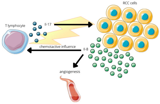
  16. Inozume, T.; Hanada, K.; Wang, Q.J.; Yang, J.C. IL-17 Secreted by Tumor Reactive T Cells Induces IL-8 Release by Human Renal Cancer Cells. J. Immunother. 2009, 32, 109–117.
  17. Witowski, J.; Książek, K.; Jörres, A. Interleukin-17: A Mediator of Inflammatory Responses. Cell. Mol. Life Sci. 2004, 61, 567–579.
  18. Kehlen, A.; Thiele, K.; Riemann, D.; Rainov, N.; Langner, J. Interleukin-17 Stimulates the Expression of IkappaB Alpha MRNA and the Secretion of IL-6 and IL-8 in Glioblastoma Cell Lines. J. Neuroimmunol. 1999, 101, 1–6.
  19. Yazawa, T.; Shibata, M.; Gonda, K.; Machida, T.; Suzuki, S.; Kenjo, A.; Nakamura, I.; Tsuchiya, T.; Koyama, Y.; Sakurai, K.; et al. Increased IL-17 Production Correlates with Immunosuppression Involving Myeloid-Derived Suppressor Cells and Nutritional Impairment in Patients with Various Gastrointestinal Cancers. Mol. Clin. Oncol. 2013, 1, 675–679.
  20. Gabrilovich, D.I.; Nagaraj, S. Myeloid-Derived Suppressor Cells as Regulators of the Immune System. Nat. Rev. Immunol. 2009, 9, 162–174.
  21. Murdoch, C.; Muthana, M.; Coffelt, S.B.; Lewis, C.E. The Role of Myeloid Cells in the Promotion of Tumour Angiogenesis. Nat. Rev. Cancer 2008, 8, 618–631.
  22. Cochaud, S.; Giustiniani, J.; Thomas, C.; Laprevotte, E.; Garbar, C.; Savoye, A.-M.; Curé, H.; Mascaux, C.; Alberici, G.; Bonnefoy, N.; et al. IL-17A Is Produced by Breast Cancer TILs and Promotes Chemoresistance and Proliferation through ERK1/2. Sci. Rep. 2013, 3, 3456.
  23. Do Thi, V.A.; Park, S.M.; Lee, H.; Kim, Y.S. The Membrane-Bound Form of IL-17A Promotes the Growth and Tumorigenicity of Colon Cancer Cells. Mol. Cells 2016, 39, 536–542.
  24. Murugaiyan, G.; Saha, B. Protumor vs Antitumor Functions of IL-17. J. Immunol. 2009, 183, 4169–4175.
  25. Ngiow, S.F.; Smyth, M.J.; Teng, M.W.L. Does IL-17 Suppress Tumor Growth? Blood 2010, 115, 2554–2557.
  26. Numasaki, M.; Lotze, M.T.; Sasaki, H. Interleukin-17 Augments Tumor Necrosis Factor-Alpha-Induced Elaboration of Proangiogenic Factors from Fibroblasts. Immunol. Lett. 2004, 93, 39–43.
  27. Wang, M.; Wang, L.; Ren, T.; Xu, L.; Wen, Z. IL-17A/IL-17RA Interaction Promoted Metastasis of Osteosarcoma Cells. Cancer Biol. Ther. 2013, 14, 155–163.
  28. Sakurai, T.; Yoshiga, D.; Ariyoshi, W.; Okinaga, T.; Kiyomiya, H.; Furuta, J.; Yoshioka, I.; Tominaga, K.; Nishihara, T. Essential Role of Mitogen-Activated Protein Kinases in IL-17A-Induced MMP-3 Expression in Human Synovial Sarcoma Cells. BMC Res. Notes 2016, 9, 68.
  29. Honorati, M.C.; Cattini, L.; Facchini, A. Possible Prognostic Role of IL-17R in Osteosarcoma. J. Cancer Res. Clin. Oncol. 2007, 133, 1017–1021.
  30. Bajpai, J.; Sharma, M.; Sreenivas, V.; Kumar, R.; Gamnagatti, S.; Khan, S.A.; Rastogi, S.; Malhotra, A.; Bakhshi, S. VEGF Expression as a Prognostic Marker in Osteosarcoma. Pediatr. Blood Cancer 2009, 53, 1035–1039.
  31. Yu, H.; Kortylewski, M.; Pardoll, D. Crosstalk between Cancer and Immune Cells: Role of STAT3 in the Tumour Microenvironment. Nat. Rev. Immunol. 2007, 7, 41–51.
  32. Lavecchia, A.; Di Giovanni, C.; Novellino, E. STAT-3 Inhibitors: State of the Art and New Horizons for Cancer Treatment. Curr. Med. Chem. 2011, 18, 2359–2375.
  33. Ren, T.; Wen, Z.-K.; Liu, Z.-M.; Liang, Y.-J.; Guo, Z.-L.; Xu, L. Functional Expression of TLR9 Is Associated to the Metastatic Potential of Human Lung Cancer Cell: Functional Active Role of TLR9 on Tumor Metastasis. Cancer Biol. Ther. 2007, 6, 1704–1709.
  34. Woessner, J.F.J. Matrix Metalloproteinases and Their Inhibitors in Connective Tissue Remodeling. FASEB J. 1991, 5, 2145–2154.
  35. Ye, S.; Eriksson, P.; Hamsten, A.; Kurkinen, M.; Humphries, S.E.; Henney, A.M. Progression of Coronary Atherosclerosis Is Associated with a Common Genetic Variant of the Human Stromelysin-1 Promoter Which Results in Reduced Gene Expression. J. Biol. Chem. 1996, 271, 13055–13060.
  36. Huang, Y.; Wang, J.; Jia, P.; Li, X.; Pei, G.; Wang, C.; Fang, X.; Zhao, Z.; Cai, Z.; Yi, X.; et al. Clonal Architectures Predict Clinical Outcome in Clear Cell Renal Cell Carcinoma. Nat. Commun. 2019, 10, 1245.
  37. Wang, D.; DuBois, R.N. Immunosuppression Associated with Chronic Inflammation in the Tumor Microenvironment. Carcinogenesis 2015, 36, 1085–1093.
  38. Chen, J.; Xia, J.; Liang, X.; Pan, K.; Wang, W.; Lv, L.; Zhao, J.; Wang, Q.; Li, Y.; Chen, S.; et al. Intratumoral Expression of IL-17 and Its Prognostic Role in Gastric Adenocarcinoma Patients. Int. J. Biol. Sci. 2011, 7, 53–60.
  39. Punt, S.; van Vliet, M.E.; Spaans, V.M.; de Kroon, C.D.; Fleuren, G.J.; Gorter, A.; Jordanova, E.S. FoxP3(+) and IL-17(+) Cells Are Correlated with Improved Prognosis in Cervical Adenocarcinoma. Cancer Immunol. Immunother. 2015, 64, 745–753.
  40. Jain, P.; Javdan, M.; Feger, F.K.; Chiu, P.Y.; Sison, C.; Damle, R.N.; Bhuiya, T.A.; Sen, F.; Abruzzo, L.V.; Burger, J.A.; et al. Th17 and Non-Th17 Interleukin-17-Expressing Cells in Chronic Lymphocytic Leukemia: Delineation, Distribution, and Clinical Relevance. Haematologica 2012, 97, 599–607.
  41. Wang, Y.-Q.; Ugai, S.-I.; Shimozato, O.; Yu, L.; Kawamura, K.; Yamamoto, H.; Yamaguchi, T.; Saisho, H.; Tagawa, M. Induction of Systemic Immunity by Expression of Interleukin-23 in Murine Colon Carcinoma Cells. Int. J. Cancer 2003, 105, 820–824.
  42. Shimozato, O.; Ugai, S.; Chiyo, M.; Takenobu, H.; Nagakawa, H.; Wada, A.; Kawamura, K.; Yamamoto, H.; Tagawa, M. The Secreted Form of the P40 Subunit of Interleukin (IL)-12 Inhibits IL-23 Functions and Abrogates IL-23-Mediated Antitumour Effects. Immunology 2006, 117, 22–28.
  43. Shan, B.E.; Hao, J.S.; Li, Q.X.; Tagawa, M. Antitumor Activity and Immune Enhancement of Murine Interleukin-23 Expressed in Murine Colon Carcinoma Cells. Cell. Mol. Immunol. 2006, 3, 47–52.
  44. Lo, C.-H.; Lee, S.-C.; Wu, P.-Y.; Pan, W.-Y.; Su, J.; Cheng, C.-W.; Roffler, S.R.; Chiang, B.-L.; Lee, C.-N.; Wu, C.-W.; et al. Antitumor and Antimetastatic Activity of IL-23. J. Immunol. 2003, 171, 600–607.
  45. Kapoor, J.; Claps, F.; Mir, M.; Ischia, J. Promising Biomarkers in Renal Cell Carcinoma. Société Int. D’urologie J. 2021, 2, 43–52.
  46. Claps, F.; Mir, M.C. Novel Expanding Renal Cell Carcinoma Biomarkers. Société Int. D’urologie J. 2021, 2, 32–42.
More
Information
Contributors MDPI registered users' name will be linked to their SciProfiles pages. To register with us, please refer to https://encyclopedia.pub/register : , , , , , , , , ,
View Times: 369
Revisions: 2 times (View History)
Update Date: 13 Sep 2022
1000/1000
Video Production Service