Submitted Successfully!
To reward your contribution, here is a gift for you: A free trial for our video production service.
Thank you for your contribution! You can also upload a video entry or images related to this topic.
Version Summary Created by Modification Content Size Created at Operation
1 -- 2643 2022-08-13 17:12:22 |
2 format change Meta information modification 2643 2022-08-16 05:49:25 |

Video Upload Options

Do you have a full video?

Confirm

Are you sure to Delete?
Cite
If you have any further questions, please contact Encyclopedia Editorial Office.
Bonelli, A.;  Paris, S.;  Nardi, M.;  Henein, M.Y.;  Agricola, E.;  Troise, G.;  Faggiano, P. Aortic Stenosis and Cardiac Amyloidosis. Encyclopedia. Available online: https://encyclopedia.pub/entry/26120 (accessed on 20 April 2024).
Bonelli A,  Paris S,  Nardi M,  Henein MY,  Agricola E,  Troise G, et al. Aortic Stenosis and Cardiac Amyloidosis. Encyclopedia. Available at: https://encyclopedia.pub/entry/26120. Accessed April 20, 2024.
Bonelli, Andrea, Sara Paris, Matilde Nardi, Michael Y. Henein, Eustachio Agricola, Giovanni Troise, Pompilio Faggiano. "Aortic Stenosis and Cardiac Amyloidosis" Encyclopedia, https://encyclopedia.pub/entry/26120 (accessed April 20, 2024).
Bonelli, A.,  Paris, S.,  Nardi, M.,  Henein, M.Y.,  Agricola, E.,  Troise, G., & Faggiano, P. (2022, August 13). Aortic Stenosis and Cardiac Amyloidosis. In Encyclopedia. https://encyclopedia.pub/entry/26120
Bonelli, Andrea, et al. "Aortic Stenosis and Cardiac Amyloidosis." Encyclopedia. Web. 13 August, 2022.
Aortic Stenosis and Cardiac Amyloidosis
Edit

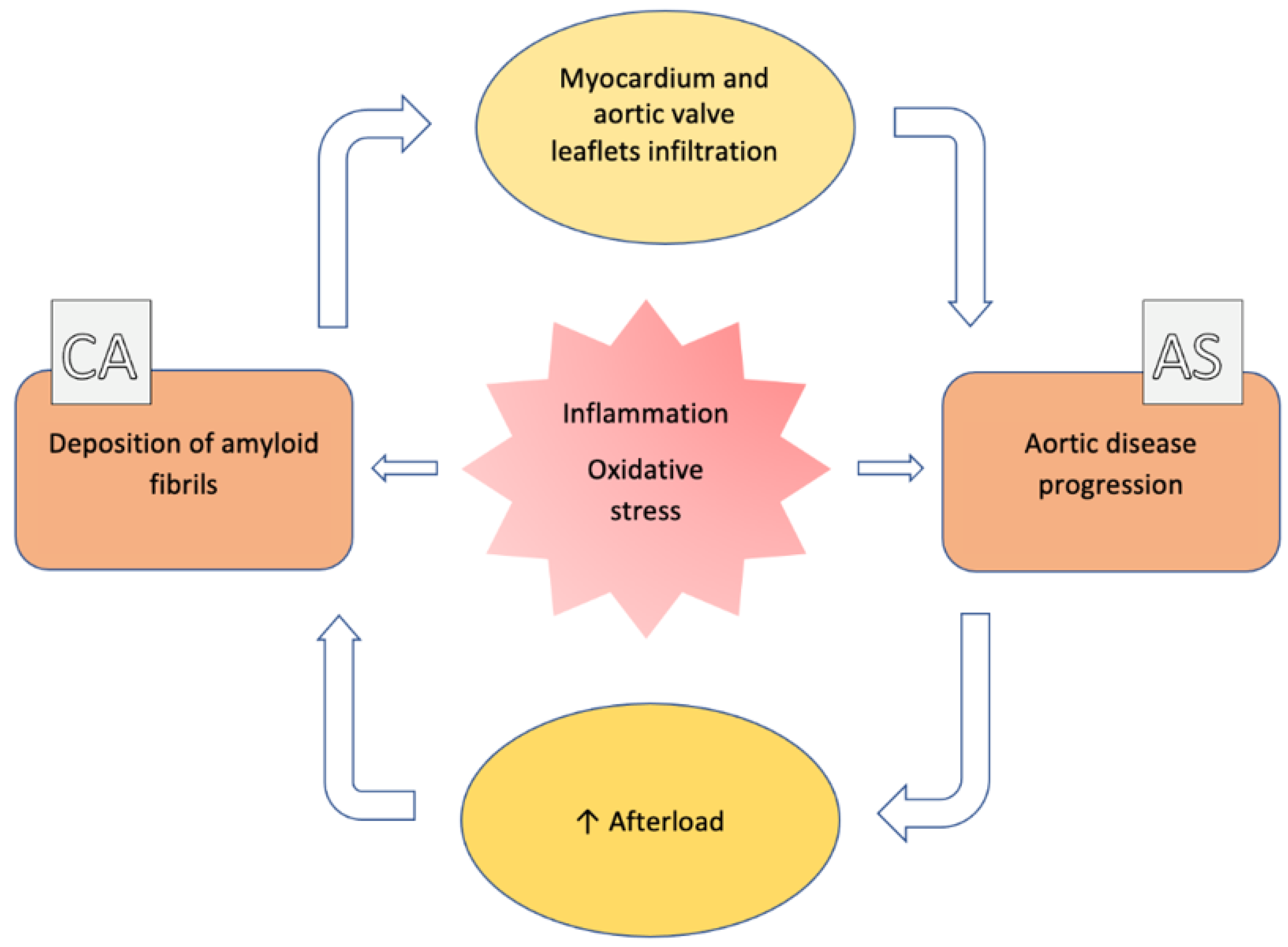
The association between aortic stenosis (AS) and cardiac amyloidosis (CA) is more frequent than expected. Albeit rare, CA, particularly the transthyretin (ATTR) form, is commonly found in elderly people. ATTR-CA is also the most prevalent form in patients with AS. These conditions share pathophysiological, clinical and imaging findings, making the diagnostic process very challenging.

multimodality imaging amyloidosis aortic valve stenosis transthyretin amyloidosis

1. Introduction

The coexistence of aortic valve stenosis (AS) and cardiac amyloidosis (CA) is not an uncommon finding in routine clinical practice. AS is the most common valvular heart disease and affects more than 4% of people aged 75 years or older [1]. Similarly, CA has a ≤25% prevalence in octogenarians, according to post-mortem studies [2].
Amyloidosis is a rare systemic disorder caused by deposition of amyloid fibrils in different organs including the heart [3]. The two predominant amyloid proteins found in the heart are transthyretin (ATTR) and immunoglobulin light chain (AL) [4] (Table 1), with ATTR amyloidosis the most prevalent form in patients with AS [1]. This combination complicates the diagnostic and therapeutic process. In fact, both conditions share epidemiological, clinical and echocardiographic features, making their management very challenging [5]. Over the last few years, given the sensitivity and specificity of bone scintigraphy, the diagnosis of ATTR-CA significantly increased, without the need for endomyocardial biopsy [6][7].
Table 1. Main amyloid types with possible cardiac involvement.

Fibril Protein

Precursor Protein

Target Organs

ATTR

Transthyretin (wild-type or variant)

Wild type: heart, carpal tunnel syndrome (bilateral), ligaments, lumbar spinal stenosis

Variant (variable): heart, PNS, ANS, ligaments, lumbar spinal stenosis, leptomeningeal, eye, gastrointestinal tract

AL

Monoclonal immunoglobulin light chain

Heart, PNS (no CNS), ANS, liver, lung, gastrointestinal tract, soft tissues (tongue), kidney, myopathy

Legend: ANS: autonomic nervous system; CNS: central nervous system; PNS: peripheral nervous system.

2. General Features (Epidemiology, Pathophysiology)

Several retrospective or prospective studies have described the presence of ATTR-CA in AS patients, with a prevalence ranging from 4% to 29% [8]. Such large variability may be explained by heterogeneity of the inclusion criteria and populations investigated. The higher prevalence was found in transcatheter aortic valve replacement (TAVR) cohorts. Interestingly, CA was found in approximately one third of patients undergoing TAVR [5].
Transthyretin is synthesized in the liver and has a role as a transporter of thyroxin and retinol-binding proteins. Its main function is the control of behavior, cognition, nerve regeneration and axonal growth [9]. The two common types are wild-type transthyretin (ATTRwt) and variant amyloidogenic (ATTRv) amyloidosis [3]. The former has a prevalence in elderly people, usually with a male preponderance. In this case, the genetic sequence of transthyretin is normal, and the aging process causes protein instability and altered aggregation. On the other hand, ATTRv has a hereditary autosomal dominant transmission, and the pathogenic mutation results in destabilization and misfolding of the ATTR protein. Several mutations have been described [10], and each of them influences the phenotype (i.e., cardiac or neurological predominance) and the severity of the disease. Moreover, the presence of mutation has important consequences on the availability and the use of specific treatment. In contrast, AL amyloidosis is a rare finding in AS patients, mainly because of the poor prognosis of the underlying disease. Indeed, AL amyloidosis is the consequence of a hematological disorder (i.e., multiple myeloma).
The pathogenetic mechanism of the two types of CA is similar. Over time, the amyloidogenic process results in amyloid fibrils aggregation and precipitation in the extracellular space and consequently causes its expansion. In the heart, this expansion causes increased biventricular wall thickness, myocardial stiffening and restrictive physiology of the left and right ventricles. In addition, amyloid fibrils may exhibit direct toxic effects on myocardial cells, impairing systolic left ventricular function [11]. While this latter mechanism is confirmed for AL amyloidosis, for ATTR it is unproven. In the heart, the progression of amyloid deposition causes severe heart failure and arrhythmias [6].
AS is the consequence of inflammatory process caused by endothelial damage due to mechanical stress and lipid penetration, eventually leading to fibrosis, leaflet thickening, sclerosis and calcification [12]. As a result, the aortic valve orifice size reduces resulting in increased pressure drop (gradient) across the valve [13]. Thus, because of long-standing pressure overload, the left ventricular myocardium thickens, and hypertrophy may mask the recognition of wall infiltration as a potential sign for the presence of an additional infiltrative disease [6].
Currently, the causative link between AS and ATTR-CA has not been demonstrated. However, there is increasing evidence supporting a central role of oxidative stress, inflammation and extracellular remodeling in the ATTR amyloidogenic process [14]. Some authors postulated that amyloid deposits could be induced or accelerated in AS, because of the pressure overload [15]. On the other side, Kristen et al. reported a high prevalence of amyloid deposits in surgically removed heart valves, mainly in AS (74% of aortic valves) [16], suggesting that amyloid deposits could induce or worsen AS (Figure 1).
Figure 1. Pathophysiologic correlation between aortic stenosis and cardiac amyloidosis. See the text for details.

3. Clinical and Imaging Assessment

Diagnosis of ATTR-CA in AS patients is challenging, as clinical and imaging features can overlap. Moreover, comorbidities such as coronary artery disease and hypertension are frequently present in the older population, making the diagnostic process, based on any risk score, even more difficult [5].
The analysis of myocardial deformation by tissue Doppler and speckle-tracking echocardiography has a crucial role in the detection of CA [17]. Reduced LV longitudinal deformation is an early marker of cardiac amyloid deposition [18][19] (Figure 2). Significantly reduced LV longitudinal deformation and global longitudinal strain has been recently reported in patients with concomitant ATTR-CA and AS [20][21] compared to lone AS. The typical pattern of ‘apical sparing’ refers to the abnormal ratios of apical to basal strain or apical to basal plus midventricular strain, which is specific in CA. The pattern reflects the more normal strain values of left ventricular apical region compared to progressively worse values at the mid and basal regions. Apical sparing has shown good diagnostic accuracy for differentiating amyloid heart disease from other etiologies [22]. Interestingly, in the study by Castano et al. [23], the apical sparing pattern was not depicted in patients with ATTR-CA and AS. This is due to elevated wall stress and increased afterload induced by AS that finally masks the reduced apical deposition of amyloid in comparison to other segments. Thus, the discriminatory power of speckle-tracking in patients with dual pathology should be elucidated, because a classical apical-sparing pattern may be hidden by the presence of AS. In contrast, the apical sparing may also be observed in patients with AS and no CA [8], with evidence suggesting that relative apical sparing becomes manifest only after TAVR [23].
Figure 2. Red flags of cardiac amyloidosis (CA). (A–E) Typical echocardiographic features of CA. (A) Left ventricle (LV) short-axis slices obtained from a 3D dataset showing severe LV hypertrophy and myocardial granular sparkling. (B) 4-Chamber view and parasternal long-axis showing an increase of left and right ventricular walls thickness, left atrial dilatation, atrial septal thickening and pericardial effusion. (C) Pulsed wave Doppler of the mitral valve inflow reveals restrictive filling pattern (grade III diastolic dysfunction) with marked reduced lateral mitral annular diastolic velocity (e′ 4.8 cm/sec at the tissue Doppler analysis); tissue Doppler signals from the lateral mitral annulus shows longitudinal systolic dysfunction with mitral Sʹ ≤ 6 cm/s. (D) Speckle tracking imaging showing a depressed LV global longitudinal strain (−12%) with apical sparing and an apex/basal longitudinal strain ratio > 2. (E) Focused right ventricle (RV) view showing RV wall thickening (≥5 mm). (FI) Typical cardiac magnetic resonance (CMR) features of CA. (F) Steady-state free precession sequences (SSFP) showing diffuse and asymmetric hypertrophy of the LV and RV. (G) T1-mapping reveals prolongation of the native relaxation time and of the extracellular volume (ECV). (H) Typical late gadolimium enhancement (LGE) pattern: LGE is extensive and circumferential, starts from the subendocardium and predominates at the basal segments with a base-to-apex gradient in a non-ischemic pattern; sub-optimal nulling of myocardium is present, and the blood pool has a signal darker than the myocardium; LGE is also evident in the RV wall, atria walls and atrial septum. (I) Frames from Look–Locker inversion recovery sequences (T1 scout) showing altered gadolinium kinetics in ATTR-CA: evidence of reverse order of sequences with the myocardium passing through the null-point before the blood pool. (J) Typical ECG findings of CA: discordance between low-voltage and LV wall thickness; discordance between the voltages in peripheral and precordial leads; pseudo-infarction pattern (Q waves) without history of myocardial infarction; right axis deviation; abnormal P wave duration and morphology reflecting slow atrial conduction. (K) 99mTc-hydroxymethylene diphosphonate scintigraphy showing strong cardiac uptake (Perugini Grade 2).
Performing bone scintigraphy with 99m technetium-labeled bisphosphonates and excluding AL by search of monoclonal light chain in blood and urine is now sufficient to confirm the suspicion of ATTR-CA [24]. In particular, grade 2 or 3 uptake on scintigraphy, using the Perugini score [25] and the absence of a monoclonal protein have specificity and a positive predictive value of 100% for ATTR-CA [24] (Figure 2). Then, genotyping is required to distinguish ATTRwt from ATTRv [21]. Clinicians should not forget that 30–50% of AL-CA patients also display cardiac uptake on scintigraphy, typically a grade 1 uptake [26]. As already stated, patients with AS mostly have ATTR-CA; however, AL-CA should be always excluded with serum/urine light chain protein analyses, because of its poor prognosis in the absence of chemotherapy [8].
Although endomyocardial biopsy and confirmation using mass spectrometry remain the gold standard for diagnosing ATTR-CA, these procedures often delay diagnosis and may not be appropriate in frail elderly adults, including those referring for TAVR. In the absence of specific CA features on imaging, a positive extracardiac biopsy is not sufficient to confirm the diagnosis. In contrast, a negative extracardiac biopsy may not exclude CA in patients with typical CA features on imaging [27]. A diagnostic flow-chart is proposed in Figure 3.
Figure 3. Diagnostic flow-chart and red flags to recognize cardiac amyloidosis involvement in AS patients. See the text for details. Legend—AF: atrial fibrillation; AS: aortic stenosis; DPD: 99mTc-3,3-diphosphono-1,2-propanodicarboxylic-acid; EMB: endomyocardial biopsy; GLS: global longitudinal strain; HF—heart failure; HMDP: 99mTc-hydroxymethylene diphosphonate; LFLG: low-flow low-gradient; LGE: late gadolinium enhancement; LV: left ventricle; Nt-proBNP: N-terminal pro-brain natriuretic peptide; PYP:99mTc-pyrophosphate, SAVR: surgical aortic valve replacement; TAVR: transcatheter aortic valve replacement.

4. Screening and Predictors

To date, there is no recommendation on whether patients with AS should be systematically screened for CA [8]. However, patients with AS requiring TAVR should be screened if there is a suspicion of concomitant ATTR-CA [5]. Recent studies have shown that patients with concomitant AS and CA are older than those with lone AS [6][23][28][29]. Age is also a significant predictor of ATTR-CA among patients referred for TAVR, even after adjustment for other variables [28][29] A history of carpal tunnel syndrome, especially if bilateral, may independently indicate the presence of amyloid deposits of ATTR [6]. NT-proBNP and high sensitivity cardiac troponin (hs-cTn) before TAVR should always be assessed, as they have been shown to be double in patients with concomitant CA and AS compared to those with lone AS, irrespective of renal function. Cardiac troponin has also been found to have a potential predictive value in this setting, but ranges were too wide to choose a cut-off [29].
ECG is another important marker of cardiac structure and function in CA and AS. Patients with concomitant ATTR-CA and AS usually exhibit more pronounced ECG abnormalities than those with solely AS. They tend to have broader QRS complex and higher prevalence of right bundle branch block [6][23]. Both these features showed a good predictive power of AS-CA at multivariate analysis [28][29]. In this context, the discordance between QRS voltage and LV hypertrophy on imaging is considered a valid element of suspicion and should help differentiating CA from hypertensive or hypertrophic cardiomyopathy [22]. Using Sokolow–Lion index to suspect cardiac involvement in patients with AS may also be helpful, as a predictor of the association between CA and AS [28][29]. This condition is also represented by another parameter, the voltage/mass ratio (VMR). The VMR combines LV mass index and signs of hypertrophy on ECG [30]. VMR values are usually lower in CA-AS patients, and this parameter has been found to efficiently discriminate between lone AS and CA-AS [6][28][29]. However, attention should be paid in case of bundle branch block or pacemaker-induced rhythm because of poor reliability on voltage quantification.
Once raised a suspicion, echocardiography with myocardial deformation analysis may add discriminatory power for the presence of CA, as initial imaging technique. Interestingly, Nitsche and colleagues showed that apical sparing was a powerful marker for diagnosing AS-CA [28]. AS-CA patients appeared to have significantly lower LV ejection fraction (LVEF), lower stroke volume index and lower trans-aortic gradient [21][23][31][32]. All these echocardiographic parameters together with high grade diastolic dysfunction, concentric hypertrophy and increased left atrial volume showed predictive power on univariate analysis [23][29]. However, only the systolic mitral annular velocity (S’) was the best predictor of ATTR-CA in a multivariable logistic regression analysis, with an AUC of 0.95, p < 0.0001, compared to the rest of the echocardiographic variables. Indeed, a cut-off value of S’ < 6 cm/s had 100% sensitivity (with a 57% specificity) in predicting a positive 99mTc-PYP (99mTc-labeled pyrophosphate) amyloid scan [23]. Thus, S’ may be used by clinicians as a valid tool for screening. Stroke volume index (SVi) was also proposed as an independent predictor of CA in patients with AS. As for low mitral annular systolic velocity, SVi values have been able to detect CA-AS with an AUC of 0.77, suggesting an additional useful tool. This finding reflects typical low flow aortic pattern for CA-AS patients [28]. The high prevalence of low-flow state is secondary to several factors: LV concentric remodeling, impairment of diastolic filling, left atrial remodeling and dysfunction and RV remodeling and dysfunction. However, 50% of patients with CA and low-flow, low-gradient AS have preserved LVEF. In this setting, stress echocardiography may be used to confirm AS severity [33]. Sometimes, in patients with CA, it may provide inconclusive results, when it fails to significantly increase LV outflow velocities [8]. In this case, the quantitation of aortic valve calcium burden using non-contrast CT may be a valid option to confirm AS severity [34].
Cardiac magnetic resonance (CMR) has demonstrated promising value for disease detection, following disease progression or monitoring response to therapy in patients with CA. However, Nitsche et al. confirmed the formerly reported low sensitivity of distinctive LGE patterns. In this study, CMR was diagnostic in only 25% of CA-AS patients [28]. Given the high cost, the availability of CMR and available findings, CMR does not seem a suitable screening tool in patients referring for TAVR.

5. Prognosis and Management

Combination of AS with CA is prognostically important. Currently, only few studies have investigated the outcome of AS-CA patients and have shown worse prognosis. Treibel and colleagues have followed 146 patients with severe AS who underwent surgical AVR for a median of 2.3 years. Of all variables assessed, the presence of ATTR-CA had the highest hazard ratio for all-cause mortality [20]. Similarly, in another group of older AS patients, CA was associated with significantly increased 1-year all-cause mortality, independently of aortic valve disease treatment. Even after adjustment for other variables including aortic valve replacement (i.e., Surgical Thoracic Society Predicted Risk of Mortality, LV ejection fraction with CMR and NYHA functional class), the presence of CA in elderly AS patients was associated with increased all-cause mortality [32]. Nevertheless, Sperry et al. found no significant difference in the 2-year mortality rate between ATTR-CA patients with and without AS [35]. Given these contrasting results, further studies are necessary to evaluate how much ATTR-CA affects outcomes, in order to choose the best therapeutic approach, especially in the TAVR era. To date, there are no recommended guidelines and no expert consensus that point out the best management of CA in patients with AS [36].

6. Conclusion

The combination of AS and CA, especially ATTR, is an important clinical problem. Its already high prevalence is destined to grow because of the aging population. However, current literature shows that CA is often underdiagnosed in old adults, resulting in underestimation of the AS-CA combination. Based on the available uncertainty of the clinical outcome of combined AS and CA, prospective multicenter studies in large cohorts are necessary to suggest an optimum road map for managing such patients.

References

  1. Coffey, S.; Cox, B.; Williams, M.J.A. The prevalence, incidence, progression, and risks of aortic valve sclerosis: A systematic review and meta-analysis. J. Am. Coll. Cardiol. 2014, 63, 2852–2861.
  2. Cornwell, G.G.; Murdoch, W.L.; Kyle, R.A.; Westermark, P.; Pitkänen, P. Frequency and distribution of senile cardiovascular amyloid. A clinicopathologic correlation. Am. J. Med. 1983, 75, 618–623.
  3. Merlini, G.; Bellotti, V. Molecular mechanisms of amyloidosis. N. Engl. J. Med. 2003, 349, 583–596.
  4. Gertz, M.A.; Dispenzieri, A.; Sher, T. Pathophysiology and treatment of cardiac amyloidosis. Nat. Rev. Cardiol. 2015, 12, 91–102.
  5. Balciunaite, G.; Rimkus, A.; Zurauskas, E.; Zaremba, T.; Palionis, D.; Valeviciene, N.; Aidietis, A.; Serpytis, P.; Rucinskas, K.; Sogaard, P.; et al. Transthyretin cardiac amyloidosis in aortic stenosis: Prevalence, diagnostic challenges, and clinical implications. Hell J. Cardiol. HJC Hell Kardiol. Ep. 2020, 61, 92–98.
  6. Nitsche, C.; Scully, P.R.; Patel, K.P.; Kammerlander, A.A.; Koschutnik, M.; Dona, C.; Wollenweber, T.; Ahmed, N.; Thornton, G.D.; Kelion, A.D.; et al. Prevalence and Outcomes of Concomitant Aortic Stenosis and Cardiac Amyloidosis. J. Am. Coll. Cardiol. 2021, 77, 128–139.
  7. Kittleson, M.M.; Maurer, M.S.; Ambardekar, A.V.; Bullock-Palmer, R.P.; Chang, P.P.; Eisen, H.J.; Nair, A.P.; Nativi-Nicolau, J.; Ruberg, F.L. American Heart Association Heart Failure and Transplantation Committee of the Council on Clinical Cardiology. Cardiac Amyloidosis: Evolving Diagnosis and Management: A Scientific Statement From the American Heart Association. Circulation 2020, 142, e7–e22.
  8. Ternacle, J.; Krapf, L.; Mohty, D.; Magne, J.; Nguyen, A.; Galat, A.; Gallet, R.; Teiger, E.; Côté, N.; Clavel, M.A.; et al. Aortic Stenosis and Cardiac Amyloidosis: JACC Review Topic of the Week. J. Am. Coll. Cardiol. 2019, 74, 2638–2651.
  9. Gargiulo, P.; Perrone-Filardi, P. Dangerous relationships: Aortic stenosis and transthyretin cardiac amyloidosis. Eur. Heart, J. 2017, 38, 2888–2889.
  10. Damy, T.; Judge, D.P.; Kristen, A.V.; Berthet, K.; Li, H.; Aarts, J. Cardiac findings and events observed in an open-label clinical trial of tafamidis in patients with non-Val30Met and non-Val122Ile hereditary transthyretin amyloidosis. J. Cardiovasc. Transl. Res. 2015, 8, 117–127.
  11. Falk, R.H.; Alexander, K.M.; Liao, R.; Dorbala, S. AL (Light-Chain) Cardiac Amyloidosis: A Review of Diagnosis and Therapy. J. Am. Coll. Cardiol. 2016, 68, 1323–1341.
  12. Dweck, M.R.; Boon, N.A.; Newby, D.E. Calcific aortic stenosis: A disease of the valve and the myocardium. J. Am. Coll. Cardiol. 2012, 60, 1854–1863.
  13. Joseph, J.; Naqvi, S.Y.; Giri, J.; Goldberg, S. Aortic Stenosis: Pathophysiology, Diagnosis, and Therapy. Am. J. Med. 2017, 130, 253–263.
  14. Zhao, L.; Buxbaum, J.N.; Reixach, N. Age-related oxidative modifications of transthyretin modulate its amyloidogenicity. Biochemistry 2013, 52, 1913–1926.
  15. Henderson, B.C.; Tyagi, N.; Ovechkin, A.; Kartha, G.K.; Moshal, K.S.; Tyagi, S.C. Oxidative remodeling in pressure overload induced chronic heart failure. Eur. J. Heart Fail. 2007, 9, 450–457.
  16. Kristen, A.V.; Schnabel, P.A.; Winter, B.; Helmke, B.M.; Longerich, T.; Hardt, S.; Koch, A.; Sack, F.U.; Katus, H.A.; Linke, R.P.; et al. High prevalence of amyloid in 150 surgically removed heart valves--a comparison of histological and clinical data reveals a correlation to atheroinflammatory conditions. Cardiovasc. Pathol. Off. J. Soc. Cardiovasc. Pathol. 2010, 19, 228–235.
  17. Yingchoncharoen, T.; Agarwal, S.; Popović, Z.B.; Marwick, T.H. Normal ranges of left ventricular strain: A meta-analysis. J. Am. Soc. Echocardiogr. Off. Publ. Am. Soc. Echocardiogr. 2013, 26, 185–191.
  18. Porciani, M.C.; Lilli, A.; Perfetto, F.; Cappelli, F.; Rao, C.M.; Del Pace, S.; Ciaccheri, M.; Castelli, G.; Tarquini, R.; Romagnani, L.; et al. Tissue Doppler and strain imaging: A new tool for early detection of cardiac amyloidosis. Amyloid. Int. J. Exp. Clin. Investig. Off. J. Int. Soc. Amyloidosis 2009, 16, 63–70.
  19. Koyama, J.; Ray-Sequin, P.A.; Falk, R.H. Longitudinal myocardial function assessed by tissue velocity, strain, and strain rate tissue Doppler echocardiography in patients with AL (primary) cardiac amyloidosis. Circulation 2003, 107, 2446–2452.
  20. Treibel, T.A.; Fontana, M.; Gilbertson, J.A.; Castelletti, S.; White, S.K.; Scully, P.R.; Roberts, N.; Hutt, D.F.; Rowczenio, D.M.; Whelan, C.J.; et al. Occult Transthyretin Cardiac Amyloid in Severe Calcific Aortic Stenosis: Prevalence and Prognosis in Patients Undergoing Surgical Aortic Valve Replacement. Circ. Cardiovasc. Imaging 2016, 9, e005066.
  21. Galat, A.; Guellich, A.; Bodez, D.; Slama, M.; Dijos, M.; Zeitoun, D.M.; Milleron, O.; Attias, D.; Dubois-Randé, J.L.; Mohty, D.; et al. Aortic stenosis and transthyretin cardiac amyloidosis: The chicken or the egg? Eur. Heart J. 2016, 37, 3525–3531.
  22. Ruberg, F.L.; Grogan, M.; Hanna, M.; Kelly, J.W.; Maurer, M.S. Transthyretin Amyloid Cardiomyopathy: JACC State-of-the-Art Review. J. Am. Coll. Cardiol. 2019, 73, 2872–2891.
  23. Castaño, A.; Narotsky, D.L.; Hamid, N.; Khalique, O.K.; Morgenstern, R.; DeLuca, A.; Rubin, J.; Chiuzan, C.; Nazif, T.; Vahl, T.; et al. Unveiling transthyretin cardiac amyloidosis and its predictors among elderly patients with severe aortic stenosis undergoing transcatheter aortic valve replacement. Eur. Heart J. 2017, 38, 2879–2887.
  24. Gillmore, J.D.; Maurer, M.S.; Falk, R.H.; Merlini, G.; Damy, T.; Dispenzieri, A.; Wechalekar, A.D.; Berk, J.L.; Quarta, C.C.; Grogan, M.; et al. Nonbiopsy Diagnosis of Cardiac Transthyretin Amyloidosis. Circulation 2016, 133, 2404–2412.
  25. Perugini, E.; Guidalotti, P.L.; Salvi, F.; Cooke, R.M.; Pettinato, C.; Riva, L.; Leone, O.; Farsad, M.; Ciliberti, P.; Reggiani, M.L.B.; et al. Noninvasive etiologic diagnosis of cardiac amyloidosis using 99mTc-3,3-diphosphono-1,2-propanodicarboxylic acid scintigraphy. J. Am. Coll. Cardiol. 2005, 46, 1076–1084.
  26. Hutt, D.F.; Quigley, A.-M.; Page, J.; Hall, M.L.; Burniston, M.; Gopaul, D.; Lane, T.; Whelan, C.J.; Lachmann, H.; Gillmore, J.D.; et al. Utility and limitations of 3,3-diphosphono-1,2-propanodicarboxylic acid scintigraphy in systemic amyloidosis. Eur. Heart J. Cardiovasc. Imaging. 2014, 15, 1289–1298.
  27. Dorbala, S.; Cuddy, S.; Falk, R.H. How to Image Cardiac Amyloidosis: A Practical Approach. JACC Cardiovasc. Imaging 2020, 13, 1368–1383.
  28. Nitsche, C.; Aschauer, S.; Kammerlander, A.A.; Schneider, M.; Poschner, T.; Duca, F.; Binder, C.; Koschutnik, M.; Stiftinger, J.; Goliasch, G.; et al. Light-chain and transthyretin cardiac amyloidosis in severe aortic stenosis: Prevalence, screening possibilities, and outcome. Eur. J. Heart Fail. 2020, 22, 1852–1862.
  29. Scully, P.R.; Patel, K.P.; A Treibel, T.; Thornton, G.D.; Hughes, R.K.; Chadalavada, S.; Katsoulis, M.; Hartman, N.; Fontana, M.; Pugliese, F.; et al. Prevalence and outcome of dual aortic stenosis and cardiac amyloid pathology in patients referred for transcatheter aortic valve implantation. Eur. Heart J. 2020, 41, 2759–2767.
  30. Carroll, J.D.; Gaasch, W.H.; McAdam, K.P. Amyloid cardiomyopathy: Characterization by a distinctive voltage/mass relation. Am. J. Cardiol. 1982, 49, 9–13.
  31. Longhi, S.; Lorenzini, M.; Gagliardi, C.; Milandri, A.; Marzocchi, A.; Marrozzini, C.; Saia, F.; Ortolani, P.; Biagini, E.; Guidalotti, P.L.; et al. Coexistence of Degenerative Aortic Stenosis and Wild-Type Transthyretin-Related Cardiac Amyloidosis. JACC Cardiovasc. Imaging 2016, 9, 325–327.
  32. Cavalcante, J.L.; Rijal, S.; Abdelkarim, I.; Althouse, A.D.; Sharbaugh, M.S.; Fridman, Y.; Soman, P.; Forman, D.E.; Schindler, J.T.; Gleason, T.G.; et al. Cardiac amyloidosis is prevalent in older patients with aortic stenosis and carries worse prognosis. J. Cardiovasc. Magn. Reson. Off. J. Soc. Cardiovasc. Magn. Reson. 2017, 19, 98.
  33. Salinger, T.; Hu, K.; Liu, D.; Herrmann, S.; Lorenz, K.; Ertl, G.; Nordbeck, P. Cardiac amyloidosis mimicking severe aortic valve stenosis—A case report demonstrating diagnostic pitfalls and role of dobutamine stress echocardiography. BMC Cardiovasc. Disord. 2017, 17, 86.
  34. Salinger, T.; Hu, K.; Liu, D.; Herrmann, S.; Lorenz, K.; Ertl, G.; Nordbeck, P. Impact of aortic valve calcification, as measured by MDCT, on survival in patients with aortic stenosis: Results of an international registry study. J. Am. Coll. Cardiol. 2014, 64, 1202–1213.
  35. Sperry, B.W.; Jones, B.M.; Vranian, M.N.; Hanna, M.; Jaber, W.A. Recognizing Transthyretin Cardiac Amyloidosis in Patients with Aortic Stenosis: Impact on Prognosis. JACC Cardiovasc. Imaging 2016, 9, 904–906.
  36. Java, A.P.; Greason, K.L.; Dispenzieri, A.; Grogan, M.; King, K.S.; Maleszewski, J.J.; Daly, R.C.; Eleid, M.F.; Pochettino, A.; Schaff, H.V. Aortic valve replacement in patients with amyloidosis. J. Thorac. Cardiovasc. Surg. 2018, 156, 98–103.
More
Information
Contributors MDPI registered users' name will be linked to their SciProfiles pages. To register with us, please refer to https://encyclopedia.pub/register : , , , , , ,
View Times: 311
Revisions: 2 times (View History)
Update Date: 16 Aug 2022
1000/1000