Submitted Successfully!
To reward your contribution, here is a gift for you: A free trial for our video production service.
Thank you for your contribution! You can also upload a video entry or images related to this topic.
Version Summary Created by Modification Content Size Created at Operation
1 -- 5403 2022-07-29 03:54:13 |
2 format correct Meta information modification 5403 2022-07-29 04:07:28 |

Video Upload Options

Do you have a full video?

Confirm

Are you sure to Delete?
Cite
If you have any further questions, please contact Encyclopedia Editorial Office.
Ng, C.Y.;  Kee, L.T.;  Al-Masawa, M.E.;  Lee, Q.H.;  Subramaniam, T.;  Kok, D.;  Ng, M.H.;  Law, J.X. Scalable Production of Extracellular Vesicles. Encyclopedia. Available online: https://encyclopedia.pub/entry/25637 (accessed on 28 March 2024).
Ng CY,  Kee LT,  Al-Masawa ME,  Lee QH,  Subramaniam T,  Kok D, et al. Scalable Production of Extracellular Vesicles. Encyclopedia. Available at: https://encyclopedia.pub/entry/25637. Accessed March 28, 2024.
Ng, Chiew Yong, Li Ting Kee, Maimonah Eissa Al-Masawa, Qian Hui Lee, Thayaalini Subramaniam, David Kok, Min Hwei Ng, Jia Xian Law. "Scalable Production of Extracellular Vesicles" Encyclopedia, https://encyclopedia.pub/entry/25637 (accessed March 28, 2024).
Ng, C.Y.,  Kee, L.T.,  Al-Masawa, M.E.,  Lee, Q.H.,  Subramaniam, T.,  Kok, D.,  Ng, M.H., & Law, J.X. (2022, July 29). Scalable Production of Extracellular Vesicles. In Encyclopedia. https://encyclopedia.pub/entry/25637
Ng, Chiew Yong, et al. "Scalable Production of Extracellular Vesicles." Encyclopedia. Web. 29 July, 2022.
Scalable Production of Extracellular Vesicles
Edit

Extracellular vesicles (EVs) are minute vesicles with lipid bilayer membranes. EVs are secreted by cells for intercellular communication. Recently, EVs have received much attention, as they are rich in biological components such as nucleic acids, lipids, and proteins that play essential roles in tissue regeneration and disease modification. In addition, EVs can be developed as vaccines against cancer and infectious diseases, as the vesicle membrane has an abundance of antigenic determinants and virulent factors. EVs for therapeutic applications are typically collected from conditioned media of cultured cells. However, the number of EVs secreted by the cells is limited. Thus, it is critical to devise new strategies for the large-scale production of EVs.

extracellular vesicle large-scale culture medium production stem cell bioreactor three-dimensional culture

1. Introduction

Extracellular vesicles (EVs) are naturally-occurring heterogeneous nano- to micro-sized lipid bilayer membrane vesicles packed with regulatory biological cargo, i.e., cytosol, lipids, proteins, and nucleic acids [1][2]. EVs are secreted by most of the cells and mediate intercellular communication in physiological and pathological conditions. EVs can be mainly categorized into three subtypes, i.e., exosomes, microvesicles (also called ectosomes), and apoptotic bodies, based on their biogenesis pathways. Each EV subtype has different sizes, cargoes, and functions [1]. Research on EVs as cell-free regenerative therapies, targeted therapies, drug carriers, diagnosis biomarkers, and cancer vaccines has grown drastically.
EVs have been widely investigated as drivers of tissue regeneration. In recent years, stem cell-derived EVs received much attention, as they possess therapeutic potential comparable to or even better than that of the parent cells [3]. In addition, stem cell EVs are nonliving vesicles with superior safety profiles to those of cells in clinical applications. Stem cell-derived EVs carry low risks of tumorigenicity and allogeneic immune rejection as well as minimal risk of microvascular occlusion during intravascular administration because of their nano size [4][5]. The regenerative potential of EVs has been reported in many preclinical studies to treat a myriad of diseases [6][7][8][9]. Additionally, positive outcomes have been reported in a few clinical trials, with many more EV-based clinical trials currently ongoing worldwide.
On the other hand, EVs of cancer cells have the potential to be used as drug carriers and cancer vaccines. Cancer cell-derived EVs have a higher affinity to cancer cells due to the unique protein and lipid composition that facilitates the binding or internalization of EVs in cancer cells [10][11][12]. Thus, they can be used for targeted delivery of chemotherapeutic agents and enhance treatment efficacy while minimizing off-target effects [13][14]. Cancer cell-derived EVs also possess large numbers of tumor antigens, which can trigger the host’s immune responses to inhibit tumor growth [15]. EVs produced by cancer cells can also be employed as diagnostic cancer biomarkers, since they contain cargo that reflects the tumor’s genetic and mutational status [16][17]. Understanding of the impact of tumor microenvironments, e.g., pH, extracellular matrix (ECM) stiffness, oxidative stress, hypoxia, and nutrient deprivation, as well as of treatment modalities, e.g., irradiation, chemotherapy, and photodynamic therapy, on the EV secretion of cancer cells has been instrumental in enhancing the EV production by cancer cells in vitro [18][19][20][21][22].
EVs can be derived from many sources, most commonly from cells and biofluids. Currently, EVs for preclinical and clinical studies are mainly produced from cultured cells, as they are easier to manipulate. However, the number of EVs produced by cells is meager in standard two-dimensional (2D) culture conditions. Thus, obtaining a sufficient EV yield is one of the major hurdles for translating EV-based therapeutics. Therefore, it is crucial to devise new strategies to stimulate cells to release more EVs for clinical applications. The manufacturing of EVs includes a series of sequential steps, beginning with the isolation and culture of source cells, which is followed by the separation and storage of EVs [23]

2. Strategies to Increase Production of Extracellular Vesicles

Several strategies have been investigated in the literature to upscale the release of EVs from cultured cells. Alteration of cell culture environments, e.g., three-dimensional (3D) culture, chemical stimulation, physical stimulation, physiological modification, and genetic manipulation of source cells, has been the most common approach to enhancing the quantity and quality of EVs secreted by cells. Alternatively, some studies have explored the use of physical techniques such as sonication, nitrogen cavitation, and porous membrane extrusion to produce EV-like vesicles, known as EV-mimetic nanovesicles, which have similar characteristics and functionalities to those of natural EVs while circumventing some of their limitations (Figure 1).
Figure 1. Scalable production of extracellular vesicles. Physical stimulation, chemical stimulation, 3D culture, physiological modification, and genetic manipulation can stimulate cells to produce more extracellular vesicles. In addition, EV-mimetic nanovesicles can be produced via nitrogen cavitation, porous membrane extrusion, and sonication. Created with BioRender.com.

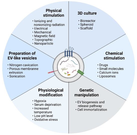
2.1. Three-Dimensional Culture

One technique for increasing EV secretion from cultured cells is using 3D cell culture systems such as bioreactors and cell spheres. Generally, 3D cell culture systems allow the expansion of a large number of cells in high density for high yields of EVs, as they provide a large surface area for cell growth [24].
The bioreactor is the most commonly used 3D cell culture system for large-scale EV production, as not only does it increase the volume of production, but the mechanical stimulus provided by the bioreactor stimulates the cultured cells to produce more EVs [25]. Generally, the use of bioreactors can increase the yield and concentration of EVs in conditioned medium and reduce the production time. In a study, the authors found that umbilical cord-derived MSCs (UC-MSCs) cultured in 3D hollow fiber bioreactors secreted 7.5 times more small EVs than cells cultured in 2D tissue culture flasks [26]. In addition, the authors found that 3D-EVs were more potent in promoting chondrocyte proliferation and migration as well as inhibiting chondrocyte apoptosis in vitro. Using an in vivo cartilage defect model, the authors proved that 3D-EVs were more effective in promoting cartilage regeneration. It was postulated that 3D-EVs modulated chondrocyte functions by activating the TGF-β1 and Smad 2/3 signaling pathways.
Cao et al. demonstrated that the protein yield of small EVs from UC-MSCs cultured in a 3D hollow-fiber bioreactor was 19.4-fold higher than that from cells cultured in a 2D conditions [27]. In vivo findings showed that 3D-EVs were more effective than 2D-EVs in ameliorating cisplatin-induced acute kidney injury, as indicated by better renal function, less severe pathological changes in renal tubules, and lower infiltration of inflammatory cells. The authors attributed the renoprotective effects of 3D-EVs to their increased uptake by tubular epithelial cells and enhanced antiinflammatory activity.
In a study comparing the small EVs secreted by MSCs derived from bone marrow (BM), adipose tissue (AT), and the umbilical cord matrix (UCM) cultured in a 3D microcarrier-based Vertical-Wheel™ Bioreactor (VWBR) and a 2D tissue culture flask, the authors found 4.0-fold, 4.4-fold, and 8.8-fold increases in small EV concentration in conditioned medium and 1.4-fold, 3.7-fold, and 3.9-fold increases in small EV productivity for the 3D cultured BM-MSCs, AT-MSCs, and UCM-MSCs, respectively, compared with the 2D cultures [28]. The therapeutic potential of the isolated EVs is unknown, as no functionality assessments were performed in this entry.
Watson et al. reported a 40-fold increase in small EV secretion when human embryonic kidney (HEK) 293 cells stably expressing hetIL-15 were cultured in a hollow fiber bioreactor than when they were cultured in a standard 2D culture [29]. Moreover, the study found that the 3D-EVs contained fewer serum protein contaminants than the 2D-EVs. Large-scale production of EVs using bioreactors has also been reported in other studies [30][31]. However, these studies did not compare the EV yield with that of 2D cultures. Nonetheless, data from these studies demonstrated the feasibility of large-scale production of cell-derived EVs using bioreactors.
In addition, MSCs cultured in collagen scaffolds were found to secrete two times more small EVs than those cultured in 2D conventional conditions [32]. The 3D-EVs were more effective in promoting neurological functional recovery of traumatic brain injury models in rats than 2D-EVs and liposomes. The 3D scaffold is often used with a bioreactor, which helps to improve the nutrient perfusion and waste removal, which is critical to keep the cells viable and healthy. Patel et al. cultured human dermal microvascular endothelial cells (HDMECs) in a 3D-printed scaffold-perfusion bioreactor to collect small EVs [33]. The authors found that 3D-cultured endothelial cells secreted 100 times (collected on day 1) and 10,000 times (collected on day 3) more small EVs than those cultured in static scaffold and tissue culture flasks as assessed by NTA. However, a mere 14-fold increase in CD63 Exo-ELISA analysis was recorded between the 3D-EVs and the 2D-EVs collected from the tissue culture flasks. Furthermore, the 3D-EVs preconditioned with ethanol demonstrated a more potent provascularization effect that was attributed to higher concentrations of the proangiogenic lncRNAs HOTAIR and MALAT1 in 3D-EVs.
Apart from bioreactors, 3D spheroid cultures have been found to increase the secretion of EVs by cells [34]. 3D hanging drop spheroids and 3D poly(2-hydroxyethyl methacrylate) spheroids produced significantly more EVs than 2D-cultured BM-MSCs [34]. Interestingly, the authors found that EV secretion reduced when the size of the 3D spheroids increased. Thus, size could be an important parameter to optimize when 3D spheroid culture is used for the large-scale production of EVs.

2.2. Physical Stimulation

Physical stimulation techniques such as irradiation, electrical stimulation, magnetic field stimulation, mechanical stimulation, and topographic cues have been explored and used to enhance EV production. Generally, physical stimulation stresses cells to produce more EVs [35]. Ionizing radiation has been reported to increase the number of EVs produced by cancer cells in a dose- and time-dependent manner [18][36][37][38]. The upregulation of EV production by cancer cells upon exposure to ionizing radiation has been linked with the DNA-damaged activated p53 signaling pathway [39][40]. These studies indicated that irradiation could increase the release of EVs from cancer cells, thus aiding in developing strategies for cancer treatment. However, a study found that ionizing radiation neither altered EV secretion by cancer cells nor modified the protein cargo of the secreted EVs [41]. Limited research has explored the effects of ionizing radiation on EV secretion by normal cells. A study found that ionizing radiation increased the small EV particle concentration of astrocytes by 1.71 times [38]. In addition, nonionizing radiation (ultraviolet radiation) [42], photodynamic therapy (Foscan® photosensitizer) [19], and acoustic irradiation (at low power and high frequency) [43] have been reported to increase the number of EVs secreted by cancer cells.
Low-level electrical stimulation applied to murine melanoma and fibroblast cells was found to stimulate EV secretion, possibly through Rho guanosine triphosphatase (GTPase) activation [44]. In another study, focal and transient electrical stimulation that induced cell membrane nanoporation increased the EV secretion of mouse embryonic fibroblasts 50-fold, while moderate increases in EV yield were detected in cells cultured in serum-depletion conditions, hypoxic conditions (1% O2) and heat stress conditions (42 °C for 2 h) [45]. Additionally, nanoporation increased the mRNA cargo of EVs. The combination of magnetic iron (III) oxide nanoparticles (Fe3O4) and a static magnetic field (SMF) was utilized to increase the small EV secretion of bone MSCs [46]. The highest EV production was recorded in the Fe3O4 + SMF group, followed by the Fe3O4 and untreated groups. In terms of functionality, the small EVs secreted by the Fe3O4 + SMF-stimulated cells showed better osteogenic and angiogenic potential than the Fe3O4 stimulated cells and naïve cells.
Several studies have reported higher EV production by cancer cells cultured on biomaterials with higher stiffness; increased ECM stiffness is one of the key changes in the tumor microenvironment [20][47]. EVs induced by stiff matrices were found to promote tumor cell migration and proliferation. Cyclic stretch increased the CD63+ EV secretion of periodontal ligament cells, and the secreted EVs demonstrated improved immunomodulatory properties to suppress IL-1β production by activated macrophages [48]. Zhang et al. cultured BM-MSCs on micro-/nanonet-textured hierarchical titanium surfaces and micro-/nanotube-textured hierarchical titanium surfaces and found that these surface topographies increased small EV secretion [49].
Multiple studies have tested nanoparticle incorporation to increase the number of EVs secreted by cultured cells. The nanoparticles found to increase EV secretion include platinum nanoparticles in human lung epithelial adenocarcinoma cancer cells (through induction of oxidative stress and ceramide pathway) [50], silver-titanium oxide nanoparticles in B16F1 mouse melanoma cells (through induction of oxidative stress) [51], poly(lactic-co-glycolic acid)-polyethyleneimine (PLGA-PEI) positively charged-surface-modified nanoparticles containing iron oxide in MSCs (through promotion of MVB formation) [52], and calcium phosphate particles in macrophage-like RAW264.7 cells and monocyte-like THP-1 cells (through promotion of MVB formation and fusion with the plasma membrane) [53]. In addition, bioactive glass upregulated the small EV production of MSCs through the activation of the nSMase and Rab GTPase pathways [54].

2.3. Chemical Stimulation

The addition of chemicals to the culture medium to boost EV secretion has been investigated in several studies. Treatment with sodium iodoacetate (IAA; glycolysis inhibitor) and 2,4-dinitrophenol (DNP; oxidative phosphorylation inhibitor) was found to increase the number of small EVs secreted by cancer cells [55]. In addition, IAA/DNP increased the number of small EVs released into the culture medium from kidney explants. In vivo validation of the in vitro and ex vivo data was achieved by injecting the IAA/DNP into mice. In comparison with that in control mice, IAA/DNP injection increased the quantity of small EVs in the blood. The study partially related the higher small EV production to higher intracellular 2′-3′-cAMP levels. Wang et al. tested the effects of several small molecules, including fenoterol, norepinephrine, N-methyldopamine, mephenesin, and forskolin, on the small EV production of BM-MSCs [56]. The findings showed that all of these small molecules could stimulate small EV secretion and that the magnitudes of the increases were affected by the concentrations of each small molecule. Synergistic improvement in exosome secretion was recorded for the combinations of norepinephrine with forskolin and norepinephrine with N-methyldopamine, but not for the combination of N-methyldopamine with forskolin. Furthermore, a multiple-component herbal combination in the Suxiao Jiuxin pill (a traditional Chinese herbal medicine) revealed a synergistic effect in promoting the small EV secretion of cardiac MSCs via a GTPase-dependent pathway [57].
In addition, adiponectin, an adipokine, increased the numbers of small EVs produced by MSCs and vascular endothelial cells through T-cadherin [58][59]. In addition, adiponectin increased the concentration of exosomes in mouse serum, and this increase was linked to the augmented cardioprotective function of primed MSCs. Docosahexaenoic acid (DHA) increased the CD63+ EV secretion of breast cancer cells. The DHA-EVs contained more antiangiogenic miRNAs (miR23b, miR-27b, and miR-320b), which aided in anticancer action [60].
Intracellular calcium ions were reported to modulate EV release [61][62][63]. A few studies explored the effects of calcium exposure on EV production [53][64]. The results indicated that calcium exposure increased EV production. In a different study, extracellular DNA and phosphorothioate CpG oligodeoxynucleotides were found to induce Alix+ EV secretion of HEK293 cells and head kidney leukocytes of Atlantic salmon [65]. The neutral and cationic liposomes were reported to stimulate the EV secretion of tumor cells in a dose-dependent manner [66]. However, the PEGylated liposomes diminished the EV production. Thus, the authors postulated that the influence of liposomes on EV production was dependent on their physicochemical properties.

2.4. Physiological Modification

The quantity and functionality of cell-secreted EVs are likely microenvironment dependent. In the physiological state, the oxygen level in peripheral tissues, known as “physoxia”, ranges between 1 and 11% [67]. Multiple studies have reported that hypoxic preconditioning augments the therapeutic efficacy of MSCs [68][69][70]. The improved therapeutic efficacy could be related to the higher EV secretion. Dong et al. showed that UC-MSCs cultured in hypoxic conditions (5% O2) demonstrated a higher proliferation rate and viability than those cultured in normoxia (21% O2) [71]. The hypoxic UC-MSCs produced more small EVs with higher potency to attenuate chronic airway inflammation and lung remodeling in ovalbumin-induced asthma mice. The results of another research group showed that hypoxic BM-MSCs secreted more small EVs with more potent therapeutic potential in promoting cartilage [72] and spinal cord regeneration [73] than those secreted by normoxic cells. Secretion of CD29+, CD44+, CD73+, CD31−, and CD45− EVs from UC-MSCs also increased in hypoxic conditions, and the angiogenic potential of the secreted EVs was superior for endothelial cells that were cultured in hypoxic conditions than those that were cultured in normoxic conditions [74]. In addition, extreme hypoxic conditions (0.5% O2) remarkably increased small EV release by MSCs [75]. In terms of functionality, hypoxic small EVs were more effective in promoting myocardial repair than normoxic small EVs, as they promoted vascularization, reduced cardiomyocyte apoptosis, reduced scar tissue formation, and enhanced the recruitment of cardiac progenitor cells. However, contradictory results were reported by Almeria et al., who found no significant difference in EV secretion by AT-MSCs cultured in normoxic and hypoxic conditions [76]. However, hypoxic priming enhanced the angiogenic potential of the secreted EVs. Apart from stem cells, hypoxic conditioning also has been found to induce EV secretion by cancer cells [22][37][42][77][78].
Since hypoxic conditioning can promote EV production, Gonzalez-King et al. overexpressed hypoxia-inducible factor-1a (HIF-1a), a vital mediator in low oxygen adaptation, in human dental pulp MSC via lentiviral transduction [79]. The HIF-1a overexpressed MSCs produced more small EVs that showed more potent angiogenic potential. Taken together, hypoxia culture appears to be more suitable for maintaining both stem cell and cancer cell cultures, as it mimics the native tissue physiological microenvironment. Stem cells and cancer cells cultured in hypoxic environments are also more biologically active, secreting more EVs.
Anderson et al. primed BM-MSCs under peripheral arterial disease (PAD)-like conditions, i.e., 0% serum and 1% oxygen [80]. They found that BM-MSCs cultured in PAD-like conditions secreted more low-density EVs and fewer high-density EVs than the control cells. Furthermore, the exosomes derived from PAD-like culture elevated the expression of several proangiogenic signaling proteins. The study showed that serum and oxygen deprivation could be used in tandem to induce low-density EV secretion. The use of serum deprivation to induce the EV secretion of mouse embryonic fibroblasts was reported by Yang et al. [45].
Enhancement of EV production via thermal stimulation was reported by Hedlund et al. [21]. Induction with thermal (40 °C for 1 h) and oxidative stress (50–100 µM H2O2 for 2 h) increased the secretion of CD63+ EVs by leukemia/lymphoma T- and B-cells [21]. Interestingly, the findings also showed that cells responded to stressors differently. The Jurkat cells were more responsive to oxidative stress, and the Raji cells were more susceptible to thermal stress by producing more CD63+ EVs in these culture conditions. Thermal stimulation of EV production by B-cells was also reported by Clayton et al., who found a small (1.25-fold) increase in EV secretion when cells were cultured at 40 °C for 3 h [81]. Harmati et al. showed that B16F1 mouse melanoma cells produced more small EVs when they were cultured at 42 °C for 2 h three times [51], and Gong et al. found that MGC-803 human gastric cancer cells released more small EVs in response to high temperatures (40 °C) [42].
Physiological and intracellular pH are important in many biological processes and cellular metabolism [82][83]. Kim et al. reported that a slight difference in the pH of the culture medium affected cell reprogramming and differentiation [84]. Besides hypoxia, extracellular acidity is another hallmark of cancer because of the accumulation of glycolytic metabolites such as lactic acid [85]. Melanoma cells secreted more Lamp-2+, CD81+, and Rab5B+ EVs that served as intercellular cross-talk mediators in acidic conditions (pH 6.0) than in buffered conditions (pH 7.4) to transport tumor-associated proteins to the other cells [12]. Higher secretion of small EVs in acidic pH (pH 4) was also reported by Gong et al. using MGC-803 human gastric cancer cells [42]. In a study using HEK293 cells, the authors reported that more CD9+, CD63+, and Hsp70+ EVs were collected from a conditioned medium of HEK293 cells cultured at pH 4 than from conditioned media of cells cultured at pH 7 and pH 11 [86]. The findings above demonstrated that cells could respond to environmental stresses and pathological conditions, such as tumor microenvironment (i.e., higher temperature, lower oxygen tension, and lower pH level), by altering their EV production. The changes in EVs produced by cells are also being studied to understand the cellular response to stress and pathological conditions.

2.5. Genetic Manipulation

Genetic modification of parental cells has been performed to modulate the signaling pathway regulating EV secretion. Rab proteins are GTPases that regulate vesicle traffic and have been identified to regulate the secretory pathway of EVs [87]. Studies by Bobrie et al. [88] and Ostrowski et al. [89] showed that silencing of RAB27A and RAB27B genes reduced multivesicular endosome (MVE) docking to the plasma membrane and reduced the small EV secretion of cancer cells. Furthermore, Rab35 depletion resulted in the accumulation of late endosomal vesicles and reduced exosome secretion in the oligodendroglial precursor cells [90]. More recently, Rab13 and Rab7a were also found to regulate the EV secretion of cancer cells [91][92].
Phospholipase D (PLD) catalyzes phosphatidylcholine hydrolysis to produce phosphatidic acid, an important lipid messenger involved in cell signaling, including exocytosis and endocytosis [93][94]. A significant increase in BODIPY-ceramide-labeled EV secretion was reported in PLD2-overexpressing RBL-2H3 cells (mast cells), while PLD2-knockout RBL-2H3 cells demonstrated poorer EV release [95]. PLD2 has been found to act as the effector of the ADP ribosylation factor 6 (ARF6) gene in regulating intraluminal vesicle (ILV) budding, thus playing an integral role in exosome production [96]. Furthermore, ARF6 has been reported to control the shedding of microvesicles in tumor cells [97]. Böker et al. found that the increased exogenous expression of tetraspanin CD9 after lentivirus transduction enhanced the secretion of small EVs in five cell lines, i.e., HEK293, SH-SY5Y, HeLa, Raji, and Jurkat, up to threefold [98]. Apart from the molecular pathways reported above, modulation of P2X7 and SNAREs receptor expression was explored to upregulate EV secretion, as these proteins have been found to influence EV formation, trafficking, and secretion in cells [99][100]. The results from these studies indicated that EV production could be modulated by targeting the key factors involved in the biogenesis and release of EVs through genetic modification of parent cells.
As discussed above, stress modulates the production of EVs by cells [51][101]. At the molecular level, p53-regulated exosome production typically occurs in response to the stress from DNA damage. Yu et al. demonstrated that upregulated transcription of tumor suppressor-activated pathway 6 (TSAP6) by activated p53 upon γ-radiation increased the small EV secretion of H460, a non-small cell lung cancer cell line [102]. It was also found that transfection of HA-tagged TSAP6 into H460 cells allowed small EV secretion without stress stimuli. Impaired EV secretion in TSAP6-knockout mice was reported by Lespagnol et al. [103]. These results indicated that TSAP6 was an essential mediator of p53-regulated exosome production. In addition, transfection of liver kinase B1 (LKB1), which is known to modulate the cell functions through the p53 pathway in lung cancer cells, was also found to increase the secretion of small EVs [104]. The small EVs secreted by LKB1-expressing cells contained fewer migration-suppressing miRNAs that inhibit cell migration.
Upregulation of eukaryotic translation initiation factor 3 subunit C (EIF3C) in human hepatocellular carcinoma cells increased the secretion of proangiogenic small EVs [105]. Knockdown of PIKfyve increased the small EV secretion of human prostate cancer epithelial cells by inhibiting MVB and autophagosome fusion with lysosomes and increasing the fusion of MVBs and autophagosomes [106].
Cell immortalization is a technique utilized to achieve the consistent production of EVs on a large scale using the desired cell source. Chen et al. generated highly expansible human ESC-MSCs by transfecting cells with the MYC gene [107]. The immortalized MYC-transformed hESC-MSCs bypassed cell senescence and could maintain high proliferation for more than 20 passages. Notably, the small EVs secreted by the immortalized MYC-transformed hESC-MSCs exhibited cardioprotective potential in an in vivo myocardial ischemia/reperfusion injury model. In another study, the same group of researchers revealed the safety of daily injection of the immortalized MYC-transformed hESC-MSC-secreted small EVs, as the small EVs did not affect tumor growth [108]. Nonetheless, the effects of EVs on cancer progression warrant further examination, as mixed results have been reported in different studies [109][110][111][112].

2.6. Preparation of EV-Mimetic Nanovesicles

Apart from stimulating the cultured cells to produce more EVs, an engineered approach has been developed to produce large-scale mimetic biologically functional nanovesicles, known as EV-mimetic nanovesicles. EV-mimetic nanovesicles are synthetic EVs that can be produced via top-down (plasma membrane fragmentation) or bottom-up (supramolecular chemistry) techniques [113]. EV-mimetic nanovesicles possess properties like those of naturally secreted EVs in terms of morphology, size, and functions [114]. Physical techniques, such as nitrogen cavitation, porous membrane extrusion, and sonication, and chemical techniques using chemical agents have been utilized to disrupt the cellular membranes and then the self-reassembly of lipids and membranes to form lipid vesicles that contain active ingredients. The advantages of this method are that it is easy to perform, increases the EV yield, and permits the production of homogenous EVs on a large scale.
Cavitation is a technical word in physics that describes the creation of many microscopic vapor-filled cavities as a result of a rapid pressure change in a liquid. When these cavities collapse, they produce a powerful shock that causes items to shatter. Nitrogen cavitation refers to the use of nitrogen gas to provide the pressure necessary for cavitation forces to occur. Gao et al. were the first to report the use of nitrogen cavitation to create synthetic EVs from white blood cells [115][116]. The cells were broken by expanding bubbles, which released cellular components into the fluid. Broken cellular membranes created vesicles with a wide range of particle sizes on their own. Gao et al. discovered that 50–75% of the cell plasma membrane generated vesicles with diameters of 180–200 nm. Extrusion of the vesicles through a membrane with 200 nm pore size resulted in EV-mimetic nanovesicles of uniform size. Nitrogen cavitation produced 16 times more EV-mimetic nanovesicles than naturally secreted EVs.
Sonication is regularly used for liposome preparation [117]. However, it can also be employed for EV-mimetic nanovesicle preparation. Thamphiwatana et al. used sonication to prepare EVs from macrophages [118]. In their protocol, the membranes of mouse macrophages were purified using a combination of hypotonic lysis, mechanical disruption, and differential centrifugation before sonication to form membrane vesicles that were later fused onto a poly(lactic-co-glycolic acid) (PLGA) core. The EV-mimetic nanovesicles retained many of the biological properties of the macrophages and were able to treat sepsis in a mouse bacteremia model. In another study, the authors prepared EV-mimetic nanovesicles through sonication of human UC-MSCs [119]. The EV-mimetic nanovesicle yield from sonication preparation was approximately 18.5-fold higher than the yield of natural EVs secreted by cells cultured in a serum-depleted medium. The EV-mimetic nanovesicles prepared by sonication were slightly larger than the naturally secreted EVs, i.e., 133.3 ± 1.8 nm vs. 122.9 ± 2.3 nm, respectively. Nonetheless, both expressed EV markers, i.e., CD9, CD63, and CD81. In terms of functionality, both EV preparations could promote wound healing in vivo using a mouse full-thickness excisional wound model.
A few studies have used the serial extrusion technique to prepare EV-mimetic nanovesicles. In general, in this technique, cells are mechanically broken down into nanosized vesicles by passing them through filters with reducing pore sizes (e.g., 10, 5, and 1 µm). The produced EV-mimetic nanovesicles can be up to 100 times more abundant than naturally secreted EVs, and they share the common features of exosomes, including size (30–200 nm) and marker expression (e.g., positive for CD9, CD63, CD81, TSG101, moesin, and β-actin) [120][121][122][123]. EV-mimetic nanovesicles could be used as drug carriers [120][121][123]. They were also more effective than naturally secreted EVs in treating emphysema [124] and could induce liver regeneration [125] in vivo.
Chemical agents, such as alkaline solutions, can be used to break down the cell membrane. Under sonication, the membrane components may self-assemble to form EV-mimetic nanovesicles after neutralizing the pH. Go et al. used human U937 monocytes to make EV-mimetic nanovesicles via sequential treatment with alkaline solution and sonication with and without the presence of dexamethasone [126]. The EV-mimetic nanovesicles exhibited identical physical properties to spontaneously released EVs. In comparison with the cell culture approach, there was a 200-fold increase in EV generation. The authors also discovered that the EV-mimetic nanovesicles lacked intracellular compartments such as cytosolic proteins and nucleic acids. In terms of functionality, the EV-mimetic nanovesicles loaded with dexamethasone were able to reduce systemic inflammatory response syndrome (SIRS) caused by the outer membrane vesicles (OMVs) of Gram-negative bacteria.
Sulfhydryl-blocking agents are known to cause cell membrane blebbing [127]. Thus, they were examined to replace physical processes to induce EV formation. Ingato et al. exposed a mouse lymphoma cell line to sulfhydryl-blocking agents, i.e., dithiothreitol (DTT) and paraformaldehyde (PFA), to prepare EVs [128]. Within 2 h, sulfhydryl blocking boosted EV production by more than tenfold compared with that by cells cultured in standard conditions for 48 h. EVs created using this approach had better cellular absorption and intracellular release of doxorubicin than liposomes. Using a mouse model, the authors showed that the doxorubicin-loaded, sulfhydryl-blocking-produced EVs were more effective in slowing down tumor growth than free doxorubicin and liposome-encapsulated doxorubicin. Doxorubicin, a chemotherapy drug, has also been identified to induce cancer cells to produce more EVs [19][51].

3. Other Factors to Consider

Apart from all the strategies mentioned above to boost the secretion of EVs, optimization of the cell culture parameters is critical for the large-scale production of EVs. Patel et al. found that small EV production was reduced when BM-MSCs were seeded at a high density of 10,000 cells/cm2 compared with the lower seeding density of 100 cells/cm2 [129]. The reduction in small EV particle concentration was very prominent at 50- to 105-fold at P2 to P5, based on the NTA data. The authors attributed the higher small EV production to higher small EV secretion due to the indirect cell–cell communication when cells are far apart in low-density culture. Direct cell–cell contact in high-density culture diminished the need for indirect cell–cell communication via EV secretion. The same observation of reduced EV production at higher cell seeding density was conserved for HDMECs, HEK cells, and human umbilical vein endothelial cells (HUVECs). In the same study, the authors found that the yield of small EVs increased when the medium collection frequency increased. Collecting EVs twice every 12 h (total 24 h), every 6 h (total 12 h), and every 3 h (total 6 h) increased the small EV yield by 1.6-fold, 2,4-fold, and 2.0-fold, respectively, compared with collecting EVs once at the later timepoints when the cells were seeded at a density of 100 cells/cm2. In terms of functionality, it was reported that the provascularization activity was reduced for small EVs collected from passage 5 cells. These findings suggested that prolonged cell expansion might diminish the therapeutic efficacy of the secreted EVs. The poorer therapeutic efficacy of the EVs secreted by the high-passage cells could be linked with cell senescence after long-term expansion. A reduction in EV secretion by cultured cells seeded at high seeding densities was also reported by Kim et al. [34], who found that BM-MSCs seeded in six-well plates at a density of 1.4 × 10⁶ cells/well produced fewer EVs than those seeded at a density of 1 × 10⁵ cells/well.
Several studies reported that stem cell differentiation affected the therapeutic potential of secreted EVs [130][131][132]. Interestingly, in one of these studies, the results showed that late osteogenic differentiated MSCs (day 21) secreted more small EVs than early osteogenic differentiated MSCs (day 3), while naïve MSCs secreted the lowest number of small EVs, even though the differences were not statistically significant [130]. The findings from this entry indicated that cell differentiation influenced not only the quality but the quantity of the EVs secreted by stem cells.
EVs are among the channels utilized by senescent cells to remove harmful molecules from the cells (such as cytoplasmic DNA) to maintain cell homeostasis [133]. Senescent cells secrete more EVs, likely in response to the higher amounts of harmful molecules produced as cells age. Increased EV secretion by senescent cells was reported in both stem cells and cancer cells [133][134][135]. One of the aforementioned studies reported that small EVs secreted by senescent normal human diploid fibroblast (HDF) TIG-3 cells promoted the proliferation of human breast cancer MCF-7 cells, but small EVs secreted by presenescent TIG-3 cells did not [135]. The uptake of senescent EVs has been shown to induce cell senescence [136] and inhibit the osteogenic differentiation of MSCs [137]. Thus, senescent cells secrete more EVs, but the secreted EVs might not be usable in the clinic. The findings from these studies clearly showed that even though some techniques and culture conditions can increase EV production, the therapeutic potential of the produced EVs may be compromised. Thus, it is critical to examine the safety and efficacy of upscaled EVs.

4. Summary

Various methods, including 3D cultures, genetic manipulation, and physical, chemical, and physiological stimulation of EV secreting cells, as well as EV-mimetic nanovesicle preparation can be used to up-scale the EV production. These manipulations not only increase EV yield but also alter EV cargo and functionality. Thus, careful evaluation of these techniques is vital to identify suitable large-scale EV production strategies that can increase yield without sacrificing efficacy or posing harmful risks. Moreover, it would be ideal if the production strategies also enhanced the therapeutic potential of the produced EVs.

References

  1. Doyle, L.M.; Wang, M.Z. Overview of Extracellular Vesicles, Their Origin, Composition, Purpose, and Methods for Exosome Isolation and Analysis. Cells 2019, 8, 727.
  2. Shao, S.; Fang, H.; Li, Q.; Wang, G. Extracellular Vesicles in Inflammatory Skin Disorders: From Pathophysiology to Treatment. Theranostics 2020, 10, 9937–9955.
  3. Jin, J.; Shi, Y.; Gong, J.; Zhao, L.; Li, Y.; He, Q.; Huang, H. Exosome Secreted from Adipose-Derived Stem Cells Attenuates Diabetic Nephropathy by Promoting Autophagy Flux and Inhibiting Apoptosis in Podocyte. Stem Cell Res. Ther. 2019, 10, 95.
  4. Vizoso, F.J.; Eiro, N.; Cid, S.; Schneider, J.; Perez-Fernandez, R. Mesenchymal Stem Cell Secretome: Toward Cell-Free Therapeutic Strategies in Regenerative Medicine. Int. J. Mol. Sci. 2017, 18, 1852.
  5. Huang, Y.-C.; Lai, L.-C. The Potential Roles of Stem Cell-Derived Extracellular Vesicles as a Therapeutic Tool. Ann. Transl. Med. 2019, 7, 693.
  6. Ng, C.Y.; Chai, J.Y.; Foo, J.B.; Mohamad Yahaya, N.H.; Yang, Y.; Ng, M.H.; Law, J.X. Potential of Exosomes as Cell-Free Therapy in Articular Cartilage Regeneration: A Review. Int. J. Nanomed. 2021, 16, 6749–6781.
  7. Huang, M.; Hong, Z.; Xiao, C.; Li, L.; Chen, L.; Cheng, S.; Lei, T.; Zheng, H. Effects of Exosomes on Neurological Function Recovery for Ischemic Stroke in Pre-Clinical Studies: A Meta-Analysis. Front. Cell. Neurosci. 2020, 14, 593130.
  8. Khan, K.; Caron, C.; Mahmoud, I.; Derish, I.; Schwertani, A.; Cecere, R. Extracellular Vesicles as a Cell-Free Therapy for Cardiac Repair: A Systematic Review and Meta-Analysis of Randomized Controlled Preclinical Trials in Animal Myocardial Infarction Models. Stem Cell Rev. Rep. 2022, 18, 1143–1167.
  9. Khalaj, K.; Figueira, R.L.; Antounians, L.; Lauriti, G.; Zani, A. Systematic Review of Extracellular Vesicle-Based Treatments for Lung Injury: Are EVs a Potential Therapy for COVID-19? J. Extracell. Vesicles 2020, 9, 1795365.
  10. Qiao, L.; Hu, S.; Huang, K.; Su, T.; Li, Z.; Vandergriff, A.; Cores, J.; Dinh, P.-U.; Allen, T.; Shen, D.; et al. Tumor Cell-Derived Exosomes Home to Their Cells of Origin and Can Be Used as Trojan Horses to Deliver Cancer Drugs. Theranostics 2020, 10, 3474–3487.
  11. Smyth, T.J.; Redzic, J.S.; Graner, M.W.; Anchordoquy, T.J. Examination of the Specificity of Tumor Cell Derived Exosomes with Tumor Cells in Vitro. Biochim. Biophys. Acta 2014, 1838, 2954–2965.
  12. Parolini, I.; Federici, C.; Raggi, C.; Lugini, L.; Palleschi, S.; De Milito, A.; Coscia, C.; Iessi, E.; Logozzi, M.; Molinari, A.; et al. Microenvironmental PH Is a Key Factor for Exosome Traffic in Tumor Cells. J. Biol. Chem. 2009, 284, 34211–34222.
  13. Chen, L.; Wang, L.; Zhu, L.; Xu, Z.; Liu, Y.; Li, Z.; Zhou, J.; Luo, F. Exosomes as Drug Carriers in Anti-Cancer Therapy. Front. Cell Dev. Biol. 2022, 10, 728616.
  14. Nie, H.; Xie, X.; Zhang, D.; Zhou, Y.; Li, B.; Li, F.; Li, F.; Cheng, Y.; Mei, H.; Meng, H.; et al. Use of Lung-Specific Exosomes for MiRNA-126 Delivery in Non-Small Cell Lung Cancer. Nanoscale 2020, 12, 877–887.
  15. Xu, Z.; Zeng, S.; Gong, Z.; Yan, Y. Exosome-Based Immunotherapy: A Promising Approach for Cancer Treatment. Mol. Cancer 2020, 19, 160.
  16. Balaj, L.; Lessard, R.; Dai, L.; Cho, Y.-J.; Pomeroy, S.L.; Breakefield, X.O.; Skog, J. Tumour Microvesicles Contain Retrotransposon Elements and Amplified Oncogene Sequences. Nat. Commun. 2011, 2, 180.
  17. Thakur, B.K.; Zhang, H.; Becker, A.; Matei, I.; Huang, Y.; Costa-Silva, B.; Zheng, Y.; Hoshino, A.; Brazier, H.; Xiang, J.; et al. Double-Stranded DNA in Exosomes: A Novel Biomarker in Cancer Detection. Cell Res. 2014, 24, 766–769.
  18. Tortolici, F.; Vumbaca, S.; Incocciati, B.; Dayal, R.; Aquilano, K.; Giovanetti, A.; Rufini, S. Ionizing Radiation-Induced Extracellular Vesicle Release Promotes AKT-Associated Survival Response in SH-SY5Y Neuroblastoma Cells. Cells 2021, 10, 107.
  19. Aubertin, K.; Silva, A.K.A.; Luciani, N.; Espinosa, A.; Djemat, A.; Charue, D.; Gallet, F.; Blanc-Brude, O.; Wilhelm, C. Massive Release of Extracellular Vesicles from Cancer Cells after Photodynamic Treatment or Chemotherapy. Sci. Rep. 2016, 6, 35376.
  20. Patwardhan, S.; Mahadik, P.; Shetty, O.; Sen, S. ECM Stiffness-Tuned Exosomes Drive Breast Cancer Motility through Thrombospondin-1. Biomaterials 2021, 279, 121185.
  21. Hedlund, M.; Nagaeva, O.; Kargl, D.; Baranov, V.; Mincheva-Nilsson, L. Thermal- and Oxidative Stress Causes Enhanced Release of NKG2D Ligand-Bearing Immunosuppressive Exosomes in Leukemia/Lymphoma T and B Cells. PLoS ONE 2011, 6, e16899.
  22. Patton, M.C.; Zubair, H.; Khan, M.A.; Singh, S.; Singh, A.P. Hypoxia Alters the Release and Size Distribution of Extracellular Vesicles in Pancreatic Cancer Cells to Support Their Adaptive Survival. J. Cell. Biochem. 2020, 121, 828–839.
  23. Staubach, S.; Bauer, F.N.; Tertel, T.; Börger, V.; Stambouli, O.; Salzig, D.; Giebel, B. Scaled Preparation of Extracellular Vesicles from Conditioned Media. Adv. Drug Deliv. Rev. 2021, 177, 113940.
  24. Hassan, M.N.F.B.; Yazid, M.D.; Yunus, M.H.M.; Chowdhury, S.R.; Lokanathan, Y.; Idrus, R.B.H.; Ng, A.M.H.; Law, J.X. Large-Scale Expansion of Human Mesenchymal Stem Cells. Stem Cells Int. 2020, 2020, 9529465.
  25. Guo, S.; Debbi, L.; Zohar, B.; Samuel, R.; Arzi, R.S.; Fried, A.I.; Carmon, T.; Shevach, D.; Redenski, I.; Schlachet, I.; et al. Stimulating Extracellular Vesicles Production from Engineered Tissues by Mechanical Forces. Nano Lett. 2021, 21, 2497–2504.
  26. Yan, L.; Wu, X. Exosomes Produced from 3D Cultures of Umbilical Cord Mesenchymal Stem Cells in a Hollow-Fiber Bioreactor Show Improved Osteochondral Regeneration Activity. Cell Biol. Toxicol. 2020, 36, 165–178.
  27. Cao, J.; Wang, B.; Tang, T.; Lv, L.; Ding, Z.; Li, Z.; Hu, R.; Wei, Q.; Shen, A.; Fu, Y.; et al. Three-Dimensional Culture of MSCs Produces Exosomes with Improved Yield and Enhanced Therapeutic Efficacy for Cisplatin-Induced Acute Kidney Injury. Stem Cell Res. Ther. 2020, 11, 206.
  28. de Almeida Fuzeta, M.; Bernardes, N.; Oliveira, F.D.; Costa, A.C.; Fernandes-Platzgummer, A.; Farinha, J.P.; Rodrigues, C.A.V.; Jung, S.; Tseng, R.-J.; Milligan, W.; et al. Scalable Production of Human Mesenchymal Stromal Cell-Derived Extracellular Vesicles Under Serum-/Xeno-Free Conditions in a Microcarrier-Based Bioreactor Culture System. Front. Cell Dev. Biol. 2020, 8, 553444.
  29. Watson, D.C.; Bayik, D.; Srivatsan, A.; Bergamaschi, C.; Valentin, A.; Niu, G.; Bear, J.; Monninger, M.; Sun, M.; Morales-Kastresana, A.; et al. Efficient Production and Enhanced Tumor Delivery of Engineered Extracellular Vesicles. Biomaterials 2016, 105, 195–205.
  30. Gobin, J.; Muradia, G.; Mehic, J.; Westwood, C.; Couvrette, L.; Stalker, A.; Bigelow, S.; Luebbert, C.C.; Bissonnette, F.S.-D.; Johnston, M.J.W.; et al. Hollow-Fiber Bioreactor Production of Extracellular Vesicles from Human Bone Marrow Mesenchymal Stromal Cells Yields Nanovesicles That Mirrors the Immuno-Modulatory Antigenic Signature of the Producer Cell. Stem Cell Res. Ther. 2021, 12, 127.
  31. Artuyants, A.; Chang, V.; Reshef, G.; Blenkiron, C.; Chamley, L.W.; Leung, E.; Hisey, C.L. Production of Extracellular Vesicles Using a CELLine Adherent Bioreactor Flask. In Bioreactors in Stem Cell Biology: Methods and Protocols; Turksen, K., Ed.; Springer: New York, NY, USA, 2022; pp. 183–192. ISBN 978-1-07-162018-2.
  32. Zhang, Y.; Chopp, M.; Zhang, Z.G.; Katakowski, M.; Xin, H.; Qu, C.; Ali, M.; Mahmood, A.; Xiong, Y. Systemic Administration of Cell-Free Exosomes Generated by Human Bone Marrow Derived Mesenchymal Stem Cells Cultured under 2D and 3D Conditions Improves Functional Recovery in Rats after Traumatic Brain Injury. Neurochem. Int. 2017, 111, 69–81.
  33. Patel, D.B.; Luthers, C.R.; Lerman, M.J.; Fisher, J.P.; Jay, S.M. Enhanced Extracellular Vesicle Production and Ethanol-Mediated Vascularization Bioactivity via a 3D-Printed Scaffold-Perfusion Bioreactor System. Acta Biomater. 2019, 95, 236–244.
  34. Kim, M.; Yun, H.-W.; Park, D.Y.; Choi, B.H.; Min, B.-H. Three-Dimensional Spheroid Culture Increases Exosome Secretion from Mesenchymal Stem Cells. Tissue Eng. Regen. Med. 2018, 15, 427–436.
  35. Piffoux, M.; Nicolás-Boluda, A.; Mulens-Arias, V.; Richard, S.; Rahmi, G.; Gazeau, F.; Wilhelm, C.; Silva, A.K.A. Extracellular Vesicles for Personalized Medicine: The Input of Physically Triggered Production, Loading and Theranostic Properties. Adv. Drug Deliv. Rev. 2019, 138, 247–258.
  36. Al-Mayah, A.; Bright, S.; Chapman, K.; Irons, S.; Luo, P.; Carter, D.; Goodwin, E.; Kadhim, M. The Non-Targeted Effects of Radiation Are Perpetuated by Exosomes. Mutat. Res. Mol. Mech. Mutagen. 2015, 772, 38–45.
  37. Wysoczynski, M.; Ratajczak, M.Z. Lung Cancer Secreted Microvesicles: Underappreciated Modulators of Microenvironment in Expanding Tumors. Int. J. Cancer 2009, 125, 1595–1603.
  38. Arscott, W.T.; Tandle, A.T.; Zhao, S.; Shabason, J.E.; Gordon, I.K.; Schlaff, C.D.; Zhang, G.; Tofilon, P.J.; Camphausen, K.A. Ionizing Radiation and Glioblastoma Exosomes: Implications in Tumor Biology and Cell Migration. Transl. Oncol. 2013, 6, 638–648.
  39. Jelonek, K.; Widlak, P.; Pietrowska, M. The Influence of Ionizing Radiation on Exosome Composition, Secretion and Intercellular Communication. Protein Pept. Lett. 2016, 23, 656–663.
  40. Pavlakis, E.; Neumann, M.; Stiewe, T. Extracellular Vesicles: Messengers of P53 in Tumor-Stroma Communication and Cancer Metastasis. Int. J. Mol. Sci. 2020, 21, 9648.
  41. Berzaghi, R.; Islam, A.; Hellevik, T.; Martinez-Zubiaurre, I. Secretion Rates and Protein Composition of Extracellular Vesicles Released by Cancer-Associated Fibroblasts after Radiation. J. Radiat. Res. 2021, 62, 401–413.
  42. Gong, C.; Zhang, X.; Shi, M.; Li, F.; Wang, S.; Wang, Y.; Wang, Y.; Wei, W.; Ma, G. Tumor Exosomes Reprogrammed by Low PH Are Efficient Targeting Vehicles for Smart Drug Delivery and Personalized Therapy against Their Homologous Tumor. Adv. Sci. 2021, 8, 2002787.
  43. Ambattu, L.A.; Ramesan, S.; Dekiwadia, C.; Hanssen, E.; Li, H.; Yeo, L.Y. High Frequency Acoustic Cell Stimulation Promotes Exosome Generation Regulated by a Calcium-Dependent Mechanism. Commun. Biol. 2020, 3, 553.
  44. Fukuta, T.; Nishikawa, A.; Kogure, K. Low Level Electricity Increases the Secretion of Extracellular Vesicles from Cultured Cells. Biochem. Biophys. Rep. 2020, 21, 100713.
  45. Yang, Z.; Shi, J.; Xie, J.; Wang, Y.; Sun, J.; Liu, T.; Zhao, Y.; Zhao, X.; Wang, X.; Ma, Y.; et al. Large-Scale Generation of Functional MRNA-Encapsulating Exosomes via Cellular Nanoporation. Nat. Biomed. Eng. 2020, 4, 69–83.
  46. Wu, D.; Chang, X.; Tian, J.; Kang, L.; Wu, Y.; Liu, J.; Wu, X.; Huang, Y.; Gao, B.; Wang, H.; et al. Bone Mesenchymal Stem Cells Stimulation by Magnetic Nanoparticles and a Static Magnetic Field: Release of Exosomal MiR-1260a Improves Osteogenesis and Angiogenesis. J. Nanobiotechnol. 2021, 19, 209.
  47. IISEV2020 Abstract Book, Journal of Extracellular Vesicles; Taylor & Francis Group: Abingdon, UK, 2020; Volume 9, p. 1784511.
  48. Wang, Z.; Maruyama, K.; Sakisaka, Y.; Suzuki, S.; Tada, H.; Suto, M.; Saito, M.; Yamada, S.; Nemoto, E. Cyclic Stretch Force Induces Periodontal Ligament Cells to Secrete Exosomes That Suppress IL-1β Production through the Inhibition of the NF-ΚB Signaling Pathway in Macrophages. Front. Immunol. 2019, 10, 1310.
  49. Zhang, Z.; Xu, R.; Yang, Y.; Liang, C.; Yu, X.; Liu, Y.; Wang, T.; Yu, Y.; Deng, F. Micro/Nano-Textured Hierarchical Titanium Topography Promotes Exosome Biogenesis and Secretion to Improve Osseointegration. J. Nanobiotechnol. 2021, 19, 78.
  50. Gurunathan, S.; Kang, M.-H.; Jeyaraj, M.; Kim, J.-H. Platinum Nanoparticles Enhance Exosome Release in Human Lung Epithelial Adenocarcinoma Cancer Cells (A549): Oxidative Stress and the Ceramide Pathway Are Key Players. Int. J. Nanomed. 2021, 16, 515–538.
  51. Harmati, M.; Gyukity-Sebestyen, E.; Dobra, G.; Janovak, L.; Dekany, I.; Saydam, O.; Hunyadi-Gulyas, E.; Nagy, I.; Farkas, A.; Pankotai, T.; et al. Small Extracellular Vesicles Convey the Stress-Induced Adaptive Responses of Melanoma Cells. Sci. Rep. 2019, 9, 15329.
  52. Park, D.J.; Yun, W.S.; Kim, W.C.; Park, J.-E.; Lee, S.H.; Ha, S.; Choi, J.S.; Key, J.; Seo, Y.J. Improvement of Stem Cell-Derived Exosome Release Efficiency by Surface-Modified Nanoparticles. J. Nanobiotechnol. 2020, 18, 178.
  53. Shyong, Y.-J.; Chang, K.-C.; Lin, F.-H. Calcium Phosphate Particles Stimulate Exosome Secretion from Phagocytes for the Enhancement of Drug Delivery. Colloids Surf. B Biointerfaces 2018, 171, 391–397.
  54. Wu, Z.; He, D.; Li, H. Bioglass Enhances the Production of Exosomes and Improves Their Capability of Promoting Vascularization. Bioact. Mater. 2021, 6, 823–835.
  55. Ludwig, N.; Yerneni, S.S.; Menshikova, E.V.; Gillespie, D.G.; Jackson, E.K.; Whiteside, T.L. Simultaneous Inhibition of Glycolysis and Oxidative Phosphorylation Triggers a Multi-Fold Increase in Secretion of Exosomes: Possible Role of 2′,3′-CAMP. Sci. Rep. 2020, 10, 6948.
  56. Wang, J.; Bonacquisti, E.E.; Brown, A.D.; Nguyen, J. Boosting the Biogenesis and Secretion of Mesenchymal Stem Cell-Derived Exosomes. Cells 2020, 9, 660.
  57. Ruan, X.; Ju, C.; Shen, Y.; Liu, Y.; Kim, I.; Yu, H.; Weintraub, N.; Wang, X.; Tang, Y. Suxiao Jiuxin Pill Promotes Exosome Secretion from Mouse Cardiac Mesenchymal Stem Cells in Vitro. Acta Pharmacol. Sin. 2018, 39, 569–578.
  58. Obata, Y.; Kita, S.; Koyama, Y.; Fukuda, S.; Takeda, H.; Takahashi, M.; Fujishima, Y.; Nagao, H.; Masuda, S.; Tanaka, Y.; et al. Adiponectin/T-Cadherin System Enhances Exosome Biogenesis and Decreases Cellular Ceramides by Exosomal Release. JCI Insight 2018, 3, e99680.
  59. Nakamura, Y.; Kita, S.; Tanaka, Y.; Fukuda, S.; Obata, Y.; Okita, T.; Nishida, H.; Takahashi, Y.; Kawachi, Y.; Tsugawa-Shimizu, Y.; et al. Adiponectin Stimulates Exosome Release to Enhance Mesenchymal Stem-Cell-Driven Therapy of Heart Failure in Mice. Mol. Ther. 2020, 28, 2203–2219.
  60. Hannafon, B.N.; Carpenter, K.J.; Berry, W.L.; Janknecht, R.; Dooley, W.C.; Ding, W.-Q. Exosome-Mediated MicroRNA Signaling from Breast Cancer Cells Is Altered by the Anti-Angiogenesis Agent Docosahexaenoic Acid (DHA). Mol. Cancer 2015, 14, 133.
  61. Savina, A.; Furlán, M.; Vidal, M.; Colombo, M.I. Exosome Release Is Regulated by a Calcium-Dependent Mechanism in K562 Cells. J. Biol. Chem. 2003, 278, 20083–20090.
  62. Savina, A.; Fader, C.M.; Damiani, M.T.; Colombo, M.I. Rab11 Promotes Docking and Fusion of Multivesicular Bodies in a Calcium-Dependent Manner. Traffic 2005, 6, 131–143.
  63. Taylor, J.; Azimi, I.; Monteith, G.; Bebawy, M. Ca(2+) Mediates Extracellular Vesicle Biogenesis through Alternate Pathways in Malignancy. J. Extracell. Vesicles 2020, 9, 1734326.
  64. Messenger, S.W.; Woo, S.S.; Sun, Z.; Martin, T.F.J. A Ca2+-Stimulated Exosome Release Pathway in Cancer Cells Is Regulated by Munc13-4. J. Cell Biol. 2018, 217, 2877–2890.
  65. Iliev, D.; Strandskog, G.; Nepal, A.; Aspar, A.; Olsen, R.; Jørgensen, J.; Wolfson, D.; Ahluwalia, B.S.; Handzhiyski, J.; Mironova, R. Stimulation of Exosome Release by Extracellular DNA Is Conserved across Multiple Cell Types. FEBS J. 2018, 285, 3114–3133.
  66. Emam, S.E.; Ando, H.; Abu Lila, A.S.; Shimizu, T.; Ukawa, M.; Okuhira, K.; Ishima, Y.; Mahdy, M.A.; Ghazy, F.S.; Ishida, T. A Novel Strategy to Increase the Yield of Exosomes (Extracellular Vesicles) for an Expansion of Basic Research. Biol. Pharm. Bull. 2018, 41, 733–742.
  67. Carreau, A.; Hafny-Rahbi, B.E.; Matejuk, A.; Grillon, C.; Kieda, C. Why Is the Partial Oxygen Pressure of Human Tissues a Crucial Parameter? Small Molecules and Hypoxia. J. Cell. Mol. Med. 2011, 15, 1239–1253.
  68. Liu, H.; Liu, S.; Li, Y.; Wang, X.; Xue, W.; Ge, G.; Luo, X. The Role of SDF-1-CXCR4/CXCR7 Axis in the Therapeutic Effects of Hypoxia-Preconditioned Mesenchymal Stem Cells for Renal Ischemia/Reperfusion Injury. PLoS ONE 2012, 7, e34608.
  69. Kim, Y.; Jin, H.J.; Heo, J.; Ju, H.; Lee, H.-Y.; Kim, S.; Lee, S.; Lim, J.; Jeong, S.Y.; Kwon, J.; et al. Small Hypoxia-Primed Mesenchymal Stem Cells Attenuate Graft-versus-Host Disease. Leukemia 2018, 32, 2672–2684.
  70. Rosová, I.; Dao, M.; Capoccia, B.; Link, D.; Nolta, J.A. Hypoxic Preconditioning Results in Increased Motility and Improved Therapeutic Potential of Human Mesenchymal Stem Cells. Stem Cells Dayt. Ohio 2008, 26, 2173–2182.
  71. Dong, L.; Wang, Y.; Zheng, T.; Pu, Y.; Ma, Y.; Qi, X.; Zhang, W.; Xue, F.; Shan, Z.; Liu, J.; et al. Hypoxic HUCMSC-Derived Extracellular Vesicles Attenuate Allergic Airway Inflammation and Airway Remodeling in Chronic Asthma Mice. Stem Cell Res. Ther. 2021, 12, 4.
  72. Rong, Y.; Zhang, J.; Jiang, D.; Ji, C.; Liu, W.; Wang, J.; Ge, X.; Tang, P.; Yu, S.; Cui, W.; et al. Hypoxic Pretreatment of Small Extracellular Vesicles Mediates Cartilage Repair in Osteoarthritis by Delivering MiR-216a-5p. Acta Biomater. 2021, 122, 325–342.
  73. Liu, W.; Rong, Y.; Wang, J.; Zhou, Z.; Ge, X.; Ji, C.; Jiang, D.; Gong, F.; Li, L.; Chen, J.; et al. Exosome-Shuttled MiR-216a-5p from Hypoxic Preconditioned Mesenchymal Stem Cells Repair Traumatic Spinal Cord Injury by Shifting Microglial M1/M2 Polarization. J. Neuroinflamm. 2020, 17, 47.
  74. Zhang, H.-C.; Liu, X.-B.; Huang, S.; Bi, X.-Y.; Wang, H.-X.; Xie, L.-X.; Wang, Y.-Q.; Cao, X.-F.; Lv, J.; Xiao, F.-J.; et al. Microvesicles Derived from Human Umbilical Cord Mesenchymal Stem Cells Stimulated by Hypoxia Promote Angiogenesis Both in Vitro and in Vivo. Stem Cells Dev. 2012, 21, 3289–3297.
  75. Zhu, J.; Lu, K.; Zhang, N.; Zhao, Y.; Ma, Q.; Shen, J.; Lin, Y.; Xiang, P.; Tang, Y.; Hu, X.; et al. Myocardial Reparative Functions of Exosomes from Mesenchymal Stem Cells Are Enhanced by Hypoxia Treatment of the Cells via Transferring MicroRNA-210 in an NSMase2-Dependent Way. Artif. Cells Nanomed. Biotechnol. 2018, 46, 1659–1670.
  76. Almeria, C.; Weiss, R.; Roy, M.; Tripisciano, C.; Kasper, C.; Weber, V.; Egger, D. Hypoxia Conditioned Mesenchymal Stem Cell-Derived Extracellular Vesicles Induce Increased Vascular Tube Formation in Vitro. Front. Bioeng. Biotechnol. 2019, 7, 292.
  77. King, H.W.; Michael, M.Z.; Gleadle, J.M. Hypoxic Enhancement of Exosome Release by Breast Cancer Cells. BMC Cancer 2012, 12, 421.
  78. Endzeliņš, E.; Ābols, A.; Bušs, A.; Zandberga, E.; Palviainen, M.; Siljander, P.I.A.; Linē, A. Extracellular Vesicles Derived from Hypoxic Colorectal Cancer Cells Confer Metastatic Phenotype to Non-Metastatic Cancer Cells. Anticancer Res. 2018, 38, 5139–5147.
  79. Gonzalez-King, H.; García, N.A.; Ontoria-Oviedo, I.; Ciria, M.; Montero, J.A.; Sepúlveda, P. Hypoxia Inducible Factor-1α Potentiates Jagged 1-Mediated Angiogenesis by Mesenchymal Stem Cell-Derived Exosomes. Stem Cells Dayt. Ohio 2017, 35, 1747–1759.
  80. Anderson, J.D.; Johansson, H.J.; Graham, C.S.; Vesterlund, M.; Pham, M.T.; Bramlett, C.S.; Montgomery, E.N.; Mellema, M.S.; Bardini, R.L.; Contreras, Z.; et al. Comprehensive Proteomic Analysis of Mesenchymal Stem Cell Exosomes Reveals Modulation of Angiogenesis via Nuclear Factor-KappaB Signaling. Stem Cells Dayt. Ohio 2016, 34, 601–613.
  81. Clayton, A.; Turkes, A.; Navabi, H.; Mason, M.D.; Tabi, Z. Induction of Heat Shock Proteins in B-Cell Exosomes. J. Cell Sci. 2005, 118, 3631–3638.
  82. Aoi, W.; Marunaka, Y. Importance of PH Homeostasis in Metabolic Health and Diseases: Crucial Role of Membrane Proton Transport. BioMed Res. Int. 2014, 2014, e598986.
  83. Liu, Y.; White, K.A.; Barber, D.L. Intracellular PH Regulates Cancer and Stem Cell Behaviors: A Protein Dynamics Perspective. Front. Oncol. 2020, 10, 1401.
  84. Kim, N.; Minami, N.; Yamada, M.; Imai, H. Immobilized PH in Culture Reveals an Optimal Condition for Somatic Cell Reprogramming and Differentiation of Pluripotent Stem Cells. Reprod. Med. Biol. 2016, 16, 58–66.
  85. Swietach, P.; Vaughan-Jones, R.D.; Harris, A.L.; Hulikova, A. The Chemistry, Physiology and Pathology of PH in Cancer. Philos. Trans. R. Soc. B Biol. Sci. 2014, 369, 20130099.
  86. Ban, J.-J.; Lee, M.; Im, W.; Kim, M. Low PH Increases the Yield of Exosome Isolation. Biochem. Biophys. Res. Commun. 2015, 461, 76–79.
  87. Goody, R.S.; Müller, M.P.; Wu, Y.-W. Mechanisms of Action of Rab Proteins, Key Regulators of Intracellular Vesicular Transport. Biol. Chem. 2017, 398, 565–575.
  88. Bobrie, A.; Krumeich, S.; Reyal, F.; Recchi, C.; Moita, L.F.; Seabra, M.C.; Ostrowski, M.; Théry, C. Rab27a Supports Exosome-Dependent and -Independent Mechanisms That Modify the Tumor Microenvironment and Can Promote Tumor Progression. Cancer Res. 2012, 72, 4920–4930.
  89. Ostrowski, M.; Carmo, N.B.; Krumeich, S.; Fanget, I.; Raposo, G.; Savina, A.; Moita, C.F.; Schauer, K.; Hume, A.N.; Freitas, R.P.; et al. Rab27a and Rab27b Control Different Steps of the Exosome Secretion Pathway. Nat. Cell Biol. 2010, 12, 19–30.
  90. Hsu, C.; Morohashi, Y.; Yoshimura, S.-I.; Manrique-Hoyos, N.; Jung, S.; Lauterbach, M.A.; Bakhti, M.; Grønborg, M.; Möbius, W.; Rhee, J.; et al. Regulation of Exosome Secretion by Rab35 and Its GTPase-Activating Proteins TBC1D10A-C. J. Cell Biol. 2010, 189, 223–232.
  91. Hinger, S.A.; Abner, J.J.; Franklin, J.L.; Jeppesen, D.K.; Coffey, R.J.; Patton, J.G. Rab13 Regulates SEV Secretion in Mutant KRAS Colorectal Cancer Cells. Sci. Rep. 2020, 10, 15804.
  92. Guerra, F.; Paiano, A.; Migoni, D.; Girolimetti, G.; Perrone, A.M.; De Iaco, P.; Fanizzi, F.P.; Gasparre, G.; Bucci, C. Modulation of RAB7A Protein Expression Determines Resistance to Cisplatin through Late Endocytic Pathway Impairment and Extracellular Vesicular Secretion. Cancers 2019, 11, 52.
  93. Thakur, R.; Panda, A.; Coessens, E.; Raj, N.; Yadav, S.; Balakrishnan, S.; Zhang, Q.; Georgiev, P.; Basak, B.; Pasricha, R.; et al. Phospholipase D Activity Couples Plasma Membrane Endocytosis with Retromer Dependent Recycling. eLife 2016, 5, e18515.
  94. Thakur, R.; Naik, A.; Panda, A.; Raghu, P. Regulation of Membrane Turnover by Phosphatidic Acid: Cellular Functions and Disease Implications. Front. Cell Dev. Biol. 2019, 7, 83.
  95. Laulagnier, K.; Grand, D.; Dujardin, A.; Hamdi, S.; Vincent-Schneider, H.; Lankar, D.; Salles, J.-P.; Bonnerot, C.; Perret, B.; Record, M. PLD2 Is Enriched on Exosomes and Its Activity Is Correlated to the Release of Exosomes. FEBS Lett. 2004, 572, 11–14.
  96. Ghossoub, R.; Lembo, F.; Rubio, A.; Gaillard, C.B.; Bouchet, J.; Vitale, N.; Slavík, J.; Machala, M.; Zimmermann, P. Syntenin-ALIX Exosome Biogenesis and Budding into Multivesicular Bodies Are Controlled by ARF6 and PLD2. Nat. Commun. 2014, 5, 3477.
  97. Muralidharan-Chari, V.; Clancy, J.; Plou, C.; Romao, M.; Chavrier, P.; Raposo, G.; D’Souza-Schorey, C. ARF6-Regulated Shedding of Tumor Cell-Derived Plasma Membrane Microvesicles. Curr. Biol. 2009, 19, 1875–1885.
  98. Böker, K.O.; Lemus-Diaz, N.; Rinaldi Ferreira, R.; Schiller, L.; Schneider, S.; Gruber, J. The Impact of the CD9 Tetraspanin on Lentivirus Infectivity and Exosome Secretion. Mol. Ther. J. Am. Soc. Gene Ther. 2018, 26, 634–647.
  99. Qu, Y.; Dubyak, G.R. P2X7 Receptors Regulate Multiple Types of Membrane Trafficking Responses and Non-Classical Secretion Pathways. Purinergic Signal. 2009, 5, 163–173.
  100. Hessvik, N.P.; Llorente, A. Current Knowledge on Exosome Biogenesis and Release. Cell. Mol. Life Sci. 2018, 75, 193–208.
  101. Kucharzewska, P.; Belting, M. Emerging Roles of Extracellular Vesicles in the Adaptive Response of Tumour Cells to Microenvironmental Stress. J. Extracell. Vesicles 2013, 2, 20304.
  102. Yu, X.; Harris, S.L.; Levine, A.J. The Regulation of Exosome Secretion: A Novel Function of the P53 Protein. Cancer Res. 2006, 66, 4795–4801.
  103. Lespagnol, A.; Duflaut, D.; Beekman, C.; Blanc, L.; Fiucci, G.; Marine, J.-C.; Vidal, M.; Amson, R.; Telerman, A. Exosome Secretion, Including the DNA Damage-Induced P53-Dependent Secretory Pathway, Is Severely Compromised in TSAP6/Steap3-Null Mice. Cell Death Differ. 2008, 15, 1723–1733.
  104. Zhang, C.; Xiao, X.; Chen, M.; Aldharee, H.; Chen, Y.; Long, W. Liver Kinase B1 Restoration Promotes Exosome Secretion and Motility of Lung Cancer Cells. Oncol. Rep. 2018, 39, 376–382.
  105. Lee, H.-Y.; Chen, C.-K.; Ho, C.-M.; Lee, S.-S.; Chang, C.-Y.; Chen, K.-J.; Jou, Y.-S. EIF3C-Enhanced Exosome Secretion Promotes Angiogenesis and Tumorigenesis of Human Hepatocellular Carcinoma. Oncotarget 2018, 9, 13193–13205.
  106. Hessvik, N.P.; Øverbye, A.; Brech, A.; Torgersen, M.L.; Jakobsen, I.S.; Sandvig, K.; Llorente, A. PIKfyve Inhibition Increases Exosome Release and Induces Secretory Autophagy. Cell. Mol. Life Sci. 2016, 73, 4717–4737.
  107. Chen, T.S.; Arslan, F.; Yin, Y.; Tan, S.S.; Lai, R.C.; Choo, A.B.H.; Padmanabhan, J.; Lee, C.N.; de Kleijn, D.P.V.; Lim, S.K. Enabling a Robust Scalable Manufacturing Process for Therapeutic Exosomes through Oncogenic Immortalization of Human ESC-Derived MSCs. J. Transl. Med. 2011, 9, 47.
  108. Tan, T.T.; Lai, R.C.; Padmanabhan, J.; Sim, W.K.; Hwa Choo, A.B.; Lim, S.K. Assessment of Tumorigenic Potential in Mesenchymal-Stem/Stromal-Cell-Derived Small Extracellular Vesicles (MSC-SEV). Pharmaceuticals 2021, 14, 345.
  109. Wu, S.; Ju, G.-Q.; Du, T.; Zhu, Y.-J.; Liu, G.-H. Microvesicles Derived from Human Umbilical Cord Wharton’s Jelly Mesenchymal Stem Cells Attenuate Bladder Tumor Cell Growth In Vitro and In Vivo. PLoS ONE 2013, 8, e61366.
  110. Rodini, C.O.; Gonçalves da Silva, P.B.; Assoni, A.F.; Carvalho, V.M.; Okamoto, O.K. Mesenchymal Stem Cells Enhance Tumorigenic Properties of Human Glioblastoma through Independent Cell-Cell Communication Mechanisms. Oncotarget 2018, 9, 24766–24777.
  111. Roccaro, A.M.; Sacco, A.; Maiso, P.; Azab, A.K.; Tai, Y.-T.; Reagan, M.; Azab, F.; Flores, L.M.; Campigotto, F.; Weller, E.; et al. BM Mesenchymal Stromal Cell-Derived Exosomes Facilitate Multiple Myeloma Progression. J. Clin. Investig. 2013, 123, 1542–1555.
  112. Weng, Z.; Zhang, B.; Wu, C.; Yu, F.; Han, B.; Li, B.; Li, L. Therapeutic Roles of Mesenchymal Stem Cell-Derived Extracellular Vesicles in Cancer. J. Hematol. Oncol. 2021, 14, 136.
  113. García-Manrique, P.; Matos, M.; Gutiérrez, G.; Pazos, C.; Blanco-López, M.C. Therapeutic Biomaterials Based on Extracellular Vesicles: Classification of Bio-Engineering and Mimetic Preparation Routes. J. Extracell. Vesicles 2018, 7, 1422676.
  114. Jia, X.; Tang, J.; Yao, C.; Yang, D. Recent Progress of Extracellular Vesicle Engineering. ACS Biomater. Sci. Eng. 2021, 7, 4430–4438.
  115. Gao, J.; Wang, S.; Wang, Z. High Yield, Scalable and Remotely Drug-Loaded Neutrophil-Derived Extracellular Vesicles (EVs) for Anti-Inflammation Therapy. Biomaterials 2017, 135, 62–73.
  116. Gao, J.; Chu, D.; Wang, Z. Cell Membrane-Formed Nanovesicles for Disease-Targeted Delivery. J. Control. Release 2016, 224, 208–216.
  117. Mendez, R.; Banerjee, S. Sonication-Based Basic Protocol for Liposome Synthesis. In Lipidomics: Methods and Protocols; Bhattacharya, S.K., Ed.; Springer: New York, NY, USA, 2017; pp. 255–260. ISBN 978-1-4939-6996-8.
  118. Thamphiwatana, S.; Angsantikul, P.; Escajadillo, T.; Zhang, Q.; Olson, J.; Luk, B.T.; Zhang, S.; Fang, R.H.; Gao, W.; Nizet, V.; et al. Macrophage-like Nanoparticles Concurrently Absorbing Endotoxins and Proinflammatory Cytokines for Sepsis Management. Proc. Natl. Acad. Sci. USA 2017, 114, 11488–11493.
  119. Wang, L.; Abhange, K.K.; Wen, Y.; Chen, Y.; Xue, F.; Wang, G.; Tong, J.; Zhu, C.; He, X.; Wan, Y. Preparation of Engineered Extracellular Vesicles Derived from Human Umbilical Cord Mesenchymal Stem Cells with Ultrasonication for Skin Rejuvenation. ACS Omega 2019, 4, 22638–22645.
  120. Jang, S.C.; Kim, O.Y.; Yoon, C.M.; Choi, D.-S.; Roh, T.-Y.; Park, J.; Nilsson, J.; Lötvall, J.; Kim, Y.-K.; Gho, Y.S. Bioinspired Exosome-Mimetic Nanovesicles for Targeted Delivery of Chemotherapeutics to Malignant Tumors. ACS Nano 2013, 7, 7698–7710.
  121. Lunavat, T.R.; Jang, S.C.; Nilsson, L.; Park, H.T.; Repiska, G.; Lässer, C.; Nilsson, J.A.; Gho, Y.S.; Lötvall, J. RNAi Delivery by Exosome-Mimetic Nanovesicles—Implications for Targeting c-Myc in Cancer. Biomaterials 2016, 102, 231–238.
  122. Choo, Y.W.; Kang, M.; Kim, H.Y.; Han, J.; Kang, S.; Lee, J.-R.; Jeong, G.-J.; Kwon, S.P.; Song, S.Y.; Go, S.; et al. M1 Macrophage-Derived Nanovesicles Potentiate the Anticancer Efficacy of Immune Checkpoint Inhibitors. ACS Nano 2018, 12, 8977–8993.
  123. Pisano, S.; Pierini, I.; Gu, J.; Gazze, A.; Francis, L.W.; Gonzalez, D.; Conlan, R.S.; Corradetti, B. Immune (Cell) Derived Exosome Mimetics (IDEM) as a Treatment for Ovarian Cancer. Front. Cell Dev. Biol. 2020, 8, 553576.
  124. Kim, Y.-S.; Kim, J.-Y.; Cho, R.; Shin, D.-M.; Lee, S.W.; Oh, Y.-M. Adipose Stem Cell-Derived Nanovesicles Inhibit Emphysema Primarily via an FGF2-Dependent Pathway. Exp. Mol. Med. 2017, 49, e284.
  125. Wu, J.-Y.; Ji, A.-L.; Wang, Z.; Qiang, G.-H.; Qu, Z.; Wu, J.-H.; Jiang, C.-P. Exosome-Mimetic Nanovesicles from Hepatocytes Promote Hepatocyte Proliferation in Vitro and Liver Regeneration in Vivo. Sci. Rep. 2018, 8, 2471.
  126. Go, G.; Lee, J.; Choi, D.-S.; Kim, S.S.; Gho, Y.S. Extracellular Vesicle–Mimetic Ghost Nanovesicles for Delivering Anti-Inflammatory Drugs to Mitigate Gram-Negative Bacterial Outer Membrane Vesicle–Induced Systemic Inflammatory Response Syndrome. Adv. Healthc. Mater. 2019, 8, 1801082.
  127. Baumgart, T.; Hammond, A.T.; Sengupta, P.; Hess, S.T.; Holowka, D.A.; Baird, B.A.; Webb, W.W. Large-Scale Fluid/Fluid Phase Separation of Proteins and Lipids in Giant Plasma Membrane Vesicles. Proc. Natl. Acad. Sci. USA 2007, 104, 3165–3170.
  128. Ingato, D.; Edson, J.A.; Zakharian, M.; Kwon, Y.J. Cancer Cell-Derived, Drug-Loaded Nanovesicles Induced by Sulfhydryl-Blocking for Effective and Safe Cancer Therapy. ACS Nano 2018, 12, 9568–9577.
  129. Patel, D.B.; Gray, K.M.; Santharam, Y.; Lamichhane, T.N.; Stroka, K.M.; Jay, S.M. Impact of Cell Culture Parameters on Production and Vascularization Bioactivity of Mesenchymal Stem Cell-Derived Extracellular Vesicles. Bioeng. Transl. Med. 2017, 2, 170–179.
  130. Wang, X.; Omar, O.; Vazirisani, F.; Thomsen, P.; Ekström, K. Mesenchymal Stem Cell-Derived Exosomes Have Altered MicroRNA Profiles and Induce Osteogenic Differentiation Depending on the Stage of Differentiation. PLoS ONE 2018, 13, e0193059.
  131. Takeda, Y.S.; Xu, Q. Neuronal Differentiation of Human Mesenchymal Stem Cells Using Exosomes Derived from Differentiating Neuronal Cells. PLoS ONE 2015, 10, e0135111.
  132. Yahao, G.; Xinjia, W. The Role and Mechanism of Exosomes from Umbilical Cord Mesenchymal Stem Cells in Inducing Osteogenesis and Preventing Osteoporosis. Cell Transplant. 2021, 30, 09636897211057465.
  133. Takahashi, A.; Okada, R.; Nagao, K.; Kawamata, Y.; Hanyu, A.; Yoshimoto, S.; Takasugi, M.; Watanabe, S.; Kanemaki, M.T.; Obuse, C.; et al. Exosomes Maintain Cellular Homeostasis by Excreting Harmful DNA from Cells. Nat. Commun. 2017, 8, 15287.
  134. Lehmann, B.D.; Paine, M.S.; Brooks, A.M.; McCubrey, J.A.; Renegar, R.H.; Wang, R.; Terrian, D.M. Senescence-Associated Exosome Release from Human Prostate Cancer Cells. Cancer Res. 2008, 68, 7864–7871.
  135. Takasugi, M.; Okada, R.; Takahashi, A.; Virya Chen, D.; Watanabe, S.; Hara, E. Small Extracellular Vesicles Secreted from Senescent Cells Promote Cancer Cell Proliferation through EphA2. Nat. Commun. 2017, 8, 15729.
  136. Borghesan, M.; Fafián-Labora, J.; Eleftheriadou, O.; Carpintero-Fernández, P.; Paez-Ribes, M.; Vizcay-Barrena, G.; Swisa, A.; Kolodkin-Gal, D.; Ximénez-Embún, P.; Lowe, R.; et al. Small Extracellular Vesicles Are Key Regulators of Non-Cell Autonomous Intercellular Communication in Senescence via the Interferon Protein IFITM3. Cell Rep. 2019, 27, 3956–3971.
  137. Weilner, S.; Schraml, E.; Wieser, M.; Messner, P.; Schneider, K.; Wassermann, K.; Micutkova, L.; Fortschegger, K.; Maier, A.B.; Westendorp, R.; et al. Secreted Microvesicular MiR-31 Inhibits Osteogenic Differentiation of Mesenchymal Stem Cells. Aging Cell 2016, 15, 744–754.
More
Information
Contributors MDPI registered users' name will be linked to their SciProfiles pages. To register with us, please refer to https://encyclopedia.pub/register : , , , , , , ,
View Times: 387
Revisions: 2 times (View History)
Update Date: 29 Jul 2022
1000/1000