Submitted Successfully!
To reward your contribution, here is a gift for you: A free trial for our video production service.
Thank you for your contribution! You can also upload a video entry or images related to this topic.
Version Summary Created by Modification Content Size Created at Operation
1 -- 3296 2022-07-28 13:24:03 |
2 format correct Meta information modification 3296 2022-07-29 03:20:11 |

Video Upload Options

Do you have a full video?

Confirm

Are you sure to Delete?
Cite
If you have any further questions, please contact Encyclopedia Editorial Office.
Otocka-Kmiecik, A. Carotenoids on Paraoxonase-1 Activity and Gene Expression. Encyclopedia. Available online: https://encyclopedia.pub/entry/25610 (accessed on 15 October 2024).
Otocka-Kmiecik A. Carotenoids on Paraoxonase-1 Activity and Gene Expression. Encyclopedia. Available at: https://encyclopedia.pub/entry/25610. Accessed October 15, 2024.
Otocka-Kmiecik, Aneta. "Carotenoids on Paraoxonase-1 Activity and Gene Expression" Encyclopedia, https://encyclopedia.pub/entry/25610 (accessed October 15, 2024).
Otocka-Kmiecik, A. (2022, July 28). Carotenoids on Paraoxonase-1 Activity and Gene Expression. In Encyclopedia. https://encyclopedia.pub/entry/25610
Otocka-Kmiecik, Aneta. "Carotenoids on Paraoxonase-1 Activity and Gene Expression." Encyclopedia. Web. 28 July, 2022.
Carotenoids on Paraoxonase-1 Activity and Gene Expression
Edit

Paraoxonase 1 (PON1) is an antioxidant enzyme attached to HDL with an anti-atherogenic potential. It protects LDL and HDL from lipid peroxidation. The enzyme is sensitive to various modulating factors, such as genetic polymorphisms as well as pharmacological, dietary (including carotenoids), and lifestyle interventions. Carotenoids are nutritional pigments with antioxidant activity. Carotenoids administered as naturally occurring nutritional mixtures may present a synergistic beneficial effect on PON1 status. The effect of carotenoids on the enzyme depends on age, ethnicity, gender, diet, and PON1 genetic variation. Carotenoids, especially astaxanthin, β-carotene, and lycopene, increase PON1 activity. This effect may be explained by their ability to quench singlet oxygen and scavenge free radicals. β-carotene and lycopene were additionally shown to upregulate PON1 gene expression.

antioxidant paraoxonase PON1 carotenoids

1. The Influence of Astaxanthin on PON1 Activity

Astaxanthin is a carotenoid pigment synthesized by plants and some bacteria, algae, and fungi and distributed in some fish such as salmon and trout as well as crustaceans [1][2]. It is an antioxidant, which serves as a free radical scavenger. It protects fatty acids and cell membranes from oxidative damage [3]. It was shown to reduce lipid peroxidation while preserving the membrane structure [4].

1.1. The Influence of Astaxanthin on PON1 Activity in Animal Studies

PON1 activity was inhibited in parallel to LDL oxidation in the serum of hypercholesterolemic rabbits. Supplementation of diet with astaxanthin restored PON1 activity [5]. This effect may be explained by a mechanism that was introduced in a dynamic model based on the Atlantic salmon system, where the antioxidant was transported in the bloodstream from LDL and VLDL to HDL [2]. Astaxanthin may have an attachment site near PON1 in the HDL particle [5]. Due to this location on HDL, it can exert its protective effect on the enzyme. Even though PON1 activity was preserved by the carotenoid, no protection of LDL oxidation was registered. This observation may have been related to a lower PON1/HDL ratio in hypercholesterolemic rabbits in comparison to a control group. It may also be due to other factors influencing LDL oxidation that are not under the control of astaxanthin [5]. Kukurt et al. described a protective effect of astaxanthin in a study on 3-nitropropionic-acid-induced ovarian damage in rats [6]. As the destruction of ovaries can be explained by an oxidative mechanism, treatment with antioxidants may decrease the negative changes in structure. Indeed, administration of astaxanthin resulted in an improvement of histopathological ovarian damage. These beneficial changes were accompanied by a restoration of PON1 activity with a concomitant rise in total antioxidant capacity, whole blood reduced glutathione, and HDL, as well as a reduction in total oxidant capacity and oxidative stress index. These changes speak for the antioxidant properties of astaxanthin.

1.2. The Influence of Astaxanthin on PON1 Activity in Clinical Studies

In human studies, 90 days of carotenoid supplementation in young elite soccer players during their training program resulted in an increase in PON1 activity and improvement of the activity towards paraoxon and diazoxon with a concomitant rise in total sulphhydryl group content [7]. These changes were not observed in a group receiving a placebo. It was previously observed that exposure of PON1 to hydroxyl radicals and superoxide anions caused a fall in PON1 activity and the number of PON1-free thiol groups [8]. The authors suggest that astaxanthin supplementation might increase total sulphhydryl group content. PON1 has a free cysteine residue (Cys284), which was shown to be important for the enzyme’s activity [9]. The rise of PON1 activity may be caused by the protection of free thiol groups in the active center of the enzyme against oxidative damage. In disease states that lead to a reduction in PON1 activity, astaxanthin was shown to restore PON1 activity. In addition, astaxanthin supplementation may be useful in augmented antioxidant demand, such as during the training season of soccer players. It may deliver additional antioxidant protection and increase PON1 activity.

2. The Influence of β-Carotene on PON1 Activity and Gene Expression
Another carotenoid, β-carotene, is found in palm fruits, squash cultivars, green vegetables, carrots, orange-fleshed sweet potato, cantaloupe, mango, and apricot [1].

The Influence of β-Carotene on PON1 Activity and Gene Expression in In Vitro Studies

β-carotene strongly induced gene expression of PON-1 in cultured human endothelial cell lines [10]. A key role in the formation of initial arteriosclerotic lesions is played by an inflammatory interleukin-1 beta (IL-1β), which induces endothelial dysfunction [11]. It was found that the promoter activities of PON1 were downregulated by IL-1β in HepG2 cells [12]. IL-1β decreased the activity of PON-1, which may have negatively impacted the protection from oxidative stress in endothelial cells. To prevent the development of atherosclerosis, it is important to inhibit IL-1β-mediated endothelial alterations and upregulate protective mechanisms [13][14]. The addition of β-carotene to confluent endothelial cells treated with IL-1β was able to reverse the effects of IL-1β on the gene expression of PON-1 via Ca(2+)/calmodulin-dependent kinase II (CaMKKII) pathway induction. It led to an increase in PON-1 protein expression [10]. Enhanced adherence of monocytic U937 cells to human aortic endothelial cells was observed after treatment with IL-1β [15]. The addition of β-carotene, lutein, and lycopene led to a reduction in the adhesion. In conclusion, β-carotene may induce PON1 activity and gene expression and reduce endothelial cell dysfunction caused by inflammatory cytokines such as IL-1β through a mechanism similar to HDL and may reinforce the effects of HDL.

3. The Influence of Lycopene on PON1 Activity and Gene Expression

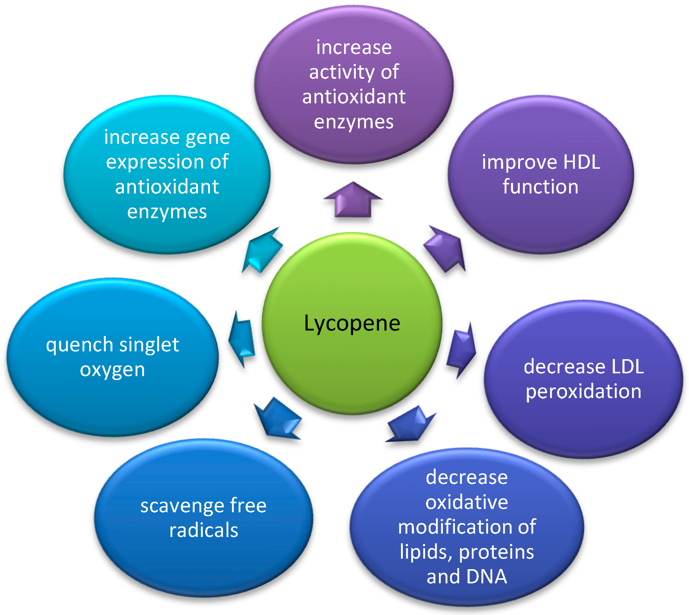
Lycopene is a carotenoid present in tomato, pitanga, pink-fleshed guava, red-fleshed papaya, and watermelon. The richest source of lycopene is the Asian gac fruit and the Spanish sarsaparilla [1]. It is considered to possess the most potent antioxidant activity of all carotenoids in accordance to the following ranking: lycopene > α-carotene > β-cryptoxanthin > zeaxanthin = β-carotene > lutein [16]. Its antioxidant effects are summarized in Figure 1.
Figure 1. Proposed mechanisms of antioxidant effects of lycopene.
Lycopene has especially high free radical scavenging properties which can be explained by a high number of conjugated double bonds with a high singlet oxygen quenching ability [17]. Experimental evidence shows that lycopene can quench singlet oxygen (102), scavenge free nitrogen dioxide (NO•2), thiyl (RS•), and sulfonyl (RSO•2) radicals [18]. Due to its ability to catch free radicals and decrease the damage caused by oxidative stress in lipids, lipoproteins, proteins, and DNA, it was suggested to prevent atherogenesis and carcinogenesis [19]. Lycopene lacks hydrophilic substituents, and therefore, it is very hydrophobic. It has been strongly associated with the ability to decrease LDL oxidation and overall lipid peroxidation [20]. High consumption of tomato products resulted in a decrease in LDL cholesterol level and an increase in LDL resistance to oxidation in healthy normocholesterolemic adults [21]. These atheroprotective changes correlated with an increase in lycopene, α-carotene, and β-carotene levels measured in serum.

3.1. The Influence of Lycopene on PON1 Activity and Gene Expression in Animal Studies

Hypercholesterolemia induces oxidative stress. In a study on hyperlipidemic rats, lycopene supplementation was shown to restore plasma antioxidant levels measured as the ferric-reducing activity of plasma (FRAP), which was accompanied by a rise in PON1 arylesterase activity [22]. The PON1 enzyme is thought to be partly responsible for the rise in FRAP. The observed improvement in PON1 activity may have been achieved by an upregulation of PON1 gene expression. PON1 expression was previously seen to be upregulated by transcription factors such as steroid regulatory element-binding protein-2 (SREBP-2), which binds to the promoter region of PON1 [23]. Apart from increasing PON1 activity, lycopene supplementation resulted in a more favorable lipid profile. It improved the concentration of HDL and caused a reduction in elevated levels of total cholesterol, triglycerides, LDL, and VLDL [22].
Lycopene supplementation for 1 month resulted in an increase in PON1 activity in non-diabetic rats [24]. In diabetic rats, lycopene consumption was able to restore PON1 activity, as its basal level was lower in diabetic rats than in a control group. A slight increase in the diabetes–lycopene group and a significant increase in the lycopene group over the control group were found. In another study, the treatment of diabetic rats with lycopene and metformin-induced PON1 activity, an effect similar to that reached by metformin or insulin [25]. The combination of these treatments has a potential beneficial effect of lowering markers of lipid peroxidation, increasing antioxidant defenses, as well as inhibiting postprandial glycemia and dyslipidemia [25]. Thus, lycopene appears as a promising therapeutic agent with the potential to be used in combination therapy to minimize the diabetic complications triggered by glycation and oxidative stress.

3.2. The Influence of Lycopene on PON1 Activity and Gene Expression in Clinical Studies

Supplementation of lycopene, as well as the implementation of a lycopene-rich diet to a group of moderately overweight middle-aged subjects, resulted in an increase in PON1 arylesterase activity in serum as well as in HDL2 and HDL3 subfractions [26]. PON1 deficient HDL is dysfunctional and not effective in preventing LDL oxidation. Lycopene may positively affect the structural and functional composition of Apo-AI and thereby restore PON1 activity in HDL particles.
A positive relationship between arylesterase activity and lycopene was also reported in a study on subjects with metabolic syndrome following an energy restriction diet [27]. This association can be explained by the capacity of lycopene to scavenge free oxygen radical products, which would otherwise engage PON1 activity and decrease it [28]. Furthermore, lycopene (and other dietary antioxidants) may exert its effects through modulation of gene expression through regulation of DNA methylation [29]. Methylation of the CpG-rich region overlapping a gene’s promoter is considered a mechanism for inhibiting a gene’s expression [30]. This mechanism was confirmed concerning the PON1 gene, as methylation of the CpG-rich region was found to inversely correlate with PON1 arylesterase activity. Inverse correlations were also observed between methylation of different CpG sites and dietary lycopene, vitamin C, and total tocopherol [27]. At the same time, all measured exogenous antioxidants correlated positively with PON1 arylesterase activity.
In conclusion, lycopene may enhance PON1 expression by inhibiting PON1 gene methylation in subjects with metabolic syndrome. Furthermore, lycopene supplementation was shown to restore PON1 activity in cases of hyperlipidemia, diabetes, obesity, and metabolic syndrome. These changes were found in serum, HDL2, and HDL3. Lycopene may favorably modify the lipid profiles in the population at risk of cardiovascular disease, as well as improve the antioxidant composition of lipoproteins and ameliorate antioxidant defense mechanisms.

4. The Effect of a Mixture of Carotenoids on PON1 Activity and LDL Oxidation

While it is easier to observe the specific effects of individual carotenoids when supplementing single compounds separately, in nature, they exist in combinations. Therefore, attempts have been made to measure the effect of carotenoids when they are administered together.
Oxidative lipid damage is a marker of the development of cardiovascular disease. PON1 hydrolyses oxidized lipids in LDL, retards atherosclerosis [31], and predicts the development of cardiovascular disease [32]. Similarly, some dietary antioxidants work in this direction, for example, β-carotene protects lipids from oxidation. In vitro and in vivo enrichment of LDL with beta-carotene protected them from cell-mediated oxidation. Surprisingly, this effect was not reached with in vivo treatment with lycopene in this study [33]. However, the administration of lycopene as tomato oleoresin (which contains a mixture of exogenous antioxidants) resulted in a strong inhibition of LDL oxidation. This gives evidence that lycopene may act as an effective antioxidant in synergism with several other natural antioxidants. It is very likely that, when given as a mixture, carotenoids do not only act additively but even synergistically, potentiating each other’s effect [16][34]. This observed effect of higher antioxidant activity of carotenoids, when supplied as mixtures, may be associated with the specific positioning of different carotenoids in membranes [16]. A single oral supplementation of alpha-tocopherol, beta-carotene, lycopene, canthaxanthin, and lutein protected LDL polyunsaturated fatty acids (PUFA) and their cholesterol moieties against oxidative modifications [35]. It has been suggested that the protection from oxidative damage and the associated cardiovascular disease is best achieved by natural antioxidants found in fruit and vegetables.

4.1. The Influence of a Mixture of Carotenoids on PON1 Activity in Animal Studies

PON1 activity has been reported to be lower in subjects with type 2 diabetes [36]. Two carotenoids, lycopene and bixin, supplemented individually increased PON1 level and HDL in streptozotocin-induced diabetic rats [37]. The treatment of rabbits on a hypocholesterolemic diet with bixin alone resulted in partial prevention of serum PON1 activity decrease [38]. Adding curcumin to lycopene or bixin led to an even more pronounced effect of decreasing biomarkers of carbohydrate and lipid disturbances, increased HDL levels, decreasing oxidized LDL, and alleviating oxidative stress [37]. Therefore, combining the two antioxidants resulted in a reduction in cardiovascular risk.

4.2. The Influence of a Mixture of Carotenoids on PON1 Activity in Clinical Studies

In a randomized controlled trial on subjects with type 2 diabetes, increasing fruit and vegetable intake for 8 weeks resulted in a rise in carotenoids (α-carotene, β-cryptoxanthin, lutein, lycopene) in serum, HDL2, and HDL3, which was accompanied by an increase in PON1 activity in serum and HDL3 [39]. The potential of a high fruit and vegetable diet rich in carotenoids to increase PON1 activity is especially valuable, as PON1 improves the anti-atherogenic mechanisms of HDL and helps to fight the potential negative complications of type 2 diabetes. A positive correlation was found between change in HDL3 β-cryptoxanthin and change in HDL3–PON1 activity, which further supports the idea of using carotenoids to induce the activity of PON1. Yet, this effect is not always observed and depends on the detailed conditions of the study. The consumption of fruits and vegetables for 6 weeks in a group of healthy subjects resulted in a decrease in PON1 activity despite an increase in carotenoids [40]. Another study showed that postprandial PON1 activity raised only after a Mediterranean-like meal together with the increase in carotenoids. On the contrary, the consumption of Western-like meal did not affect postprandial PON1 activity or carotenoids [41]. The effect of dietary modification on carotenoids and PON1 activity in healthy individuals was also assessed by DiMarco et al. While consumption of 2–3 eggs/day increased plasma lutein and zeaxanthin and caused improvements in HDL function, the intake of 3 eggs/day had the additional beneficial effect of inducing PON1 activity [42].
Carotenoids were found to exert their antioxidant effect by the protection of PON1. Yet, the level of PON1 activity preservation by the studied compounds varied in the observed populations. PON1 activity’s correlations with β-carotene, lycopene, lutein, and zeaxanthin were found in Greek but not in Anglo-Celtic subjects [43]. Yet, a different dietary intake of fruits and vegetables, which transferred to a different baseline level of carotenoids, was registered in these two groups, which could influence the outcome. These results also suggest that ethnicity may determine the influence of carotenoids on PON1 activity. Possibly, other factors such as food sources of carotenoids or different preparation methods may affect this relationship. In particular, olive oil usage with vegetables, which was registered to be higher in Greek subjects, may be a confounding factor due to its protective activity towards PON1. Virgin olive oils increased PON1-associated specific activities in a randomized study [44]. Furthermore, after stratification, the observed relationship was significant only in subjects with the R-allele of PON1-192 polymorphism [43].
In other studies, the influence of PON1 gene polymorphism on the modulation of antioxidant activity by dietary antioxidants was also noted. A higher intake of oleic acid was related to an increased PON1 activity only in the PON1-192 RR genotype group [45]. Furthermore, in a study on elderly volunteers, where antioxidant protection offered by components of tomato juice (especially β-carotene and lycopene) was more advantageous in subjects with the R-allele [46]. PON1 activity increased in all volunteers, including the control group. However, antioxidant status improved and LDL-oxidation decreased only in R-allele carriers but not in the QQ genotype group. The same group of authors observed the effect of tomato (as a source of β-carotene and lycopene) and carrot juice (as a source of β-carotene and α-carotene) consumption on PON1 activity and lipid peroxidation in healthy young volunteers for 2 weeks preceded by 2 weeks of low-carotenoid intake. In this setting, as opposed to the previous study, neither of the juices affected PON1 activity. However, tomato juice consumption resulted in a reduction in lipid peroxidation in R-allele carriers in comparison to QQ subjects. Carrot juice did not affect lipid peroxidation regardless of the PON1-192 genotype [46]. Again, the QQ homozygous subjects did not gain any additional antioxidant protection of the lipids with this nutritional intervention. These results suggest that there may be a higher potential for improving the antioxidant defense of PON1 and protection from atherosclerosis through the modulation of HDL function by using dietary antioxidants in subjects with R-allele than in the QQ-genotype. Interestingly, it is the PON1 isoenzyme corresponding to the RR genotype that has a low hydrolyzing activity towards lipid hydroperoxides [47]. Additionally, given that in some populations, subjects of the RR genotype or with R-allele were shown to be at increased risk of coronary artery disease [48][49], it would be indeed very valuable to find ways of enhancement of PON1 activity, particularly in these individuals. PON1 polymorphism modifies the effect of carotenoids on different diseases related to oxidative stress, not necessarily related to atherosclerosis. The distribution of PON1 polymorphism is known to vary between different populations. For the PON1-192 polymorphism, the R-allele was most widely distributed among Mexican (51.7–43.7% depending on the ethnic group) [50], Japanese (65.2%) [51], and Chinese (64.8%) [52] people. Among the Mexican population, the Mestizos have the highest frequency of the RR genotype. Important differences were reported after the comparison of the Mexican and Asian populations to Caucasians. The lowest R-allele frequency was observed in German (22.5%) [53], British (29%) [54], and French (29%) [55][56] populations. Taking into account the allele distribution frequency may help assess the target population, in which carotenoid supplementation improves antioxidant status and limits lipid peroxidation. Another area in which carotenoids may be useful is the osteoporosis risk attenuation. Yet this effect may also be affected by PON1 polymorphism. High serum lycopene was associated with lower bone resorption markers only with subjects with the LL genotype and Q allele [57]. Dietary interventions may be a therapeutic option, applied especially in groups where they offer the greatest advantage. Further research should be encouraged to identify these groups.
In a study on a cohort of 60 Australian Aboriginal people, PON1 arylesterase activity correlated with total carotenoid concentration, as well as the individual carotenoids β-carotene, lycopene, cryptoxanthin, and lutein plus zeaxanthin [58]. In addition, correlations of paraoxonase activity with plasma total carotenoids concentration, due mainly to a strong correlation with lycopene concentration, were found. Dietary and lifestyle intervention in this study increased PON1 activity, homocysteine, and carotenoid concentration. Change in PON1 activity correlated with the change in HDL cholesterol, but the increased HDL cholesterol could not account for all PON1 activity rise. Correlation between carotenoid concentration and PON1 activity were detected at baseline and after the intervention. Yet the authors were not able to find a correlation between change in carotenoids and change in PON1 activity. Not all studies prove that carotenoids influence PON1 activity. Ferre et al., in a study on 388 individuals, found no correlation between β-carotene intake and PON1 activity [59]. The participants of this study were randomly selected with a wide age range (18–75 years) with an equal proportion of men. Kleemola et al. describe an inverse relationship between β-carotene and PON1 activity, but the volunteers in this study were young and healthy university students and employees, mostly women [60]. These conflicting results can be explained, at least in part, by differences in the studied populations.

4.3. Conclusion on the Effect of a Mixture of Carotenoids on PON1 Activity

In most of the studies, supplementation of a mixture of carotenoids in their natural form with food increases PON1 activity in serum and/or HDL3. Studies focused on the determination of correlations of individual mixtures of carotenoids with PON1 activity suggest that the relationship between these antioxidants exists only in some populations. While this is observed for most carotenoids and carotenoid mixtures, β-carotene exceptionally shows no correlation or even an inverse correlation with PON1 activity. PON1-192 polymorphism was found to modify the effect of carotenoids on antioxidant status improvement, lipid peroxidation, LDL-oxidation, and bone turnover markers.

References

  1. Rodriguez-Amaya, D.B. Natural Food Pigments and Colorants. In Bioactive Molecules in Food. Reference Series in Phytochemistry; Mérillon, J.M., Ramawat, K., Eds.; Springer: Cham, Switzerland, 2018; pp. 1–35. ISBN 978-3-319-54528-8.
  2. Rajasingh, H.; Øyehaug, L.; Våge, D.I.; Omholt, S.W. Carotenoid Dynamics in Atlantic Salmon. BMC Biol. 2006, 4, 10.
  3. Kurashige, M.; Okimasu, E.; Inoue, M.; Utsumi, K. Inhibition of Oxidative Injury of Biological Membranes by Astaxanthin. Physiol. Chem. Phys. Med. NMR 1990, 22, 27–38.
  4. McNulty, H.; Jacob, R.F.; Mason, R.P. Biologic Activity of Carotenoids Related to Distinct Membrane Physicochemical Interactions. Am. J. Cardiol. 2008, 101, S20–S29.
  5. Augusti, P.R.; Quatrin, A.; Somacal, S.; Conterato, G.M.M.; Sobieski, R.; Ruviaro, A.R.; Maurer, L.H.; Duarte, M.M.F.; Roehrs, M.; Emanuelli, T. Astaxanthin Prevents Changes in the Activities of Thioredoxin Reductase and Paraoxonase in Hypercholesterolemic Rabbits. J. Clin. Biochem. Nutr. 2012, 51, 42–49.
  6. Kükürt, A.; Karapehlivan, M. Protective Effect of Astaxanthin on Experimental Ovarian Damage in Rats. J. Biochem. Mol. Toxicol. 2022, 36, e22966.
  7. Baralic, I.; Djordjevic, B.; Dikic, N.; Kotur-Stevuljevic, J.; Spasic, S.; Jelic-Ivanovic, Z.; Radivojevic, N.; Andjelkovic, M.; Pejic, S. Effect of Astaxanthin Supplementation on Paraoxonase 1 Activities and Oxidative Stress Status in Young Soccer Players. Phyther. Res. 2013, 27, 1536–1542.
  8. Jaouad, L.; De Guise, C.; Berrougui, H.; Cloutier, M.; Isabelle, M.; Fulop, T.; Payette, H.; Khalil, A. Age-Related Decrease in High-Density Lipoproteins Antioxidant Activity Is Due to an Alteration in the PON1’s Free Sulfhydryl Groups. Atherosclerosis 2006, 185, 191–200.
  9. Sorenson, R.C.; Primo-Parmo, S.L.; Kuo, C.L.; Adkins, S.; Lockridge, O.; La Du, B.N. Reconsideration of the Catalytic Center and Mechanism of Mammalian Paraoxonase/Arylesterase. Proc. Natl. Acad. Sci. USA 1995, 92, 7187–7191.
  10. Yamagata, K.; Tanaka, N.; Matsufuji, H.; Chino, M. β-Carotene Reverses the IL-1β-Mediated Reduction in Paraoxonase-1 Expression via Induction of the CaMKKII Pathway in Human Endothelial Cells. Microvasc. Res. 2012, 84, 297–305.
  11. Loppnow, H.; Buerke, M.; Werdan, K.; Rose-John, S. Contribution of Vascular Cell-Derived Cytokines to Innate and Inflammatory Pathways in Atherogenesis. J. Cell. Mol. Med. 2011, 15, 484–500.
  12. Kumon, Y.; Suehiro, T.; Ikeda, Y.; Hashimoto, K. Human Paraoxonase-1 Gene Expression by HepG2 Cells Is Downregulated by Interleukin-1beta and Tumor Necrosis Factor-Alpha, but Is Upregulated by Interleukin-6. Life Sci. 2003, 73, 2807–2815.
  13. Tedgui, A.; Mallat, Z. Anti-Inflammatory Mechanisms in the Vascular Wall. Circ. Res. 2001, 88, 877–887.
  14. Libby, P. Interleukin-1 Beta as a Target for Atherosclerosis Therapy: The Biological Basis of CANTOS and Beyond. J. Am. Coll. Cardiol. 2017, 70, 2278–2289.
  15. Martin, K.R.; Wu, D.; Meydani, M. The Effect of Carotenoids on the Expression of Cell Surface Adhesion Molecules and Binding of Monocytes to Human Aortic Endothelial Cells. Atherosclerosis 2000, 150, 265–274.
  16. Heber, D.; Lu, Q.Y. Overview of Mechanisms of Action of Lycopene. Exp. Biol. Med. 2002, 227, 920–923.
  17. Di Mascio, P.; Kaiser, S.; Sies, H. Lycopene as the Most Efficient Biological Carotenoid Singlet Oxygen Quencher. Arch. Biochem. Biophys. 1989, 274, 532–538.
  18. Srinivasan, M.; Sudheer, A.R.; Pillai, K.R.; Kumar, P.R.; Sudhakaran, P.R.; Menon, V.P. Lycopene as a Natural Protector against Gamma-Radiation Induced DNA Damage, Lipid Peroxidation and Antioxidant Status in Primary Culture of Isolated Rat Hepatocytes in Vitro. Biochim. Biophys. Acta 2007, 1770, 659–665.
  19. Rao, A.V.; Agarwal, S. Role of Antioxidant Lycopene in Cancer and Heart Disease. J. Am. Coll. Nutr. 2000, 19, 563–569.
  20. Agarwal, S.; Rao, A.V. Tomato Lycopene and Low Density Lipoprotein Oxidation: A Human Dietary Intervention Study. Lipids 1998, 33, 981–984.
  21. Silaste, M.L.; Alfthan, G.; Aro, A.; Kesäniemi, Y.A.; Hörkkö, S. Tomato Juice Decreases LDL Cholesterol Levels and Increases LDL Resistance to Oxidation. Br. J. Nutr. 2007, 98, 1251–1258.
  22. Sultan Alvi, S.; Ansari, I.A.; Khan, I.; Iqbal, J.; Khan, M.S. Potential Role of Lycopene in Targeting Proprotein Convertase Subtilisin/Kexin Type-9 to Combat Hypercholesterolemia. Free Radic. Biol. Med. 2017, 108, 394–403.
  23. Deakin, S.; Leviev, I.; Guernier, S.; James, R.W. Simvastatin Modulates Expression of the PON1 Gene and Increases Serum Paraoxonase: A Role for Sterol Regulatory Element-Binding Protein-2. Arterioscler. Thromb. Vasc. Biol. 2003, 23, 2083–2089.
  24. Yegin, S.Ç.; Yur, F.; Ceylan, E. Effect of Lycopene Application in Rats with Experimental Diabetes Using Lipoprotein, Paraoxonase and Cytokines. J. Membr. Biol. 2013, 246, 621–626.
  25. Figueiredo, I.D.; Lima, T.F.O.; Inácio, M.D.; Costa, M.C.; Assis, R.P.; Brunetti, I.L.; Baviera, A.M. Lycopene Improves the Metformin Effects on Glycemic Control and Decreases Biomarkers of Glycoxidative Stress in Diabetic Rats. Diabetes. Metab. Syndr. Obes. 2020, 13, 3117–3135.
  26. McEneny, J.; Wade, L.; Young, I.S.; Masson, L.; Duthie, G.; McGinty, A.; McMaster, C.; Thies, F. Lycopene Intervention Reduces Inflammation and Improves HDL Functionality in Moderately Overweight Middle-Aged Individuals. J. Nutr. Biochem. 2013, 24, 163–168.
  27. de la Iglesia, R.; Mansego, M.L.; Sánchez-Muniz, F.J.; Angeles Zulet, M.; Alfredo Martinez, J. Arylesterase Activity Is Associated with Antioxidant Intake and Paraoxonase-1 (PON1) Gene Methylation in Metabolic Syndrome Patients Following an Energy Restricted Diet. EXCLI J. 2014, 13, 416–426.
  28. Tsakiris, S.; Karikas, G.A.; Parthimos, T.; Tsakiris, T.; Bakogiannis, C.; Schulpis, K.H. Alpha-Tocopherol Supplementation Prevents the Exercise-Induced Reduction of Serum Paraoxonase 1/Arylesterase Activities in Healthy Individuals. Eur. J. Clin. Nutr. 2009, 63, 215–221.
  29. Malireddy, S.; Kotha, S.R.; Secor, J.D.; Gurney, T.O.; Abbott, J.L.; Maulik, G.; Maddipati, K.R.; Parinandi, N.L. Phytochemical Antioxidants Modulate Mammalian Cellular Epigenome: Implications in Health and Disease. Antioxid. Redox Signal. 2012, 17, 327–339.
  30. Vanderkraats, N.D.; Hiken, J.F.; Decker, K.F.; Edwards, J.R. Discovering High-Resolution Patterns of Differential DNA Methylation That Correlate with Gene Expression Changes. Nucleic Acids Res. 2013, 41, 6816–6827.
  31. Mackness, B.; Durrington, P.; McElduff, P.; Yarnell, J.; Azam, N.; Watt, M.; Mackness, M. Low Paraoxonase Activity Predicts Coronary Events in the Caerphilly Prospective Study. Circulation 2003, 107, 2775–2779.
  32. Poh, R.; Muniandy, S. Paraoxonase 1 Activity as a Predictor of Cardiovascular Disease in Type 2 Diabetes. Southestern Asian J. Trop. Med. Public Health 2010, 41, 1231–1246.
  33. Dugas, T.R.; Morel, D.W.; Harrison, E.H. Dietary Supplementation with Beta-Carotene, but Not with Lycopene, Inhibits Endothelial Cell-Mediated Oxidation of Low-Density Lipoprotein. Free Radic. Biol. Med. 1999, 26, 1238–1244.
  34. Fuhrman, B.; Volkova, N.; Rosenblat, M.; Aviram, M. Lycopene Synergistically Inhibits LDL Oxidation in Combination with Vitamin E, Glabridin, Rosmarinic Acid, Carnosic Acid, or Garlic. Antioxid. Redox Signal. 2000, 2, 491–506.
  35. Linseisen, J.; Hoffmann, J.; Riedl, J.; Wolfram, G. Effect of a Single Oral Dose of Antioxidant Mixture (Vitamin E, Carotenoids) on the Formation of Cholesterol Oxidation Products after Ex Vivo LDL Oxidation in Humans. Eur. J. Med. Res. 1998, 3, 5–12.
  36. Shokri, Y.; Variji, A.; Nosrati, M.; Khonakdar-Tarsi, A.; Kianmehr, A.; Kashi, Z.; Bahar, A.; Bagheri, A.; Mahrooz, A. Importance of Paraoxonase 1 (PON1) as an Antioxidant and Antiatherogenic Enzyme in the Cardiovascular Complications of Type 2 Diabetes: Genotypic and Phenotypic Evaluation. Diabetes Res. Clin. Pract. 2020, 161, 108067.
  37. Assis, R.; Arcaro, C.; Gutierres, V.; Oliveira, J.; Costa, P.; Baviera, A.; Brunetti, I. Combined Effects of Curcumin and Lycopene or Bixin in Yoghurt on Inhibition of LDL Oxidation and Increases in HDL and Paraoxonase Levels in Streptozotocin-Diabetic Rats. Int. J. Mol. Sci. 2017, 18, 332.
  38. Somacal, S.; Figueiredo, C.G.; Quatrin, A.; Ruviaro, A.R.; Conte, L.; Augusti, P.R.; Roehrs, M.; Denardin, I.T.; Kasten, J.; da Veiga, M.L.; et al. The Antiatherogenic Effect of Bixin in Hypercholesterolemic Rabbits Is Associated to the Improvement of Lipid Profile and to Its Antioxidant and Anti-Inflammatory Effects. Mol. Cell. Biochem. 2015, 403, 243–253.
  39. Daniels, J.A.; Mulligan, C.; McCance, D.; Woodside, J.V.; Patterson, C.; Young, I.S.; McEneny, J. A Randomised Controlled Trial of Increasing Fruit and Vegetable Intake and How This Influences the Carotenoid Concentration and Activities of PON-1 and LCAT in HDL from Subjects with Type 2 Diabetes. Cardiovasc. Diabetol. 2014, 13, 16.
  40. Freese, R.; Alfthan, G.; Jauhiainen, M.; Basu, S.; Erlund, I.; Salminen, I.; Aro, A.; Mutanen, M. High Intakes of Vegetables, Berries, and Apples Combined with a High Intake of Linoleic or Oleic Acid Only Slightly Affect Markers of Lipid Peroxidation and Lipoprotein Metabolism in Healthy Subjects. Am. J. Clin. Nutr. 2002, 76, 950–960.
  41. Blum, S.; Aviram, M.; Ben-Amotz, A.; Levy, Y. Effect of a Mediterranean Meal on Postprandial Carotenoids, Paraoxonase Activity and C-Reactive Protein Levels. Ann. Nutr. Metab. 2006, 50, 20–24.
  42. DiMarco, D.M.; Norris, G.H.; Millar, C.L.; Blesso, C.N.; Fernandez, M.L. Intake of up to 3 Eggs per Day Is Associated with Changes in HDL Function and Increased Plasma Antioxidants in Healthy, Young Adults. J. Nutr. 2017, 147, 323–329.
  43. Chee, T.L.; Rowley, K.; Jenkins, A.J.; O’Dea, K.; Itsiopoulos, C.; Stoney, R.M.; Su, Q.; Giles, G.G.; Best, J.D. Paraoxonase Activity in Greek Migrants and Anglo-Celtic Persons in the Melbourne Collaborative Cohort Study: Relationship to Dietary Markers. Eur. J. Nutr. 2005, 44, 223–230.
  44. Fernández-Castillejo, S.; García-Heredia, A.I.; Solà, R.; Camps, J.; López de la Hazas, M.C.; Farràs, M.; Pedret, A.; Catalán, Ú.; Rubió, L.; Motilva, M.J.; et al. Phenol-Enriched Olive Oils Modify Paraoxonase-Related Variables: A Randomized, Crossover, Controlled Trial. Mol. Nutr. Food Res. 2017, 61, 1600932.
  45. Tomás, M.; Sentí, M.; Elosua, R.; Vila, J.; Sala, J.; Masià, R.; Marrugat, J. Interaction between the Gln-Arg 192 Variants of the Paraoxonase Gene and Oleic Acid Intake as a Determinant of High-Density Lipoprotein Cholesterol and Paraoxonase Activity. Eur. J. Pharmacol. 2001, 432, 121–128.
  46. Bub, A.; Barth, S.; Watzl, B.; Briviba, K.; Herbert, B.M.; Lührmann, P.M.; Neuhäuser-Berthold, M.; Rechkemmer, G. Paraoxonase 1 Q192R (PON1-192) Polymorphism Is Associated with Reduced Lipid Peroxidation in R-Allele-Carrier but Not in QQ Homozygous Elderly Subjects on a Tomato-Rich Diet. Eur. J. Nutr. 2002, 41, 237–243.
  47. Mackness, M.I.; Mackness, B.; Durrington, P.N.; Fogelman, A.M.; Berliner, J.; Lusis, A.J.; Navab, M.; Shih, D.; Fonarow, G.C. Paraoxonase and Coronary Heart Disease. Curr. Opin. Lipidol. 1998, 9, 319–324.
  48. Mendonça, M.I.; Dos Reis, R.P.; Freitas, A.I.; Sousa, A.C.; Pereira, A.; Faria, P.; Gomes, S.; Silva, B.; Santos, N.; Serrão, M.; et al. Human Paraoxonase Gene Polymorphisms and Coronary Artery Disease Risk. Rev. Port. Cardiol. 2008, 27, 1539–1555.
  49. Vaisi-Raygani, A.; Ghaneialvar, H.; Rahimi, Z.; Tavilani, H.; Pourmotabbed, T.; Shakiba, E.; Vaisi-Raygani, A.; Kiani, A.; Aminian, M.; Alibakhshi, R.; et al. Paraoxonase Arg 192 Allele Is an Independent Risk Factor for Three-Vessel Stenosis of Coronary Artery Disease. Mol. Biol. Rep. 2011, 38, 5421–5428.
  50. Gamboa, R.; Zamora, J.; Rodríguez-Pérez, J.M.; Fragoso, J.M.; Cardoso, G.; Posadas-Romero, C.; Vargas-Alarcón, G. Distribution of Paraoxonase PON1 Gene Polymorphisms in Mexican Populations. Its Role in the Lipid Profile. Exp. Mol. Pathol. 2006, 80, 85–90.
  51. Imai, Y.; Morita, H.; Kurihara, H.; Sugiyama, T.; Kato, N.; Ebihara, A.; Hamada, C.; Kurihara, Y.; Shindo, T.; Oh-Hashi, Y.; et al. Evidence for Association between Paraoxonase Gene Polymorphisms and Atherosclerotic Diseases. Atherosclerosis 2000, 149, 435–442.
  52. Wang, X.; Fan, Z.; Huang, J.; Su, S.; Yu, Q.; Zhao, J.; Hui, R.; Yao, Z.; Shen, Y.; Qiang, B.; et al. Extensive Association Analysis between Polymorphisms of PON Gene Cluster with Coronary Heart Disease in Chinese Han Population. Arterioscler. Thromb. Vasc. Biol. 2003, 23, 328–334.
  53. Gardemann, A.; Philipp, M.; Heß, K.; Katz, N.; Tillmanns, H.; Haberbosch, W. The Paraoxonase Leu-Met54 and Gln-Arg191 Gene Polymorphisms Are Not Associated with the Risk of Coronary Heart Disease. Atherosclerosis 2000, 152, 421–431.
  54. Mackness, B.; Davies, G.K.; Turkie, W.; Lee, E.; Roberts, D.H.; Hill, E.; Roberts, C.; Durrington, P.N.; Mackness, M.I. Paraoxonase Status in Coronary Heart Disease: Are Activity and Concentration More Important than Genotype? Arterioscler. Thromb. Vasc. Biol. 2001, 21, 1451–1457.
  55. Cohen, E.; Aviram, M.; Khatib, S.; Artoul, F.; Rabin, A.; Mannheim, D.; Karmeli, R.; Salamon, T.; Vaya, J. Human Carotid Plaque Phosphatidylcholine Specifically Interacts with Paraoxonase 1, Increases Its Activity, and Enhances Its Uptake by Macrophage at the Expense of Its Binding to HDL. Free Radic. Biol. Med. 2014, 76, 14–24.
  56. Helbecque, N.; Cottel, D.; Meirhaeghe, A.; Dallongeville, P.; Arveiler, D.; Wagner, A.; Ruidavets, J.B.; Ferrieres, J. Paraoxonase (Gln192-Arg) Polymorphism in French Type 2 Diabetics. Atherosclerosis 1999, 147, 415–416.
  57. MacKinnon, E.S.; El-Sohemy, A.; Rao, A.V.; Rao, L.G. Paraoxonase 1 Polymorphisms 172T→A and 584A→G Modify the Association between Serum Concentrations of the Antioxidant Lycopene and Bone Turnover Markers and Oxidative Stress Parameters in Women 25-70 Years of Age. J. Nutrigenet. Nutr. 2010, 3, 1–8.
  58. Cohen, J.; Jenkins, A.J.; Karschimkus, C.; Qing, S.; Lee, C.T.; O’Dea, K.; Best, J.D.; Rowley, K.G. Paraoxonase and Other Coronary Risk Factors in a Community-Based Cohort. Redox Rep. 2002, 7, 304–307.
  59. Ferrè, N.; Camps, J.; Fernández-Ballart, J.; Arija, V.; Murphy, M.M.; Ceruelo, S.; Biarnés, E.; Vilella, E.; Tous, M.; Joven, J. Regulation of Serum Paraoxonase Activity by Genetic, Nutritional, and Lifestyle Factors in the General Population. Clin. Chem. 2003, 49, 1491–1497.
  60. Kleemola, P.; Freese, R.; Jauhiainen, M.; Pahlman, R.; Alfthan, G.; Mutanen, M. Dietary Determinants of Serum Paraoxonase Activity in Healthy Humans. Atherosclerosis 2002, 160, 425–432.
More
Information
Contributor MDPI registered users' name will be linked to their SciProfiles pages. To register with us, please refer to https://encyclopedia.pub/register :
View Times: 418
Revisions: 2 times (View History)
Update Date: 02 Aug 2022
1000/1000
ScholarVision Creations