Submitted Successfully!
To reward your contribution, here is a gift for you: A free trial for our video production service.
Thank you for your contribution! You can also upload a video entry or images related to this topic.
Version Summary Created by Modification Content Size Created at Operation
1 -- 5953 2022-07-07 14:27:57 |
2 format change -490 word(s) 5463 2022-07-08 03:09:35 |

Video Upload Options

Do you have a full video?

Confirm

Are you sure to Delete?
Cite
If you have any further questions, please contact Encyclopedia Editorial Office.
Souza, I.D.;  Ramalho, M.C.C.;  Guedes, C.B.;  Osawa, I.Y.A.;  Monteiro, L.K.S.;  Gomes, L.R.;  Rocha, C.R.R. Ferroptosis Modulation as Therapeutic Target for Glioblastoma Treatment. Encyclopedia. Available online: https://encyclopedia.pub/entry/24908 (accessed on 03 July 2024).
Souza ID,  Ramalho MCC,  Guedes CB,  Osawa IYA,  Monteiro LKS,  Gomes LR, et al. Ferroptosis Modulation as Therapeutic Target for Glioblastoma Treatment. Encyclopedia. Available at: https://encyclopedia.pub/entry/24908. Accessed July 03, 2024.
Souza, Izadora De, Maria Carolina Clares Ramalho, Camila Banca Guedes, Isabeli Yumi Araújo Osawa, Linda Karolynne Seregni Monteiro, Luciana Rodrigues Gomes, Clarissa Ribeiro Reily Rocha. "Ferroptosis Modulation as Therapeutic Target for Glioblastoma Treatment" Encyclopedia, https://encyclopedia.pub/entry/24908 (accessed July 03, 2024).
Souza, I.D.,  Ramalho, M.C.C.,  Guedes, C.B.,  Osawa, I.Y.A.,  Monteiro, L.K.S.,  Gomes, L.R., & Rocha, C.R.R. (2022, July 07). Ferroptosis Modulation as Therapeutic Target for Glioblastoma Treatment. In Encyclopedia. https://encyclopedia.pub/entry/24908
Souza, Izadora De, et al. "Ferroptosis Modulation as Therapeutic Target for Glioblastoma Treatment." Encyclopedia. Web. 07 July, 2022.
Ferroptosis Modulation as Therapeutic Target for Glioblastoma Treatment
Edit

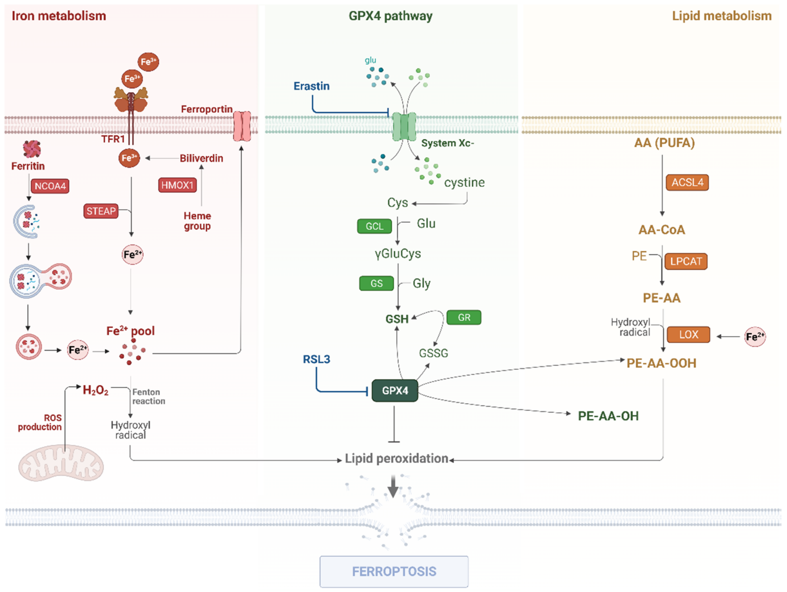
Glioblastoma multiforme is a lethal disease and represents the most common and severe type of glioma. Drug resistance and the evasion of cell death are the main characteristics of its malignancy, leading to a high percentage of disease recurrence and the patients’ low survival rate. Exploiting the modulation of cell death mechanisms could be an important strategy to prevent tumor development and reverse the high mortality and morbidity rates in glioblastoma patients. Ferroptosis is a recently described type of cell death, which is characterized by iron accumulation, high levels of polyunsaturated fatty acid (PUFA)-containing phospholipids, and deficiency in lipid peroxidation repair. Several studies have demonstrated that ferroptosis has a potential role in cancer treatment and could be a promising approach for glioblastoma patients.

ferroptosis glioma treatment cell death

1. Introduction

Glioblastomas (GBM, WHO IV) are the most aggressive type of glioma due to their peculiar characteristics such as cellular heterogeneity, high proliferation rate, diffuse infiltration capacity, and more importantly, high resistance to chemotherapeutic drugs [1]. Therefore, the standard treatment with temozolomide (TMZ) has low effectiveness, leading to a poor prognosis with a median overall survival of not more than 15 months, and a five-year survival rate of only 5% [2]. Indeed, it has been a challenge to sensitize this type of tumor due to its malignant attributes, and therefore, strategies involving the induction of cell death have been widely studied. Undoubtedly, one of the most studied cell death targets for tumor therapy is apoptosis, however, glioma cells have developed several mechanisms of resistance toward this type of cell death [3][4]. Thus, focusing on studying different types of cell death that still have not yet been completely characterized is a relevant tool in glioma treatment strategies.
At its most basic, cell death is an important biological mechanism for physiological homeostasis, which may occur in response to stress conditions [5]. Upon oxidative stress, a specific group of effector molecules is activated and triggers a cell death signaling pathway in a programmed manner to preserve the organism’s integrity. Thus, cell death is extremely important to eliminate damaged cells, and to regulate the state of danger signaling. According to Nomenclature Committee on Cell Death, there are several types of regulated cell death including apoptosis, necroptosis, autophagy, and ferroptosis. Regulatory failure in these different types of cell death can lead to human disorders [6]. Thus, the research on the molecular mechanisms of cell death is an important strategy to better understand the pathologies and promote new treatment options for aggressive diseases such as glioblastoma.
Studies have revealed an emergent type of regulated cell death identified as ferroptosis (Figure 1), which may play an important role in cancer treatment [7]. Ferroptosis has been initially described in a study by Dixon et al., in which the researchers used several lethal molecular compounds to kill RAS-mutated cancer cell lines [8]. As a consequence, two main compounds, namely, Erastin and RSL3, were identified. They promote non-apoptotic regulated cell death, and the process was termed ferroptosis by the authors. In that study, the authors distinguished this new type of cell death both morphologically and biochemically from other regulated cell deaths [8].
Figure 1. The molecular mechanisms of the ferroptosis pathway. Ferroptosis is triggered by three main regulatory pathways: iron metabolism, the GPX4 pathway, and lipid metabolism. In the iron metabolism, Fe3+ (ferric iron) is transported into the cell by TfR1 (Transferrin receptor) or obtained through the conversion of the heme group to biliverdin by HMOX1 activity. Then, Fe3+ is reduced to Fe2+ (ferrous iron) by STEAP3. Ferritin degradation by NCOA4 via autophagy also contributes to the labile iron pool. Once in the cytosol, Fe2+ can react with ROS and it generates the hydroxyl radical, promoting PUFA oxidation. The GPX4 pathway is responsible for controlling the lipoperoxidation levels through the reduction of lipid peroxides (PE-AA-OOH) to lipid alcohol (PE-AA-OH), thus GPX4 blockage by RSL3 induces ferroptosis. GPX4 utilizes GSH as a cofactor, therefore the GSH synthesis pathway is directly related to GPX4 activity, and xCT system blockage by Erastin leads to ferroptotic cell death. In the lipid metabolism, AA (as well as other PUFAs) are metabolized by ACSL4 and esterified by LPCAT3. Then, LOXs oxidize PUFAs using Fe2+ as a cofactor, which produces lipid peroxides.

2. Ferroptosis Modulation on Glioma

2.1. Iron Metabolism

Iron is commonly associated with its role in oxygen transport in the blood, however, this element is involved in many other biological processes including nucleic acid repair, DNA synthesis, cell growth, and cell death [9]. The level of iron is maintained by the action of several regulatory proteins. Initially, for intestinal absorption to occur, dietary iron is reduced to Fe2+ (ferrous iron) by ferric reductase duodenal cytochrome B (DCYTB) activity. After its reduction, Fe2+ is absorbed by a divalent metal transporter (DMT1), located in enterocytes, and it can be redirected to three distinct purposes: (1) the storage in ferritin protein; (2) the execution of biological processes in the cells; or (3) the release in the blood circulation [9].
Iron is exported from enterocytes by ferroportin (FPN1) and oxidized to Fe3+ (ferric iron) by the Hephaestin protein (Hp), enabling its binding to the glycoprotein transferrin (Tf). In cells where Transferrin receptors (TfR1) are present such as erythroblast precursors, Tf is internalized. The iron is released in the endosome and, by the action of metalloreductase six-transmembrane epithelial antigen of the prostate 3 (STEAP3), it is reduced to Fe2+, allowing its cytosol exportation by DMT1. When it is in cytosol, ferrous iron can be used in hemoglobin heme biosynthesis or it can be stored in ferritin, depending on the cellular needs [9]. To maintain adequate iron levels, the ferritin-stored iron can be released by ferritinophagy, an autophagic process mediated by NCOA4 proteins [10].
Despite its participation in several essential biological processes, excess iron can be harmful to cells. Indeed, iron can promote the oxidation of biomolecules, generating reactive oxygen species (ROS). Furthermore, iron is capable of reacting with ROS such as hydrogen peroxide (H2O2) and oxygen (O2), promoting the formation of hydroxyl radicals (OH) and anion superoxide (O2) by the Fenton chain reaction [11]. This increase in hydroxyl radicals, resulting from iron accumulation, promotes PUFA oxidation, which is a well-established ferroptosis hallmark [12].
An expanded labile iron pool (LIP) has been considered as a notable characteristic that distinguishes cancer cells from normal cells, since all cancer cells are more dependent on iron than normal tissues to support the intense energetic demand that comes with indefinite proliferation. Due to this, ferroaddiction in cancer has been explored in LIP-targeted therapies that induce oxidative stress to trigger ferroptosis. This approach requires biomarkers to point out those tumors with the most elevated LIP, and thus most likely to respond to LIP-targeted therapies [13][14]. In this sense, a radiotracer was developed, named 18F-TRX, to assess LIP in situ with PET, and showed that among other types of cancer, glioma harbors a wide range of LIP concentrations. U251 cells had the highest 18F-TRX uptake and were highly sensitive to treatment with TRX-CBI—a LIP-activated prodrug—suggesting a role for LIP-targeted therapies in the treatment of glioma [14].
It is known that GBM cells accumulate iron by altering the expression levels of many proteins and enzymes related to iron metabolism, promoting physiological processes such as tumor initiation, progression, and metastasis [13]. One way to increase the iron content in cancer cells is through TMZ treatment. According to a recently published study, TMZ, which is a standard chemotherapeutic drug utilized in glioblastoma treatment, drives ferroptosis by upregulating DMT1, a transporter related to iron metabolism. Thus, DMT1 could be a crucial target in GBM [15]. In this context, Zhang and collaborators revealed that the overexpression of the Coatomer protein complex subunit zeta 1 (COPZ1), a component of the coatomer protein complex I, was associated with increasing tumor grade and poor prognosis in glioma patients [16]. The referred study demonstrated that COPZ1 knockdown induced ferritinophagy, and ultimately, activated cell death via ferroptosis in the U87MG, U251, and P3#GBM cultured cell lines. Additionally, the deficiency of COPZ1 led to increased levels of the proteins NCOA4, autophagy flux marker LC3B-II, and ATG7 (autophagy-related 7), resulting in the degradation of the intracellular iron storage protein ferritin through autophagy, and consequently, augmented levels of ferrous iron. Therefore, iron accumulation generated high levels of intracellular H2O2 and superoxide, triggering the Fenton reaction and leading to lipid peroxidation [16].
In the same way, STEAP2 and STEAP3, identified as potential prognostic-related genes in GBM, were found to be downregulated and upregulated, respectively, in these tumors [17]. As members of the six-transmembrane epithelial antigen of prostate (STEAP) family, these proteins play a significant role in maintaining iron homeostasis, reducing ferric iron to ferrous iron to increase cellular iron uptake [17][18]. Considering the link between iron metabolism and ferroptosis, the Pearson’s correlation analysis indicated that STEAP2 and STEAP3 were correlated with genes involved in ferroptosis [19] such as ACSL4, ALOX5, CBS, FANCD2, GCLM, HMGCR, HSPB1, NFE2L2, PTGS2, and SAT1 [17]. Additionally, via Gene Ontology (GO) enrichment analysis, both genes were associated with immune regulation and cell cycle transition in the initiation and progression of GBM, but the mechanisms involving this association require further investigation [17].
According to Zhang and co-workers, ferroptosis has a therapeutic effect in glioblastoma, and synergistic effects when combined with chemotherapeutic agents [20]. This report found that gallic acid (GA), a natural compound extracted from gallnut, can complex with Fe2+ to form nanoparticles (GA/Fe2+ nanoparticles, GFNPs), which can lead to GBM cell ferroptosis by promoting the Fenton reaction. In addition, GA can effectively reduce Fe3+ to Fe2+, inducing the Fenton reaction even further [20]. Based on the highly stable Fenton catalytic activity of GFNPs, a biocompatible nanodrug known as cRGD/Pt + DOX@GFNPs (RPDGs) was designed as a potential anticancer strategy, combining the action of two types of cell death: apoptosis and ferroptosis. The nanoparticle consists of the cyclic Arg-Gly-Asp peptide (cRGD) that can mediate the nanodrug endocytosis by binding to αvβ3 integrin, which is highly expressed on the surfaces of the tumor cells and neovascular endothelial cells; Pt (IV) which is reduced to Pt (II), causing the depletion of GSH and a significant increase in the intracellular ROS levels in the process; doxorubicin (DOX), a broad-spectrum chemotherapeutic drug; and the GFNPs. Both cRGD and Pt (IV) were functionalized with DSPE-PEG (2000). Other than elevating the levels of Fe2+ and triggering the Fenton reaction, the RPDGs increased the intracellular ROS levels and generated lipid peroxidation, inducing significant ferroptosis [20].
Furthermore, a study revealed that upon endoplasmic reticulum (ER) stress-induced by brucine, activating transcription factor 3 (ATF3) was upregulated and translocated to the nucleus of glioma cells. Then, ATF3 contributed to the intracellular accumulation of H2O2 by upregulating NOX4 and SOD1, and downregulating xCT and catalase. As a member of the NADPH oxidase family, NOX4 is responsible for the conversion of superoxide to H2O2 in the presence of cytoplasmic SOD1 and it was found to be overexpressed in human gliomas. In the end, high levels of H2O2 led to brucine-induced ferroptosis in glioma cells through the upregulation of TFR and the consequent iron overload, which also caused lipid peroxidation [21].

2.2. Lipid Metabolism

Since PUFAs play a pivotal role in ferroptosis, lipid metabolism is directly related to the ferroptosis pathway. This type of fatty acid is much more susceptible to oxidation than saturated fatty acids or monounsaturated fatty acids due to the bis-allylic hydrogen atoms present within its molecular structure [22]. The polyunsaturated fatty acid chain can be incorporated into phospholipids of the cellular membrane, becoming susceptible to oxidation. It has been established that this incorporation is mediated by ACSL4 and LPCAT3 and the deletion of these genes can prevent ferroptosis [23]. ACSL4 catalyzes arachidonic acid (AA) and adrenic acid (AdA) reactions and produces acyl Co-A. Then, LPCAT3 is responsible for esterifying these fatty acids into phosphatidylethanolamines (AA-PE and AdA-PE), which are oxidized by ALOX15, generating lipid hydroperoxides [24][25][26].
ACSL4 knockout cells can lose their sensitivity to ferroptosis upon supplementation with exogenous AA and AdA [25]. Recently, it has been demonstrated that the microRNA-670-3p can suppress ferroptosis through ACSL4 inhibition in human glioblastoma cell lines U87MG and A172, therefore miR-670-3p inhibitors increased the sensitivity to temozolomide treatment [27]. Additionally, ACSL4 overexpression has promoted high levels of lipid peroxides and cell viability reduction in glioma cells, suggesting a central regulatory role of this gene in ferroptosis modulation on brain tumors, which may serve as a potential target [28]. Important roles have also been assigned to lipoxygenase enzymes (LOXs) in cancer development in different pathways. In ferroptosis, these enzymes oxidized fatty acids, triggering lipid peroxide formation using Fe2+ as a cofactor [29]. ALOXE3 inhibition by miR-18a activity promotes the resistance to ferroptosis in GBM cells, which increases cell survival and migration [30].
Other lipid-related genes have also promoted ferroptosis modulation. For instance, CYP2E1 activity produces acetaldehyde and ROS, increasing lipid peroxidation and promoting ferroptosis. Glioma patients with low levels of CYP2E1 have a poor prognosis since the downregulation of this gene affects lipid metabolism and prevents ferroptosis in tumor tissues, leading to glioma progression [31]. Additionally, MDM2 and MDMX promote ferroptosis in a p53-independent manner by modulating lipid activity through PPARα regulation and by inhibiting the activity of lipophilic antioxidants via FSP1 protein regulation [32].

2.3. The GPX4 Pathway

The defect in the repair system that removes lipid hydroperoxides of PUFA-PLs is another important ferroptosis hallmark. Glutathione peroxidase 4 (GPX4), one enzyme member of the GPX family, stands out due to its phospholipid hydroperoxidase activity, protecting the cells from oxidative damage caused by ROS and thus maintaining cellular lipid homeostasis [12]. Currently, GPX4 is an important regulator of ferroptosis, since it converts lipid hydroperoxides to lipid alcohol, preventing these molecules from triggering the process of lipid peroxidation [33]. In order to eliminate lipid hydroperoxides, GPX4 reduces GSH to oxidized gluthatione (GSSG), thus GSH-depleted cells usually die on account of ferroptosis [34]. GSH is an essential cellular antioxidant, acting in the reduction of oxygen radicals and maintaining the cell’s redox balance. The synthesis of GSH occurs in two steps, catalyzed by γ-glutamylcysteine ligase (GCL) and GSH synthetase. GCL is formed by two subunits, one catalytic (GCLC) and one reductive (GCLM) [35]. Low GSH activity can increase the oxidative stress and lead to cell death [36].
Recent studies have shown that some neuroprotectors such as tert-butylhydroquinone (tBHQ), 15-deoxy-prostaglandin J2, curcumin, and melatonin are involved in the increased production of GCLC, a precursor of glutathione. These molecules protect cells from oxidative stress, which is one of the features of ferroptosis induction [36]. Therefore, compounds that influence GSH synthesis are associated with cell resistance, mediating ferroptosis. Recently, it has also been demonstrated that gastrodin can diminish lipid peroxidation and prevent ferroptosis in glioma [37], indicating new targets to ferroptosis modulation.
The increase in intracellular Fe2+ associated with GSH depletion results in an increase in ROS and lipid peroxidation, which are crucial factors in the induction of ferroptosis [38]. Higher GPX4 expression was observed in human glioma cells (U251 and U87) compared to normal glial cells, suggesting that ferroptosis sensitivity is reduced in this type of tumor [39]. Assuming that the inhibition of GPX4 leads to the accumulation of lipid peroxides, which in turn cause damage to cellular lipid membranes, inducing ferroptosis, the modulation of GPX4 is a potential therapeutic strategy against many cancer types including glioblastoma. Therefore, the GPX4 blockage or inhibition of GSH synthesis is a relevant mechanism to be studied [33][40].
Cysteine depletion is another factor that results in lipid peroxides. Cystine arrives in the cell through the system xCT, where it will be used as a substrate to produce reduced GSH. The system xCT is an antiporter of glutamate-cysteine, through which the cell internalizes cystine in exchange for glutamate. In turn, cystine is reduced to cysteine, which is essential in the production of GSH. Due to its important participation in the antioxidant defense, xCT has become a promising therapeutic target in gliomas [41]. Thus, one of the strategies for this is the blockage of the cystine capture, which presents an indirect way to inhibit GPX4 through the inhibition of xCT [42]. The system xCT—or “cystine/glutamate antiporter system xc-”—is formed by the regulatory subunit SLC3A2 (also known as a 4F2 heavy chain, 4F2hc, or CD98) SLC7A11 gene that encode the transport subunit xCT [42][43][44].
Recently, pharmacological ferroptosis inducers for glioblastoma treatment that operate through the blockage of the system xCT such as Erastin [45], sulfasalazine [46], or sorafenib [47] have been explored [48]. Interestingly, such ferroptosis inducers could potentiate temozolomide toxicity [49][50]. In addition, some studies have shown that xCT expression could be modulated by ATF4 [51]. The activating transcription factor 4 (ATF4) is related to cellular homeostasis, protecting the cell against oxidative stress. Studies have demonstrated that its high expression is associated with glioma malignancy, increasing angiogenesis and tumor proliferation [51]. The glutamate antiporter (xCT) is highly expressed when ATF4 is activated, and the higher level of xCT induces greater resistance to TMZ [52]. It was observed that the cells became more sensitive to the ferroptosis inducers, sorafenib, Erastin, and RSL3, with the knockdown of ATF4 [51]. ATF4 is described as a chemoresistance gene in gliomas, because its high expression promotes glioma resistance to TMZ [52]. Recent studies have demonstrated that cystine deprivation induces ferroptosis in T98G and A172 cells [53]. Curiously, cell lines treated with BSO escaped from cell death when they received inducers to increase the level of intracellular iron such as ferrous ammonium sulfate (FAS) or hemin. Thus, this suggests that ferroptosis-induced cystine deprivation requires not only a decrease in GSH, but also intracellular iron accumulation [53].
Another factor that can be considered as one of the keys to ferroptosis regulation is the transcription factor NRF2 due to its role in controlling the expression of intracellular redox-balancing proteins including GPX4 and SLC7A11 [54]. NRF2 also represents a potent mechanism of resistance in glioma [55][56]. Of note, NRF2 overexpression promoted higher proliferation, oncogenic transformation, and ferroptosis resistance in glioma cells through xCT upregulation [41]. However, NRF2 could play a contrasting role in ferroptosis by ABCC1/MRP1 upregulation, a pro-ferroptotic target that could induce ferroptosis by GSH depletion, or by the HMOX1 regulation, promoting high iron levels [57][58] [unpublished observations]. Altogether, these findings demonstrate that Nrf2 is an interesting pathway to be further explored in ferroptosis modulation in glioma.
Different studies have associated the compound RSL3 with the inhibition of the GPX4 activity. Recent data associated RSL3 activity with the activation of the NF-kB pathway and the depletion of GPX4, which induced lipid peroxidation in glioblastoma cells, reducing proliferation [59]. Xuanzhong Wang and collaborators demonstrated that this compound decreases the cellular viability of glioblastoma in vitro and in vivo.

3. Non-Canonical Pathways

3.1. LncRNAs, CircRNAs, and miRNAs

Currently, many authors have established ferroptosis-related lncRNA signatures to predict the prognosis of glioma patients, enabling a more individualized treatment based on the risk score. This approach is fundamental for the development of therapies that target these lncRNAs. In one particular study, 14 long non-coding RNAs (lncRNAs) were found to develop a signature capable of estimating tumor progression in glioma patients. This panel includes lncRNAs, most of which are related to cell migration and invasion, proliferation, and tumor progression [60]. In another study, it was also demonstrated that a ferroptosis-related lncRNA signature could predict an immune landscape and radiotherapy response in all types of glioma patients [61]. According to transcriptomic data, 15 lncRNAs were selected to compose a signature that predicts survival outcomes. The high-risk patient’s group had upregulation of risky lncRNAs, while the low-risk patients demonstrated upregulation of protective lncRNAs and also showed a better response to radiotherapy and a different immune landscape [61].
The lncRNAs’ differential expression may also be related to chemoresistance and the biological behavior of cancer cells. Overexpression of TMEM161B-AS1 in U87 and U251 cells is associated with cell proliferation, migration, and invasion. TMEM161B-AS1 regulates the expression of FANCD2 (Fanconi anemia complementation group D2) and CD44, both ferroptosis-related genes, by sponging hsa-miR-27a-3p, promoting a malignant biological behavior and temozolomide (TMZ) resistance [62].
Circular RNAs (circRNAs) are also biomarkers in glioma for presenting an essential function in tumorigenesis regulation. Studies have shown an upregulation of circ-TTBK2 in glioma cells and tissues, accompanied by an increase in ITGB8 (integrin subunit beta 8) and downregulation of miR-761. Knockdown of circ-TTBK2 promotes a decrease in cell proliferation and invasion and the induction of ferroptosis. Circ-TTBK2 is a sponge for miR-761 to modulate ITGB8, explaining the network between these genes [63]. The circular RNA circCDK14 was associated with lower ferroptosis sensitivity, tumoral progression, and malignant behavior in glioma. CircCDK14 promoted the upregulation of PDGFRA, an oncogenic gene, via sponging of miR-3938 [64].
Human glioblastoma tissue presented an increase in the levels of miR-670-3p, but upon ferroptotic stimulation, these levels decreased. This microRNA suppressed ferroptosis by targeting ACSL4, a pro-ferroptotic enzyme [25]. It was demonstrated that the use of miR-670-3p inhibitors promoted the suppression of cell growth and an increase in chemosensitivity to TMZ [27]. In this way, the establishment of a ferroptosis-related lncRNA gene signature to predict the prognosis of glioma patients [61][62][63] as well as the modulation of some circRNAs [63][64] and microRNAs [27] are promising strategies that deserve to be better explored as alternatives to the patients’ resistance to standard therapies.

3.2. Autophagy

Autophagy is a lysosome-dependent degradation mechanism essential in maintaining cellular homeostasis. This pathway is activated upon nutrient deprivation, oxidative stress, and DNA damage, for instance, and plays an important role in cellular survival, removing misfolded proteins or damaged macromolecules, organelles, and pathogens. Ferritinophagy is a type of NCOA4-mediated autophagy, which promotes the degradation of the iron storage protein ferritin, increasing the intracellular iron levels [65]. Ferritinophagy has been described as playing a role in cystine deprivation-induced cell death once the inhibition of GSH synthesis is not sufficient to induce ferroptosis in glioblastoma cells [66].
Recently, it was demonstrated that RSL3 and Erastin causes ferroptosis in an autophagy-dependent manner [40][67]. Likewise, novel ferroptotic inducers such as polyphenol amentoflavone (AF) require autophagy activity in order to induce cell death, which was prevented by Ferrostatin-1 [68]. Similarly, the 35G8-induced cell death in U87MG glioblastoma cells was attributed to autophagy and ferroptosis. 35G8 was reported as a nanomolar potent inhibitor of protein disulfide isomerase (PDI), which is an endoplasmic reticulum (ER) oxidoreductase of the thioredoxin superfamily that assists protein folding in the ER and is overexpressed in glioblastoma [50].
Furthermore, some studies have established a gene signature that enables the prognosis prediction of glioma patients based on an autophagy-ferroptosis gene profile. So far, 23 autophagy-ferroptosis-related genes including ATG7, ATG5, LAMP2A, and BECN1 have been validated to analyze the prognosis of GBM isocitrate dehydrogenase (IDH) mutated and wild-type as well as oligodendroglioma II/anaplastic oligodendrocytoma III [69]. Another study focused on elucidating the tumor immune escape of patients with glioma based on the autophagy-dependent ferroptosis-related gene (AD-FRG) signature [70]. The establishment of a gene signature is a promising strategy to enable the determination of therapeutic targets and improve the treatment effectiveness in glioma.

4. Targeting Ferroptosis for Glioblastoma Treatment and Prognosis

Currently, the GBM treatment consists of surgery for the maximum removal of the tumor, followed by radiotherapy and chemotherapeutic treatment with TMZ, which increases the patient prognosis by approximately 2 months [71]. However, treatment remains inefficient mainly due to drug resistance. Thus, a wide body of research has aimed to target cell death mechanisms, especially apoptosis [72]. However, cells end up developing mechanisms of resistance to this type of cell death. In this sense, ferroptosis modulation becomes an alternative in the treatment of glioblastoma as detailed below and is schematically shown in Figure 2. Recent studies have shown that the increase in ferroptosis in glioblastoma cells was directly correlated with the reduction in tumor growth, providing better outcomes [73]. Likewise, the use of biomarker genes for a better prognosis is also an interesting approach for glioma patients. Several studies have found gene signatures related to ferroptosis to establish a risk assessment of glioblastoma and predict the prognosis [67][74].
Figure 2. The schematic view of new potential targets for glioblastoma treatment by ferroptosis modulation. The red box indicates the interesting new mechanisms for iron accumulation by the upregulation of DMT1 and STEAP genes, or by the downregulation of COPZ1. Additionally, the administration of gallic acid and brucine could induce a labile iron pool, promoting ferroptotic cell death in glioblastoma cells. Targeting GPX4 inhibition is also an interesting approach for glioblastoma treatment; this effect could be triggered by xCT downregulation, or by AZL003 and dihydroartemisinin (DHA) administration, as demonstrated in the green box. The yellow box shows that the PUFAs accumulation could be induced by the upregulation of several genes of lipid metabolism such as ACSL4 and LOXs, or by CYP2E1 and MDM2 upregulation. In the blue box is detailed that the regulation of lncRNA, circRNAs, and miRNAs could also be an interesting target for glioblastoma treatment. Finally, in the purple box, ferroptosis-inducers such as Erastin and RSL3, NCOA4 regulation, polyphenol amentoflavone (AF), and 35G8 administration promote autophagy induction, leading to ferroptotic cell death in glioma cells. Abbreviations: up: upregulation; down: downregulation. Created with BioRender.

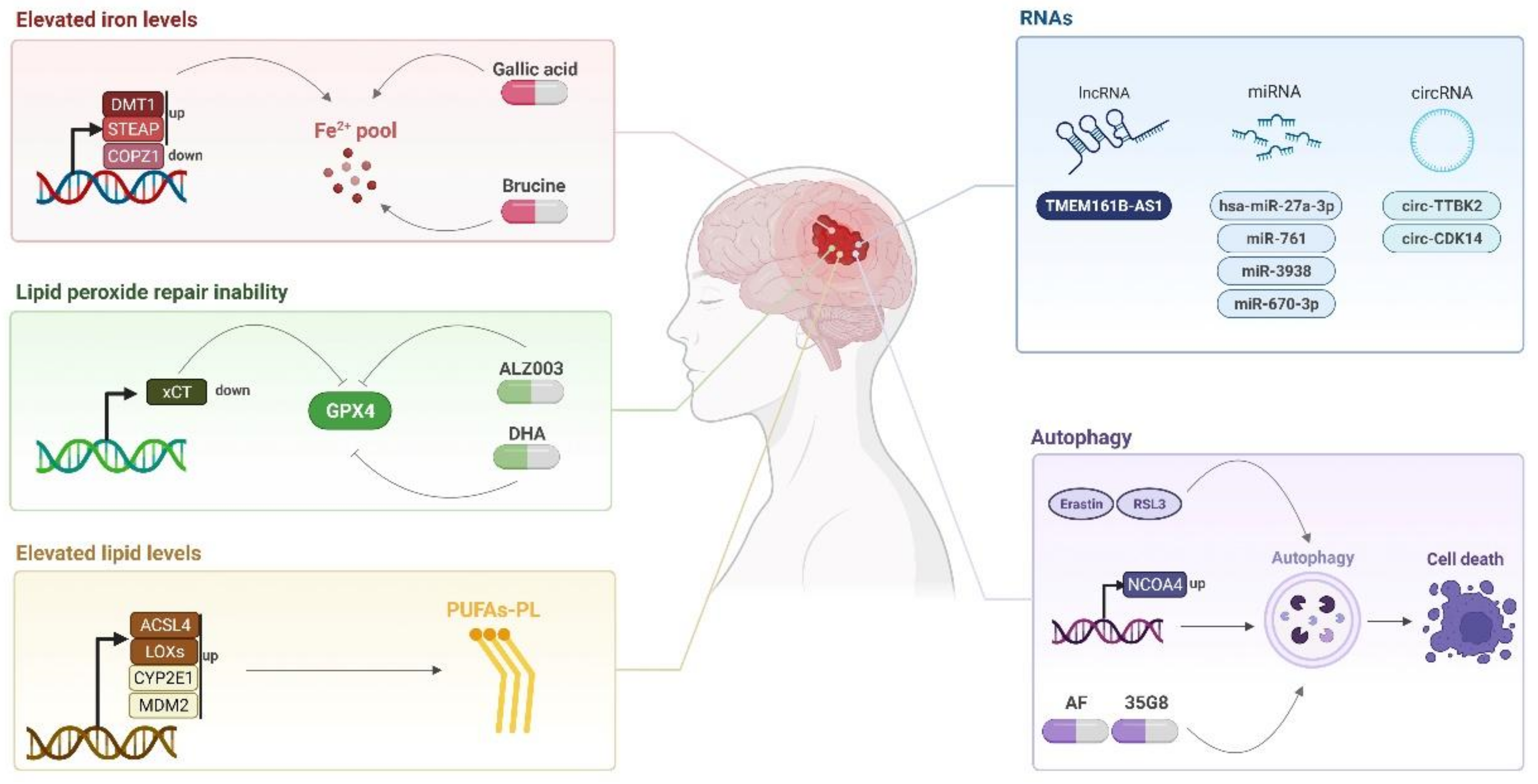
4.1. Ferroptosis-Inducing Compounds

Compounds capable of inducing ferroptosis can direct new treatments for glioblastoma such as brucine and cRGD/Pt + DOX@GFNPs nanoformulation, which promotes ferroptosis mediated by the iron pathway [20][21] as well as AF and 35G8, which induced ferroptosis in an autophagy dependent manner [50][68], above-as mentioned. 
The compound ALZ003 (a curcumin analog), is a negative regulator of GPX4, which promotes a reduction in the GSH/GSSG levels and therefore generates the accumulation of lipid peroxidation and high ROS levels, leading to ferroptosis in the U87MG cell line [75]. Interestingly, natural plant extracts are prospective therapies in many types of cancer including glioblastoma. Artemisinin is an active ingredient extracted from the natural plant Artemisia annua, and this metabolic form in vivo with the most potential action is referred to as Dihydroartemisinin (DHA). A study with U87 and A172 cell lines analyzing protein expression patterns suggests that DHA activates ferroptosis through the inhibition of GPX4 in glioblastoma. Using DCFH-DA and BODIPY-C11 probes, the combination of ferrostatin-1 and DHA resulted in a reversion of the increased intracellular ROS and lipid peroxidation levels caused by DHA single treatment, corroborating the ferroptosis action in these cells after DHA treatment [76].
Accordingly, another study with DHA using U251 and U373 cell lines demonstrated that this drug can induce ferroptosis by causing stress in the endoplasmic reticulum (ER) in glioma cells. However, ER stress causes unfolded protein response (UPR), which could be responsible for providing drug-resistant capacity and more tumorigenicity. UPR signaling can be mediated by the PERK protein, and this pathway may mitigate the effects of DHA in glioma cell lines. Therefore, the treatment with this drug also activated, in parallel, a pathway that protects glioma cells from this cell death type: a feedback pathway of ferroptosis. This process is dependent on the PERK (protein kinase R-like ER kinase) activity, which induced HSPA5 (heat shock protein family A member 5) expression through ATF4 activation; therefore, the PERK-ATF4 pathway resulted in the induction of HSPA5 expression, and this led to the expression and activity of GPX4. Thus, ER stress induced by DHA causes the activation of this molecular cascade (PERK-ATF4-HSPA5-GPX4), which inhibits ferroptosis, through the prevention of lipid peroxidation. In essence, the blockage of the PERK-ATF4-HSPA5-GPX4 pathway using siRNA or small molecules could improve the DHA effect in glioma cells, increasing ferroptosis in vitro and in vivo and carrying out antitumor activity [77].
15,16-Dihidrotanshinone I (DHI) is another natural herbal compound (extracted from Salvia miltiorrhiza Bunge). Currently, it is used to treat cardiovascular disease and has been studied due to its therapeutic effects on some types of cancer cells. It was observed that DHI decreases the cell proliferation in a dose- and time-dependent manner in the U251 and U87 cell lines. The results indicate that cell death occurred by GPX4 inhibition and ACSL4 increase. Furthermore, the cellular antioxidant system GSH/GSSG levels and mitochondrial membrane potential (MMP) was reduced, promoting ferroptosis [39]. Pseudolaric acid B (a diterpene acid isolated from the root and trunk bark of Cortex pseudolaricis, known as PAB) can induce glioma cell death both in vitro and in vivo due to excessive H2O2 production and lipid peroxide formation, generated mainly by the iron-activated Nox4 [78]. Moreover, it was demonstrated that these effects are also generated by the depletion of GSH and cysteine through the activation of p53, which inhibits the xCT pathway [79].
Among the prospective drugs that can contribute to ferroptosis, ibuprofen can exert antitumor effects in many different tumor cells including glioblastoma. This anti-inflammatory drug is a potential therapeutic strategy due to its ability to induce ferroptosis through the inhibition/dysregulation of the NRF2 signaling pathway, thereby generating increased intracellular lipid peroxidation, leading to decreased viability of glioblastoma cells in vitro and in vivo. According to Gao et al., the treatment with increasing concentrations of ibuprofen decreases NRF2GPX4, and SLC7A11 expression in the glioma cells, resulting in ferroptosis induction [78].
As aforementioned, Kyani et al. described a novel nanomolar PDI inhibitor, 1,3,6-trimethylpyrimido[5,4-e][1,2,4]triazine-5,7-dione (35G8), which is toxic to human glioblastoma cell lines U87MG, U118MG, A172, and NU04. Interestingly, PDI is overexpressed in multiple cancer types, but particularly in glioblastoma. Thus, targeting PDI inhibitors can be a promising strategy in the treatment of the disease. The results obtained from their study pointed out that the 35G8-induced cell death in U87MG glioblastoma cells was due to autophagy and ferroptosis, since treatment with apoptosis (Z-VAD-FMK,) and necroptosis (necrostatin-1) inhibitors did not prevent cell death. 35G8 promoted the upregulation of NRF2 response genes including two genes related to ferroptosis, HMOX1 and SLC7A11, (which are known to mediate the response to oxidative stress (ROS). Protein levels of HMOX1 and SLC7A11 were also increased. Interestingly, when U87MG received treatment by DFO, the action of 35G8 was less potent than in the absence of these, indicating that 35G8 can induce ferroptosis [50].
Koike et al. suggested the application of the compound 2-nitroimidazoles in hypoxic glioma stem cells (GSCs). The group observed that 2-nitroimidazole doranidazol could induce GSC death by mitochondrial dysfunction and ferroptosis through ROS accumulation. Thus, it could be a potential target and inducer of ferroptosis in these cell types [80].
Certainly, it is extremely urgent to find therapeutic alternatives for the treatment of glioblastoma, considering its devasting effect on patients. Ferroptosis-inducing compounds have played an interesting role in reversing this scenario, however, it is also important to take into account the potential long-term side effects that these drugs could trigger in brain tissues. Indeed, it was identified that lipid peroxidation, glutamate abnormal levels, and elevated iron levels are common features among ferroptosis and many neurodegenerative diseases such as Parkinson’s, Huntington’s disease, motor neuron disease, and multiple sclerosis [81][82]. Notably, it was observed that Erastin treatment could sensitize neuronal cells and elevate the iron deposition in the brain as a side-effect in vivo [83]. Neuronal cells also presented vulnerability to RSL3, and GPX4 depletion caused hippocampal degeneration, lipid peroxidation, and mitochondrial damage [84]. In this sense, ferroptosis inhibitors such as Ferrostatin-1 can partially rescue these effects of GPX4 depletion [84]. Therefore, ferroptosis inducers that block GPX4 can worsen neuronal pathologies and affect brain tissues through ferroptosis.
Since there are many compounds able to potentially reverse drug resistance through ferroptosis induction in brain tumors, an intriguing question is whether these drugs could be triggering a collateral effect in normal brain tissues. Interestingly, it was demonstrated that DHA promoted a decrease in the aggregation of amyloid β plaque and neuronal loss, and therefore it is considered as a promising therapeutic drug for hypoxic-ischemic brain damage (HIBD), and Alzheimer’s disease patients [85][86]. Similarly, ibuprofen and polyphenol amentoflavone have been suggested to play potential neuroprotective effects against neurological diseases [87][88]. Considering that some ferroptosis-inducing compounds have positive as well as negative effects in normal brain tissues, the results above-mentioned demonstrate that ferroptosis has a dual effect in cancer and neurodegenerative diseases, pointing out the demand for more studies in the area.

4.2. Potential Biomarkers

It is known that ferroptosis could play a crucial role in several phases of the tumor [89] such as the tumorigenesis and progression [90], cell death, and drug resistance [91]. Thus, the discovery and validation of ferroptosis biomarkers are critical in order to predict tumor prognosis [67]. In this sense, studies have demonstrated the existence of metabolic changes in glioblastoma tumors, for instance, dysfunctions in cellular respiration and in the glutamine and lipid metabolism [67][92].
For example, CD44 is a transmembrane molecule known to facilitate glioma growth and proliferation due to interactions with the tumor microenvironment, and studies have revealed that it promotes the suppression of ferroptosis in cancer cells and may cause chemoresistance [93]. FADS2 (fatty acid desaturase 2) is an enzyme responsible for the desaturation of fatty acids and is upregulated in glioblastoma [94]; Yamane et al. have already described it as a key determinant of cellular sensitivity to ferroptosis in the hepatitis C virus [95]. HSPB1 (or heat shock protein 27 ‘HSP27′) activates G6PD in response to oxidative stress or DNA damage, and together, they promote the glioma development; also, HSPB1 has another role in the activation of G6PD, because it contributes to the production of cellular NADPH and pentose in glioma cells [96]. These pathways have been correlated with ferroptosis, and therefore require further studies.

References

  1. Davis, M. Glioblastoma: Overview of Disease and Treatment. Clin. J. Oncol. Nurs. 2016, 20, S2–S8.
  2. Venur, V.A.; Peereboom, D.M.; Ahluwalia, M.S. Current medical treatment of glioblastoma. Cancer Treat. Res. 2015, 163, 103–115.
  3. Singh, N.; Miner, A.; Hennis, L.; Mittal, S. Mechanisms of temozolomide resistance in glioblastoma—A comprehensive review. Cancer Drug Resist. 2020, 4, 17–43.
  4. Woo, P.; Li, Y.; Chan, A.; Ng, S.; Loong, H.; Chan, D.; Wong, G.; Poon, W.-S. A multifaceted review of temozolomide resistance mechanisms in glioblastoma beyond O-6-methylguanine-DNA methyltransferase. Glioma 2019, 2, 68–82.
  5. Galluzzi, L.; Pedro, J.M.B.-S.; Kepp, O.; Kroemer, G. Regulated cell death and adaptive stress responses. Cell. Mol. Life Sci. 2016, 73, 2405–2410.
  6. Galluzzi, L.; Vitale, I.; Aaronson, S.A.; Abrams, J.M.; Adam, D.; Agostinis, P.; Alnemri, E.S.; Altucci, L.; Amelio, I.; Andrews, D.W.; et al. Molecular mechanisms of cell death: Recommendations of the Nomenclature Committee on Cell Death 2018. Cell Death Differ. 2018, 25, 486–541.
  7. Liang, C.; Zhang, X.; Yang, M.; Dong, X. Recent Progress in Ferroptosis Inducers for Cancer Therapy. Adv. Mater. 2019, 31, e1904197.
  8. Dixon, S.J.; Lemberg, K.M.; Lamprecht, M.R.; Skouta, R.; Zaitsev, E.M.; Gleason, C.E.; Patel, D.N.; Bauer, A.J.; Cantley, A.M.; Yang, W.S.; et al. Ferroptosis: An Iron-Dependent Form of Nonapoptotic Cell Death. Cell 2012, 149, 1060–1072.
  9. Vogt, A.-C.S.; Arsiwala, T.; Mohsen, M.; Vogel, M.; Manolova, V.; Bachmann, M.F. On Iron Metabolism and Its Regulation. Int. J. Mol. Sci. 2021, 22, 4591.
  10. Mancias, J.D.; Wang, X.; Gygi, S.P.; Harper, J.W.; Kimmelman, A.C. Quantitative proteomics identifies NCOA4 as the cargo receptor mediating ferritinophagy. Nature 2014, 509, 105–109.
  11. Lei, P.; Bai, T.; Sun, Y. Mechanisms of Ferroptosis and Relations With Regulated Cell Death: A Review. Front. Physiol. 2019, 10, 139.
  12. Dixon, S.J.; Stockwell, B.R. The Hallmarks of Ferroptosis. Annu. Rev. Cancer Biol. 2019, 3, 35–54.
  13. Torti, S.V.; Manz, D.H.; Paul, B.T.; Blanchette-Farra, N.; Torti, F.M. Iron and Cancer. Annu. Rev. Nutr. 2018, 38, 97–125.
  14. Zhao, Z.; Zhang, K.-N.; Wang, Q.; Li, G.; Zeng, F.; Zhang, Y.; Wu, F.; Chai, R.; Wang, Z.; Zhang, C.; et al. Ferronostics: Measuring Tumoral Ferrous Iron with PET to Predict Sensitivity to Iron-Targeted Cancer Therapies. J. Nucl. Med. Off. Publ. Soc. Nucl. Med. 2021, 62, 949–955.
  15. Song, Q.; Peng, S.; Sun, Z.; Heng, X.; Zhu, X. Temozolomide Drives Ferroptosis via a DMT1-Dependent Pathway in Glioblastoma Cells. Yonsei Med. J. 2021, 62, 843.
  16. Zhang, Y.; Kong, Y.; Ma, Y.; Ni, S.; Wikerholmen, T.; Xi, K.; Zhao, F.; Zhao, Z.; Wang, J.; Huang, B.; et al. Loss of COPZ1 induces NCOA4 mediated autophagy and ferroptosis in glioblastoma cell lines. Oncogene 2021, 40, 1425–1439.
  17. Chen, H.; Xu, C.; Yu, Q.; Zhong, C.; Peng, Y.; Chen, J.; Chen, G. Comprehensive landscape of STEAP family functions and prognostic prediction value in glioblastoma. J. Cell. Physiol. 2021, 236, 2988–3000.
  18. Ohgami, R.S.; Campagna, D.R.; McDonald, A.; Fleming, M.D. The Steap proteins are metalloreductases. Blood 2006, 108, 1388–1394.
  19. Stockwell, B.R.; Friedmann Angeli, J.P.; Bayir, H.; Bush, A.I.; Conrad, M.; Dixon, S.J.; Fulda, S.; Gascón, S.; Hatzios, S.K.; Kagan, V.E.; et al. Ferroptosis: A Regulated Cell Death Nexus Linking Metabolism, Redox Biology, and Disease. Cell 2017, 171, 273–285.
  20. Zhang, Y.; Xi, K.; Fu, X.; Sun, H.; Wang, H.; Yu, D.; Li, Z.; Ma, Y.; Liu, X.; Huang, B.; et al. Versatile metal-phenolic network nanoparticles for multitargeted combination therapy and magnetic resonance tracing in glioblastoma. Biomaterials 2021, 278, 121163.
  21. Lu, S.; Wang, X.-Z.; He, C.; Wang, L.; Liang, S.-P.; Wang, C.-C.; Li, C.; Luo, T.-F.; Feng, C.-S.; Wang, Z.-C.; et al. ATF3 contributes to brucine-triggered glioma cell ferroptosis via promotion of hydrogen peroxide and iron. Acta Pharmacol. Sin. 2021, 42, 1690–1702.
  22. Gaschler, M.M.; Stockwell, B.R. Lipid peroxidation in cell death. Biochem. Biophys. Res. Commun. 2017, 482, 419–425.
  23. Dixon, S.J.; Winter, G.E.; Musavi, L.S.; Lee, E.D.; Snijder, B.; Rebsamen, M.; Superti-Furga, G.; Stockwell, B.R. Human Haploid Cell Genetics Reveals Roles for Lipid Metabolism Genes in Nonapoptotic Cell Death. ACS Chem. Biol. 2015, 10, 1604–1609.
  24. Sparvero, L.J. PEBP1 Wardens Ferroptosis by Enabling Lipoxygenase Generation of Lipid Death Signals. Cell 2017, 171, 628–641.e26.
  25. Doll, S.; Proneth, B.; Tyurina, Y.Y.; Panzilius, E.; Kobayashi, S.; Ingold, I.; Irmler, M.; Beckers, J.; Aichler, M.; Walch, A.; et al. ACSL4 dictates ferroptosis sensitivity by shaping cellular lipid composition. Nat. Chem. Biol. 2017, 13, 91–98.
  26. Kagan, V.E.; Mao, G.; Qu, F.; Angeli, J.P.F.; Doll, S.; Croix, C.S.; Dar, H.H.; Liu, B.; Tyurin, V.A.; Ritov, V.B.; et al. Oxidized arachidonic and adrenic PEs navigate cells to ferroptosis. Nat. Chem. Biol. 2017, 13, 81–90.
  27. Bao, C.; Zhang, J.; Xian, S.Y.; Chen, F. MicroRNA-670-3p suppresses ferroptosis of human glioblastoma cells through targeting. Free Radic. Res. 2021, 55, 853–864.
  28. Cheng, J.; Fan, Y.Q.; Liu, B.H.; Zhou, H.; Wang, J.M.; Chen, Q.X. ACSL4 suppresses glioma cells proliferation via activating ferroptosis. Oncol. Rep. 2019, 43, 147–158.
  29. Yang, W.S.; Kim, K.J.; Gaschler, M.M.; Patel, M.; Shchepinov, M.S.; Stockwell, B.R. Peroxidation of polyunsaturated fatty acids by lipoxygenases drives ferroptosis. Proc. Natl. Acad. Sci. USA 2016, 113, E4966–E4975.
  30. Yang, X.; Liu, J.; Wang, C.; Cheng, K.K.-Y.; Xu, H.; Li, Q.; Hua, T.; Jiang, X.; Sheng, L.; Mao, J.; et al. miR-18a promotes glioblastoma development by down-regulating ALOXE3-mediated ferroptotic and anti-migration activities. Oncogenesis 2021, 10, 15.
  31. Ye, L.; Xu, Y.; Wang, L.; Zhang, C.; Hu, P.; Tong, S.A.; Liu, Z.; Tian, D. Downregulation of CYP2E1 is associated with poor prognosis and tumor progression of gliomas. Cancer Med. 2021, 10, 8100–8113.
  32. Venkatesh, D.; O’Brien, N.A.; Zandkarimi, F.; Tong, D.R.; Stokes, M.E.; Dunn, D.E.; Kengmana, E.S.; Aron, A.T.; Klein, A.M.; Csuka, J.M.; et al. MDM2 and MDMX promote ferroptosis by PPARα-mediated lipid remodeling. Genes Dev. 2020, 34, 526–543.
  33. Forcina, G.C.; Dixon, S.J. GPX4 at the Crossroads of Lipid Homeostasis and Ferroptosis. Proteomics 2019, 19, e1800311.
  34. Sun, Y.; Zheng, Y.; Wang, C.; Liu, Y. Glutathione depletion induces ferroptosis, autophagy, and premature cell senescence in retinal pigment epithelial cells. Cell Death Dis. 2018, 9, 1–15.
  35. Seelig, G.F.; Simondsen, R.P.; Meister, A. Reversible dissociation of gamma-glutamylcysteine synthetase into two subunits. J. Biol. Chem. 1984, 259, 9345–9347.
  36. Ikawa, T.; Sato, M.; Oh-hashi, K.; Furuta, K.; Hirata, Y. Oxindole–curcumin hybrid compound enhances the transcription of γ-glutamylcysteine ligase. Eur. J. Pharmacol. 2021, 896, 173898.
  37. Jiang, T.; Chu, J.; Chen, H.; Cheng, H.; Su, J.; Wang, X.; Cao, Y.; Tian, S.; Li, Q. Gastrodin Inhibits H2O2-Induced Ferroptosis through Its Antioxidative Effect in Rat Glioma Cell Line C6. Biol. Pharm. Bull. 2020, 43, 480–487.
  38. Wei, R.; Qiu, H.; Xu, J.; Mo, J.; Liu, Y.; Gui, Y.; Huang, G.; Zhang, S.; Yao, H.; Huang, X.; et al. Expression and prognostic potential of GPX1 in human cancers based on data mining. Ann. Transl. Med. 2020, 8, 124.
  39. Tan, S.; Hou, X.; Mei, L. Dihydrotanshinone I inhibits human glioma cell proliferation via the activation of ferroptosis. Oncol. Lett. 2020, 20, 122.
  40. Wang, X.; Lu, S.; He, C.; Wang, C.; Wang, L.; Piao, M.; Chi, G.; Luo, Y.; Ge, P. RSL3 induced autophagic death in glioma cells via causing glycolysis dysfunction. Biochem. Biophys. Res. Commun. 2019, 518, 590–597.
  41. Fan, Z.; Wirth, A.-K.; Chen, D.; Wruck, C.J.; Rauh, M.; Buchfelder, M.; Savaskan, N. Nrf2-Keap1 pathway promotes cell proliferation and diminishes ferroptosis. Oncogenesis 2017, 6, e371.
  42. Koppula, P.; Zhuang, L.; Gan, B. Cystine transporter SLC7A11/xCT in cancer: Ferroptosis, nutrient dependency, and cancer therapy. Protein Cell 2021, 12, 599–620.
  43. Sato, H.; Tamba, M.; Ishii, T.; Bannai, S. Cloning and Expression of a Plasma Membrane Cystine/Glutamate Exchange Transporter Composed of Two Distinct Proteins. J. Biol. Chem. 1999, 274, 11455–11458.
  44. Umans, R.A.; Martin, J.; Harrigan, M.E.; Patel, D.C.; Chaunsali, L.; Roshandel, A.; Iyer, K.; Powell, M.D.; Oestreich, K.; Sontheimer, H. Transcriptional Regulation of Amino Acid Transport in Glioblastoma Multiforme. Cancers 2021, 13, 6169.
  45. Chen, L.; Li, X.; Liu, L.; Yu, B.; Xue, Y.; Liu, Y. Erastin sensitizes glioblastoma cells to temozolomide by restraining xCT and cystathionine-γ-lyase function. Oncol. Rep. 2015, 33, 1465–1474.
  46. Sugiyama, A.; Ohta, T.; Obata, M.; Takahashi, K.; Seino, M.; Nagase, S. xCT inhibitor sulfasalazine depletes paclitaxel-resistant tumor cells through ferroptosis in uterine serous carcinoma. Oncol. Lett. 2020, 20, 2689–2700.
  47. Dixon, S.J.; Patel, D.N.; Welsch, M.; Skouta, R.; Lee, E.D.; Hayano, M.; Thomas, A.G.; Gleason, C.E.; Tatonetti, N.P.; Slusher, B.S.; et al. Pharmacological inhibition of cystine–glutamate exchange induces endoplasmic reticulum stress and ferroptosis. eLife 2014, 3, e02523.
  48. Feng, H.; Stockwell, B.R. Unsolved mysteries: How does lipid peroxidation cause ferroptosis? PLoS Biol. 2018, 16, e2006203.
  49. Buccarelli, M.; Marconi, M.; Pacioni, S.; De Pasqualis, I.; D’Alessandris, Q.G.; Martini, M.; Ascione, B.; Malorni, W.; Larocca, L.M.; Pallini, R.; et al. Inhibition of autophagy increases susceptibility of glioblastoma stem cells to temozolomide by igniting ferroptosis. Cell Death Dis. 2018, 9, 841.
  50. Kyani, A.; Tamura, S.; Yang, S.; Shergalis, A.; Samanta, S.; Kuang, Y.; Ljungman, M.; Neamati, N. Discovery and Mechanistic Elucidation of a Class of Protein Disulfide Isomerase Inhibitors for the Treatment of Glioblastoma. ChemMedChem 2018, 13, 177.
  51. Chen, D.; Fan, Z.; Rauh, M.; Buchfelder, M.; Eyupoglu, I.Y.; Savaskan, N. ATF4 promotes angiogenesis and neuronal cell death and confers ferroptosis in a xCT-dependent manner. Oncogene 2017, 36, 5593–5608.
  52. Chen, D.; Rauh, M.; Buchfelder, M.; Eyupoglu, I.Y.; Savaskan, N. The oxido-metabolic driver ATF4 enhances temozolamide chemo-resistance in human gliomas. Oncotarget 2017, 8, 51164–51176.
  53. Hayashima, K.; Kimura, I.; Katoh, H. Role of ferritinophagy in cystine deprivation-induced cell death in glioblastoma cells. Biochem. Biophys. Res. Commun. 2021, 539, 56–63.
  54. Dodson, M.; Castro-Portuguez, R.; Zhang, D.D. NRF2 plays a critical role in mitigating lipid peroxidation and ferroptosis. Redox Biol. 2019, 23, 101107.
  55. Rocha, C.R.R.; Rocha, A.R.; Silva, M.M.; Gomes, L.R.; Latancia, M.T.; Andrade-Tomaz, M.; De Souza, I.; Monteiro, L.K.S.; Menck, C.F.M. Revealing Temozolomide Resistance Mechanisms via Genome-Wide CRISPR Libraries. Cells 2020, 9, 2573.
  56. Rocha, C.R.R.; Kajitani, G.S.; Quinet, A.; Fortunato, R.; Menck, C.F.M. NRF2 and glutathione are key resistance mediators to temozolomide in glioma and melanoma cells. Oncotarget 2016, 7, 48081–48092.
  57. Cao, J.Y.; Poddar, A.; Magtanong, L.; Lumb, J.H.; Mileur, T.R.; Reid, M.A.; Dovey, C.M.; Wang, J.; Locasale, J.W.; Stone, E.; et al. A Genome-wide Haploid Genetic Screen Identifies Regulators of Glutathione Abundance and Ferroptosis Sensitivity. Cell Rep. 2019, 26, 1544–1556.e8.
  58. Hassannia, B.; Wiernicki, B.; Ingold, I.; Qu, F.; Van Herck, S.; Tyurina, Y.Y.; Bayır, H.; Abhari, B.A.; Angeli, J.P.F.; Choi, S.M.; et al. Nano-targeted induction of dual ferroptotic mechanisms eradicates high-risk neuroblastoma. J. Clin. Investig. 2018, 128, 3341–3355.
  59. Li, S.; He, Y.; Chen, K.; Sun, J.; Zhang, L.; He, Y.; Yu, H.; Li, Q. RSL3 Drives Ferroptosis through NF-κB Pathway Activation and GPX4 Depletion in Glioblastoma. Oxidative Med. Cell. Longev. 2021, 2021, 1–10.
  60. He, Y.; Ye, Y.; Tian, W.; Qiu, H. A Novel lncRNA Panel Related to Ferroptosis, Tumor Progression, and Microenvironment is a Robust Prognostic Indicator for Glioma Patients. Front. Cell Dev. Biol. 2021, 9, 788451.
  61. Zheng, J.; Zhou, Z.; Qiu, Y.; Wang, M.; Yu, H.; Wu, Z.; Wang, X.; Jiang, X. A Prognostic Ferroptosis-Related lncRNAs Signature Associated With Immune Landscape and Radiotherapy Response in Glioma. Front. Cell Dev. Biol. 2021, 9, 675555.
  62. Chen, Q.; Wang, W.; Wu, Z.; Chen, S.; Chen, X.; Zhuang, S.; Song, G.; Lv, Y.; Lin, Y. Over-expression of lncRNA TMEM161B-AS1 promotes the malignant biological behavior of glioma cells and the resistance to temozolomide via up-regulating the expression of multiple ferroptosis-related genes by sponging hsa-miR-27a-3p. Cell Death Discov. 2021, 7, 1–12.
  63. Zhang, H.Y.; Zhang, B.W.; Zhang, Z.B.; Deng, Q.J. Circular RNA TTBK2 regulates cell proliferation, invasion and ferroptosis via miR-761/ITGB8 axis in glioma. Eur. Rev. Med. Pharmacol. Sci. 2020, 24, 2585–2600.
  64. Chen, S.; Zhang, Z.; Zhang, B.; Huang, Q.; Liu, Y.; Qiu, Y.; Long, X.; Wu, M.; Zhang, Z. CircCDK14 Promotes Tumor Progression and Resists Ferroptosis in Glioma by Regulating PDGFRA. Int. J. Biol. Sci. 2022, 18, 841–857.
  65. Latunde-Dada, G.O. Ferroptosis: Role of lipid peroxidation, iron and ferritinophagy. Biochim. Biophys. Acta (BBA) Gen. Subj. 2017, 1861, 1893–1900.
  66. Gao, M.; Monian, P.; Pan, Q.; Zhang, W.; Xiang, J.; Jiang, X. Ferroptosis is an autophagic cell death process. Cell Res. 2016, 26, 1021–1032.
  67. Liu, H.-J.; Hu, H.-M.; Li, G.-Z.; Zhang, Y.; Wu, F.; Liu, X.; Wang, K.-Y.; Zhang, C.-B.; Jiang, T. Ferroptosis-Related Gene Signature Predicts Glioma Cell Death and Glioma Patient Progression. Front. Cell Dev. Biol. 2020, 8, 538.
  68. Chen, Y.; Li, N.; Wang, H.; Wang, N.; Peng, H.; Wang, J.; Li, Y.; Liu, M.; Li, H.; Zhang, Y.; et al. Amentoflavone suppresses cell proliferation and induces cell death through triggering autophagy-dependent ferroptosis in human glioma. Life Sci. 2020, 247, 117425.
  69. Zhou, L.; Jiang, Z.; Shi, Z.; Zhao, W.; Lu, Z.; Xie, Y.; Zhang, B.; Lu, H.; Tan, G.; Wang, Z. New Autophagy-Ferroptosis Gene Signature Predicts Survival in Glioma. Front. Cell Dev. Biol. 2021, 9, 739097.
  70. Sun, W.; Yan, J.; Ma, H.; Wu, J.; Zhang, Y. Autophagy-Dependent Ferroptosis-Related Signature is Closely Associated with the Prognosis and Tumor Immune Escape of Patients with Glioma. Int. J. Gen. Med. 2022, 15, 253–270.
  71. Lee, S.Y. Temozolomide resistance in glioblastoma multiforme. Genes Dis. 2016, 3, 198–210.
  72. Hu, Z.; Mi, Y.; Qian, H.; Guo, N.; Yan, A.; Zhang, Y.; Gao, X. A Potential Mechanism of Temozolomide Resistance in Glioma–Ferroptosis. Front. Oncol. 2020, 10, 897.
  73. Mitre, A.-O.; Florian, A.I.; Buruiana, A.; Boer, A.; Moldovan, I.; Soritau, O.; Florian, S.I.; Susman, S. Ferroptosis Involvement in Glioblastoma Treatment. Medicina 2022, 58, 319.
  74. Zhuo, S.; Chen, Z.; Yang, Y.; Zhang, J.; Tang, J.; Yang, K. Clinical and Biological Significances of a Ferroptosis-Related Gene Signature in Glioma. Front. Oncol. 2020, 10, 590861.
  75. Chen, T.-C.; Chuang, J.-Y.; Ko, C.-Y.; Kao, T.-J.; Yang, P.-Y.; Yu, C.-H.; Liu, M.-S.; Hu, S.-L.; Tsai, Y.-T.; Chan, H.; et al. AR ubiquitination induced by the curcumin analog suppresses growth of temozolomide-resistant glioblastoma through disrupting GPX4-Mediated redox homeostasis. Redox Biol. 2020, 30, 101413.
  76. Yi, R.; Wang, H.; Deng, C.; Wang, X.; Yao, L.; Niu, W.; Fei, M.; Zhaba, W. Dihydroartemisinin initiates ferroptosis in glioblastoma through GPX4 inhibition. Biosci. Rep. 2020, 40, BSR20193314.
  77. Chen, Y.; Mi, Y.; Zhang, X.; Ma, Q.; Song, Y.; Zhang, L.; Wang, D.; Xing, J.; Hou, B.; Li, H.; et al. Dihydroartemisinin-induced unfolded protein response feedback attenuates ferroptosis via PERK/ATF4/HSPA5 pathway in glioma cells. J. Exp. Clin. Cancer Res. 2019, 38, 402.
  78. Gao, X.; Guo, N.; Xu, H.; Pan, T.; Lei, H.; Yan, A.; Mi, Y.; Xu, L. Ibuprofen induces ferroptosis of glioblastoma cells via downregulation of nuclear factor erythroid 2-related factor 2 signaling pathway. Anti-Cancer Drugs 2020, 31, 34.
  79. Wang, Z.; Ding, Y.; Wang, X.; Lu, S.; Wang, C.; He, C.; Wang, L.; Piao, M.; Chi, G.; Luo, Y.; et al. Pseudolaric acid B triggers ferroptosis in glioma cells via activation of Nox4 and inhibition of xCT. Cancer Lett. 2018, 428, 33.
  80. Koike, N.; Kota, R.; Naito, Y.; Hayakawa, N.; Matsuura, T.; Hishiki, T.; Onishi, N.; Fukada, J.; Suematsu, M.; Shigematsu, N.; et al. 2-Nitroimidazoles induce mitochondrial stress and ferroptosis in glioma stem cells residing in a hypoxic niche. Commun. Biol. 2020, 3, 1–13.
  81. Abdalkader, M.; Lampinen, R.; Kanninen, K.M.; Malm, T.M.; Liddell, J.R. Targeting Nrf2 to Suppress Ferroptosis and Mitochondrial Dysfunction in Neurodegeneration. Front. Neurosci. 2018, 12, 466.
  82. Masaldan, S.; Bush, A.I.; Devos, D.; Rolland, A.S.; Moreau, C. Striking while the iron is hot: Iron metabolism and ferroptosis in neurodegeneration. Free. Radic. Biol. Med. 2019, 133, 221–233.
  83. Zhao, J.; Xu, B.; Xiong, Q.; Feng, Y.; Du, H. Erastin-induced ferroptosis causes physiological and pathological changes in healthy tissues of mice. Mol. Med. Rep. 2021, 24, 1–8.
  84. Hambright, W.S.; Fonseca, R.S.; Chen, L.; Na, R.; Ran, Q. Ablation of ferroptosis regulator glutathione peroxidase 4 in forebrain neurons promotes cognitive impairment and neurodegeneration. Redox Biol. 2017, 12, 8–17.
  85. Xiong, Q.; Li, X.; Xia, L.; Yao, Z.; Shi, X.; Dong, Z. Dihydroartemisinin attenuates hypoxic-ischemic brain damage in neonatal rats by inhibiting oxidative stress. Mol. Brain 2022, 15, 1–10.
  86. Zhao, Y.; Long, Z.; Ding, Y.; Jiang, T.; Liu, J.; Li, Y.; Liu, Y.; Peng, X.; Wang, K.; Feng, M.; et al. Dihydroartemisinin Ameliorates Learning and Memory in Alzheimer’s Disease Through Promoting Autophagosome-Lysosome Fusion and Autolysosomal Degradation for Aβ Clearance. Front. Aging Neurosci. 2020, 12, 47.
  87. Shin, D.H.; Bae, Y.C.; Kim-Han, J.S.; Lee, J.H.; Choi, I.Y.; Son, K.H.; Kang, S.S.; Kim, W.-K.; Han, B.H. Polyphenol amentoflavone affords neuroprotection against neonatal hypoxic-ischemic brain damage via multiple mechanisms. J. Neurochem. 2006, 96, 561–572.
  88. Le, T.T.; Kuplicki, R.; Yeh, H.-W.; Aupperle, R.L.; Khalsa, S.S.; Simmons, W.K.; Paulus, M.P. Effect of Ibuprofen on BrainAGE: A Randomized, Placebo-Controlled, Dose-Response Exploratory Study. Biol. Psychiatry Cogn. Neurosci. Neuroimaging 2018, 3, 836–843.
  89. Hu, Y.; Tu, Z.; Lei, K.; Huang, K.; Zhu, X. Ferroptosis-related gene signature correlates with the tumor immune features and predicts the prognosis of glioma patients. Biosci. Rep. 2021, 41, BSR20211640.
  90. Zhao, J.; Liu, Z.; Zheng, X.; Gao, H.; Li, L. Prognostic Model and Nomogram Construction Based on a Novel Ferroptosis-Related Gene Signature in Lower-Grade Glioma. Front. Genet. 2021, 12, 753680.
  91. Elgendy, S.M.; Alyammahi, S.K.; Alhamad, D.W.; Abdin, S.M.; Omar, H.A. Ferroptosis: An emerging approach for targeting cancer stem cells and drug resistance. Crit. Rev. Oncol. Hematol. 2020, 155, 103095.
  92. Liu, Y.; Xu, Z.; Jin, T.; Xu, K.; Liu, M.; Xu, H. Ferroptosis in Low-Grade Glioma: A New Marker for Diagnosis and Prognosis. Med. Sci. Monit. 2020, 26, e921947.
  93. Mooney, K.L.; Choy, W.; Sidhu, S.; Pelargos, P.; Bui, T.T.; Voth, B.; Barnette, N.; Yang, I. The role of CD44 in glioblastoma multiforme. J. Clin. Neurosci. 2016, 34, 1–5.
  94. Affronti, H.C.; Wellen, K.E. Epigenetic Control of Fatty-Acid Metabolism Sustains Glioma Stem Cells. Cancer Discov. 2019, 9, 1161–1163.
  95. Yamane, D.; Hayashi, Y.; Matsumoto, M.; Nakanishi, H.; Imagawa, H.; Kohara, M.; Lemon, S.M.; Ichi, I. FADS2-dependent fatty acid desaturation dictates cellular sensitivity to ferroptosis and permissiveness for hepatitis C virus replication. Cell Chem. Biol. 2021, 29, 799–810.e4.
  96. Ye, H.; Huang, H.; Cao, F.; Chen, M.; Zheng, X.; Zhan, R. HSPB1 Enhances SIRT2-Mediated G6PD Activation and Promotes Glioma Cell. PLoS ONE 2016, 11, e0164285.
More
Information
Subjects: Oncology; Cell Biology
Contributors MDPI registered users' name will be linked to their SciProfiles pages. To register with us, please refer to https://encyclopedia.pub/register : , , , , , ,
View Times: 411
Entry Collection: Biopharmaceuticals Technology
Revisions: 2 times (View History)
Update Date: 08 Jul 2022
1000/1000
Video Production Service