Submitted Successfully!
To reward your contribution, here is a gift for you: A free trial for our video production service.
Thank you for your contribution! You can also upload a video entry or images related to this topic.
Version Summary Created by Modification Content Size Created at Operation
1 -- 1892 2022-06-30 12:04:43 |
2 format correct Meta information modification 1892 2022-07-04 10:25:38 |

Video Upload Options

Do you have a full video?

Confirm

Are you sure to Delete?
Cite
If you have any further questions, please contact Encyclopedia Editorial Office.
Hernandez, E.P.; , A.;  Alim, M.A.;  Kawada, H.;  Kwofie, K.D.;  Ladzekpo, D.;  Koike, Y.;  Inoue, T.;  Sasaki, S.;  Mikami, F.; et al. Vector Competency and Oxidative Stress in Arthropods. Encyclopedia. Available online: https://encyclopedia.pub/entry/24778 (accessed on 30 April 2024).
Hernandez EP,  A,  Alim MA,  Kawada H,  Kwofie KD,  Ladzekpo D, et al. Vector Competency and Oxidative Stress in Arthropods. Encyclopedia. Available at: https://encyclopedia.pub/entry/24778. Accessed April 30, 2024.
Hernandez, Emmanuel Pacia, Anisuzzaman , Md Abdul Alim, Hayato Kawada, Kofi Dadzie Kwofie, Danielle Ladzekpo, Yuki Koike, Takahiro Inoue, Sana Sasaki, Fusako Mikami, et al. "Vector Competency and Oxidative Stress in Arthropods" Encyclopedia, https://encyclopedia.pub/entry/24778 (accessed April 30, 2024).
Hernandez, E.P., , A.,  Alim, M.A.,  Kawada, H.,  Kwofie, K.D.,  Ladzekpo, D.,  Koike, Y.,  Inoue, T.,  Sasaki, S.,  Mikami, F.,  Matsubayashi, M.,  Tanaka, T.,  Tsuji, N., & Hatta, T. (2022, July 04). Vector Competency and Oxidative Stress in Arthropods. In Encyclopedia. https://encyclopedia.pub/entry/24778
Hernandez, Emmanuel Pacia, et al. "Vector Competency and Oxidative Stress in Arthropods." Encyclopedia. Web. 04 July, 2022.
Vector Competency and Oxidative Stress in Arthropods
Edit

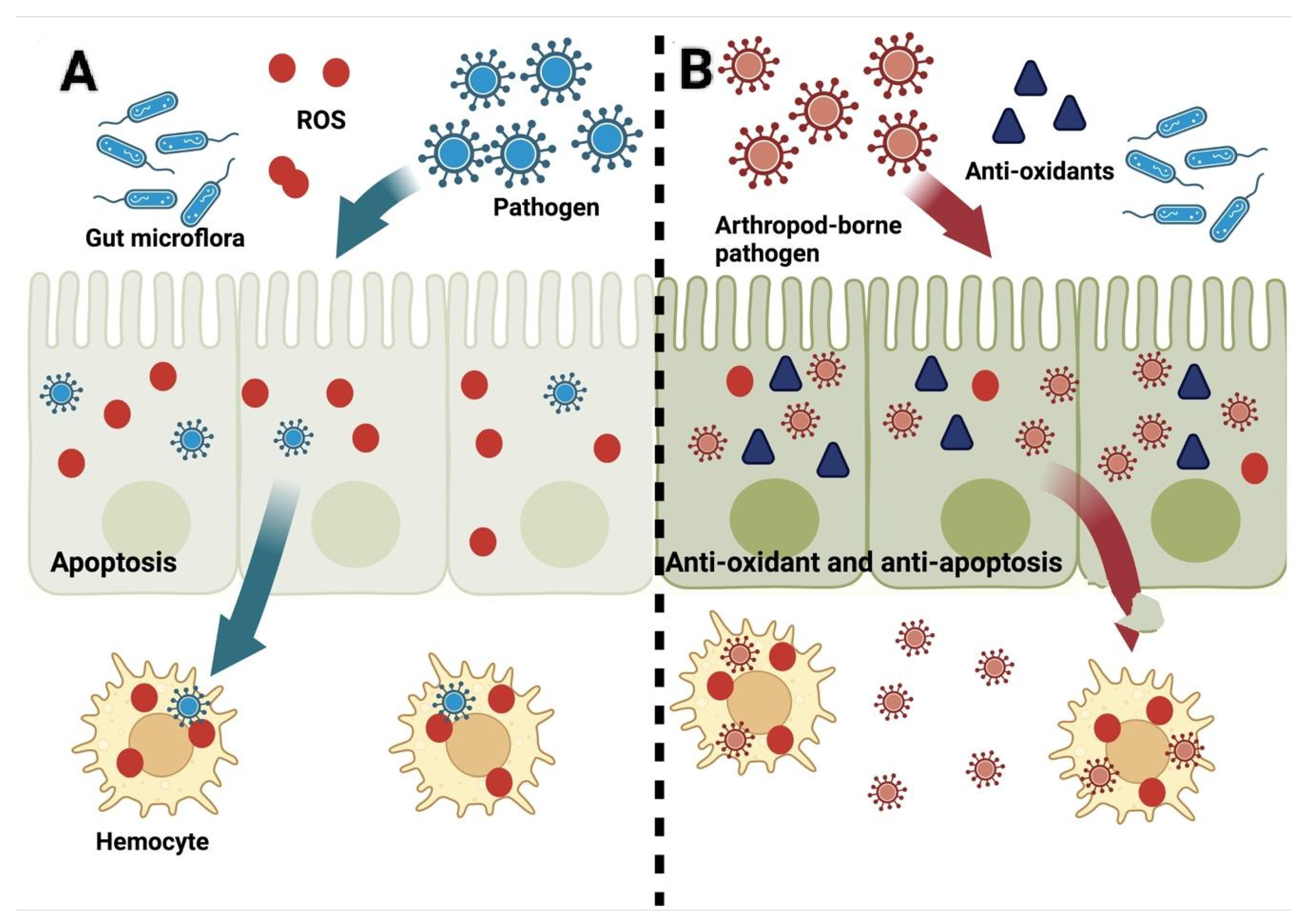
Blood-feeding arthropods, particularly ticks and mosquitoes are considered the most important vectors of arthropod-borne diseases affecting humans and animals. Vector competence (also termed vector potential) refers to the ability of arthropods to transmit pathogens, which is greatly influenced by the genetic and/or other intrinsic factors of arthropod vectors. Additionally, it is also governed by the factors exerted by hosts themselves during pathogen inoculation, development, and propagation in particular hosts. During an infection, ROS have pivotal roles in the triangular relationship among vectors, pathogens, and hosts and may influence the triad either positively or negatively. A pluripotent molecule isolated from the salivary glands of H. longicornis, called longistatin, plays a central role in the feeding and development of ticks  and has been elegantly shown to ameliorate cellular ROS production in human endothelial cells, making it a key molecule in the survival of hard ticks. On the other hand, the acquisition of pathogens into a vector also induces modification of the normal ROS production resulting in oxidative stress to arthropod cells, which ultimately is being utilized by hematophagous arthropods to eliminate invading pathogens.

ticks mosquitoes oxidative stress ROS

1. ROS and Arthropod’s Innate Immunity

To ensure their own survival and existence, arthropods use ROS to eliminate invading pathogens as well as to mount a better immune response during infection (Figure 1A) [1][2][3]Anopheles gambiae, for example, can survive better at higher levels of systemic ROS when challenged with Micrococcus and Escherichia. Furthermore, the supplementation of antioxidants in the diet results in significantly higher mortality during bacterial infection [4], indicating that ROS and oxidative stress play a critical role in the arthropods’ survival during the acquisition and transmission of infections. On the other hand, a Plasmodium refractory strain of Ano. gambiae was observed to be in a chronic state of oxidative stress, while the same parasite would survive if antioxidants were provided in the diet [5][6]. The same effects of oxidative stress are observed in the Rh. microplus (BME26) cell line. During infection with Rickettsia ricketsii or exposure to heat-killed microorganisms, upregulation of genes encoding for ROS production was observed, while antioxidant genes were downregulated [7]. Oxidative burst by macrophages efficiently eliminates pathogens basically by ROS, which are either toxic to the pathogen or work together with hydrolases, reactive nitrogen species, and the NADPH oxidase system (NOX). Supporting the above notion, bacterial infections have been shown to increase ROS in the ticks’ hemocytes [8]. ROS play a role to block pathogen transmission by melanotic encapsulation, where invading pathogens are encapsulated to help the prevention of transmission. Melanotic encapsulation of Plasmodium has been shown in Ano. gambiae. The melanocytic capsule of the refractory strains of Ano. gambiae constructed around Plasmodium can block parasite development in the mosquitoes’ midgut and the strain was observed to have higher levels of ROS [6]. On the other hand, in mosquitoes, ROS also act as a signaling molecule for the mitogen-activated protein kinase (MAPK)–dependent cascade and phosphatidylinositol 3-kinase (PI3K)/Akt-dependent pathway, which has been shown to regulate innate immunity and affects the physiology and development of the malarial parasite [9][10].
Figure 1. Schematic diagram of the life cycle of the pathogen (A) after infection of the arthropod versus the arthropod-borne pathogen’s (B) life cycle and its interaction with ROS. Created with Biorender.com (accessed on 10 May 2022).

2. ROS after the Establishment of Infection in Hosts

While ROS are an important component of defense by the host against the pathogen, ROS are also generated during the establishment of a pathogen within a host and continue to be produced throughout the progression of the disease, for example, flaviviruses are known to induce the production of ROS, which are linked to apoptosis and are, thus, involved in the killing of infected cells together with the pathogen and eventually support the survival of the remaining non-infected cells. Through the production of ROS, infected individuals battle against pathogens to eliminate invading microbes in the early stage of invasion to prevent the progression of damage to the adjacent cells caused by the pathogens themselves and by the triggered inflammatory insults as well (Figure 1A) [11][12]. Therefore, this cellular response could affect the vectorial capacity of the arthropods. However, the coevolution of the arthropods with the pathogens carried by them has led to their coadaptation with each other’s immune responses (Figure 1B). Pathogens have devised various protective shields to evade host responses to ensure their transmission, colonization, and survival in a hostile environment within vertebrate hosts, until either the recovery or the death of the host [13][14][15].
During dengue virus (DENV) infection, apoptosis is the usual outcome [16]. Apoptosis is usually brought about by the production of viral proteins, which disrupts the function of the endoplasmic reticulum (ER), resulting in the accumulation of misfolded and unfolded proteins. The presence of these misfolded and unfolded proteins activates the unfolded protein response (UPR). Even with the UPR, the mitigation of the effect of ER stress may not be addressed within a specific time and will still result in apoptosis [17][18]. However, in a mosquito cell line, it was found that mosquito cells were neither severely damaged nor subjected to apoptosis, rather the infection persisted in this setting, and ROS were detected. Interestingly, a p53 paralogue was upregulated during infection. The p53 is a transcription factor that selectively transcribes the catalase gene, which alleviates ROS accumulation within the cells, therefore reducing the rate of apoptosis (Figure 1B). In experiments that reduced the expression of the p53 gene, ROS accumulated in the infected cells [16]. In Ae. aegypti, knockdown of the catalase gene also resulted in reduced oviposition and lifespan with H2O2 challenge and reduced virus titer in the midgut upon infection with DENV [19]. Aside from the catalase activity, glutathione S-transferase (GST) activity was also higher in DENV-infected cells. GST suppression resulted in an earlier release of superoxide ions and higher cell mortality. Interestingly, this upregulation was not observed in mammalian cells infected with the same virus, indicating that this phenomenon may be limited to only mosquito cells [20]. Besides GST activity, an additive anti-apoptotic activity was observed due to the upregulation of the inhibitor of apoptosis (IAP) [17]. ROS and oxidative stress are also believed to be controlled by the proper refolding of misfolded proteins, and this is usually achieved through the production of the X-box binding protein 1 (XBP1), which is presumed to be a critical transcription factor for various chaperones, including the BiP/GRP 78 mRNA [18]. In contrast, West Nile virus, another flavivirus, also induces ROS production. However, the exact mechanism of this ROS production is still unknown. Mosquito cells infected with this virus-induced upregulation of Nrf2- and NRF1-mediated antioxidant genes, eventually result in elevated reduced glutathione (GSH) levels. This ultimately increased the oxidative capacity of the cells to withstand the oxidative stress elicited by the virus infection [21].
In contrast, transfection with the nonstructural protein 1 of the flavivirus, e.g., tick-borne encephalitis virus, induces oxidative stress in HEK293T cells and activates the antioxidant defense of these cells [22]. Moreover, in tick cells, the knockdown of the antioxidant GST molecule with subsequent infection of the Langat virus, another member of Flaviviridae, resulted in increased mortality, decreased proliferation, and decreased viral titer [23]. Furthermore, infection of LGTV in tick cells indicates a possible correction of the protein folding as seen by the upregulation of chaperone proteins, specifically heat shock proteins (HSPs) 90 and 70 [24][25]. These HSPs help in the refolding of misfolded proteins or are related to the degradation of terminally misfolded proteins to prevent protein aggregation, thereby creating an anti-apoptotic environment within cellular niches [12][26]. This corrective response was also observed in Anaplasma phagocytophilum infection in ticks and tick cells, wherein HSP20, HSP70, and HSP90 expressions have been upregulated [12][27][28]. Metabolomics also indicates that terminally misfolded proteins tend to prevent ER stress and apoptosis. Accordingly, HSP70 knockdown decreases Ana. phagocytophilum titer in ticks [27].
Furthermore, Ana. marginale infection upregulated genes closely related to the generation of antioxidants. Simultaneous knockdown of catalase, glutathione peroxidase, and thioredoxin together with oxidative resistance 1 gene favored the colonization of Ana. marginale in BME26 cells, strongly supporting that the oxidant response is involved in the control of infection and the maintenance of cell survival [7]. Additionally, mitochondrial ROS production also increases in response to Ana. phagocytophilum to control the parasite. Conversely, to maintain the parasite’s fitness and maintain the infection, other alternative ROS production and apoptosis pathways are also inhibited [29].
Another group of molecules that are also upregulated during infection are selenoproteins. They are a group of proteins that both catalyze and regulate several redox reactions [30]. It has been proposed that pathogens can induce the production of selenoproteins that not only allow their proliferation and transmission but also play a key role in pathogen acquisition. In Borrelia burgdorferi infection, Salp25D, a tick selenoprotein, is utilized against the oxidative stress from the inflammatory process at the biting site [31]. Knockdown of selenoprotein M reduces the titer of Ana. marginale and inhibits the development of the infective stage of Ana. marginale [32]. Selenoprotein P (SelP) is upregulated in the salivary glands of the Ri. parkeri–infected ticks to ameliorate oxidative stress during feeding. Furthermore, the knockdown of SelP genes also reduced the transovarial transmission of pathogens [1]. In addition to the direct control of ROS through antioxidant enzymes, the generation of ROS is also controlled by regulating free cations such as iron, which augments ROS productions. Ferritins that sequester free iron are upregulated in Dermacentor variabilis during Escherichia coli infection [33]. Bacterial iron-binding proteins could also sequester iron in the blood meal, which is expressed in the infective stage of Ana. phagocytophilum [34].

3. ROS and Arthropod Microbiome

Microbiome refers to the overall genome of microorganisms in a certain niche, which has been shown to shape the phenome [35][36]. Therefore, attaining a balance between the natural microbiota and potential pathogens is very crucial. One way to maintain this balance is through the dual oxidase (Duox)–dependent ROS generation system [37]. Duox is a protein that mainly functions in the generation of ROS; however, the Duox-ROS pathway remains inactive unless proliferation occurs and bacteria come in contact with the mucosal barrier. In this manner, ROS produced from the Duox pathways attack invading pathogens through the mucosal barrier, particularly by H2O2. These attacks can disrupt the tyrosine phosphorylation network of invading pathogens and, thereby, reduce their fitness. Enterobacteria dominate in the midgut by maintaining gut homeostasis [38][39][40], and these bacterial species are adapted to survive within blood-feeding arthropods (e.g., ticks) as they are resistant to ROS killing. Since enterobacterial species are ROS-generating bacteria, during blood feeding, the gut environment would favor the growth of these bacterial species, overcoming the possible proliferation of Plasmodium and other pathogenic organisms [41][42][43][44]. Challenge with pathogens, therefore, accelerates the expansion of bacterial populations during blood feeding and they escape the constraints of the Duox system. One way for the natural microbiota to escape the Duox system is by avoiding contact with the gut epithelium. To achieve this, bacteria are encaged in a blood bolus during blood feeding. The formation of a dityrosine network (DTN) on the luminal surface of the gut epithelium also makes it difficult for the soluble immune mediators to penetrate the blood bolus; thus, the microbiota avoids activation of the immune responses [45]. The protective effects of this DTN are not only beneficial for the microbiota but also for the Plasmodium parasite [46]. The same DTN is also found in ticks and has been proved to maintain B. burgdorferi infection.
Furthermore, in ticks, some pathogenic organisms have already adapted to this strategy by altering their transcription mechanism and ameliorating the antioxidant mechanisms, including selenoproteins, to maintain the favorable levels of ROS that allow for their survival and growth. The knockdown of the SelP gene increases oxidative stress, thus, decreasing Ri. parkeri loads and increasing levels of Francisella-like symbionts such as Candidatus Midichloria mitochondrii and other bacteria [1][41].

References

  1. Budachetri, K.; Crispell, G.; Karim, S. Amblyomma maculatum SECIS binding protein 2 and putative selenoprotein P are indispensable for pathogen replication and tick fecundity. Insect Biochem. Mol. Biol. 2017, 88, 37–47.
  2. Ha, E.M.; Oh, C.T.; Ryu, J.H.; Bae, Y.S.; Kang, S.W.; Jang, I.H.; Brey, P.T.; Lee, W.J. An antioxidant system required for host protection against gut infection in Drosophila. Dev. Cell 2005, 8, 125–132.
  3. Muralidharan, S.; Mandrekar, P. Cellular stress response and innate immune signaling: Integrating pathways in host defense and inflammation. J. Leukoc. Biol. 2013, 94, 1167–1184.
  4. Molina-Cruz, A.; DeJong, R.J.; Charles, B.; Gupta, L.; Kumar, S.; Jaramillo-Gutierrez, G.; Barillas-Mury, C. Reactive oxygen species modulate Anopheles gambiae immunity against bacteria and Plasmodium. J. Biol. Chem. 2008, 283, 3217–3223.
  5. Graça-Souza, A.V.; Maya-Monteiro, C.; Paiva-Silva, G.O.; Braz, G.R.C.; Paes, M.C.; Sorgine, M.H.F.; Oliveira, M.F.; Oliveira, P.L. Adaptations against heme toxicity in blood-feeding arthropods. Insect Biochem. Mol. Biol. 2006, 36, 322–335.
  6. Kumar, S.; Christophides, G.K.; Cantera, R.; Charles, B.; Han, Y.S.; Meister, S.; Dimopoulos, G.; Kafatos, F.C.; Barillas-Mury, C. The role of reactive oxygen species on Plasmodium melanotic encapsulation in Anopheles gambiae. Proc. Natl. Acad. Sci. USA 2003, 100, 14139–14144.
  7. Kalil, S.P.; da Rosa, R.D.; Capelli-Peixoto, J.; Pohl, P.C.; de Oliveira, P.L.; Fogaça, A.C.; Daffre, S. Immune-related redox metabolism of embryonic cells of the tick Rhipicephalus microplus (BME26) in response to infection with Anaplasma marginale. Parasit. Vectors 2017, 10, 613.
  8. Pereira, L.S.; Oliveira, P.L.; Barja-Fidalgo, C.; Daffre, S. Production of reactive oxygen species by hemocytes from the cattle tick Boophilus microplus. Exp. Parasitol. 2001, 99, 66–72.
  9. Surachetpong, W.; Pakpour, N.; Cheung, K.W.; Luckhart, S. Reactive oxygen species-dependent cell signaling regulates the mosquito immune response to Plasmodium falciparum. Antioxid. Redox Signal. 2011, 14, 943–955.
  10. Corby-Harris, V.; Drexler, A.; Watkins de Jong, L.; Antonova, Y.; Pakpour, N.; Ziegler, R.; Ramberg, F.; Lewis, E.E.; Brown, J.M.; Luckhart, S.; et al. Activation of Akt signaling reduces the prevalence and intensity of malaria parasite infection and lifespan in Anopheles stephensi mosquitoes. PLoS Pathog. 2010, 6, e1001003.
  11. Ashida, H.; Mimuro, H.; Ogawa, M.; Kobayashi, T.; Sanada, T.; Kim, M.; Sasakawa, C. Cell death and infection: A double-edged sword for host and pathogen survival. J. Cell Biol. 2011, 195, 931–942.
  12. Villar, M.; Ayllón, N.; Alberdi, P.; Moreno, A.; Moreno, M.; Tobes, R.; Mateos-Hernández, L.; Weisheit, S.; Bell-Sakyi, L.; de la Fuente, J. Integrated Metabolomics, Transcriptomics and Proteomics Identifies Metabolic Pathways Affected by Anaplasma phagocytophilum Infection in Tick Cells. Mol. Cell Proteom. 2015, 14, 3154–3172.
  13. de la Fuente, J.; Antunes, S.; Bonnet, S.; Cabezas-Cruz, A.; Domingos, A.G.; Estrada-Peña, A.; Johnson, N.; Kocan, K.M.; Mansfield, K.L.; Nijhof, A.M.; et al. Tick-Pathogen Interactions and Vector Competence: Identification of Molecular Drivers for Tick-Borne Diseases. Front. Cell Infect. Microbiol. 2017, 7, 114.
  14. Hajdušek, O.; Síma, R.; Ayllón, N.; Jalovecká, M.; Perner, J.; de la Fuente, J.; Kopáček, P. Interaction of the tick immune system with transmitted pathogens. Front. Cell Infect. Microbiol. 2013, 3, 26.
  15. Villar, M.; Popara, M.; Bonzón-Kulichenko, E.; Ayllón, N.; Vázquez, J.; de la Fuente, J. Characterization of the tick-pathogen interface by quantitative proteomics. Ticks Tick Borne Dis. 2012, 3, 154–158.
  16. Chen, T.H.; Wu, Y.J.; Hou, J.N.; Chiang, Y.H.; Cheng, C.C.; Sifiyatun, E.; Chiu, C.H.; Wang, L.C.; Chen, W.J. A novel p53 paralogue mediates antioxidant defense of mosquito cells to survive dengue virus replication. Virology 2018, 519, 156–169.
  17. Chen, T.H.; Lo, Y.P.; Yang, C.F.; Chen, W.J. Additive protection by antioxidant and apoptosis-inhibiting effects on mosquito cells with dengue 2 virus infection. PLoS Negl. Trop. Dis. 2012, 6, e1613.
  18. Chen, T.H.; Chiang, Y.H.; Hou, J.N.; Cheng, C.C.; Sofiyatun, E.; Chiu, C.H.; Chen, W.J. XBP1-Mediated BiP/GRP78 Upregulation Copes with Oxidative Stress in Mosquito Cells during Dengue 2 Virus Infection. Biomed Res. Int. 2017, 2017, 3519158.
  19. Oliveira, J.H.M.; Talyuli, O.A.C.; Goncalves, R.L.S.; Paiva-Silva, G.O.; Sorgine, M.H.F.; Alvarenga, P.H.; Oliveira, P.L. Catalase protects Aedes aegypti from oxidative stress and increases midgut infection prevalence of dengue but not Zika. PLoS Negl. Trop. Dis. 2017, 11, e0005525.
  20. Chen, T.H.; Tang, P.; Yang, C.F.; Kao, L.H.; Lo, Y.P.; Chuang, C.K.; Shih, Y.T.; Chen, W.J. Antioxidant defense is one of the mechanisms by which mosquito cells survive dengue 2 viral infection. Virology 2011, 410, 410–417.
  21. Basu, M.; Courtney, S.C.; Brinton, M.A. Arsenite-induced stress granule formation is inhibited by elevated levels of reduced glutathione in West Nile virus-infected cells. PLoS Pathog. 2017, 13, e1006240.
  22. Kuzmenko, Y.V.; Smirnova, O.A.; Ivanov, A.V.; Starodubova, E.S.; Karpov, V.L. Nonstructural Protein 1 of Tick-Borne Encephalitis Virus Induces Oxidative Stress and Activates Antioxidant Defense by the Nrf2/ARE Pathway. Intervirology 2016, 59, 111–117.
  23. Hernandez, E.P.; Talactac, M.R.; Vitor, R.J.S.; Yoshii, K.; Tanaka, T. An Ixodes scapularis glutathione S-transferase plays a role in cell survival and viability during Langat virus infection of a tick cell line. Acta Trop. 2021, 214, 105763.
  24. Grabowski, J.M.; Perera, R.; Roumani, A.M.; Hedrick, V.E.; Inerowicz, H.D.; Hill, C.A.; Kuhn, R.J. Changes in the Proteome of Langat-Infected Ixodes scapularis ISE6 Cells: Metabolic Pathways Associated with Flavivirus Infection. PLoS Negl. Trop. Dis. 2016, 10, e0004180.
  25. Grabowski, J.M.; Gulia-Nuss, M.; Kuhn, R.J.; Hill, C.A. RNAi reveals proteins for metabolism and protein processing associated with Langat virus infection in Ixodes scapularis (black-legged tick) ISE6 cells. Parasit. Vectors 2017, 10, 24.
  26. Fulda, S.; Gorman, A.M.; Hori, O.; Samali, A. Cellular stress responses: Cell survival and cell death. Int. J. Cell Biol. 2010, 2010, 214074.
  27. Busby, A.T.; Ayllón, N.; Kocan, K.M.; Blouin, E.F.; de la Fuente, G.; Galindo, R.C.; Villar, M.; de la Fuente, J. Expression of heat shock proteins and subolesin affects stress responses, Anaplasma phagocytophilum infection and questing behaviour in the tick, Ixodes scapularis. Med. Vet. Entomol. 2012, 26, 92–102.
  28. Villar, M.; Ayllón, N.; Busby, A.T.; Galindo, R.C.; Blouin, E.F.; Kocan, K.M.; Bonzón-Kulichenko, E.; Zivkovic, Z.; Almazán, C.; Torina, A.; et al. Expression of Heat Shock and Other Stress Response Proteins in Ticks and Cultured Tick Cells in Response to Anaplasma spp. Infection and Heat Shock. Int. J. Proteom. 2010, 2010, 657261.
  29. Alberdi, P.; Cabezas-Cruz, A.; Prados, P.E.; Rayo, M.V.; Artigas-Jerónimo, S.; de la Fuente, J. The redox metabolic pathways function to limit Anaplasma phagocytophilum infection and multiplication while preserving fitness in tick vector cells. Sci. Rep. 2019, 9, 13236.
  30. Reeves, M.A.; Hoffmann, P.R. The human selenoproteome: Recent insights into functions and regulation. Cell Mol. Life Sci. 2009, 66, 2457–2478.
  31. Narasimhan, S.; Sukumaran, B.; Bozdogan, U.; Thomas, V.; Liang, X.; De Ponte, K.; Marcantonio, N.; Koski, R.A.; Anderson, J.F.; Kantor, F.; et al. A tick antioxidant facilitates the Lyme disease agent’s successful migration from the mammalian host to the arthropod vector. Cell Host Microbe 2007, 2, 7–18.
  32. Kocan, K.M.; Zivkovic, Z.; Blouin, E.F.; Naranjo, V.; Almazán, C.; Mitra, R.; de la Fuente, J. Silencing of genes involved in Anaplasma marginale-tick interactions affects the pathogen developmental cycle in Dermacentor variabilis. BMC Dev. Biol. 2009, 9, 42.
  33. Galay, R.L.; Umemiya-Shirafuji, R.; Mochizuki, M.; Fujisaki, K.; Tanaka, T. Iron metabolism in hard ticks (Acari: Ixodidae): The antidote to their toxic diet. Parasitol. Int. 2015, 64, 182–189.
  34. Kahlon, A.; Ojogun, N.; Ragland, S.A.; Seidman, D.; Troese, M.J.; Ottens, A.K.; Mastronunzio, J.E.; Truchan, H.K.; Walker, N.J.; Borjesson, D.L.; et al. Anaplasma phagocytophilum Asp14 is an invasin that interacts with mammalian host cells via its C terminus to facilitate infection. Infect. Immun. 2013, 81, 65–79.
  35. Hooper, L.V.; Gordon, J.I. Commensal host-bacterial relationships in the gut. Science 2001, 292, 1115–1118.
  36. Kinross, J.M.; Darzi, A.W.; Nicholson, J.K. Gut microbiome-host interactions in health and disease. Genome Med. 2011, 3, 14.
  37. Chaitanya, R.K.; Shashank, K.; Sridevi, P. Oxidative stress in invertebrate systems. In Free Radicals and Diseases; Ahmad, R., Ed.; InTech: London, UK, 2016.
  38. Budachetri, K.; Browning, R.E.; Adamson, S.W.; Dowd, S.E.; Chao, C.-C.; Ching, W.M.; Karim, S. An insight into the microbiome of the Amblyomma maculatum (Acari: Ixodidae). J. Med. Entomol. 2014, 51, 119–129.
  39. Cirimotich, C.M.; Ramirez, J.L.; Dimopoulos, G. Native microbiota shape insect vector competence for human pathogens. Cell Host Microbe 2011, 10, 307–310.
  40. Cirimotich, C.M.; Dong, Y.; Clayton, A.M.; Sandiford, S.L.; Souza-Neto, J.A.; Mulenga, M.; Dimopoulos, G. Natural microbe-mediated refractoriness to Plasmodium infection in Anopheles gambiae. Science 2011, 332, 855–858.
  41. Budachetri, K.; Kumar, D.; Crispell, G.; Beck, C.; Dasch, G.; Karim, S. The tick endosymbiont Candidatus Midichloria mitochondrii and selenoproteins are essential for the growth of Rickettsia parkeri in the Gulf Coast tick vector. Microbiome 2018, 6, 141.
  42. Crispell, G.; Budachetri, K.; Karim, S. Rickettsia parkeri colonization in Amblyomma maculatum: The role of superoxide dismutases. Parasit. Vectors 2016, 9, 291.
  43. Kumar, D.; Budachetri, K.; Meyers, V.C.; Karim, S. Assessment of tick antioxidant responses to exogenous oxidative stressors and insight into the role of catalase in the reproductive fitness of the Gulf Coast tick, Amblyomma maculatum. Insect Mol. Biol. 2016, 25, 283–294.
  44. Lee, W.J. Bacterial-modulated signaling pathways in gut homeostasis. Sci. Signal. 2008, 1, pe24.
  45. Champion, C.J.; Xu, J. The impact of metagenomic interplay on the mosquito redox homeostasis. Free Radic. Biol. Med. 2017, 105, 79–85.
  46. Romoli, O.; Gendrin, M. The tripartite interactions between the mosquito, its microbiota and Plasmodium. Parasit. Vectors 2018, 11, 200.
More
Information
Subjects: Entomology
Contributors MDPI registered users' name will be linked to their SciProfiles pages. To register with us, please refer to https://encyclopedia.pub/register : , , , , , , , , , , , , ,
View Times: 271
Revisions: 2 times (View History)
Update Date: 04 Jul 2022
1000/1000