 +1 credit
| Version | Summary | Created by | Modification | Content Size | Created at | Operation |
|---|---|---|---|---|---|---|
| 1 | Kuan-Yu Chu | + 3120 word(s) | 3120 | 2022-06-21 13:27:42 | | | |
| 2 | Kuan-Yu Chu | Meta information modification | 3120 | 2022-06-22 02:26:03 | | | | |
| 3 | Conner Chen | Meta information modification | 3120 | 2022-06-22 08:26:01 | | | | |
| 4 | Conner Chen | Meta information modification | 3120 | 2022-06-22 08:32:45 | | | | |
| 5 | Conner Chen | Meta information modification | 3120 | 2022-06-22 08:33:20 | | | | |
| 6 | Conner Chen | Meta information modification | 3120 | 2022-06-22 08:46:04 | | | | |
| 7 | Conner Chen | Meta information modification | 3120 | 2022-06-22 08:47:05 | | | | |
| 8 | Conner Chen | Meta information modification | 3120 | 2022-06-22 08:48:52 | | | | |
| 9 | Conner Chen | Meta information modification | 3120 | 2022-06-22 08:52:44 | | | | |
| 10 | Conner Chen | Meta information modification | 3120 | 2022-06-22 08:53:44 | | | | |
| 11 | Conner Chen | Meta information modification | 3120 | 2022-06-30 08:36:56 | | |
Video Upload Options
Autoimmune bullous skin disorders are a group of disorders characterized by the formation of numerous blisters and erosions on the skin and/or the mucosal membrane, arising from autoantibodies against the intercellular adhesion molecules and the structural proteins. They can be classified into intraepithelial or subepithelial autoimmune bullous dermatoses based on the location of the targeted antigens. These dermatoses are extremely debilitating and fatal in certain cases, depending on the degree of cutaneous and mucosal involvement. Effective treatments should be implemented promptly. Glucocorticoids serve as the first-line approach due to their rapid onset of therapeutic effects and remission of the acute phase. Nonetheless, long-term applications may lead to major adverse effects that outweigh the benefits. Hence, other adjuvant therapies are mandatory to minimize the potential harm and ameliorate the quality of life.
1. Intraepithelial Autoimmune Bullous Skin Disorders
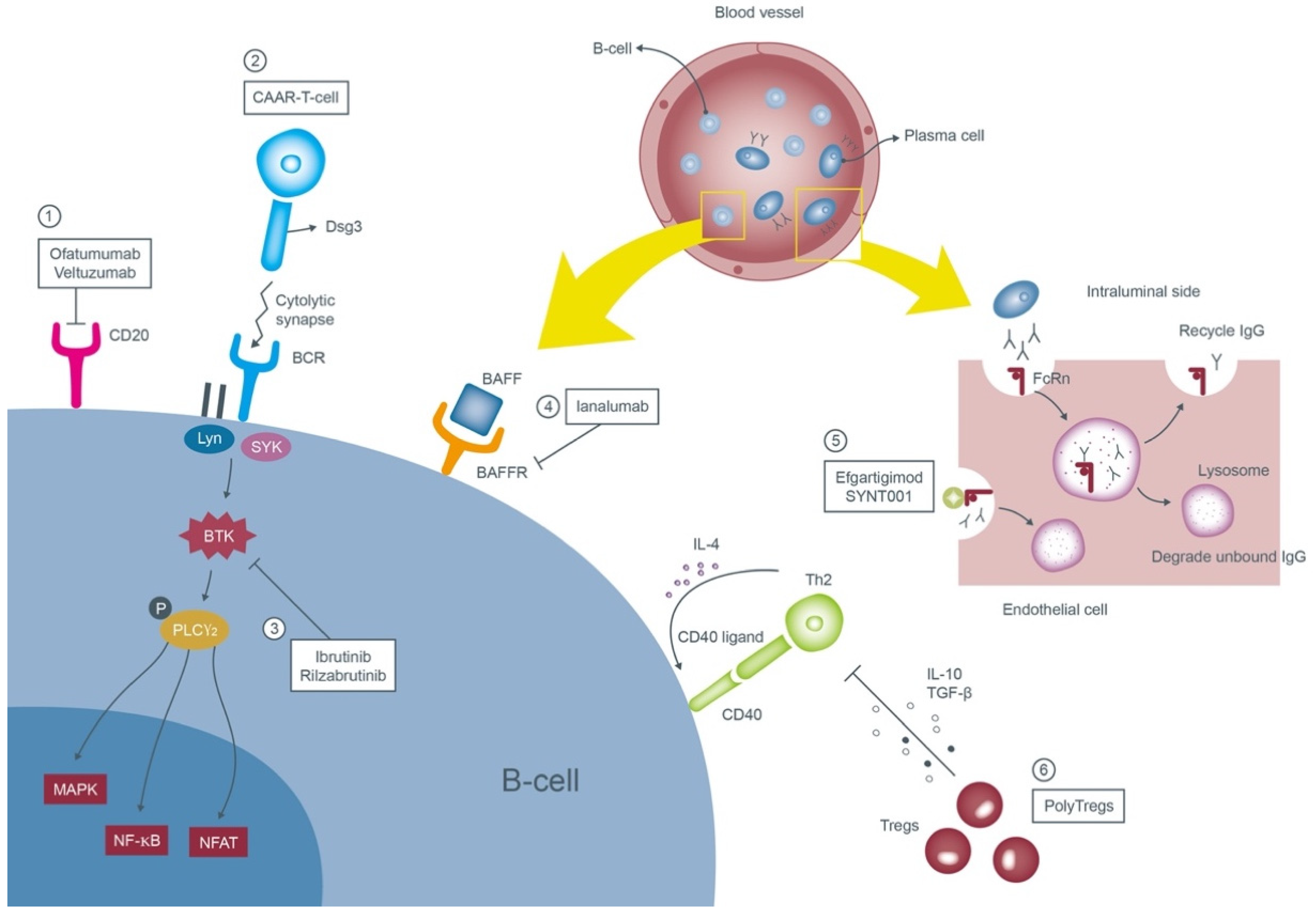
1.1. Pemphigus Vulgaris (PV)
First-Line Therapies
Second-Line Therapies
Emerging Options
1.2. Pemphigus Foliaceus (PF)
1.3. Pemphigus Erythematosus (PE)
1.4. IgA Pemphigus
1.5. Paraneoplastic Pemphigus (PNP)
Conventional Treatments
Emerging Options
2. Subepithelial Autoimmune Bullous Skin Disorders
2.1. Bullous Pemphigoid (BP)
First-Line Therapies
Second-Line Therapies
Emerging Options
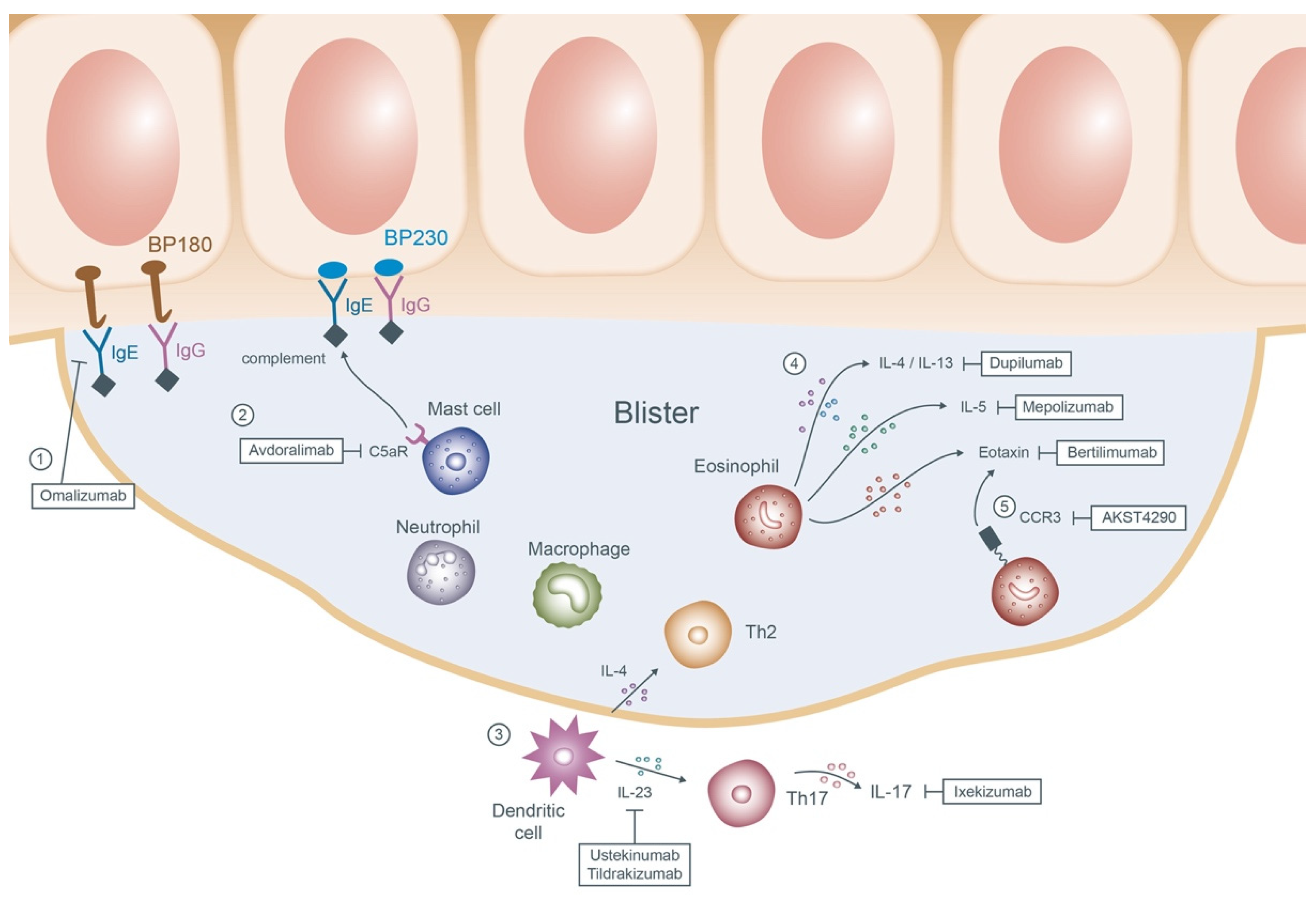
Anti-IgE monoclonal antibody: Omalizumab
Anti-C5a receptor (C5aR) antibody: Avdoralimab
Interleukin-17A (IL-17A) and IL-23 antagonist: Ixekizumab/Ustekinumab/Tildrakizumab
Inhibiting the eosinophil cytokines and chemokines: Dupilumab/Bertilimumab/Mepolizumab
C-C chemokine receptor 3 (CCR3) antagonist: AKST4290
2.2. Mucous Membrane Pemphigoid (MMP)
2.3. Linear IgA Bullous Dermatosis
2.4. Epidermolysis Bullosa Acquisita (EBA) Conventional Treatments
Management of EBA is exceptionally arduous. High-dose systemic GCs ranging from 1 to 1.5 mg/kg/day remain the preferred regimen. Dapsone or colchicine can be applied together with GCs to accelerate remission. The administration of other adjuvant therapies such as immunosuppressive agents (e.g., cyclosporine, azathioprine, CYP, MTX, and MMF), IVIG, and rituximab is utilized in the refractory cases [90][91][92][93].
Emerging Options
Anti-CD25 monoclonal antibody: Daclizumab
The production of autoantibodies in EBA patients is highly associated with T-cells. CD25, a significant component of the IL-2 receptor, regulates the survival and activation of T-cells. Daclizumab, through blocking CD25, facilitates a rapid and continuous reduction in lymphocyte CD25 expression and was proven to be effective in one EBA patient [94].
2.5. Dermatitis Herpetiformis (DH)
A strict gluten-free diet takes a chief role in dealing with DH. Slow titration of dapsone is the most popular first-line therapy showing immediate clinical improvement. Pharmacotherapy with sulfa-based regimens (sulfapyridine, sulfasalazine and sulfamethoxypyridazine) is suitable for those who cannot tolerate prior treatments [95][96][97]. Topical GCs may soothe the pruritus; nevertheless, systemic GCs are not warranted in DH.
2.6. Laminin γ1 Pemphigoid
The therapeutic information is sparse due to its rarity. Topical GCs can be applied to those with milder symptoms. With higher disease severity, some of the popular selections include systemic GCs (starting from 0.5 mg/kg/day) in collaboration with dapsone (1–1.5 mg/kg/day), cyclosporine, or azathioprine [98][99]. Other documented managements include doxycycline, high-dose IVIG, colchicine, and ustekinumab [99].
References
- Dumas, V.; Roujeau, J.C.; Wolkenstein, P.; Revuz, J.; Cosnes, A. The treatment of mild pemphigus vulgaris and pemphigus foliaceus with a topical corticosteroid. Br. J. Dermatol. 1999, 140, 1127–1129.
- Bystryn, J.C.; Steinman, N.M. The adjuvant therapy of pemphigus: An update. Arch. Dermatol. 1996, 132, 203–212.
- Santi, C.G.; Gripp, A.C.; Roselino, A.M.; Mello, D.S.; Gordilho, J.O.; Marsillac, P.F.; Porro, A.M. Consensus on the treatment of autoimmune bullous dermatoses: Bullous pemphigoid, mucous membrane pemphigoid and epidermolysis bullosa acquisita—Brazilian Society of Dermatology. An. Bras. Dermatol. 2019, 94, 33–47.
- Eming, R.; Sticherling, M.; Hofmann, S.C.; Hunzelmann, N.; Kern, J.S.; Kramer, H.; Pfeiffer, C.; Schuster, V.; Zillikens, D.; Goebeler, M.; et al. S2k guidelines for the treatment of pemphigus vulgaris/foliaceus and bullous pemphigoid. J. Dtsch. Dermatol. Ges. J. Ger. Soc. Dermatol. JDDG 2015, 13, 833–844.
- Rao, P.N.; Lakshmi, T.S. Pulse therapy and its modifications in pemphigus: A six year study. Indian J. Dermatol. Venereol. Leprol. 2003, 69, 329–333.
- Chams-Davatchi, C.; Esmaili, N.; Daneshpazhooh, M.; Valikhani, M.; Balighi, K.; Hallaji, Z.; Barzegari, M.; Akhyani, M.; Ghodsi, S.Z.; Seirafi, H.; et al. Randomized controlled open-label trial of four treatment regimens for pemphigus vulgaris. J. Am. Acad. Dermatol. 2007, 57, 622–628.
- Joly, P.; Maho-Vaillant, M.; Prost-Squarcioni, C.; Hebert, V.; Houivet, E.; Calbo, S.; Caillot, F.; Golinski, M.L.; Labeille, B.; Picard-Dahan, C.; et al. First-line rituximab combined with short-term prednisone versus prednisone alone for the treatment of pemphigus (Ritux 3): A prospective, multicentre, parallel-group, open-label randomised trial. Lancet 2017, 389, 2031–2040.
- Huang, A.; Madan, R.K.; Levitt, J. Future therapies for pemphigus vulgaris: Rituximab and beyond. J. Am. Acad. Dermatol. 2016, 74, 746–753.
- Cianchini, G.; Lupi, F.; Masini, C.; Corona, R.; Puddu, P.; De Pità, O. Therapy with rituximab for autoimmune pemphigus: Results from a single-center observational study on 42 cases with long-term follow-up. J. Am. Acad. Dermatol. 2012, 67, 617–622.
- Heelan, K.; Al-Mohammedi, F.; Smith, M.J.; Knowles, S.; Lansang, P.; Walsh, S.; Shear, N.H. Durable Remission of Pemphigus With a Fixed-Dose Rituximab Protocol. JAMA Dermatol. 2014, 150, 703–708.
- Kanwar, A.J.; Vinay, K.; Sawatkar, G.U.; Dogra, S.; Minz, R.W.; Shear, N.H.; Koga, H.; Ishii, N.; Hashimoto, T. Clinical and immunological outcomes of high- and low-dose rituximab treatments in patients with pemphigus: A randomized, comparative, observer-blinded study. Br. J. Dermatol. 2014, 170, 1341–1349.
- Buch, M.H.; Smolen, J.S.; Betteridge, N.; Breedveld, F.C.; Burmester, G.; Dörner, T.; Ferraccioli, G.; Gottenberg, J.E.; Isaacs, J.; Kvien, T.K.; et al. Updated consensus statement on the use of rituximab in patients with rheumatoid arthritis. Ann. Rheum. Dis. 2011, 70, 909–920.
- Wu, K.-J.; Wei, K.-C. Venous thromboembolism in a case with pemphigus vulgaris after infusion of rituximab plus systemic glucocorticoids and azathioprine: A possible adverse effect of rituximab? Derm. Sin. 2021, 39, 103–104.
- Schiavo, A.L.; Puca, R.V.; Ruocco, V.; Ruocco, E. Adjuvant drugs in autoimmune bullous diseases, efficacy versus safety: Facts and controversies. Clin. Derm. 2010, 28, 337–343.
- Hertl, M.; Jedlickova, H.; Karpati, S.; Marinovic, B.; Uzun, S.; Yayli, S.; Mimouni, D.; Borradori, L.; Feliciani, C.; Ioannides, D.; et al. Pemphigus. S2 Guideline for diagnosis and treatment—Guided by the European Dermatology Forum (EDF) in cooperation with the European Academy of Dermatology and Venereology (EADV). J. Eur. Acad. Dermatol. Venereol. JEADV 2015, 29, 405–414.
- Meurer, M. Immunosuppressive therapy for autoimmune bullous diseases. Clin. Derm. 2012, 30, 78–83.
- Li, N.; Zhao, M.; Hilario-Vargas, J.; Prisayanh, P.; Warren, S.; Diaz, L.A.; Roopenian, D.C.; Liu, Z. Complete FcRn dependence for intravenous Ig therapy in autoimmune skin blistering diseases. J. Clin. Investig. 2005, 115, 3440–3450.
- Hoffmann, J.H.O.; Enk, A.H. High-Dose Intravenous Immunoglobulin in Skin Autoimmune Disease. Front. Immunol. 2019, 10, 1090.
- Daoud, Y.J.; Amin, K.G. Comparison of cost of immune globulin intravenous therapy to conventional immunosuppressive therapy in treating patients with autoimmune mucocutaneous blistering diseases. Int. Immunopharmacol. 2006, 6, 600–606.
- Keskin, D.B.; Stern, J.N.; Fridkis-Hareli, M.; Razzaque Ahmed, A. Cytokine profiles in pemphigus vulgaris patients treated with intravenous immunoglobulins as compared to conventional immunosuppressive therapy. Cytokine 2008, 41, 315–321.
- Lever, W.F.; Schaumburg-Lever, G. Treatment of pemphigus vulgaris. Results obtained in 84 patients between 1961 and 1982. Arch. Dermatol. 1984, 120, 44–47.
- Becker, B.A.; Gaspari, A.A. Pemphigus vulgaris and vegetans. Dermatol. Clin. 1993, 11, 429–452.
- Ruocco, E.; Wolf, R.; Ruocco, V.; Brunetti, G.; Romano, F.; Lo Schiavo, A. Pemphigus: Associations and management guidelines: Facts and controversies. Clin. Derm. 2013, 31, 382–390.
- Sinha, A.A.; Hoffman, M.B.; Janicke, E.C. Pemphigus vulgaris: Approach to treatment. Eur. J. Dermatol. EJD 2015, 25, 103–113.
- Kasperkiewicz, M.; Shimanovich, I.; Meier, M.; Schumacher, N.; Westermann, L.; Kramer, J.; Zillikens, D.; Schmidt, E. Treatment of severe pemphigus with a combination of immunoadsorption, rituximab, pulsed dexamethasone and azathioprine/mycophenolate mofetil: A pilot study of 23 patients. Br. J. Dermatol. 2012, 166, 154–160.
- Zillikens, D.; Derfler, K.; Eming, R.; Fierlbeck, G.; Goebeler, M.; Hertl, M.; Hofmann, S.C.; Karlhofer, F.; Kautz, O.; Nitschke, M.; et al. Recommendations for the use of immunoapheresis in the treatment of autoimmune bullous diseases. J. Dtsch. Dermatol. Ges. J. Ger. Soc. Dermatol. JDDG 2007, 5, 881–887.
- Behzad, M.; Möbs, C.; Kneisel, A.; Möller, M.; Hoyer, J.; Hertl, M.; Eming, R. Combined treatment with immunoadsorption and rituximab leads to fast and prolonged clinical remission in difficult-to-treat pemphigus vulgaris. Br. J. Dermatol. 2012, 166, 844–852.
- Robak, T.; Robak, E. New anti-CD20 monoclonal antibodies for the treatment of B-cell lymphoid malignancies. BioDrugs Clin. Immunother. Biopharm. Gene Ther. 2011, 25, 13–25.
- Klufas, D.M.; Amerson, E.; Twu, O.; Clark, L.; Shinkai, K. Refractory pemphigus vulgaris successfully treated with ofatumumab. JAAD Case Rep. 2020, 6, 734–736.
- Du, F.H.; Mills, E.A.; Mao-Draayer, Y. Next-generation anti-CD20 monoclonal antibodies in autoimmune disease treatment. Auto-Immun. Highlights 2017, 8, 12.
- Ellebrecht, C.T.; Choi, E.J.; Allman, D.M.; Tsai, D.E.; Wegener, W.A.; Goldenberg, D.M.; Payne, A.S. Subcutaneous veltuzumab, a humanized anti-CD20 antibody, in the treatment of refractory pemphigus vulgaris. JAMA Derm. 2014, 150, 1331–1335.
- Ellebrecht, C.T.; Bhoj, V.G.; Nace, A.; Choi, E.J.; Mao, X.; Cho, M.J.; Di Zenzo, G.; Lanzavecchia, A.; Seykora, J.T.; Cotsarelis, G.; et al. Reengineering chimeric antigen receptor T cells for targeted therapy of autoimmune disease. Science 2016, 353, 179–184.
- Crofford, L.J.; Nyhoff, L.E.; Sheehan, J.H.; Kendall, P.L. The role of Bruton’s tyrosine kinase in autoimmunity and implications for therapy. Expert Rev. Clin. Immunol. 2016, 12, 763–773.
- Corneth, O.B.J.; Klein Wolterink, R.G.J.; Hendriks, R.W. BTK Signaling in B Cell Differentiation and Autoimmunity. Curr. Top. Microbiol. Immunol. 2016, 393, 67–105.
- Campbell, R.; Chong, G.; Hawkes, E.A. Novel Indications for Bruton’s Tyrosine Kinase Inhibitors, beyond Hematological Malignancies. J. Clin. Med. 2018, 7, 62.
- Didona, D.; Maglie, R.; Eming, R.; Hertl, M. Pemphigus: Current and Future Therapeutic Strategies. Front. Immunol. 2019, 10, 1418.
- Patsatsi, A.; Murrell, D.F. Bruton Tyrosine Kinase Inhibition and Its Role as an Emerging Treatment in Pemphigus. Front. Med. 2021, 8, 708071.
- Lee, A.; Sandhu, S.; Imlay-Gillespie, L.; Mulligan, S.; Shumack, S. Successful use of Bruton’s kinase inhibitor, ibrutinib, to control paraneoplastic pemphigus in a patient with paraneoplastic autoimmune multiorgan syndrome and chronic lymphocytic leukaemia. Australas J. Derm. 2017, 58, e240–e242.
- Vidal-Crespo, A.; Rodriguez, V.; Matas-Cespedes, A.; Lee, E.; Rivas-Delgado, A.; Giné, E.; Navarro, A.; Beà, S.; Campo, E.; López-Guillermo, A.; et al. The Bruton tyrosine kinase inhibitor CC-292 shows activity in mantle cell lymphoma and synergizes with lenalidomide and NIK inhibitors depending on nuclear factor-κB mutational status. Haematologica 2017, 102, e447–e451.
- Murrell, D.F.; Patsatsi, A.; Stavropoulos, P.; Baum, S.; Zeeli, T.; Kern, J.S.; Roussaki-Schulze, A.V.; Sinclair, R.; Bassukas, I.D.; Thomas, D.; et al. Proof of concept for the clinical effects of oral rilzabrutinib, the first Bruton tyrosine kinase inhibitor for pemphigus vulgaris: The phase II BELIEVE study. Br. J. Dermatol. 2021, 185, 745–755.
- Izumi, K.; Bieber, K.; Ludwig, R.J. Current Clinical Trials in Pemphigus and Pemphigoid. Front. Immunol. 2019, 10, 978.
- Mackay, F.; Browning, J.L. BAFF: A fundamental survival factor for B cells. Nat. Rev. Immunol. 2002, 2, 465–475.
- Lesley, R.; Xu, Y.; Kalled, S.L.; Hess, D.M.; Schwab, S.R.; Shu, H.B.; Cyster, J.G. Reduced competitiveness of autoantigen-engaged B cells due to increased dependence on BAFF. Immunity 2004, 20, 441–453.
- Matsushita, T.; Hasegawa, M.; Matsushita, Y.; Echigo, T.; Wayaku, T.; Horikawa, M.; Ogawa, F.; Takehara, K.; Sato, S. Elevated serum BAFF levels in patients with localized scleroderma in contrast to other organ-specific autoimmune diseases. Exp. Derm. 2007, 16, 87–93.
- Groom, J.; Kalled, S.L.; Cutler, A.H.; Olson, C.; Woodcock, S.A.; Schneider, P.; Tschopp, J.; Cachero, T.G.; Batten, M.; Wheway, J.; et al. Association of BAFF/BLyS overexpression and altered B cell differentiation with Sjögren’s syndrome. J. Clin. Investig. 2002, 109, 59–68.
- Cheema, G.S.; Roschke, V.; Hilbert, D.M.; Stohl, W. Elevated serum B lymphocyte stimulator levels in patients with systemic immune-based rheumatic diseases. Arthritis Rheum. 2001, 44, 1313–1319.
- Kuo, T.T.; Baker, K.; Yoshida, M.; Qiao, S.W.; Aveson, V.G.; Lencer, W.I.; Blumberg, R.S. Neonatal Fc receptor: From immunity to therapeutics. J. Clin. Immunol. 2010, 30, 777–789.
- Howard, J.F., Jr.; Bril, V.; Burns, T.M.; Mantegazza, R.; Bilinska, M.; Szczudlik, A.; Beydoun, S.; Garrido, F.; Piehl, F.; Rottoli, M.; et al. Randomized phase 2 study of FcRn antagonist efgartigimod in generalized myasthenia gravis. Neurology 2019, 92, e2661–e2673.
- Newland, A.C.; Sánchez-González, B.; Rejtő, L.; Egyed, M.; Romanyuk, N.; Godar, M.; Verschueren, K.; Gandini, D.; Ulrichts, P.; Beauchamp, J.; et al. Phase 2 study of efgartigimod, a novel FcRn antagonist, in adult patients with primary immune thrombocytopenia. Am. J. Hematol. 2020, 95, 178–187.
- Ulrichts, P.; Guglietta, A.; Dreier, T.; van Bragt, T.; Hanssens, V.; Hofman, E.; Vankerckhoven, B.; Verheesen, P.; Ongenae, N.; Lykhopiy, V.; et al. Neonatal Fc receptor antagonist efgartigimod safely and sustainably reduces IgGs in humans. J. Clin. Investig. 2018, 128, 4372–4386.
- Schmidt, T.; Willenborg, S.; Hünig, T.; Deeg, C.A.; Sonderstrup, G.; Hertl, M.; Eming, R. Induction of T regulatory cells by the superagonistic anti-CD28 antibody D665 leads to decreased pathogenic IgG autoantibodies against desmoglein 3 in a HLA-transgenic mouse model of pemphigus vulgaris. Exp. Derm. 2016, 25, 293–298.
- Zwang, N.A.; Leventhal, J.R. Cell Therapy in Kidney Transplantation: Focus on Regulatory T Cells. J. Am. Soc. Nephrol. 2017, 28, 1960–1972.
- Wang, W.-E.; Wu, R.-W.; Chang, C.-H. An elderly female with pemphigus foliaceus possibly induced by losartan/hydrochlorothiazide. Derm. Sin. 2021, 39, 107–108.
- Kneisel, A.; Hertl, M. Autoimmune bullous skin diseases. Part 1: Clinical manifestations. J. Dtsch. Dermatol. Ges. 2011, 9, 844–856.
- Kasperkiewicz, M.; Ellebrecht, C.T.; Takahashi, H.; Yamagami, J.; Zillikens, D.; Payne, A.S.; Amagai, M. Pemphigus. Nat. Rev. Dis. Primers 2017, 3, 17026.
- Hofmann, S.C.; Juratli, H.A.; Eming, R. Bullous autoimmune dermatoses. J. Dtsch. Dermatol. Ges. J. Ger. Soc. Dermatol. JDDG 2018, 16, 1339–1358.
- Hong, H.; Chang, T.; Wu, C.; Chang, Y. Intraepidermal neutrophilic dermatosis-type immunoglobulin A pemphigus. Derm. Sin. 2021, 39, 47–48.
- Gruss, C.; Zillikens, D.; Hashimoto, T.; Amagai, M.; Kroiss, M.; Vogt, T.; Landthaler, M.; Stolz, W. Rapid response of IgA pemphigus of subcorneal pustular dermatosis type to treatment with isotretinoin. J. Am. Acad. Dermatol. 2000, 43, 923–926.
- Ruiz-Genao, D.P.; Hernández-Núñez, A.; Hashimoto, T.; Amagai, M.; Fernández-Herrera, J.; García-Díez, A. A case of IgA pemphigus successfully treated with acitretin. Br. J. Dermatol. 2002, 147, 1040–1042.
- Howell, S.M.; Bessinger, G.T.; Altman, C.E.; Belnap, C.M. Rapid response of IgA pemphigus of the subcorneal pustular dermatosis subtype to treatment with adalimumab and mycophenolate mofetil. J. Am. Acad. Dermatol. 2005, 53, 541–543.
- Sehgal, V.N.; Srivastava, G. Paraneoplastic pemphigus/paraneoplastic autoimmune multiorgan syndrome. Int. J. Dermatol. 2009, 48, 162–169.
- Heizmann, M.; Itin, P.; Wernli, M.; Borradori, L.; Bargetzi, M.J. Successful treatment of paraneoplastic pemphigus in follicular NHL with rituximab: Report of a case and review of treatment for paraneoplastic pemphigus in NHL and CLL. Am. J. Hematol. 2001, 66, 142–144.
- Zhao, Y.; Su, H.; Shen, X.; Du, J.; Zhang, X.; Zhao, Y. The immunological function of CD52 and its targeting in organ transplantation. Inflamm. Res. 2017, 66, 571–578.
- Ruck, T.; Bittner, S.; Wiendl, H.; Meuth, S.G. Alemtuzumab in Multiple Sclerosis: Mechanism of Action and Beyond. Int. J. Mol. Sci. 2015, 16, 16414–16439.
- Paolino, G.; Didona, D.; Magliulo, G.; Iannella, G.; Didona, B.; Mercuri, S.R.; Moliterni, E.; Donati, M.; Ciofalo, A.; Granata, G.; et al. Paraneoplastic Pemphigus: Insight into the Autoimmune Pathogenesis, Clinical Features and Therapy. Int. J. Mol. Sci. 2017, 18, 2532.
- Hohwy, T.; Bang, K.; Steiniche, T.; Peterslund, N.A.; d’Amore, F. Alemtuzumab-induced remission of both severe paraneoplastic pemphigus and leukaemic bone marrow infiltration in a case of treatment-resistant B-cell chronic lymphocytic leukaemia. Eur. J. Haematol. 2004, 73, 206–209.
- Bech, R.; Baumgartner-Nielsen, J.; Peterslund, N.A.; Steiniche, T.; Deleuran, M.; d’Amore, F. Alemtuzumab is effective against severe chronic lymphocytic leukaemia-associated paraneoplastic pemphigus. Br. J. Dermatol. 2013, 169, 469–472.
- Joly, P.; Roujeau, J.C.; Benichou, J.; Picard, C.; Dreno, B.; Delaporte, E.; Vaillant, L.; D’Incan, M.; Plantin, P.; Bedane, C.; et al. A comparison of oral and topical corticosteroids in patients with bullous pemphigoid. N. Engl. J. Med. 2002, 346, 321–327.
- Joly, P.; Roujeau, J.C.; Benichou, J.; Delaporte, E.; D’Incan, M.; Dreno, B.; Bedane, C.; Sparsa, A.; Gorin, I.; Picard, C.; et al. A comparison of two regimens of topical corticosteroids in the treatment of patients with bullous pemphigoid: A multicenter randomized study. J. Invest. Derm. 2009, 129, 1681–1687.
- Kirtschig, G.; Middleton, P.; Bennett, C.; Murrell, D.F.; Wojnarowska, F.; Khumalo, N.P. Interventions for bullous pemphigoid. Cochrane Database Syst. Rev. 2010, 2010, Cd002292.
- Daniel, B.S.; Borradori, L.; Hall, R.P., 3rd; Murrell, D.F. Evidence-based management of bullous pemphigoid. Dermatol. Clin. 2011, 29, 613–620.
- Kimura, K.; Kawai, K. Doxycycline as an initial treatment of bullous pemphigoid in Japanese patients. J. Cutan. Immunol. Allergy 2020, 3, 80–85.
- Van Beek, N.; Lüttmann, N.; Huebner, F.; Recke, A.; Karl, I.; Schulze, F.S.; Zillikens, D.; Schmidt, E. Correlation of Serum Levels of IgE Autoantibodies Against BP180 With Bullous Pemphigoid Disease Activity. JAMA Dermatol. 2017, 153, 30–38.
- Yu, K.K.; Crew, A.B.; Messingham, K.A.; Fairley, J.A.; Woodley, D.T. Omalizumab therapy for bullous pemphigoid. J. Am. Acad. Dermatol. 2014, 71, 468–474.
- Balakirski, G.; Alkhateeb, A.; Merk, H.F.; Leverkus, M.; Megahed, M. Successful treatment of bullous pemphigoid with omalizumab as corticosteroid-sparing agent: Report of two cases and review of literature. J. Eur. Acad. Dermatol. Venereol. JEADV 2016, 30, 1778–1782.
- De, D.; Kaushik, A.; Handa, S.; Mahajan, R.; Schmidt, E. Omalizumab: An underutilized treatment option in bullous pemphigoid patients with co-morbidities. J. Eur. Acad. Dermatol. Venereol. JEADV 2021, 35, e469–e472.
- Heimbach, L.; Li, Z.; Berkowitz, P.; Zhao, M.; Li, N.; Rubenstein, D.S.; Diaz, L.A.; Liu, Z. The C5a receptor on mast cells is critical for the autoimmune skin-blistering disease bullous pemphigoid. J. Biol. Chem. 2011, 286, 15003–15009.
- Plée, J.; Le Jan, S.; Giustiniani, J.; Barbe, C.; Joly, P.; Bedane, C.; Vabres, P.; Truchetet, F.; Aubin, F.; Antonicelli, F.; et al. Integrating longitudinal serum IL-17 and IL-23 follow-up, along with autoantibodies variation, contributes to predict bullous pemphigoid outcome. Sci. Rep. 2015, 5, 18001.
- Wakugawa, M.; Nakamura, K.; Hino, H.; Toyama, K.; Hattori, N.; Okochi, H.; Yamada, H.; Hirai, K.; Tamaki, K.; Furue, M. Elevated levels of eotaxin and interleukin-5 in blister fluid of bullous pemphigoid: Correlation with tissue eosinophilia. Br. J. Dermatol. 2000, 143, 112–116.
- Rico, M.J.; Benning, C.; Weingart, E.S.; Streilein, R.D.; Hall, R.P., 3rd. Characterization of skin cytokines in bullous pemphigoid and pemphigus vulgaris. Br. J. Dermatol. 1999, 140, 1079–1086.
- Fabbri, P.; Caproni, M.; Berti, S.; Bianchi, B.; Amato, L.; De Pità, O.; Frezzolini, A. The role of T lymphocytes and cytokines in the pathogenesis of pemphigoid gestationis. Br. J. Dermatol. 2003, 148, 1141–1148.
- Abdat, R.; Waldman, R.A.; de Bedout, V.; Czernik, A.; McLeod, M.; King, B.; Gordon, S.; Ahmed, R.; Nichols, A.; Rothe, M.; et al. Dupilumab as a novel therapy for bullous pemphigoid: A multicenter case series. J. Am. Acad. Dermatol. 2020, 83, 46–52.
- Maglie, R.; Hertl, M. Pharmacological advances in pemphigoid. Curr. Opin. Pharm. 2019, 46, 34–43.
- Simon, D.; Yousefi, S.; Cazzaniga, S.; Bürgler, C.; Radonjic, S.; Houriet, C.; Heidemeyer, K.; Klötgen, H.W.; Kozlowski, E.; Borradori, L.; et al. Mepolizumab failed to affect bullous pemphigoid: A randomized, placebo-controlled, double-blind phase 2 pilot study. Allergy 2020, 75, 669–672.
- Grimaldi, J.C.; Yu, N.X.; Grunig, G.; Seymour, B.W.; Cottrez, F.; Robinson, D.S.; Hosken, N.; Ferlin, W.G.; Wu, X.; Soto, H.; et al. Depletion of eosinophils in mice through the use of antibodies specific for C-C chemokine receptor 3 (CCR3). J. Leukoc. Biol. 1999, 65, 846–853.
- Taylor, J.; McMillan, R.; Shephard, M.; Setterfield, J.; Ahmed, R.; Carrozzo, M.; Grando, S.; Mignogna, M.; Kuten-Shorrer, M.; Musbah, T.; et al. World Workshop on Oral Medicine VI: A systematic review of the treatment of mucous membrane pemphigoid. Oral Surg. Oral Med. Oral Pathol. Oral Radiol. 2015, 120, 161–171.e120.
- Lee, H.Y.; Blazek, C.; Beltraminelli, H.; Borradori, L. Oral mucous membrane pemphigoid: Complete response to topical tacrolimus. Acta Derm. Venereol. 2011, 91, 604–605.
- Ujiie, H.; Iwata, H.; Yamagami, J.; Nakama, T.; Aoyama, Y.; Ikeda, S.; Ishii, N.; Iwatsuki, K.; Kurosawa, M.; Sawamura, D.; et al. Japanese guidelines for the management of pemphigoid (including epidermolysis bullosa acquisita). J. Derm. 2019, 46, 1102–1135.
- Alajlan, A.; Al-Khawajah, M.; Al-Sheikh, O.; Al-Saif, F.; Al-Rasheed, S.; Al-Hoqail, I.; Hamadah, I.R. Treatment of linear IgA bullous dermatosis of childhood with flucloxacillin. J. Am. Acad. Dermatol. 2006, 54, 652–656.
- Gürcan, H.M.; Ahmed, A.R. Current concepts in the treatment of epidermolysis bullosa acquisita. Expert Opin. Pharm. 2011, 12, 1259–1268.
- Sami, N. Mycophenolate mofetil (MMF) in the treatment of epidermolysis bullosa acquisita (EBA) long-term follow-up. JAAD Case Rep. 2015, 1, 321–323.
- Caldwell, J.B.; Yancey, K.B.; Engler, R.J.; James, W.D. Epidermolysis bullosa acquisita: Efficacy of high-dose intravenous immunoglobulins. J. Am. Acad. Dermatol. 1994, 31, 827–828.
- Sadler, E.; Schafleitner, B.; Lanschuetzer, C.; Laimer, M.; Pohla-Gubo, G.; Hametner, R.; Hintner, H.; Bauer, J.W. Treatment-resistant classical epidermolysis bullosa acquisita responding to rituximab. Br. J. Dermatol. 2007, 157, 417–419.
- Egan, C.A.; Brown, M.; White, J.D.; Yancey, K.B. Treatment of epidermolysis bullosa acquisita with the humanized anti-Tac mAb daclizumab. Clin. Immunol. 2001, 101, 146–151.
- Fry, L. Dermatitis herpetiformis: Problems, progress and prospects. Eur. J. Dermatol. EJD 2002, 12, 523–531.
- McFadden, J.P.; Leonard, J.N.; Powles, A.V.; Rutman, A.J.; Fry, L. Sulphamethoxypyridazine for dermatitis herpetiformis, linear IgA disease and cicatricial pemphigoid. Br. J. Dermatol. 1989, 121, 759–762. [Google Scholar]
- Willsteed, E.; Lee, M.; Wong, L.C.; Cooper, A. Sulfasalazine and dermatitis herpetiformis. Australas J. Derm. 2005, 46, 101–103.
- Wozniak, K.; Hashimoto, T.; Fukuda, S.; Ohyama, B.; Ishii, N.; Koga, H.; Dainichi, T.; Kowalewski, C. IgA Anti-p200 Pemphigoid. Arch. Dermatol. 2011, 147, 1306–1310.
- Goletz, S.; Hashimoto, T.; Zillikens, D.; Schmidt, E. Anti-p200 pemphigoid. J. Am. Acad. Dermatol. 2014, 71, 185–191.

