You're using an outdated browser. Please upgrade to a modern browser for the best experience.
Inguinal Hernia in Nonhuman Primates
Edit

A herniation is a condition in which there is a protrusion of an organ, fascia, fat, or omentum through the wall of the cavity in which it is contained. A hernia may be classified into different categories based on the cause, location, size, recurrence, reducibility, contents, and symptoms. Inguinal hernia is described as a bulge of the peritoneum through a defect (congenital or acquired) in the muscular and fascial structures of the abdominal wall; a defect in the myofascial plane of the oblique and transversalis muscles and fascia. Inguinal hernias are classified into (1) indirect hernia, (2) direct hernia, (3) scrotal or giant hernia, (4) femoral hernia, and (5) others, i.e., rare hernias such as Spigelian hernias. Inguinal hernias are relatively common in both humans and domestic animal species, and surgery to repair an inguinal hernia is a nonurgent, routine procedure. However, every hernia carries a hazard of incarceration and strangulation, warranting immediate surgical treatment.

emergency incarceration inguinal hernia

1. Epidemiology and Anamnesis

The lifetime risk of developing an inguinal hernia is around 27% for males and 3% for females [1][2][3]. Curiously, 1 male in 5 and 1 female in 50 will eventually develop an inguinal hernia in their lifetime; nevertheless, the etiology of inguinal hernias remains unresolved. In humans, inguinal hernias have a hereditary factor, with a complex inheritance pattern [1][4][5].
The current incidence and prevalence of inguinal hernias in nonhuman primates (NHPs) are unknown. Records of zoological gardens dating from 1934, which included several thousand monkeys, showed that the percentage of hernias (all types of hernias, not exclusively inguinal hernias) was 0.37% [6]. More recent literature reported that inguinal hernias are relatively common in primates; however, no data to support their statements are provided [7][8][9]. It was revealed only nine reported cases of inguinal hernias in NHPs. Their incident is likely much higher, as most inguinal hernias are of low clinical significance and thus underreported. Fowler [10] reported, without providing specific details, that lion-tailed macaques (Macaca silenus) are predisposed to the development of inguinal hernias. Here, it was conducted to calculate hernia incidence at the Biomedical Primate Research Centre (BPRC, Rijswijk, The Netherlands). Data were obtained in retrospect from the electronic health record database of the BPRC. The dataset used for the analysis covered the period from January 2021 to December 2021. Marmosets (Callithrix jacchus) < 5 months of age or weighing < 200 gr, rhesus monkeys (Macaca mulatta) < 6 months of age, and cynomolgus monkeys (Macaca fascicularis) < 8 months of age were not sedated for their physical examination and were therefore excluded from this analysis. Data from the physical examination were analyzed on the presence of inguinal hernia at the moment of examination, in combination with age, sex, and body condition score (BCS). Physical examinations are routinely performed yearly, including a thorough physical examination, a tuberculosis screening test, complete blood count (CBC), and serum biochemistry. No animals were sedated solely for purpose. Ethical approval was not required. All animals were housed in accordance with Dutch law and international ethical and scientific standards and guidelines (EU Directive 63/2010). All husbandry procedures were compliant with the above standards and legislation. The animal care at BPRC is in accordance with programs accredited by AAALAC International. Data included 897 NHPs: 88 common marmosets, 225 cynomolgus monkeys, and 582 rhesus monkeys. No inguinal hernias were observed in the marmosets (38 males and 50 females). In the 225 examined cynomolgus monkeys (74 males and 151 females), 6 adult males were observed with an inguinal hernia. Those males had a BCS of 3–3.5, age range between 2 and 12 years. In the 582 examined rhesus monkeys (166 males and 416 females), 2 adult males and 1 adult female were observed with an inguinal hernia (Figure 1A,B). One involved a 4-year-old male with a BCS of 2.5, the other animal was an 8-year-old male with a BCS of 5, and the female was a 14-year-old with a BCS of 3. The percentage of inguinal hernia was 1.00%.
Figure 1. (A) Several examples of inguinal hernias observed during physical examination in male rhesus monkeys (photographs provided by Biomedical Primate Research Centre); (B) example of an inguinal hernia in a female rhesus monkey. Hernias are present in both left and right inguinal regions (photographs provided by Biomedical Primate Research Centre).
It is suggested that inguinal hernias in NHPs occur secondary to the following factors:
  • Trauma [11]. Although the exact role of trauma in the occurrence and progress of inguinal hernia remains unclear, accidents such as a fall from height while hopping from one tree to another may play a role;
  • Congenital weakness of muscles of the groin region or other congenital anomalies from the time of birth [11];
  • In utero lead exposure [12].
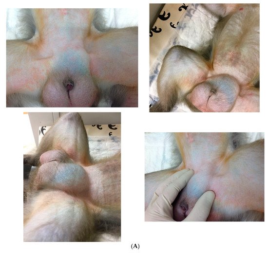
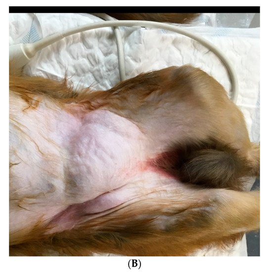
Berg et al. [13] explored the role of inheritance in macaques by conducting a four-generation pedigree analysis. No hereditary component or inheritance pattern was revealed. It is assumed, similar to men, that factors that may contribute to increased intraabdominal pressure, including obesity, chronic cough, and straining, are predisposing factors for the development of inguinal hernias in male NHPs [14]. In humans, other reported conditions associated with an increased incidence of inguinal hernias are varied and may include prematurity, hydrops, meconium peritonitis, chylous ascites, liver disease with ascites, ambiguous genitalia, hypospadias, epispadias, exstrophy of bladder, cloaca, cryptorchid testes, cystic fibrosis, connective tissue disorders, ventriculoperitoneal shunt, continuous ambulatory peritoneal dialysis, Hunter–Hurler syndrome, and mucopolysaccharidosis [15]. Appendectomy, abdominal surgeries, and parturitions are not associated with inguinal hernias in women. Interestingly, in women, both high sports activity and obesity are described to be protective against inguinal hernia [16]. Although not investigated in NHPs, these factors may also be influential.

2. Clinical Signs

In most cases, this is an incidental finding discovered in healthy animals during routine physical examinations, manifesting as a bulge in the groin (inguinal) area. The clinical appearance varies widely—from uni- to bilateral, reducible to nonreducible, immobile to mobile, and firm to soft. Hernias in the inguinal region may present as a mass in the femoral area near the vessels or as a scrotal mass. When intestines appear within the hernia defect, peristalsis of fluid in the hernia sac is palpable [17]. Abdominal pain, absence of flatus or feces, and abdominal distension are symptoms preceding shock that may be indications of strangulation [18]. An incarcerated hernia is a part of the intestine or abdominal tissue that is trapped in the sac of a hernia. When the trapped tissue involves intestines that are strangulated as a result, the animal may show symptoms of intestinal obstruction, including nausea, vomiting, and obstipation [17]. If the strangulation is not resolved immediately, tissue necrosis follows, and the animal will exhibit signs of severe pain, focal or generalized peritonitis, and sepsis (hypotension, tachycardia). Edema and localized inflammation are consistent with a possible strangulated hernia. Acutely incarcerated hernias may appear erythematous, indurated, swollen, and painful on palpation.

3. Diagnostics

Inguinal hernia is often diagnosed based on the presence of a bulge in the inguinal region during physical examination (Figure 1A,B). However, small hernias are not always clinically detectable, and a patent processus vaginalis is not apparent on a physical examination. Furthermore, examination of NHPs in dorsal recumbency facilitates reduction in the contents of the hernia and hernial ring palpation. It is even more challenging to diagnose incarceration and strangulation and evaluate their severity.
Plain or contrast X-rays, ultrasonography (US), magnetic resonance imaging (MRI), and computed tomography (CT) provide information about the anatomical characteristics of the suspected region, i.e., the content of the hernial sac and the integrity of the anatomical structures, allowing for a diagnosis and treatment [14][19][20][21][22][23]. Although cheap and harmless, the US is not very reliable for hernia detection due to its accuracy being user-dependent [24]. However, the US can be helpful in cases in which recurrent hernia, postsurgical complications, or other causes of groin pain is suspected. MRI has higher sensitivity and specificity compared to US and CT and is, therefore, the definitive radiologic examination for diagnosing occult hernias [19][20][24].
In the event of strangulation, acute bowel ischemia may occur. Angiography and CT directly assess mesenteric vascularity, with CT having a high expectation of revealing early findings of bowel ischemia. CT provides a roadmap to assist veterinarians with the restoration of intestinal blood flow as early and fast as possible [25].

4. Differential Diagnosis

A combination of the clinical signs and imaging results will assist veterinarians in the diagnosis of the mass in the groin region. These groin masses can be defined as being inguinal hernias, neoplasms, infectious or inflammatory processes, vascular conditions, as well as congenital abnormalities. Therefore, the differential diagnosis should include appendicitis, adhesions, abscesses, inflammatory bowel diseases, urinary tract infection, hip pathologies, pelvic pathologies, undescended testicles, hematoma, lymphadenopathy, lipoma, metastatic neoplasia, hydrocele, and vascular aneurysm [17][21][26][27].

5. Medical Management

In humans, inguinal hernias are common and were in the past believed to all require surgical repair (herniorrhaphy) regardless of the presence or severity of symptoms, in order to avoid complications [2][3][28][29][30][31]. However, an increasing number have revealed that minor symptomatic, first occurrence hernias do not necessarily require repair, and these patients may be followed expectantly (watchful waiting) [24][27][32][33][34]. In humans, delaying surgical repair until symptoms increase is described to be acceptable as acute hernia incarcerations are rare [24].
In NHPs, inguinal herniation is usually of no significant consequence. Therefore, close monitoring is an acceptable course of action for NHPs in asymptomatic or minimally symptomatic patients. Surgery is necessary for acutely incarcerated hernias or those that cause significant discomfort, i.e., pain or physical limitations. Precise determination of the actual risk of serious consequences of an inguinal hernia in NHPs does not exist due to a lack of published data. In humans, the change rate in the cumulative probability of strangulation increases quickly over the first 3 months of the existence of a hernia [35]. Incarceration of inguinal hernia occurs in around 10% of the patients, which, in turn, can result in intestinal obstruction, strangulation, and infarction [36][37]. Strangulation is the severest complication, even with potentially lethal sequela.
Many different techniques for surgical repair of inguinal hernias have been described in human medicine, mainly characterized as mesh reinforcement or suturing of defects, performed as an open or laparoscopic approach [2][3][23][29][30][31][32][38]. It is crucial to understand the differences in outcomes of different approaches (e.g., postoperative pain, recovery period, recurrence rate, and cost-effectiveness) and how best they fit each patient in deciding upon a technique. The treatment of choice fluctuates widely with regard to various factors such as anamnesis, hernia type, and the preference of the surgeon [27][32][39]. Surgical skills and proper training of the surgeon are essential in minimizing the risk of peri- and postoperative complications [29].
In humans, mesh repairs demonstrated lower recurrence rates, compared with sutured repairs, presumably due to tension-free repair [32][40]. There is also a quicker return to normal activity for patients, notwithstanding that the use of mesh risks the formation of adhesions between the underlying viscera and the mesh repair. Options for mesh selection include nonabsorbable, absorbable, and biologic materials [41]. Each mesh material and brand possess variations in intrinsic properties, such as tensile strength, weight, pore size, constitution, shrinkage, reactivity/biocompatibility, and elasticity, which may affect the outcome [42].
Laparoscopy is an alternative method to an open surgical approach [32][38][43][44]. Laparoscopic hernia repair is associated with a lower recurrence rate, dehiscence, and postoperative pain or infection, compared with the open approach. Totally extraperitoneal endoscopic inguinal repair (TEP) combines the benefits of minor access surgery and mesh reinforcement of the inguinal region [44][45]. TEP is related to a short recovery period and a very low recurrence rate.
When performing surgery in NHPs, the surgical site should be clipped and shaved and asepsis achieved by prepping with disinfectants such as 70% alcohol and povidone-iodine [46][47]. The NHP species variability, as well as the clinical presentation of the animal, will greatly influence the drug choice and dosages needed [48]. Comfortable positioning, maintaining appropriate body temperature, and lubrication of the eyes with ophthalmic ointment should be provided [46][47]. Endotracheal intubation should be performed, allowing respiratory support, control of the anesthetic plane, and effective emergency responses, if necessary [48]. Moreover, the insertion of an intravenous (IV) catheter is endorsed for fluid administration and immediate access to the administration of emergency drugs. Multimodal analgesia should be provided pre-, peri-, and postoperatively. Surgical treatment is performed in the same way as that in humans. The repair process can incorporate either suture or mesh techniques, and the approach can be either open or laparoscopic [2][23][29][30][31][38][49]. In veterinary medicine, suture repairs are used irrespective of body size, whereas meshes are mostly used for larger animals. Some postoperative discomfort would be expected in NHPs. This can be alleviated with the administration of routine analgesics. In humans, postoperative movements (five to eight days) [50], and abdominal pressure is advised to be limited to reduce mechanical constraints applied to the inguinal region, to prevent wound dehiscence. However, minimal mobility is encouraged to prevent adhesions at the surgical site. In NHPs, this would be much more difficult to achieve due to their housing conditions and unique animal characteristics. Close postoperative assessments in NHPs are necessary to ascertain that sutures remain in place, as they tend to pick as incision sites.
In case of strangulation, adequate exposure to the hernial content is crucial to visually determine its integrity and viability before closing the defect [17]. A wide variety of organs can be incarcerated, including the omentum, bowel, and in female animals, even the uterus, ovaries, and fallopian tubes [7][13]. When dark purple bowels are present, the mesenteric pulse and intestinal motility should be checked. Hernia repair will restore blood supply, which may result in reperfusion injuries [51][52]. Potential strategies to overcome ischemia–reperfusion injuries include several modalities: (1) ischemic preconditioning, (2) antioxidants; (3) nitrous oxide supplementation; (4) anticomplement therapy; (5) antileukocyte therapy; (6) perfluorocarbons; (7) enteral feeding; (8) glutamine supplementation; and (9) glycine supplementation [51]. An IV bolus of alpha-1 agonists can be administered to restore a mesenteric pulse and prevent vasoplegia [18]. Dysmotility, poor peripheral pulse or increased capillary refill time following hernia repair, and adrenergic drug administration imply nonviable small bowel segments requiring enterectomy. Enterectomy can be performed, as it is well-described in companion animals [47][53]. The untreated nonviable intestine will result in multisystem organ dysfunction and ultimately lead to death [54]. The objective of surgical intervention includes re-establishment of blood supply to the ischemic bowel, resection of all nonviable regions, and preservation of all viable bowel.

References

  1. Öberg, S.; Andresen, K.; Rosenberg, J. Etiology of Inguinal Hernias: A Comprehensive Review. Front. Surg. 2017, 4, 52.
  2. Köckerling, F.; Simons, M.P. Current Concepts of Inguinal Hernia Repair. Visc. Med. 2018, 34, 145–150.
  3. Primatesta, P.; Goldacre, M.J. Inguinal hernia repair: Incidence of elective and emergency surgery, readmission and mortality. Int. J. Epidemiol. 1996, 25, 835–839.
  4. Burcharth, J.; Pommergaard, H.C.; Rosenberg, J. The inheritance of groin hernia: A systematic review. Hernia 2013, 17, 183–189.
  5. Zoller, B.; Ji, J.; Sundquist, J.; Sundquist, K. Shared and nonshared familial susceptibility to surgically treated inguinal hernia, femoral hernia, incisional hernia, epigastric hernia, and umbilical hernia. J. Am. Coll. Surg. 2013, 217, 289–299.
  6. Andrews, E.; Bissell, A. Comparative Studies of Hernia in Man and Animals. J. Urol. 1934, 31, 839–866.
  7. Cline, M.J.; Brignolo, L.; Ford, E.W. Chapter 10: Urogenital System. In Nonhuman Primates in Biomedical Research, Volume II: Diseases, 2nd ed.; Abee, C.R., Mansfield, K., Tardif, S., Morris, T., Eds.; Academic Press: San Diego, CA, USA, 2012.
  8. Valverde, C.R.; Christe, K.L. Chapter 22: Radiographic imaging of nonhuman primates. In The Laboratory Primate; Wolfe-Coote, S., Ed.; Elsevier Academic Press: San Diego, CA, USA, 2005.
  9. Butler, T.M.; Brown, B.G.; Dysko, R.C.; Ford, E.W.; Hoskins, D.E.; Klein, H.J.; Levin, J.L.; Murray, K.A.; Rosenberg, D.P.; Southers, J.L.; et al. Chapter 13: Medical management. In Nonhuman Primates in Biomedical Research: Biology and Management; Bennett, T.B., Abee, C.R., Henrickson, R., Eds.; Academic Press: San Diego, CA, USA, 1995.
  10. Fowler, M.E. Chapter 37: New world and old world monkeys, In Fowler’s Zoo and Wild Animal Medicine; Miller, R.E., Fowler, M.E., Eds.; Saunders: St. Louis, Mo, USA, 2014; Volume 8, p. 307.
  11. Kumar, V.; Raj, A. Surgical management of unilateral inguinoscrotal hernia in a male rhesus macaque. J. Vet. Sci. Technol. 2012, 1, 1–4.
  12. Krugner-Higby, L.; Rosenstein, A.; Handschke, L.; Luck, M.; Laughlin, N.K.; Mahvi, D.; Gendron, A. Inguinal hernias, endometriosis, and other adverse outcomes in rhesus monkeys following lead exposure. Neurotoxicol. Teratol. 2003, 25, 561–570.
  13. Berg, M.R.; MacAllister, R.P.; Martin, L.D. Nonreducible Inguinal Hernia Containing the Uterus and Bilateral Adnexa in a Rhesus Macaque (Macaca mulatta). Comp. Med. 2017, 67, 537–540.
  14. Bush, M.; Heller, R.; Gray, C.W.; Oh, K.S.; James, A.E. Peritoneography and Herniography in Non-Human Primates. Vet. Radiol. 1975, 15, 77–82.
  15. Grosfeld, J.L. Current concepts in inguinal hernia in infants and children. World J. Surg. 1989, 13, 506–515.
  16. Liem, M.S.; van der Graaf, Y.; Zwart, R.C.; Geurts, I.; van Vroonhoven, T.J. Risk factors for inguinal hernia in women: A case control study. Am. J. Epidemiol. 1997, 146, 721–726.
  17. Pastorino, A.; Alshuqayfi, A.A. Strangulated Hernia. In StatPearls ; StatPearls Publishing: Treasure Island, FL, USA, January 2022. Available online: https://www.ncbi.nlm.nih.gov/books/NBK555972/ (accessed on 28 December 2021).
  18. Sadoughi, B.; Dirheimer, M.; Regnard, P.; Wanert, F. Surgical management of a strangulated inguinal hernia in a Cynomolgus Monkey (Macaca fascicularis): A case report with discussion of diagnosis, and review of literature. Rev. De Primatol. 2019, 9, 1–11.
  19. Shakil, A.; Aparicio, K.; Barta, E.; Munez, K. Inguinal Hernias: Diagnosis and Management. Am. Fam. Physician 2020, 102, 487–492.
  20. Miller, J.; Cho, J.; Michael, M.J.; Saouaf, R.; Towfigh, S. Role of imaging in the diagnosis of occult hernias. JAMA Surg. 2014, 149, 1077–1080.
  21. Robinson, A.; Light, D.; Kasim, A.; Nice, C. A systematic review and meta-analysis of the role of radiology in the diagnosis of occult inguinal hernia. Surg. Endosc. 2013, 27, 11–18.
  22. James, A.E., Jr.; Heller, R.M., Jr.; Bush, M.; Gray, C.W.; Oh, K.S. Positive contrast peritoneography and herniography in primate animals. With special reference to indirect inguinal hernias. J. Med. Primatol. 1975, 4, 114–119.
  23. Kingsnorth, A.; LeBlanc, K. Hernias: Inguinal and incisional. Lancet 2003, 362, 1561–1571.
  24. Montgomery, J.; Dimick, J.B.; Telem, D.A. Management of Groin Hernias in Adults-2018. JAMA 2018, 320, 1029–1030.
  25. Srisajjakul, S.; Prapaisilp, P.; Bangchokdee, S. Comprehensive review of acute small bowel ischemia: CT imaging findings, pearls, and pitfalls. Emerg. Radiol. 2022, 29, 531–544.
  26. Gandhi, J.; Zaidi, S.; Suh, Y.; Joshi, G.; Smith, N.L.; Ali Khan, S. An index of inguinal and inguinofemoral masses in women: Critical considerations for diagnosis. Transl. Res. Anat. 2018, 12, 1–10.
  27. LeBlanc, K.E.; LeBlanc, L.L.; LeBlanc, K.A. Inguinal hernias: Diagnosis and management. Am. Fam. Physician 2013, 87, 844–848.
  28. Holzheimer, R.G. Inguinal Hernia: Classification, diagnosis and treatment--classic, traumatic and Sportsman’s hernia. Eur. J. Med. Res. 2005, 10, 121–134.
  29. Kouhia, S. Complication and Cost Analysis of Inguinal Hernia Surgery: Comparison of Open and Laparoscopic Techniques. Ph.D. Thesis, Faculty of Health Sciences Publications of the University of Eastern Finland, University of Eastern Finland, Joensuu, Finland, 2016.
  30. HerniaSurge Group. International guidelines for groin hernia management. Hernia 2018, 22, 1–165.
  31. Hammoud, M.; Gerken, J. Inguinal Hernia. In StatPearls ; StatPearls Publishing: Treasure Island, FL, USA, 2022. Available online: https://www.ncbi.nlm.nih.gov/books/NBK513332/ (accessed on 22 August 2021).
  32. Kulacoglu, H. Current options in inguinal hernia repair in adult patients. Hippokratia 2011, 15, 223–231.
  33. van den Heuvel, B.; Dwars, B.J.; Klassen, D.R.; Bonjer, H.J. Is surgical repair of an asymptomatic groin hernia appropriate? A review. Hernia 2011, 15, 251–259.
  34. Fitzgibbons, R.J., Jr.; Giobbie-Hurder, A.; Gibbs, J.O.; Dunlop, D.D.; Reda, D.J.; McCarthy, M., Jr.; Neumayer, L.A.; Barkun, J.S.; Hoehn, J.L.; Murphy, J.T.; et al. Watchful waiting vs repair of inguinal hernia in minimally symptomatic men: A randomized clinical trial. JAMA 2006, 295, 285–292.
  35. Gallegos, N.C.; Dawson, J.; Jarvis, M.; Hobsley, M. Risk of strangulation in groin hernias. Br. J. Surg. 1991, 178, 1171–1173.
  36. McFadyen, B.V.; Mathis, C.R. Inguinal herniorraphy: Complications and recurrences. Semin. Laparosc. Surg. 1994, 1, 128–140.
  37. Bali, C.; Tsironis, A.; Zikos, N.; Mouselimi, M.; Katsamakis, N. An unusual case of a strangulated right inguinal hernia containing the sigmoid colon. Int. J. Surg. Case. Rep. 2011, 2, 53–55.
  38. Bax, T.; Sheppard, B.C.; Crass, R.A. Surgical options in the management of groin hernias. Am, Fam, Physician 1999, 59, 893–906.
  39. Itani, K.M.F.; Fitzgibbons, R. Approach to Groin Hernias. JAMA Surg. 2019, 154, 551–552.
  40. Kokotovic, D.; Bisgaard, T.; Helgstrand, F. Long-term Recurrence and Complications Associated With Elective Incisional Hernia Repair. JAMA 2016, 316, 1575–1582.
  41. Brown, C.N.; Finch, J.G. Which mesh for hernia repair? Ann. R Coll. Surg. Engl. 2010, 92, 272–278.
  42. Kempton, S.J.; Israel, J.S.; Capuano, S., III; Poore, S.O. Repair of a Large Ventral Hernia in a Rhesus Macaque (Macaca mulatta) by Using an Abdominal Component Separation Technique. Comp. Med. 2018, 68, 177–181.
  43. Gudigopuram, S.V.R.; Raguthu, C.C.; Gajjela, H.; Kela, I.; Kakarala, C.L.; Hassan, M.; Belavadi, R.; Sange, I. Inguinal Hernia Mesh Repair: The Factors to Consider When Deciding Between Open Versus Laparoscopic Repair. Cureus 2021, 13, e19628.
  44. Crawford, D.L.; Phillips, E.H. Laparoscopic repair and groin hernia surgery. Surg. Clin. North Am. 1998, 78, 1047–1062.
  45. Tamme, C.; Scheidbach, H.; Hampe, C.; Schneider, C.; Köckerling, F. Totally extraperitoneal endoscopic inguinal hernia repair (TEP). Surg. Endosc. 2003, 17, 190–195.
  46. Fossum, T.W. Chapter 6: Preparation of the operative site. In Small Animal Surgery, 2nd ed.; Fossum, T.W., Hedlund, C.S., Hulse, D.A., Johnson, A.L., Seim, H.B., Willard, M.D., Carroll, G.L., Eds.; Mosby Inc.: St. Louis, Mo, USA, 2002.
  47. Fossum, T.W. Chapter 20 Surgery of the abdominal cavity. In Small Animal Surgery, 2nd ed.; Fossum, T.W., Hedlund, C.S., Hulse, D.A., Johnson, A.L., Seim, H.B., Willard, M.D., Carroll, G.L., Eds.; Mosby Inc.: St. Louis, Mo, USA, 2002.
  48. Popilskis, S.J.; Kohn, D.F. Chapter 11: Anesthesia and Analgesia in Nonhuman Primates. In Anesthesia and Analgesia in Laboratory Animals; Academic Press: San Diego, CA, USA, 1997; pp. 233–255.
  49. Pizarro, A.I.; Amarasekaran, B.; Brown, D.; Pizzi, R. Laparoscopic repair of an umbilical hernia in a Western chimpanzee (Pan troglodytes verus) rescued in Sierra Leone. J. Med. Primatol. 2019, 48, 189–191.
  50. Jenkins, J.T.; O’Dwyer, P.J. Inguinal hernias. Br. Med. J. 2008, 336, 269–272.
  51. Mallick, I.H.; Yang, W.; Winslet, M.C.; Seifalian, A.M. Ischemia-reperfusion injury of the intestine and protective strategies against injury. Dig. Dis. Sci. 2004, 49, 1359–1377.
  52. Khalil, A.A.; Aziz, F.A.; Hall, J.C. Reperfusion Injury. Plast. Reconstr. Surg. 2006, 117, 1024–1033.
  53. Smeak, D.D.; Monnet, E. Chapter 25: Enterectomy. In Gastrointestinal Surgical Techniques in Small Animals; Monnet, E., Smeak, D.D., Eds.; Wiley-Blackwell: Hoboken, NJ, USA, 2020; pp. 187–202.
  54. Bala, M.; Kashuk, J.; Moore, E.E.; Kluger, Y.; Biffl, W.; Augusto Gomes, C.; Ben-Ishay, O.; Rubinstein, C.; Balogh, J.Z.; Civil, I.; et al. Acute mesenteric ischemia: Guidelines of the World Society of Emergency Surgery. World J. Emerg. Surg. 2017, 12, 1–11.
More
Upload a video for this entry
Information
Contributors MDPI registered users' name will be linked to their SciProfiles pages. To register with us, please refer to https://encyclopedia.pub/register : , ,
View Times: 935
Revisions: 2 times (View History)
Update Date: 22 Jun 2022
Academic Video Service