Submitted Successfully!
To reward your contribution, here is a gift for you: A free trial for our video production service.
Thank you for your contribution! You can also upload a video entry or images related to this topic.
Version Summary Created by Modification Content Size Created at Operation
1 -- 3100 2022-06-20 12:23:03 |
2 format corrected. -1 word(s) 3099 2022-06-21 03:27:50 |

Video Upload Options

Do you have a full video?

Confirm

Are you sure to Delete?
Cite
If you have any further questions, please contact Encyclopedia Editorial Office.
Shaghayegh, G.;  Cooksley, C.;  Ramezanpour, M.;  Wormald, P.;  Psaltis, A.;  Vreugde, S. Chronic Rhinosinusitis, S. aureus Biofilm and Secreted Products. Encyclopedia. Available online: https://encyclopedia.pub/entry/24220 (accessed on 29 March 2024).
Shaghayegh G,  Cooksley C,  Ramezanpour M,  Wormald P,  Psaltis A,  Vreugde S. Chronic Rhinosinusitis, S. aureus Biofilm and Secreted Products. Encyclopedia. Available at: https://encyclopedia.pub/entry/24220. Accessed March 29, 2024.
Shaghayegh, Gohar, Clare Cooksley, Mahnaz Ramezanpour, Peter-John Wormald, Alkis Psaltis, Sarah Vreugde. "Chronic Rhinosinusitis, S. aureus Biofilm and Secreted Products" Encyclopedia, https://encyclopedia.pub/entry/24220 (accessed March 29, 2024).
Shaghayegh, G.,  Cooksley, C.,  Ramezanpour, M.,  Wormald, P.,  Psaltis, A., & Vreugde, S. (2022, June 20). Chronic Rhinosinusitis, S. aureus Biofilm and Secreted Products. In Encyclopedia. https://encyclopedia.pub/entry/24220
Shaghayegh, Gohar, et al. "Chronic Rhinosinusitis, S. aureus Biofilm and Secreted Products." Encyclopedia. Web. 20 June, 2022.
Chronic Rhinosinusitis, S. aureus Biofilm and Secreted Products
Edit

Chronic rhinosinusitis (CRS) is a persistent inflammation of the nasal cavity and paranasal sinuses associated with tissue remodelling, dysfunction of the sinuses’ natural defence mechanisms, and induction of different inflammatory clusters. The etiopathogenesis of CRS remains elusive, and both environmental factors, such as bacterial biofilms and the host’s general condition, are thought to play a role. Bacterial biofilms have significant clinical relevance due to their potential to cause resistance to antimicrobial therapy and host defenses. Despite substantial medical advances, some CRS patients suffer from recalcitrant disease that is unresponsive to medical and surgical treatments. Those patients often have nasal polyps with tissue eosinophilia, S. aureus-dominant mucosal biofilm, comorbid asthma, and a severely compromised quality of life.

chronic rhinosinusitis S. aureus biofilm/virulence factor inflammatory cells/endotypes

1. Chronic Rhinosinusitis

Chronic rhinosinusitis (CRS) is a persistent inflammation of the nasal cavity and paranasal sinuses for more than 12 weeks. CRS is associated with tissue remodelling, dysfunction of the sinus’s natural defence mechanisms, and induction of different inflammatory clusters [1][2]. Common symptoms of CRS include nasal congestion, rhinorrhoea, sinus pain/pressure, and a reduced sense of smell. Fever, sense of fatigue, ear fullness, foul taste/odour, and disturbance of sleep have also been reported, which lead to a considerable impairment of a person’s quality of life [3]. CRS afflicts up to 10% of the general population, with the greater prevalence reported in developed countries, male patients, the elderly, and asthmatics, thus imposing a considerable direct and indirect burden on the healthcare system and economies globally [1][4]. CRS encompasses a heterogeneous condition in clinical manifestation, histopathology, and therapeutic response, demonstrating a wide spectrum of disease entities with inconsistent pathophysiology [5]. The disease is phenotypically classified into two broad categories, based on the presence (CRSwNP) or absence (CRSsNP) of nasal polyps on nasal endoscopy or computed tomography (CT) imaging [2]. Nasal polyps are noncancerous inflammatory lesions arising from the ethmoid sinus projecting into the nasal airway [6]. Nasal polyps can block the ostiomeatal complex, interfering with paranasal sinus ventilation and drainage [3]. Even though only about 30% of patients with CRS develop nasal polyps, these polyps are linked to higher disease severity and negatively affect patients’ health-related quality of life and productivity [7].

2. Aetiology of CRS

CRS is a multifactorial disease with numerous systemic, host-related, and environmental triggers contributing to its pathophysiology. Systemic factors comprise genetic disorders, such as cystic fibrosis, autoimmune disease, immunodeficiency disorders, idiopathic conditions such as Samter’s triad, and gastroesophageal reflux disease (GORD). Major host factors include anatomical abnormalities affecting the ostiomeatal complex (such as nasal septal deviation and concha bullosa of the nasal cavities), sinonasal drainage abnormalities, iatrogenic conditions such as postsurgical sinus scarring, and the presence of foreign bodies in the nose. Potential environmental triggers include the existence of bacterial biofilms and its associated infection, fungal infection, allergies, environmental pollutants, and smoking [8]. Despite the high prevalence and substantial health impact of CRS, its aetiopathogenesis has remained incompletely understood.

3. CRS and Asthma

The prevalence of asthma in CRS patients has been reported to range from 4 to 44% [9][10][11][12][13][14][15]. CRSwNP patients have a much higher comorbidity rate of asthma than CRSsNP patients [16]. CRSwNP and asthma are strongly linked, coexist epidemiologically, clinically, and pathophysiologically [14][17], and influence each other bidirectionally [18]. In Europe, about 20 to 60% of CRSwNP patients have asthma [19][20]. Asthmatic CRSwNP patients have more severe sinonasal symptoms and worse quality of life; their condition is more challenging to treat, both medically and surgically [21]. They are characterised by tissue eosinophilia, upregulation of type 2 cytokines, and high local IgE levels [16][22]. The pathophysiological resemblances of the upper and lower airways have significant implications for both the diagnosis and management of these common comorbidities [21]. In light of the growing understanding of the pathophysiology of concurrent chronic upper and lower airway diseases, the rationale for targeted therapy that focuses on the underlying immune mechanisms of both diseases becomes more compelling.

4. CRS and Dysbiosis

Originally, the sinuses of healthy individuals were believed to be sterile environments, with CRS emerging as a consequence of bacterial infection [1]. Growing focus on the human microbiome has led to a paradigm shift, and it is now well-understood that diverse bacterial communities colonise healthy sinuses, where they act in symbiosis [23][24].
Research aimed at distinguishing the sinus microbiome in healthy subjects and CRS patients found it to be heterogeneous, with a dramatic decrease in bacterial diversity, as well as a remarkable change in the percentage of specific taxa in CRS patients at post-surgical states, compared to healthy controls [25][26][27]. Feazel et al. revealed that an enhanced relative abundance of S. aureus is associated with a diminished total bacterial biodiversity. Their study also demonstrated that elevated exposure to antibiotics is associated with a lower diversity of bacteria. These outcomes suggest that frequent antibiotic use contributes to a constant disturbance of the sinus microbiome, resulting in chronic S. aureus colonization [25]. Additionally, the total bacterial burden was reported to be the same in both CRS and control subjects in several studies; however, a noticeable expansion of pathogenic bacteria, particularly S. aureus and anaerobes, was revealed in CRS patients. Furthermore, patients with CRSwNP, particularly those with comorbid asthma, possess an increased relative abundance of S. aureus [26][28][29].
A long-term analysis, by Koutsourelakis et al., of the microbiome in CRS sufferers revealed that approximately 25% of the sinonasal bacterial taxa remain noticeably constant over time [30]. These stably abundant taxa existed in both the healthy control and CRS subjects. These findings were in line with a more recent study by Paramasivan et al., in which the sinonasal microbiome in a large, multicentre, international cohort of 410 CRS patients and healthy controls was investigated. They showed that the core microbiome within the middle meatus of patients with or without CRS is composed of the genera Corynebacterium, Staphylococcus, Streptococcus, Moraxella, and Haemophilus [31]. Nevertheless, these main bacterial taxa are accompanied by numerous less abundant taxa that are believed to be responsible for modifying the community dynamics of the microbial niche [30][32]. It is thought that these low-abundant bacteria are crucial for maintaining microbial homeostasis, and the abundance of S. aureus becomes clinically evident in a state of chronic inflammation and prolonged use of steroids or antibiotics [33].
Analysing differences in the nasal microbiome within CRS patients is also essential, as nasal polyps might supply niche microenvironments for bacterial colonisation. CRSwNP is strikingly linked to the elevated presence of S. aureus, compared to CRSsNP [27][34][35][36][37]. An enhanced abundance of pathogenic bacteria and loss of protective/commensal bacterial strains might be a driving factor in the local immune response observed in CRS sufferers. Interestingly, some bacterial species, including S. aureus, have been proposed to have a protective function in the sinus microbiome under normal circumstances; nonetheless, in the context of dysbiosis, their presence is linked with an intense local immune response, as well as disease severity [37]. Hence, an imbalanced sinus microbiome or loss of microbiome diversity appears to be a crucial factor in CRS; however, whether this dysbiosis is a causative or propagative mechanism of inflammation remains controversial. Dysbiosis might contribute to stimulating an inflammatory response, whereas inflammation itself can establish an environment that encourages alterations in the local bacterial residents. A comprehensive analysis of host-microbiome association/interactions, including the analysis of the correlation/effect of microbial metabolites on host immunity, might shed light on the inflammatory responses of CRS patients [38].

5. Staphylococcus aureus

Diverse areas of the human body, such as the sinonasal mucosa, have their own microbiome comprising numerous microorganisms in low abundance. Any interruption in this balance by a single bacterium’s overpopulation and suppression of other bacterial communities can cause a pathologic state. Common infectious agents of the upper respiratory tract include S. aureus, Haemophilus influenzae, Pseudomonas aeruginosa, and Moraxella catharralis [36]. Even though no specific bacterial species has been considered the initial aetiologic factor in CRS, a strong emphasis has been laid on the possible effect of S. aureus and its enterotoxins [39]. S. aureus is an important human pathogen that is responsible for a broad spectrum of diseases, ranging from minor skin and soft tissue infections to life-threatening conditions, such as endocarditis, osteomyelitis, toxic shock syndrome, and medical device-related infections [40][41]. The asymptomatic carriage of S. aureus by humans is the primary natural reservoir, and the anterior nasal mucosa and skin have been thought to be the major ecological niche in more than 50% of the general population [42]. The precise prevalence of S. aureus colonisation in human sinuses is not entirely known; however, it has been reported that about 64% of CRSwNP sufferers exhibit nasal cavity colonisation with S. aureus, compared to only 33% and 20% of CRSsNP and healthy control subjects, respectively [35]. CRS patients colonised with particular pathogenic strains of S. aureus tend to maintain the same strain for a long period of time, despite frequent antibiotic treatments, implying either a resistance to antibacterial agents or presence of a reservoir for bacterial recolonization [43].

6. Staphylococcal Biofilm

S. aureus is notorious as the most frequent agent that causes hospital-acquired infections, and the emergence of antibiotic-resistant strains, such as the methicillin-resistant S. aureus (MRSA), challenges healthcare systems worldwide. Strains of S. aureus with increased virulence, known as community-acquired MRSA (CA-MRSA), can also pose a threat to healthy individuals [44]. Thus far, no candidate vaccine has proven effective against S. aureus infections. This highlights the urgent need to better understand how the bacterium interacts with the host immune system, in order to avoid or prevent protective immunity [45]. The remarkable success of S. aureus as a pathogen might be due to the numerous measures it takes to protect itself against the host’s immune system, including the biofilm’s mode of existence. Bacteria present in biofilm express different genes and proteins from their planktonic counterparts [46][47], and they are more resistant to antimicrobial therapy and host defenses [48].
Biofilm forms when planktonic bacteria organise into three-dimensional, multilayered colonies. Biofilm is the ideal mode of existence for an estimated 99% of bacteria. There are several significant differences between the bacteria that establish a biofilm and their planktonic counterparts, with respect to growth dynamics and genetic expression [49][50][51]. The formation of a bacterial biofilm is an intricate process. Primarily, sessile planktonic bacteria attach to a surface and create microcolonies [52]. Initial adherence is shaped by feeble van der Waals forces and might require bacterial flagella [53]. Upregulation in the expression of cell adhesion structures, such as pili, creates robust and permanent interaction [54]. Once bacteria attach to a surface, they initiate the proliferation and secretion of an extracellular polymeric substance (EPS) matrix, consisting mainly of polysaccharides, proteins, and extracellular nucleic acids [55]. The EPS matrix protects the biofilm inhabitants against environmental stress. As the biofilm grows, the concentrations of some signalling molecules, such as cyclic di-guanosine monophosphate (c-di-GMP), increase and lead to alterations in intracellular signalling. This molecule functions to trigger biofilm maturation through the modulation of cell-to-cell adhesion, quorum sensing, metabolic activity, stress response, and the phenotypic conversion from the planktonic form to the biofilm form [56]. Upon biofilm maturation, bacteria within the biofilm transcribe DNA in a synchronised manner, demonstrating the features of a single multicellular organism that can colonise host tissues. Next, biofilms spread by dispersing free-floating planktonic bacteria [57]. These bacteria can attach to distant spots in the host [58].
Due to their highly efficient adaptation mechanisms to changing environments, bacterial biofilms have mastered coordinated defence mechanisms that render them over 1000-fold more resistant to antimicrobial therapy and host defenses than that of their planktonic form [48]. The prevalence of bacterial biofilms in the paranasal sinuses of CRS patients has been reported in about 42–80% of patients, with a notably higher prevalence in CRSwNP [51][59][60][61][62]. The most frequently detected organisms in the composition of CRS biofilms are S. aureus, Haemophilus influenzae, and Pseudomonas aeruginosa [63][64]. Clinically, biofilm-positive CRS sufferers tend to have a higher severity of disease preoperatively, as well as a persistence of postoperative symptoms, infection, and inflammation of the sinonasal mucosa [48][65]. CRS biofilms, particularly those dominated by S. aureus, are associated with an unfavourable prognosis and disease recalcitrance, and existing medical therapies fail to eliminate the mucosal bacterial biofilms [66][67][68]. Additionally, a recent study by Cirkovic et al. evaluated the bacterial biofilm production in CRSwNP patients, and S. aureus resulted in being a stronger biofilm-producing bacterium, compared to other bacterial species that exist in patients’ polymicrobial flora [69]. On the other hand, bacterial biofilms have also been found in healthy individuals’ sinonasal mucosa, implying that these biofilms might be a normal component of the regular respiratory mucosal blanket [70]. However, with the lack of precise, convincing evidence that the inflammation in CRS is triggered by bacterial biofilms, the presence of inflammation might be considered a secondary consequence of chronic mucosal immune dysfunction and/or mucociliary impairment [5].

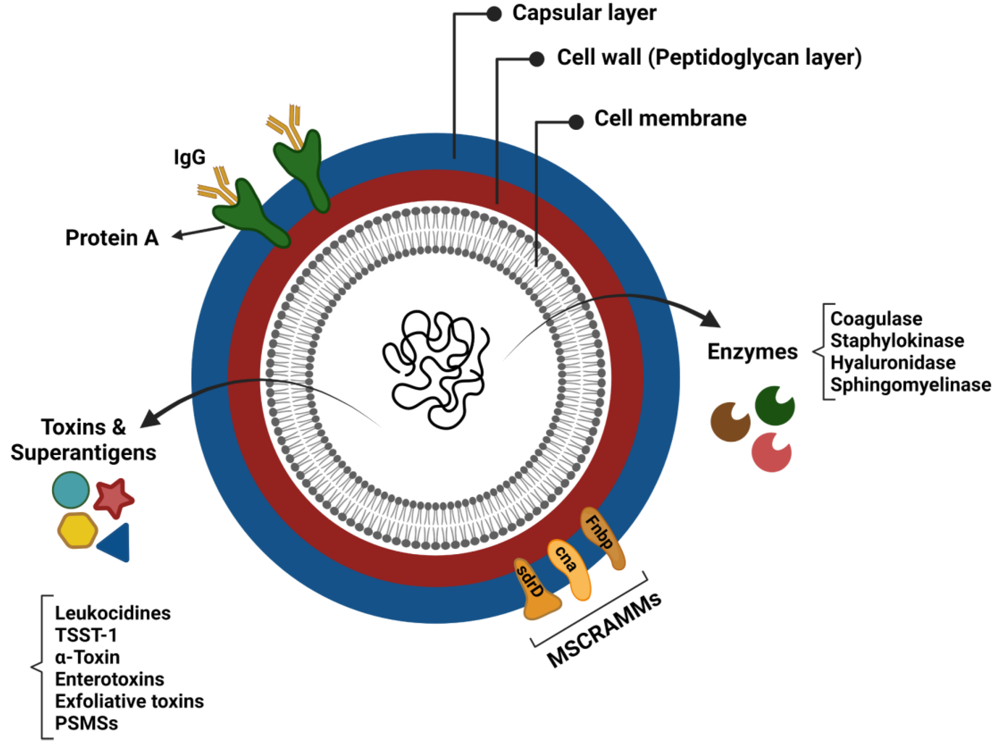
7. Staphylococcal Virulence Factors

The virulence of Staphylococcus aureus is generally considered to be multifactorial, due to the combined activity of an arsenal of virulence determinants that promote tissue adhesion, immune evasion, and host cell damage [71]. These virulence factors consist of structural factors and secreted molecules (exoproteins) (Figure 1) [72]. In addition to S. aureus biofilm, a better understanding of each virulence factor’s functions and mechanisms of action is essential for enhancing the prognosis of patients suffering from CRS.
Figure 1. Virulence factors of Staphylococcus aureus.

8. Immune Response in CRS

CRS is an inflammatory disease, and various cells, including epithelial cells, endothelial cells, fibroblasts, mast cells, neutrophils, eosinophils, dendritic cells, T cells, and B cells, have been demonstrated to be involved in its immune-inflammatory network. These cells normally exert their effect by secreting various mediators, such as cytokines, chemokines, antibodies, and eicosanoids [73].
Failure in the protective measures of the upper respiratory system can lead to the persistence of microbial colonisation, secretion of cytokines and chemokines, recruitment of various immune cells, and activation of inflammatory pathways [74]. The innate immune system represents the first line of defence against inhaled pathogens and foreign substances, and it relies on a large family of pattern recognition receptors (PRRs), which identify distinct evolutionarily conserved structures on pathogens, termed pathogen-associated molecular patterns (PAMPs). The most widely studied PRRs are known as toll-like receptors (TLRs). The binding of PAMPs, including foreign nucleic acids, chemical products, or physical structures, to the ligand-domain of TLRs triggers downstream signal transduction, thus leading to secretion of proinflammatory molecules, such as chemokines and cytokines, which boost the antigen presentation, induction of co-stimulatory molecules of dendritic cells, recruitment of immune cells, and finally, orchestrate the early host response to infection [75].

9. CRS Inflammatory Endotypes

Endotypes of CRS are commonly characterised based on underlying immune responses and cellular differentiation, specifically CD4+ T helper (Th) cells, CD8+ cytotoxic T (Tc) lymphocytes, and ILCs, which regulate the expression of various chemokines and cytokines [16][73][76]. The type 1 response is mainly associated with CRSsNP and is predominately defined by the increased neutrophils linked to myeloperoxidase and elevated secretion of IFN-γ, IL-2, and TNF-α from ILC1, Tc1, and Th1 cells [77][78]. Type 2 inflammation is associated with CRSwNP in Caucasian patients, and it is primarily characterised by high levels of eosinophils and increased quantities of IL-4, IL-5, and IL-13 from ILC2, Tc2, and Th2 cells, as well as large amounts of eosinophil cationic protein (ECP). Total IgE and S. aureus enterotoxin-specific IgE are also increased in patients with CRSwNP [16][79][80]. Finally, type 3 immune response, which has recently been reported to be dominant in Asian patients with CRSwNP, is associated with elevated IL-17 and IL-22 cytokines from ILC3, Tc17 cells, and Th17 cells [79][81].

10. Current Therapeutic Strategies for CRS

CRS patients are primarily treated with standard medical therapy, based on the current consensus guidelines [82]. Nasal irrigation and intranasal steroids are considered the backbones of the pharmacological therapy of CRS, while systemic steroids and antibiotics function as the main relievers during exacerbation onset. Topical steroids have demonstrated beneficial effects in reducing inflammation in CRS patients, with limited side effects. Strong evidence asserts that intranasal corticosteroids efficiently diminish CRS symptoms and polyp development in the nasal cavity [83]. Adjuvant medical therapies in CRS include low-dose macrolides, leukotriene antagonists, topical antibiotics, and oral anti-fungal medicines [84].
Surgery is commonly executed when all attempts at successful medical intervention fail [85]. Diverse surgery options exist, varying from simple polypectomy to the complete removal of the polypoid mucosal tissue from the sinuses [86]. Endoscopic sinus surgery (ESS) is a minimally invasive surgical procedure for CRS that aims to restore sinus ventilation and drainage by opening the main areas and maintaining the sinus mucosa [1]. Furthermore, effective avoidance measures for the target allergen and allergen immunotherapy (AIT) are other well-known strategies [87]. AIT is a highly effective therapeutic approach for allergic disorders, and it induces a long-lasting allergen tolerance by altering the disease course [7].
Numerous potential CRS biomarkers have also been described in the literature but have yet to be clinically validated as indicators of severity or treatment outcome. Eosinophils, IL-4, IL-5, IL-13, and IgE are well-known biomarkers of type 2 inflammation, and some of these are targets for the current biological therapies. Regulatory T cells, IL-25, IL-33, and TSLP are other promising candidates; however, further research is required to validate their role as type 2 biomarkers [88][89]. Despite discovering many potential biomarkers, it is unclear how they can be translated to the bedside [90]. A phase 3 study of duplilumab, a monoclonal antibody (mAb) against IL-4Rα, reported considerable benefits for patients, regardless of the peripheral eosinophil count [91]. The results of phase 2 mepolizumab (anti-IL-5 mAb) found that baseline peripheral eosinophils do not predict improvement in CRSwNP [92]. Dexpramipexole (an anti-eosinophilic synthetic aminobenzothiazole) reduced the number of eosinophils in peripheral blood and nasal polyp tissue; however, the size and symptom scores of nasal polyps did not change [93]. The use of omalizumab (anti-IgE mAb) reduced the size of nasal polyps and improved sinus CT scores but had no significant impact on nasal IgE [94]. The use of duplilumab and omalizumab has been approved by the FDA for difficult-to-treat CRSwNP [95]. However, their effects on patients with mixed inflammatory patterns are unknown, and factors such as long-term safety [96] and cost-effectiveness [97] need to be considered. On the other hand, there are currently no clinical biomarkers indicative of non-type 2 inflammation, which remains an unresolved issue.
Considering that recalcitrant CRS is often found in association with S. aureus biofilms, therapeutic strategies targeting this bacterium’s biofilm or virulence factors might be beneficial. Increasing antibiotic resistance among S. aureus strains emphasises the necessity for alternative treatments. Anti-virulence treatments, including antibodies, nanoparticles, RNAIII-inhibiting peptides, antimicrobial peptides (AMPs), natural compounds, and vaccines that directly or indirectly neutralise S. aureus toxins, have been investigated, and some of them have shown promising effects [98]. However, there is currently no vaccine against S. aureus. As mentioned earlier, S. aureus secretes a broad spectrum of toxins during the colonisation and infection of the host, making vaccine development challenging. IBT-VO2, as a promising multivalent vaccine, is currently under investigation. α-toxin, PVL, LukS, LukF, LukAB, enterotoxins A and B, and TSST1 toxoids are all included in this vaccine. After completing the encouraging pre-clinical phase, it has entered a phase I clinical study [99].

References

  1. Fokkens, W.J.; Lund, V.J.; Mullol, J.; Bachert, C.; Alobid, I.; Baroody, F.; Cohen, N.; Cervin, A.; Douglas, R.; Gevaert, P. EPOS 2012: European position paper on rhinosinusitis and nasal polyps 2012. A summary for otorhinolaryngologists. Rhinology 2012, 50, 1–12.
  2. Rosenfeld, R.M.; Piccirillo, J.F.; Chandrasekhar, S.S.; Brook, I.; Ashok Kumar, K.; Kramper, M.; Orlandi, R.R.; Palmer, J.N.; Patel, Z.M.; Peters, A. Clinical practice guideline (update) adult sinusitis executive summary. Otolaryngol.-Head Neck Surg. 2015, 152, 598–609.
  3. Brook, I. Microbiology of chronic rhinosinusitis. Eur. J. Clin. Microbiol. Infect. Dis. 2016, 35, 1059–1068.
  4. DeConde, A.S.; Soler, Z.M. Chronic rhinosinusitis: Epidemiology and burden of disease. Am. J. Rhinol. Allergy 2016, 30, 134–139.
  5. Lee, S.; Lane, A.P. Chronic rhinosinusitis as a multifactorial inflammatory disorder. Curr. Infect. Dis. Rep. 2011, 13, 159–168.
  6. Stevens, W.W.; Schleimer, R.P.; Kern, R.C. Chronic rhinosinusitis with nasal polyps. J. Allergy Clin. Immunol. Pract. 2016, 4, 565–572.
  7. Kucuksezer, U.C.; Ozdemir, C.; Akdis, M.; Akdis, C.A. Chronic rhinosinusitis: Pathogenesis, therapy options, and more. Expert Opin. Pharmacother. 2018, 19, 1805–1815.
  8. Fastenberg, J.H.; Hsueh, W.D.; Mustafa, A.; Akbar, N.A.; Abuzeid, W.M. Biofilms in chronic rhinosinusitis: Pathophysiology and therapeutic strategies. World J. Otorhinolaryngol.-Head Neck Surg. 2016, 2, 219–229.
  9. Chapurin, N.; Pynnonen, M.A.; Roberts, R.; Schulz, K.; Shin, J.J.; Witsell, D.L.; Parham, K.; Langman, A.; Carpenter, D.; Vambutas, A. CHEER national study of chronic rhinosinusitis practice patterns: Disease comorbidities and factors associated with surgery. Otolaryngol.–Head Neck Surg. 2017, 156, 751–756.
  10. Frendø, M.; Håkansson, K.; Schwer, S.; Rix, I.; Ravn, A.T.; Backer, V.; von Buchwald, C. Asthma in ear, nose, and throat primary care patients with chronic rhinosinusitis with nasal polyps. Am. J. Rhinol. Allergy 2016, 30, e67–e71.
  11. Benninger, M.S.; Sindwani, R.; Holy, C.E.; Hopkins, C. Impact of medically recalcitrant chronic rhinosinusitis on incidence of asthma. Proc. Int. Forum Allergy Rhinol. 2016, 6, 124–129.
  12. Chung, S.D.; Chen, P.Y.; Lin, H.C.; Hung, S.H. Comorbidity profile of chronic rhinosinusitis: A population-based study. Laryngoscope 2014, 124, 1536–1541.
  13. Kariyawasam, H.H.; Rotiroti, G. Allergic rhinitis, chronic rhinosinusitis and asthma: Unravelling a complex relationship. Curr. Opin. Otolaryngol. Head Neck Surg. 2013, 21, 79–86.
  14. Jarvis, D.; Newson, R.; Lotvall, J.; Hastan, D.; Tomassen, P.; Keil, T.; Gjomarkaj, M.; Forsberg, B.; Gunnbjornsdottir, M.; Minov, J. Asthma in adults and its association with chronic rhinosinusitis: The GA2LEN survey in Europe. Allergy 2012, 67, 91–98.
  15. Yoshimura, K.; Kawata, R.; Haruna, S.; Moriyama, H.; Hirakawa, K.; Fujieda, S.; Masuyama, K.; Takenaka, H. Clinical epidemiological study of 553 patients with chronic rhinosinusitis in Japan. Allergol. Int. 2011, 60, 491–496.
  16. Tomassen, P.; Vandeplas, G.; Van Zele, T.; Cardell, L.-O.; Arebro, J.; Olze, H.; Förster-Ruhrmann, U.; Kowalski, M.L.; Olszewska-Ziąber, A.; Holtappels, G. Inflammatory endotypes of chronic rhinosinusitis based on cluster analysis of biomarkers. J. Allergy Clin. Immunol. 2016, 137, 1449–1456.e4.
  17. Langdon, C.; Mullol, J. Nasal polyps in patients with asthma: Prevalence, impact, and management challenges. J. Asthma Allergy 2016, 9, 45.
  18. Ryu, G.; Min, C.; Park, B.; Choi, H.G.; Mo, J.-H. Bidirectional association between asthma and chronic rhinosinusitis: Two longitudinal follow-up studies using a national sample cohort. Sci. Rep. 2020, 10, 9589.
  19. Zhang, N.; Van Zele, T.; Perez-Novo, C.; Van Bruaene, N.; Holtappels, G.; DeRuyck, N.; Van Cauwenberge, P.; Bachert, C. Different types of T-effector cells orchestrate mucosal inflammation in chronic sinus disease. J. Allergy Clin. Immunol. 2008, 122, 961–968.
  20. Klossek, J.M.; Neukirch, F.; Pribil, C.; Jankowski, R.; Serrano, E.; Chanal, I.; El Hasnaoui, A. Prevalence of nasal polyposis in France: A cross-sectional, case–control study. Allergy 2005, 60, 233–237.
  21. Laidlaw, T.M.; Mullol, J.; Woessner, K.M.; Amin, N.; Mannent, L.P. Chronic rhinosinusitis with nasal polyps and asthma. J. Allergy Clin. Immunol. Pract. 2021, 9, 1133–1141.
  22. Bachert, C.; Zhang, N.; Holtappels, G.; De Lobel, L.; Van Cauwenberge, P.; Liu, S.; Lin, P.; Bousquet, J.; Van Steen, K. Presence of IL-5 protein and IgE antibodies to staphylococcal enterotoxins in nasal polyps is associated with comorbid asthma. J. Allergy Clin. Immunol. 2010, 126, 962–968.e966.
  23. Ramakrishnan, V.R.; Feazel, L.M.; Gitomer, S.A.; Ir, D.; Robertson, C.E.; Frank, D.N. The microbiome of the middle meatus in healthy adults. PLoS ONE 2013, 8, e85507.
  24. Bordin, A.; Sidjabat, H.E.; Cottrell, K.; Cervin, A. Chronic rhinosinusitis: A microbiome in dysbiosis and the search for alternative treatment options. Microbiol. Aust. 2016, 37, 149–152.
  25. Feazel, L.M.; Robertson, C.E.; Ramakrishnan, V.R.; Frank, D.N. Microbiome complexity and Staphylococcus aureus in chronic rhinosinusitis. Laryngoscope 2012, 122, 467–472.
  26. Abreu, N.A.; Nagalingam, N.A.; Song, Y.; Roediger, F.C.; Pletcher, S.D.; Goldberg, A.N.; Lynch, S.V. Sinus microbiome diversity depletion and Corynebacterium tuberculostearicum enrichment mediates rhinosinusitis. Sci. Transl. Med. 2012, 4, 151ra124.
  27. Choi, E.B.; Hong, S.W.; Kim, D.K.; Jeon, S.; Kim, K.R.; Cho, S.H.; Gho, Y.; Jee, Y.K.; Kim, Y.K. Decreased diversity of nasal microbiota and their secreted extracellular vesicles in patients with chronic rhinosinusitis based on a metagenomic analysis. Allergy 2014, 69, 517–526.
  28. Ramakrishnan, V.R.; Feazel, L.M.; Abrass, L.J.; Frank, D.N. Prevalence and abundance of Staphylococcus aureus in the middle meatus of patients with chronic rhinosinusitis, nasal polyps, and asthma. Proc. Int. Forum Allergy Rhinol. 2013, 3, 267–271.
  29. Ramakrishnan, V.R.; Hauser, L.J.; Feazel, L.M.; Ir, D.; Robertson, C.E.; Frank, D.N. Sinus microbiota varies among chronic rhinosinusitis phenotypes and predicts surgical outcome. J. Allergy Clin. Immunol. 2015, 136, 334–342.e1.
  30. Koutsourelakis, I.; Halderman, A.; Khalil, S.; Hittle, L.E.; Mongodin, E.F.; Lane, A.P. Temporal instability of the post-surgical maxillary sinus microbiota. BMC Infect. Dis. 2018, 18, 441.
  31. Paramasivan, S.; Bassiouni, A.; Shiffer, A.; Dillon, M.R.; Cope, E.K.; Cooksley, C.; Ramezanpour, M.; Moraitis, S.; Ali, M.J.; Bleier, B. The international sinonasal microbiome study: A multicentre, multinational characterization of sinonasal bacterial ecology. Allergy 2020, 75, 2037–2049.
  32. Frank, D.N.; Zhu, W.; Sartor, R.B.; Li, E. Investigating the biological and clinical significance of human dysbioses. Trends Microbiol. 2011, 19, 427–434.
  33. Vickery, T.W.; Ramakrishnan, V.R.; Suh, J.D. The role of Staphylococcus aureus in patients with chronic sinusitis and nasal polyposis. Curr. Allergy Asthma Rep. 2019, 19, 1–8.
  34. Chalermwatanachai, T.; Zhang, N.; Holtappels, G.; Bachert, C. Association of mucosal organisms with patterns of inflammation in chronic rhinosinusitis. PLoS ONE 2015, 10, e0136068.
  35. Van Zele, T.; Gevaert, P.; Watelet, J.-B.; Claeys, G.; Holtappels, G.; Claeys, C.; van Cauwenberge, P.; Bachert, C. Staphylococcus aureus colonization and IgE antibody formation to enterotoxins is increased in nasal polyposis. J. Allergy Clin. Immunol. 2004, 114, 981–983.
  36. Schmidt, F.; Meyer, T.; Sundaramoorthy, N.; Michalik, S.; Surmann, K.; Depke, M.; Dhople, V.; Salazar, M.G.; Holtappels, G.; Zhang, N. Characterization of human and Staphylococcus aureus proteins in respiratory mucosa by in vivo-and immunoproteomics. J. Proteom. 2017, 155, 31–39.
  37. Schwartz, J.S.; Peres, A.G.; Endam, L.M.; Cousineau, B.; Madrenas, J.; Desrosiers, M. Topical probiotics as a therapeutic alternative for chronic rhinosinusitis: A preclinical proof of concept. Am. J. Rhinol. Allergy 2016, 30, e202–e205.
  38. Skelly, A.N.; Sato, Y.; Kearney, S.; Honda, K. Mining the microbiota for microbial and metabolite-based immunotherapies. Nat. Rev. Immunol. 2019, 19, 305–323.
  39. Seiberling, K.A.; Conley, D.B.; Tripathi, A.; Grammer, L.C.; Shuh, L.; Haines, G.K., III; Schleimer, R.; Kern, R.C. Superantigens and chronic rhinosinusitis: Detection of staphylococcal exotoxins in nasal polyps. Laryngoscope 2005, 115, 1580–1585.
  40. Monaco, M.; de Araujo, F.P.; Cruciani, M.; Coccia, E.M.; Pantosti, A. Worldwide epidemiology and antibiotic resistance of Staphylococcus aureus. In Staphylococcus aureus; Springer: New York, NY, USA, 2016; pp. 21–56.
  41. Thammavongsa, V.; Missiakas, D.M.; Schneewind, O. Staphylococcus aureus degrades neutrophil extracellular traps to promote immune cell death. Science 2013, 342, 863–866.
  42. Diep, B.A.; Gill, S.R.; Chang, R.F.; Phan, T.H.; Chen, J.H.; Davidson, M.G.; Lin, F.; Lin, J.; Carleton, H.A.; Mongodin, E.F. Complete genome sequence of USA300, an epidemic clone of community-acquired meticillin-resistant Staphylococcus aureus. Lancet 2006, 367, 731–739.
  43. Drilling, A.; Coombs, G.W.; Tan, H.l.; Pearson, J.C.; Boase, S.; Psaltis, A.; Speck, P.; Vreugde, S.; Wormald, P.J. Cousins, siblings, or copies: The genomics of recurrent Staphylococcus aureus infections in chronic rhinosinusitis. Proc. Int. Forum Allergy Rhinol. 2014, 4, 953–960.
  44. Grumann, D.; Nübel, U.; Bröker, B.M. Staphylococcus aureus toxins–their functions and genetics. Infect. Genet. Evol. 2014, 21, 583–592.
  45. Teymournejad, O.; Montgomery, C.P. Evasion of immunological memory by S. aureus infection: Implications for vaccine design. Front. Immunol. 2021, 12, 430.
  46. Flemming, H.-C.; Neu, T.R.; Wozniak, D.J. The EPS matrix: The “house of biofilm cells”. J. Bacteriol. 2007, 189, 7945–7947.
  47. Gil, C.; Solano, C.; Burgui, S.; Latasa, C.; García, B.; Toledo-Arana, A.; Lasa, I.; Valle, J. Biofilm matrix exoproteins induce a protective immune response against Staphylococcus aureus biofilm infection. Infect. Immun. 2014, 82, 1017–1029.
  48. Singhal, D.; Psaltis, A.J.; Foreman, A.; Wormald, P.-J. The impact of biofilms on outcomes after endoscopic sinus surgery. Am. J. Rhinol. Allergy 2010, 24, 169–174.
  49. Donlan, R.M.; Costerton, J.W. Biofilms: Survival mechanisms of clinically relevant microorganisms. Clin. Microbiol. Rev. 2002, 15, 167–193.
  50. Costerton, J.W.; Lewandowski, Z.; Caldwell, D.E.; Korber, D.R.; Lappin-Scott, H.M. Microbial biofilms. Annu. Rev. Microbiol. 1995, 49, 711–745.
  51. Suh, J.D.; Ramakrishnan, V.; Palmer, J.N. Biofilms. Otolaryngol. Clin. N. Am. 2010, 43, 521–530.
  52. Donné, J.; Dewilde, S. The challenging world of biofilm physiology. In Advances in Microbial Physiology; Elsevier: Amsterdam, The Netherlands, 2015; Volume 67, pp. 235–292.
  53. Belas, R. Biofilms, flagella, and mechanosensing of surfaces by bacteria. Trends Microbiol. 2014, 22, 517–527.
  54. Donlan, R.M. Biofilms: Microbial life on surfaces. Emerg. Infect. Dis. 2002, 8, 881.
  55. Flemming, H.-C.; Wingender, J.; Szewzyk, U.; Steinberg, P.; Rice, S.A.; Kjelleberg, S. Biofilms: An emergent form of bacterial life. Nat. Rev. Microbiol. 2016, 14, 563–575.
  56. Boyd, C.D.; O’Toole, G.A. Second messenger regulation of biofilm formation: Breakthroughs in understanding c-di-GMP effector systems. Annu. Rev. Cell Dev. Biol. 2012, 28, 439–462.
  57. Romeo, T. When the party is over: A signal for dispersal of Pseudomonas aeruginosa biofilms. J. Bacteriol. 2006, 188, 7325–7327.
  58. Ramakrishnan, Y.; Shields, R.; Elbadawey, M.; Wilson, J. Biofilms in chronic rhinosinusitis: What is new and where next? J. Laryngol. Otol. 2015, 129, 744–751.
  59. Singh, P.; Mehta, R.; Agarwal, S.; Mishra, P. Bacterial biofilm on the sinus mucosa of healthy subjects and patients with chronic rhinosinusitis (with or without nasal polyposis). J. Laryngol. Otol. 2015, 129, 46–49.
  60. Sanderson, A.R.; Leid, J.G.; Hunsaker, D. Bacterial biofilms on the sinus mucosa of human subjects with chronic rhinosinusitis. Laryngoscope 2006, 116, 1121–1126.
  61. Sanclement, J.A.; Webster, P.; Thomas, J.; Ramadan, H.H. Bacterial biofilms in surgical specimens of patients with chronic rhinosinusitis. Laryngoscope 2005, 115, 578–582.
  62. Dlugaszewska, J.; Leszczynska, M.; Lenkowski, M.; Tatarska, A.; Pastusiak, T.; Szyfter, W. The pathophysiological role of bacterial biofilms in chronic sinusitis. Eur. Arch. Oto-Rhino-Laryngol. 2016, 273, 1989–1994.
  63. Prince, A.A.; Steiger, J.D.; Khalid, A.N.; Dogrhamji, L.; Reger, C.; Claire, S.E.; Chiu, A.G.; Kennedy, D.W.; Palmer, J.N.; Cohen, N.A. Prevalence of biofilm-forming bacteria in chronic rhinosinusitis. Am. J. Rhinol. 2008, 22, 239–245.
  64. Foreman, A.; Psaltis, A.J.; Tan, L.W.; Wormald, P.-J. Characterization of bacterial and fungal biofilms in chronic rhinosinusitis. Am. J. Rhinol. Allergy 2009, 23, 556–561.
  65. Psaltis, A.J.; Weitzel, E.K.; Ha, K.R.; Wormald, P.-J. The effect of bacterial biofilms on post-sinus surgical outcomes. Am. J. Rhinol. 2008, 22, 1–6.
  66. Pratt, E.; Collins, A.M.; Sewell, W.A.; Harvey, R.J. Antigen selection in IgE antibodies from individuals with chronic rhinosinusitis with nasal polyps. Am. J. Rhinol. Allergy 2010, 24, 416–421.
  67. Cryer, J.; Schipor, I.; Perloff, J.R.; Palmer, J.N. Evidence of bacterial biofilms in human chronic sinusitis. J. Oto-Rhino-Laryngol. 2004, 66, 155–158.
  68. Bachert, C.; Claeys, S.E.; Tomassen, P.; Van Zele, T.; Zhang, N. Rhinosinusitis and asthma: A link for asthma severity. Curr. Allergy Asthma Rep. 2010, 10, 194–201.
  69. Cirkovic, I.; Pavlovic, B.; Bozic, D.D.; Jotic, A.; Bakic, L.; Milovanovic, J. Antibiofilm effects of topical corticosteroids and intranasal saline in patients with chronic rhinosinusitis with nasal polyps depend on bacterial species and their biofilm-forming capacity. Eur. Arch. Oto-Rhino-Laryngol. 2017, 274, 1897–1903.
  70. Mladina, R.; Skitarelić, N.; Musić, S.; Ristić, M. A biofilm exists on healthy mucosa of the paranasal sinuses: A prospectively performed, blinded, scanning electron microscope study. Clin. Otolaryngol. 2010, 35, 104–110.
  71. Powers, M.E.; Wardenburg, J.B. Igniting the fire: Staphylococcus aureus virulence factors in the pathogenesis of sepsis. PLoS Pathog. 2014, 10, e1003871.
  72. Bien, J.; Sokolova, O.; Bozko, P. Characterization of virulence factors of Staphylococcus aureus: Novel function of known virulence factors that are implicated in activation of airway epithelial proinflammatory response. J. Pathog. 2011, 2011, 601905.
  73. Liu, Y.; Yao, Y.; Wang, Z.C.; Ning, Q.; Liu, Z. Novel innate and adaptive lymphocytes: The new players in the pathogenesis of inflammatory upper airway diseases. Clin. Exp. Allergy 2018, 48, 620–631.
  74. Stevens, W.W.; Lee, R.J.; Schleimer, R.P.; Cohen, N.A. Chronic rhinosinusitis pathogenesis. J. Allergy Clin. Immunol. 2015, 136, 1442–1453.
  75. Hirschberg, A.; Kiss, M.; Kadocsa, E.; Polyanka, H.; Szabo, K.; Razga, Z.; Bella, Z.; Tiszlavicz, L.; Kemeny, L. Different activations of toll-like receptors and antimicrobial peptides in chronic rhinosinusitis with or without nasal polyposis. Eur. Arch. Oto-Rhino-Laryngol. 2016, 273, 1779–1788.
  76. Van Zele, T.; Claeys, S.; Gevaert, P.; Van Maele, G.; Holtappels, G.; Van Cauwenberge, P.; Bachert, C. Differentiation of chronic sinus diseases by measurement of inflammatory mediators. Allergy 2006, 61, 1280–1289.
  77. Bachert, C.; Zhang, N.; Hellings, P.W.; Bousquet, J. Endotype-driven care pathways in patients with chronic rhinosinusitis. J. Allergy Clin. Immunol. 2018, 141, 1543–1551.
  78. Ahern, S.; Cervin, A. Inflammation and endotyping in chronic rhinosinusitis—A paradigm shift. Medicina 2019, 55, 95.
  79. Annunziato, F.; Romagnani, C.; Romagnani, S. The 3 major types of innate and adaptive cell-mediated effector immunity. J. Allergy Clin. Immunol. 2015, 135, 626–635.
  80. De Greve, G.; Hellings, P.W.; Fokkens, W.J.; Pugin, B.; Steelant, B.; Seys, S.F. Endotype-driven treatment in chronic upper airway diseases. Clin. Transl. Allergy 2017, 7, 22.
  81. Bachert, C.; Zhang, N. Medical algorithm: Diagnosis and treatment of chronic rhinosinusitis. Allergy 2019, 75, 240–242.
  82. Fokkens, W.J.; Lund, V.; Luong, A.U.; Orlandi, R.R. A comparison of international guidelines for rhinosinusitis. J. Allergy Clin. Immunol. Pract. 2022, 10, 1418–1422.
  83. Chong, L.Y.; Head, K.; Hopkins, C.; Philpott, C.; Schilder, A.G.; Burton, M.J. Intranasal steroids versus placebo or no intervention for chronic rhinosinusitis. Cochrane Database Syst. Rev. 2016, 26, CD011996.
  84. Bachert, C.; Pawankar, R.; Zhang, L.; Bunnag, C.; Fokkens, W.J.; Hamilos, D.L.; Jirapongsananuruk, O.; Kern, R.; Meltzer, E.O.; Mullol, J. ICON: Chronic rhinosinusitis. World Allergy Organ. J. 2014, 7, 25.
  85. Rudmik, L.; Soler, Z.M. Medical therapies for adult chronic sinusitis: A systematic review. J. Am. Med. Assoc. 2015, 314, 926–939.
  86. Sharma, R.; Lakhani, R.; Rimmer, J.; Hopkins, C. Surgical interventions for chronic rhinosinusitis with nasal polyps. Cochrane Database Syst. Rev. 2014, 12, CD006991.
  87. Cardona, V.; Luengo, O.; Labrador-Horrillo, M. Immunotherapy in allergic rhinitis and lower airway outcomes. Allergy 2017, 72, 35–42.
  88. Mann, C.; Schmidtmann, I.; Bopp, T.; Brieger, J.; Fruth, K. Treg activation and their role in different subtypes of chronic rhinosinusitis. Allergy 2020, 75, 2687–2689.
  89. Hong, H.; Liao, S.; Chen, F.; Yang, Q.; Wang, D.Y. Role of IL-25, IL-33, and TSLP in triggering united airway diseases toward type 2 inflammation. Allergy 2020, 75, 2794–2804.
  90. Staudacher, A.G.; Peters, A.T.; Kato, A.; Stevens, W.W. Use of endotypes, phenotypes, and inflammatory markers to guide treatment decisions in chronic rhinosinusitis. Ann. Allergy Asthma Immunol. 2020, 124, 318–325.
  91. Bachert, C.; Han, J.K.; Desrosiers, M.; Hellings, P.W.; Amin, N.; Lee, S.E.; Mullol, J.; Greos, L.S.; Bosso, J.V.; Laidlaw, T.M. Efficacy and safety of dupilumab in patients with severe chronic rhinosinusitis with nasal polyps (LIBERTY NP SINUS-24 and LIBERTY NP SINUS-52): Results from two multicentre, randomised, double-blind, placebo-controlled, parallel-group phase 3 trials. Lancet 2019, 394, 1638–1650.
  92. Bachert, C.; Sousa, A.R.; Lund, V.J.; Scadding, G.K.; Gevaert, P.; Nasser, S.; Durham, S.R.; Cornet, M.E.; Kariyawasam, H.H.; Gilbert, J. Reduced need for surgery in severe nasal polyposis with mepolizumab: Randomized trial. J. Allergy Clin. Immunol. 2017, 140, 1024–1031.e14.
  93. Laidlaw, T.M.; Prussin, C.; Panettieri, R.A.; Lee, S.; Ferguson, B.J.; Adappa, N.D.; Lane, A.P.; Palumbo, M.L.; Sullivan, M.; Archibald, D. Dexpramipexole depletes blood and tissue eosinophils in nasal polyps with no change in polyp size. Laryngoscope 2019, 129, E61–E66.
  94. Gevaert, P.; Calus, L.; Van Zele, T.; Blomme, K.; De Ruyck, N.; Bauters, W.; Hellings, P.; Brusselle, G.; De Bacquer, D.; Van Cauwenberge, P. Omalizumab is effective in allergic and nonallergic patients with nasal polyps and asthma. J. Allergy Clin. Immunol. 2013, 131, 110–116.e1.
  95. Fokkens, W.J.; Lund, V.; Bachert, C.; Mullol, J.; Bjermer, L.; Bousquet, J.; Canonica, G.W.; Deneyer, L.; Desrosiers, M.; Diamant, Z. EUFOREA consensus on biologics for CRSwNP with or without asthma. Allergy 2019, 74, 2312–2319.
  96. Kim, C.; Han, J.; Wu, T.; Bachert, C.; Fokkens, W.; Hellings, P.; Hopkins, C.; Lee, S.; Mullol, J.; Lee, J.T. Role of biologics in chronic rhinosinusitis with nasal polyposis: State of the art review. Otolaryngol.–Head Neck Surg. 2021, 164, 57–66.
  97. Brown, W.C.; Senior, B. A critical look at the efficacy and costs of biologic therapy for chronic rhinosinusitis with nasal polyposis. Curr. Allergy Asthma Rep. 2020, 20, 16.
  98. Ahmad-Mansour, N.; Loubet, P.; Pouget, C.; Dunyach-Remy, C.; Sotto, A.; Lavigne, J.-P.; Molle, V. Staphylococcus aureus toxins: An update on their pathogenic properties and potential treatments. Toxins 2021, 13, 677.
  99. Karauzum, H.; Venkatasubramaniam, A.; Adhikari, R.P.; Kort, T.; Holtsberg, F.W.; Mukherjee, I.; Mednikov, M.; Ortines, R.; Nguyen, N.T.; Doan, T. IBT-V02: A multicomponent toxoid vaccine protects against primary and secondary skin infections caused by Staphylococcus aureus. Front. Immunol. 2021, 12, 475.
More
Information
Contributors MDPI registered users' name will be linked to their SciProfiles pages. To register with us, please refer to https://encyclopedia.pub/register : , , , , ,
View Times: 322
Revisions: 2 times (View History)
Update Date: 21 Jun 2022
1000/1000