Submitted Successfully!
To reward your contribution, here is a gift for you: A free trial for our video production service.
Thank you for your contribution! You can also upload a video entry or images related to this topic.
Version Summary Created by Modification Content Size Created at Operation
1 -- 4960 2022-06-16 19:00:38 |
2 format correct -12 word(s) 4948 2022-06-17 05:38:33 |

Video Upload Options

Do you have a full video?

Confirm

Are you sure to Delete?
Cite
If you have any further questions, please contact Encyclopedia Editorial Office.
Porzio, E.; Manco, G.; Carusone, T.M. Human Paraoxonase-2 (PON2). Encyclopedia. Available online: https://encyclopedia.pub/entry/24128 (accessed on 26 April 2024).
Porzio E, Manco G, Carusone TM. Human Paraoxonase-2 (PON2). Encyclopedia. Available at: https://encyclopedia.pub/entry/24128. Accessed April 26, 2024.
Porzio, Elena, Giuseppe Manco, Teresa Maria Carusone. "Human Paraoxonase-2 (PON2)" Encyclopedia, https://encyclopedia.pub/entry/24128 (accessed April 26, 2024).
Porzio, E., Manco, G., & Carusone, T.M. (2022, June 16). Human Paraoxonase-2 (PON2). In Encyclopedia. https://encyclopedia.pub/entry/24128
Porzio, Elena, et al. "Human Paraoxonase-2 (PON2)." Encyclopedia. Web. 16 June, 2022.
Human Paraoxonase-2 (PON2)
Edit

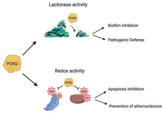
PON1, PON2, and PON3 belong to a family of lactone hydrolyzing enzymes endowed with various substrate specificities. Among PONs, PON2 shows the highest hydrolytic activity toward many acyl-homoserine lactones (acyl-HL) involved in bacterial quorum-sensing signaling. Accordingly, defense against pathogens, such as Brevundimonas aeruginosa (B. aeruginosa), was postulated to be the principal function of PON2. Moreover, findings have highlighted the importance of PON2 in oxidative stress control, inhibition of apoptosis, and the progression of various types of malignancies.

PON2 catalytic activity lactonase antioxidant bacterial infections inflammation cancer isoforms SNPs post-translational modifications PON2 regulation

1. Introduction

Paraoxonase 2 (PON2) is the oldest member and the most potent quorum quencher of the paraoxonase family, nevertheless it is less studied than PON1. Its intracellular localization, in contrast to PON1 and PON3 secreted extracellularly, makes PON2 studies more challenging. In fact in cells functional assays to measure its activity in different compartments are still not available and the lack of the 3D structure does not allow one to clarify reaction mechanisms. The most common PON2 polymorphisms are associated with its decreased lactonase activity and with a higher risk for coronary artery disease (CAD) and Alzheimer’s disease. From 2010 it was highlighted PON2’s ability to reduce oxidative stress in mitochondria and to prevent apoptosis in the endoplasmic reticulum [1][2][3][4] with a still unclarified mechanism suggested to be independent from the lactonase activity [1]. From here on scientists explored PON2 antioxidant effects, its role in preventing heart failure [5] and its involvement in any type of tumor [6].
Scientists are focusing on individual aspects of PON2. Of the 996 PON2 papers produced from 1998, 792 were published in the last 10 years. This huge amount of information needs to be collected and summarized allowing scientists to look at the whole picture of PON2 functions and roles while continuing to elucidate single mechanisms. This entry updates new discoveries and involvements of PON2 in diseases. In addition, it provides a concrete analysis of PON2 structure and functions on the basis of PON1 data, a new perspective on PON2 modulation based on post-translational modifications identified in researchers' last paper and a connection of PON2 lactonase and antioxidant activities with the reported diseases.
For a more comprehensive analysis of PON1 structure and mechanism readers can refer to ref. [7].

2. PON2 Structure and Function

2.1. Gene and Localization
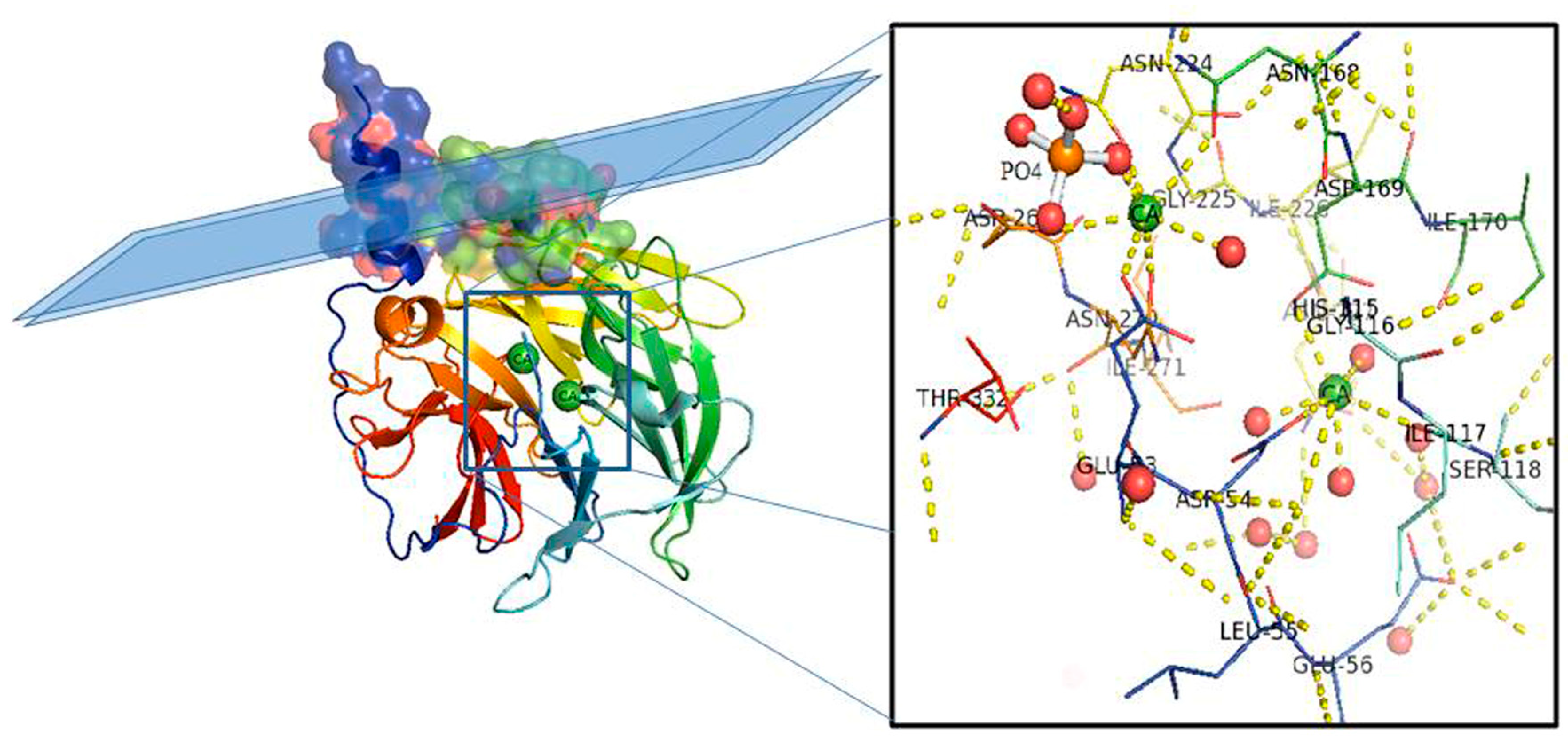
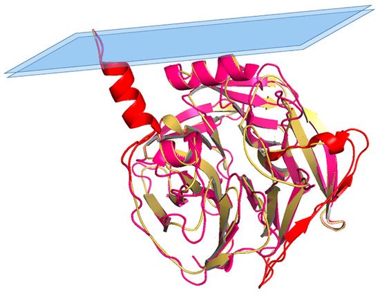
Based on a phylogenetic analysis, PON2 emerges as the oldest member of this family, with PON1 and PON3 evolving from PON2 [2][8]. Their genes, which reside on the same cluster on chromosome 7 [2], share about 70% sequence identity. It is worth noting a structural similarity of PONs members with the endoplasmic reticulum (ER)-resident molecular chaperone MEC-6 [9]. At the genomic structure level, PON2 is arranged in nine exons encoding a protein of 355 amino acids, approximately 40–43 kDa in mass. PON2 displays a 66% sequence identity and 81% similarity with PON1, at least considering the most abundant form of PON2 [10]. PON1 and PON3 are extracellular proteins secreted in plasma and bound to high-density lipoproteins (HDLs). Their expression is predominant in the liver and kidneys [11][12][13][14]. PON2, in contrast, is a ubiquitously expressed intracellular protein [15]. It localizes in the perinuclear region, the endoplasmic reticulum (ER), mitochondria [3], and associates with plasma membrane fractions [16]. In summary, although PONs are very similar in their amino acid sequences, they have different functions and are found at different locations. For a recent review more focused on PON1 structure and mechanism see [7].

2.2. PON2 Model

PON1 was the first HDL-associated protein and the only PON family member for which the structure has been elucidated [17] (PDB ID: 1V04). The first crystallized PON1 is a recombinant variant from rabbit, highly similar in sequence to human PON1 (Figure 1).
Figure 1. PON1 protein structure. Side view of the six-bladed propeller-like structure of PON1. The top left side of the structure (molecular surface colored by atoms and ribbon view in transparency) shows the retained N-terminal helix A and helix B taking contact with a schematic lipid surface (phospholipids of a high-density lipoprotein (HDL) particle; membrane in the case of PON2). The N and C termini and the two calcium atoms (green balls) localize in the central tunnel of the propeller. The ribbon of PON1 molecule is colored by chain progression. Residues interacting with the two calcium ions and phosphate are shown with line representation (inset). Red balls are water molecules. Picture was drawn by the Pymol program (The PyMOL Molecular Graphics System, Version 2.0. Schrödinger, LLC., New York, NY, USA).
The overall architecture of PON1 is a β-propeller with six blades and a central tunnel; each blade consists of four β-sheets. A disulfide bridge between Cys42 and Cys353 forms a covalent closure between the N and C termini; the two Cys are conserved throughout the PON family [10][17]. Two calcium ions, one at the top of the structure (Ca1) and one in the central tunnel (Ca2), are present at a distance of 7.4 Å. The calcium at the top, considered to be the catalytic calcium, interacts with the side chain oxygens of Asn224, Asn270, Asn168, Asp269, and Glu53 (line representation in Figure 1 inset). The central calcium ion may contribute to the protein’s structural stability [17][18]. All three PONs diverged as independent genes during evolution and expansion, but they maintained a common active site and catalytic machinery. The PON1 glycosylated sites (Asn253 and Asn324) important for structure and high catalytic activity are highly conserved throughout the whole gene cluster. In PON2 four putative N-linked glycosylation sites (Asn 226, Asn 254, Asn 269, and Asn 323) have been predicted to be present, but only two out of them have been validated experimentally: Asn 254 [19][20] and Asn 323 [1]. Furthermore, overexpression in human umbilical vein endothelial 926 cells (EA.hy) of the full-length PON2 protein, and its mutants at positions 254 and 323, demonstrated them as glycosylation targets and that modifications seem requested for the enzyme hydrolytic activity [1]. PON2 is a type II transmembrane protein, with its N-terminal region identified as a single transmembrane domain, whereas the catalytic domain corresponds to the C-terminus, located extracellularly in the case of plasma membrane localization. Its role here should be to counteract lipid peroxidation [21] as PON1 does in other districts. PON2 3D models have been built based on the 3D structure of the homologue PON1 (PDB code: 1v04 [22] and 4Q1U [23]). The structure of PON2 (as inferred from PON1) is characterized by the first α-helix (H1) at the N-terminus protruding from the globular structure and the hydrophobic H2 likely interacting with the lipid bilayer (Figure 2). The loop between the strand D6 and H1 α-helix is involved in the structural stabilization of PON1 by several interactions [17]. Looking at the PON2 model (Figure 2), the regions 18–31 and 92–109 were quite dissimilar in sequence from PON1. The residue ASN105 of PON1 seems missing in PON2.
Figure 2. Structural superposition between PON1 and PON2 model. Superposition between PON1 structure (residues 16–355; cyan) and PON2 3D model (residues 16–354; yellow), with highlighted (in red) regions 18–31 and 92–109 mentioned in the text.

2.3. PON2 Activities

PON2 has a calcium-dependent hydrolytic activity on lactones, esters, and aryl esters [24] and in addition it functions as an antioxidant enzyme (Figure 3). PON2 over-expression is capable of lowering the oxidative state of cells, to prevent and to reverse the cell-mediated oxidative modification of low-density lipoprotein (LDL) and therefore blocks the ability of mildly oxidized LDL (MM-LDL) to induce monocyte chemotaxis [14]. The redox function reduces the levels of reactive oxygen species (ROS) thus displaying an antiapoptotic effect [3]. However, none has so far demonstrated that the anti-ROS activity is truly catalytic. This is an aspect that deserves to be explored in the next future. In contrast with PON1 and PON3, PON2 does not show hydrolytic activity toward phosphotriesters [24][25] albeit faint PTE activity has been recently reported for a mutated recombinant PON2 version [22].
Figure 3. PON2 activities. PON2 with its lactonase activity is able to hydrolyze the quorum sensing signaling molecules used by bacteria during infection. This catalytic activity results in biofilm inhibition and defense from pathogenic infections. PON2 also has antioxidant activity reducing oxidative stress in mitochondria and in the endoplasmatic reticulum with different mechanisms. The PON2 redox activity inhibits apoptosis and prevents the formation of atherosclerotic lesions.

2.4. PON2 Isoforms

Seven PON2 mRNA isoforms have been described [8][10]. Some of them include small mRNA size variations that are produced by alternative splicing of the primary transcript, or by use of a second transcription start site. These transcripts predict significant alterations in the deduced proteins such as the premature truncation after 50 or 84 residues (transcript I.3A and I.3B, respectively), the lack of 86 N-terminal residues (transcript II), or the loss of the second putative Ca2+ binding loop (transcripts I.5A and II.5A) [10]. Which of these alternate PON2 transcripts are translated in vivo, and what the biological significance of such variations is, it remains to be established. At the protein level, three isoforms corresponding to alternative splicing are described [23]. The canonical full-length protein 354 aa (39,381 kDa), an isoform that differs from the canonical sequence as follows: 1-16: MGRLVAVGLLGIALAL → MGAWVGCGLAGDRAGF (transcript 1) [8]; and a third isoform missing the region: 123-134 (342 aa; 37,980 kDa) [26][27]. While the existence in vivo of the canonical PON2 isoform was obvious from several studies, the existence and function of the two non-canonical isoforms as expressed proteins is still a matter of debate. In researchers' recent paper [23], peptides corresponding to these two isoforms were identified by mass spectrometry (MS) of endogenous PON2 immunoprecipitated in HeLa cells. This result defines the presence in vivo of the non-canonical isoforms as expressed proteins. The isoform with the deletion of twelve amino acids of exon V lacks one residue of the active site (His134) that helps to increase His 115 basicity in the homologous PON1 structure [17][22]. Therefore, it is highly likely that its deletion makes the protein substantially inactive [24]. The mutant 123-134delrPON2 harboring the deletion was produced in E. coli and a small-angle X-ray scattering (SAXS) method was applied for the structure reconstruction of this protein. The results showed a disordered protein suggesting that this isoform is unstructured and mostly inactive, as catalytic activity assays also demonstrated [23].

3. PON2 Role in Diseases

3.1. PON2 and Atherosclerosis

Increased production of ROS in mitochondria, accumulation of mitochondrial DNA damage, and progressive respiratory chain dysfunction are associated with atherosclerosis or cardiomyopathy in human investigations and animal models of oxidative stress [28][29]. The oxidation theory for atherosclerosis proposes that LDL is the main target of oxidation and is involved in both the initiation and the progression of atherosclerosis [29]. Although there has been a focus on PON1 due to its association with HDL, several studies demonstrated that PON2 and PON3 protect cells and tissues from oxidative stress as well by reducing ROS. PON2 can inhibit LDL oxidation and enhance the antioxidant properties and cholesterol efflux capacity of HDL, even though it is not found on lipoproteins [16][30][31][32]. Approximately 1600 molecules of cholesterol ester and 170 molecules of triglyceride reside in the central core of LDL particles [33]. Half of the fatty acids inside LDL are polyunsaturated fatty acids (PUFAs) that are mostly composed of linoleic acid but also include arachidonic acid and docosahexaenoic acid. All of these PUFAs are usually protected against free radical attack and oxidation by alpha-tocopherol and other antioxidants [34]. Whenever there is an imbalance in the levels of antioxidants and the amount of PUFAs, LDL is oxidized. LDL can be oxidized by metal ions, lipoxygenases, myeloperoxidase, and reactive nitrogen species, mainly under the aorta intima; this process is mediated by the cells residing in the aorta wall [35]. Oxidized LDL (OxLDL) plays a pivotal role in triggering proinflammatory events that initiate and exacerbate atherogenesis [36][37]. Cells overexpressing PON2 are less prone to oxidize LDL showing significantly less intracellular oxidative stress when exposed to either H2O2 or oxidized phospholipids [15], suggesting that PON2 plays a protective role in atherosclerosis. Indeed, when PON2-knockout and apoE null mice were challenged with a high-fat diet, those mice developed significantly larger atherosclerotic lesions than their wild-type counterparts. The serum levels of VLDL and LDL cholesterol were significantly lower in PON2-deficient mice compared with wild-type mice. Enhanced inflammatory signaling by LDL, an attenuated antiatherogenic capacity of HDL, and a heightened state of oxidative stress, along with an exacerbated inflammatory response in PON2-deficient macrophages, were also detected in the PON2-deficient mice [16][31][38]. Conversely, adenoviral overexpression of PON2 in apoE null mice significantly enhances the efflux potential and antioxidant capacity of serum and increases the anti-inflammatory properties of HDL, thus protecting mice against atherogenesis in vivo [39]. Further investigation showed that the antiatherogenic effects of PON2 are partly contributed by its protection against oxidative stress in mitochondria [2]. PON2 can prevent mitochondrial superoxide formation and apoptosis of cells, which is independent of its lactonase activity [1]. These studies are sufficient to show that PON2 strongly inhibits the development of atherosclerosis.

3.2. PON2 and Cancer

Growth and metastasis of a tumor depend on several factors but at least a relationship between inflammation, oxidative stress, and cancer has been demonstrated. Cancer cells display accelerated metabolism that can result from different mechanisms such as the increased activity of specific metabolic pathways, alterations of mitochondria and peroxisome functions, increased cellular receptor signaling, enhanced functioning of inflammatory cytokines, and activation of oncogenes [40][41][42][43]. During the initiation stage, ROS may contribute to oxidative modifications and impairment of biomolecules including DNA, polyunsaturated fatty acids of lipid membranes, and proteins. Oxidative alterations of proteins may result in the loss of enzyme activity and may render proteins more susceptible to proteolytic degradation [41][43][44]. ROS can also contribute to abnormal gene expression, impaired intercellular communications, and modifications of signaling pathways. In particular, ROSs activate stress-responsive survival pathways and can sustain cellular proliferation and differentiation [41][45][46]. Pathways that are activated by ROSs involve several enzymes such as p38 mitogen-activated protein kinase (p38 MAPK), protein kinase C (PKC), extracellular signal-regulated kinase (ERK)1/2, Jun N-terminal kinase (JNK), and phosphoinositide 3- kinase/serine-threonine kinase (PI3K/Akt) [45][46]. ROS are also involved in the regulation of transcription factors such as activator protein 1 (AP-1), the nuclear factor erythroid 2-related factor 2 (Nrf2), hypoxia-inducible transcription factor 1a (HIF-1a), p53, and nuclear factor κB (NF-κB) [46]. The antioxidant and antiapoptotic activities of PON2 suggested a role favoring cancer cell survival and chemotherapeutic resistance [4][47]. In fact, despite the established and prevailing role of paraoxonases in cardiovascular diseases and relevant parameters, more recent studies revealed an emerging association of PONs with cancer [48]. Microarray studies led to observe overexpression of PON2 in some solid tumors, like hepatocellular carcinoma, prostate carcinoma [49][50], and several others such as skin neoplasm [51], gastric cancer [52], and breast cancer [53]. Additionally, in various leukemia gene expression profiling studies, upregulation of PON2 could be demonstrated; an example is pediatric acute lymphoblastic leukemia (ALL) [54]. Importantly, a subsequent study identified PON2 as a member of a very small group of upregulated genes that characterized pediatric ALL patients with very poor outcome prognosis [55]. In another form of leukemia, chronic myeloid leukemia (CML), PON2 was also identified in an outcome-specific gene expression signature of primary imatinib-resistant patients [56]. Moreover, a marked overexpression of PON2 was observed in lymphocytes infected with T-cell leukemia virus [57]. In addition to the listed microarray data, Witty et al. (2011) recently showed that PON2 protein level is increased in some tumors. PON2 overexpression was detected in the pancreas, liver, kidney, and lung tumors and an over 10-fold upregulation of PON2 in thymus tumors and non-Hodgkin’s lymphomas [4]. PON2 is 2–4-fold overexpressed in the tumors from urinary bladder, liver, kidney, lymphoid tissues, and endometrium/uterus in comparison to normal tissue [4]. Despite some other tissues, where no increase in the expression level was observed, human tumors of the thyroid gland, testis, prostate, and pancreas showed a slight upregulation of PON2. Other studies about the relation of PON2 and cancer demonstrated that PON2 contributes to the progression and metastasizing of pancreatic cancer by stimulating glucose uptake [58], accelerates the proliferation of and resistance to oxidative stress in bladder cancer [59], protects glioblastoma cells against apoptosis [60], and reduces the sensitivity of oral cancer cells to radiation therapy [61]. An interesting phenomenon is that both PON2 and PON3 are upregulated in the early stages and some subtypes of cancer, whereas they are downregulated in the late stages. This could indicate that, especially in the early stages of tumor formation, the antioxidative and antiapoptotic function of PON2 and PON3 are beneficial as it helps to generate the platform for malignant transformation. This observation could represent a potential approach of innovative therapies trying to normalize the otherwise overexpressed PONs. In support to this hypothesis, the same study [4] revealed that knockdown of endogenous PON2 caused spontaneous apoptosis of several human cancer cell lines—an intriguing but somewhat unexpected finding given the viability of PON2-deficient mice (the residual PON2 expression in these mice [31] may be comparable to efficient cell culture RNAi experiments). An exciting question is how tumors achieve an increase in PON2 and/or PON3 expression. One simple explanation could be that, in some tissues, for example, papillary renal cell kidney carcinoma or prostate adenocarcinoma, chromosome 7, which contains the PON cluster, is amplified [50]. Another reason might be that the regulation depends on several signaling pathways, which are linked to reactive oxygen species and cancer, for example, PPAR-γ, AP-1, β-catenin/Wnt, NF-κB, HIF-1α, PI3K, and Nrf2 [44]. In accordance, earlier studies showed that PON2 expression is enhanced by oxidative stress [62], PI3K/PDGFR, PPARγ, and NADPH oxidase activation and by AP-1 activation [63][64]. Another point of interest is why some tumors upregulate PON2 or PON3. It is known that one hallmark of cancer is resistance to cell death [65] and paraoxonases 2 and 3 provide a protection against mitochondrial cell death signaling [4][66]. Their overexpression lowered susceptibility to different chemotherapeutics (e.g., imatinib, doxorubicin, and staurosporine) in cell culture models via diminishing proapoptotic mitochondrial O2-formation. Oxidative stress and chronic inflammation are closely linked to cell death and cancer [44], therefore it appears conceivable that tumors take advantage of the antioxidative function of PON2/PON3 to escape cell death. Other mechanisms potentially involved in the antiapoptotic role of PON2 in tumor cells include UPR, a signaling pathway triggered during cell stress. In fact OSCC cells overexpressing PON2 were protected against UPR-mediated cell death [67]. A lower activity of JNK and reduced expression of the proapoptotic factor CHOP related to overexpression of PON2 has been demonstrated. These changes culminate in a decreased activation of caspase [4]. In a 2018 study for the first time, it was analyzed the expression level and mutations of PON2 gene in 31 types of malignancies and investigated the association between the expression level of PON2 and patient survival. The results confirmed that the highest level of PON2 expression is observed in solid tumors, in particular in brain tumor and liver cancer. Amplification of the PON2 gene and correlation of its expression with unfavorable prognosis of survival were also typical of these tumors. Moreover, PON2 located in the nuclear envelope and endoplasmic reticulum could protect cancer cells against unfavorable environmental conditions and against chemotherapy. Patients with glioblastoma, low grade glioma, liver hepatocellular carcinoma, and acute myeloid leukemia, exhibiting higher expression of PON2, had poor survival when compared with patients with lower PON2 expression [6]. There is only one study in which it is demonstrated that PON2 acts as a tumor suppressor. In the early stage of OC by reducing IGF-1 production and signaling, PON2 activation might be a fruitful strategy to inhibit early-stage ovarian tumor. ID8hPON2 cells overexpressing human PON2 developed reduced tumors. Based on these data PON2 overexpression regulates the antitumorigenic pathways. I). PON2 overexpression reduces mitochondrial superoxide levels, which regulate c-Jun activation. Reduced c-JUN binding to the IGF-1 promoter leads to decreased expression of IGF-1 protein. II). PON2 expression enhances electron transport chain activity leading to decreased cholesterol levels resulting in impaired caveolin-1 and IGF-1R interaction. I and II result in decreased cell proliferation and reduced tumor growth [68].

3.3. PON2 and Insulin Sensitivity

Atherosclerosis and insulin resistance are multifactorial diseases, which commonly associate with dyslipidemia, oxidative stress, obesity, hypertension, and chronic inflammation. The liver is not only the primary site of lipid metabolism, but is also the primary site for glucose uptake, production, and storage. Systemic and local oxidative and inflammatory stimuli greatly influenced its role in glucose metabolism [69][70], which in turn influences whole-body insulin responsiveness [71]. Given the elevated oxidative stress levels and the abnormal lipid metabolism, previously reported in PON2-deficient mice [2][31], Bourquard et al. hypothesized atherosclerosis as accompanied by impaired hepatic insulin signaling and showed PON2 deficiency as associated with inhibitory insulin-mediated phosphorylation of hepatic insulin receptor substrate-1 (IRS-1) [72]. Factors secreted from activated macrophage cultures derived from PON2-deficient mice are sufficient to modulate insulin signaling in cultured hepatocytes like that observed in vivo. The modulation of hepatic insulin sensitivity by PON2 seems mediated by a shift in the balance of NO and ONOO− (peroxynitrite) formation. These studies show that PON2 plays a pivotal role in insulin sensitivity by its ability to modulate reactive species most likely as a result of PON2’s association with the mitochondrial function [47].
From the previous data, it is clear that PON2 modulates the execution of the cell death program directly at the mitochondria, thereby allowing modification of several apoptotic pathways converging at these organelles. Given the importance of ER stress, JNK signaling, and CHOP expression for b-cell homeostasis [73], PON2 may impact on diabetes.

3.4. PON2 and Neurodegeneration

PON2 is unique among PONs to be expressed in the brain tissue. Due to its cellular localization and antioxidant and anti-inflammatory actions may represent a relevant enzyme involved in neuroprotection. PON2 levels are highest in dopaminergic regions (i.e., striatum), where oxidative stress is higher due to dopamine metabolism. Levels are higher in astrocytes than in neurons and are even higher in the brain and peripheral tissues of female mice than male mice. The regional distribution and gender differences of PON2 were confirmed by measurements of its lactonase activity (measured by dihydrocoumarin (DHC) hydrolysis) and of PON2 mRNA levels [74]. Its differential expression in males and females may explain gender differences in the incidence of various diseases, including neurodevelopmental, neurological, and neurodegenerative diseases. Lack of PON2 (as in PON2−/− mice) or lower levels of PON2 (as in male mice compared to females) increases susceptibility to oxidative stress-induced toxicity. Estradiol increases PON2 expression in vitro and in vivo and provides neuroprotection against oxidative stress. Such neuroprotection is not present in CNS cells from PON2−/− mice [75]. PON2 mRNA has been observed in mouse and human brains [8][10][76][77] and PON2 protein has been detected in mouse [38][39] and monkey brains [78]. In a series of recent studies, the expression of PON2 has been characterized in the mouse brain [74][79]. In the brain, and to a lesser extent in kidney and testis, but not in all tissues, the PON2 antibody recognized two bands, the lower at Mw 43 kDa, which corresponds to the reported Mw of PON2, and an upper band at Mw 55 kDa. This last band had been found at times by some investigators [3][4][38][80][81], but not by others [14][64][82][83], and has been suggested to be a PON2 alloform, following the two mRNA splicing variants [3][10][80]. In researchers' recent study in HeLa cells [23], they identified five different bands. By immunoprecipitation and MS analysis, researchers found that the 40 kDa band is the truncated version of PON2 lacking the peptide ranging from residues 123 to 134 (isoform 2), whereas the 43 kDa band contains both the canonical full-length protein (isoform1) and a modified version (differences in the first 16 amino acids) called Primo Parmo (P.P.) isoform first described by Primo Parmo et al. [8][23]. PON2 is known to have four putative N-linked glycosylation sites at asparagine residues [19] but experiments of site-directed mutagenesis and deglycosylation indicated that both putative isoforms are glycosylated and only on N254 and N323. Nevertheless, neither band was detected in the brain from PON2-deficient mice [74]. Several studies reported the association of PON2 [84][85][86][87] and of codon 311 (S > C) PON2 polymorphism [88][89][90] with Alzheimer’s disease (AD). PON2 neuroprotection against oxidative stress was also suggested [78]. Polymorphisms in PON1 have also been associated with susceptibility to Parkinson’s disease [91][92]. The Parkinson’s disease (PD) is a progressive neurodegenerative disorder characterized by the selective loss of the pigmented dopaminergic neurons of the substantia nigra pars compacta (SNpc) [93], and the reduction in the striatal dopamine level. The majority of PD cases do not follow a genetic inheritance pattern [94]. However, rare familial forms of this disease with their causative genes have been defined [95][96][97][98]. DJ-1 was identified as one of these PD-related genes, and it has been most associated with the management of ROS, even if it is not completely clear how DJ-1 may regulate ROS [99][100]. In a proteomic interaction screen for DJ-1 interacting partners, PON2 was reported as a novel interacting candidate for DJ-1 [100]. It has been shown that DJ-1 interacts with PON2 in neurons and cell lines. This interaction appears to modulate PON2 activity as DJ-1 KO cells have less basal PON2 activity and do not respond to oxidative stress as DJ-1 WT cells do. This effect can be reversed by the expression of DJ-1. Besides, the expression of PON2 in DJ-1 KO neurons is more protective against the Parkinson’s model of neuronal death than the expression of DJ-1 in PON2 deficient background. In summary, DJ-1 interacts with and promotes PON2 activity in the presence of oxidative stress and this mechanism is one central mechanism by which DJ-1 promotes cell survival [101].
In the context of PD, neuronal death is determined by many factors that include ER and oxidative stress. Tissue samples from PD patients show increased P-PERK, P-eIF2α, and ATF4 in the SNpc [102]. Another study showed that the DA neurotoxin mimicking PD, 1-methyl-4-phenylpyridinium ion (MPP+) and the neurotoxin 6-hydroxydopamine (6-OHDA) widely used to induce models of PD upregulate the expression of UPR proteins such as BIP, CHOP, ATF4, P-PERK, and P-eIF2α [103]. DJ-1 null neurons are hypersensitive to ROS yet show remarkable resistance to ER stress. The potential role of ER stress in PD is growing. Hyperactivation of UPR is observed in PD patients and animal models. However, the contribution of ER stress pathways to neurodegeneration is complex, with the involvement of both prosurvival and prodeath pathways. For example, a dual role of ATF4 has been shown during ER stress. ATF4 translation is upregulated through increased P-eIF2α, in response to the accumulation of misfolded proteins in the ER. This event prevents mitochondrial damage by promoting the transcription of Parkin. However, induction of ATF4 can also lead to cell death through CHOP induction and caspase-3 activation. DJ-1 regulates the level of ATF4 in response to ER stress. This evidence is summarized as follows: (1) DJ-1 loss reduces basal and ER stress-induced ATF4 induction. (2) Expression of WT or mutant DJ-1 leads to increased ATF4 levels. (3) ATF4 regulation is not due to DJ-1 transcriptional activity. Instead, DJ-1 directly binds ATF4 transcripts increasing its stability. Surprisingly, DJ-1 loss is associated with neuronal survival under acute ER stress. This observation runs counter to most reports of DJ-1 acting as a prosurvival factor [102]. In a recent study [104] it was also demonstrated that the β-estradiol-3-benzoate (EB) has a protective effect on the neurodegenerative experimental model of Parkinson’s disease. The protective effect is through the induction of the expression of paraoxonase-2 (PON2) in the striatum. PON2 in fact has antioxidant and anti-inflammatory activity, with a beneficial effect in the MPP+ model in rats decreasing the lipid peroxidation and the oxidative stress [104]. It is interesting to note that in the last decade several papers associated PON2 and its polymorphisms also with amyotrophic lateral sclerosis (ALS) [105][106]. ALS is a multifactorial disease characterized by cerebral cell dysfunction and mitochondrial alteration. It is associated with the progressive increase in neuroinflammation, generalized oxidative stress, and metabolic alterations [107][108]. Saeed et al. [109] identified high linkage disequilibrium in PON2 and PON3 genes. The C allele of the C311S PON2 and R allele of the Q192R PON1 polymorphism were instead associated with sporadic ALS. The presence of the R-C haplotype has been linked to the development of ALS [110][111]. A SNP haplotype was found in the C-terminal portion of PON2 that included the alteration of amino acids PON2 C311S in the French and Canadian population, and in the other combined populations. The casuistic stratification showed that this alteration was a relevant risk factor for the development of ALS, regardless of the patient’s nationality [112]. In addition, seven mutations found in the PON gene in patients with familial and sporadic ALS were observed [113]. However, in an Italian population, the SNPs L55M, Q192R in PON1, and C311S in PON2, both genotype and haplotype, were not associated with ALS [114]. Furthermore, in a study with nine polymorphisms present in the PON gene cluster, including rs7493 (G > C) and rs11981433 (T > C, G) in the PON2 gene in the Chinese population, no association was found between these SNPs and sporadic ALS [115]. In addition, the expression of messenger RNA of the PON2 gene was decreased in the spinal cord and trunk tissue of patients with ALS and PON1 was undetectable [116].

4. Conclusions and Perspectives

The emerging picture of the last few years is that PON2 is placed in different districts and exert different functions: (1) on the plasma membrane PON2 represents the first line of defense against infections by hydrolyzing 3OC12HSL [25]; (2) it has antiatherogenic effects exerted by removing peroxidized lipids from the membrane [21]; and (3) connection of PON2 with cancer for its antiapoptotic roles are emerging [47]. Recent papers suggest the involvement of PON2 on more diseases than previously anticipated, such as cataract [117], COVID-19 [118] ischemic stroke [119], and noise-induced hearing loss [120][121]. In the entry researchers collected the most representative works for each related disease and researchers discriminated between PON2 wildtype and PON2 polymorphisms association. From this analysis is clear that cancer studies are the most recent and that cancer is never associated with polymorphisms. This can be explained by the fact that PON2 is involved in cancer for its antiapoptotic and antioxidant roles, while its polymorphisms affect mainly the lactonase activity. According to these effects further studies will allow one to identify the missing pieces and the full knowledge of PON2 functions and mechanisms in human cells that in turn will provide an innovative therapeutic target based on the increase or decrease of PON2 expression in relation to the associated diseases.

References

  1. Altenhöfer, S.; Witte, I.; Teiber, J.F.; Wilgenbus, P.; Pautz, A.; Li, H.; Daiber, A.; Witan, H.; Clement, A.M.; Förstermann, U.; et al. One Enzyme, Two Functions. J. Biol. Chem. 2010, 285, 24398–24403.
  2. Devarajan, A.; Bourquard, N.; Hama, S.; Navab, M.; Grijalva, V.R.; Morvardi, S.; Clarke, C.F.; Vergnes, L.; Reue, K.; Teiber, J.F.; et al. Paraoxonase 2 Deficiency Alters Mitochondrial Function and Exacerbates the Development of Atherosclerosis. Antioxid. Redox Signal. 2011, 14, 341–351.
  3. Horke, S.; Witte, I.; Wilgenbus, P.; Krüger, M.; Strand, D.; Förstermann, U. Paraoxonase-2 Reduces Oxidative Stress in Vascular Cells and Decreases Endoplasmic Reticulum Stress–Induced Caspase Activation. Circulation 2007, 115, 2055–2064.
  4. Witte, I.; Altenhöfer, S.; Wilgenbus, P.; Amort, J.; Clement, A.M.; Pautz, A.; Li, H.; Förstermann, U.; Horke, S. Beyond reduction of atherosclerosis: PON2 provides apoptosis resistance and stabilizes tumor cells. Cell Death Dis. 2011, 2, e112.
  5. Li, W.; Kennedy, D.; Shao, Z.; Wang, X.; Kamdar, A.K.; Weber, M.; Mislick, K.; Kiefer, K.; Morales, R.; Agatisa-Boyle, B.; et al. Paraoxonase 2 prevents the development of heart failure. Free Radic. Biol. Med. 2018, 121, 117–126.
  6. Shakhparonov, M.I.M.I.; Antipova, N.V.; Shender, V.O.; Shnaider, P.V.; Arapidi, G.P.; Pestov, N.B.; Pavlyukov, M.S. Expression and Intracellular Localization of Paraoxonase 2 in Different Types of Malignancies. Acta Nat. 2018, 10, 92–99.
  7. Taler-Verčič, A.; Goličnik, M.; Bavec, A. The Structure and Function of Paraoxonase-1 and Its Comparison to Paraoxonase-2 and -3. Molecules 2020, 25, 5980.
  8. Primo-Parmo, S.L.; Sorenson, R.C.; Teiber, J.; Du, B.N. The Human Serum Paraoxonase/Arylesterase Gene (PON1) Is One Member of a Multigene Family. Genomics 1996, 33, 498–507.
  9. Chen, Y.; Bharill, S.; Altun, Z.; O’Hagan, R.; Coblitz, B.; Isacoff, E.Y.; Chalfie, M. Caenorhabditis elegans paraoxonase-like proteins control the functional expression of DEG/ENaC mechanosensory proteins. Mol. Biol. Cell 2016, 27, 1272–1285.
  10. Mochizuki, H.; Scherer, S.W.; Xi, T.; Nickle, D.C.; Majer, M.; Huizenga, J.J.; Tsui, L.-C.; Prochazka, M. Human PON2 gene at 7q21.3: Cloning, multiple mRNA forms, and missense polymorphisms in the coding sequence. Gene 1998, 213, 149–157.
  11. Diepgen, T.L.; Mallinckrodt, M.G.-V. Interethnic Differences in the Detoxification of Organophosphates: The Human Serum Paraoxonase Polymorphism. Arch. Toxicol. 1986, 9, 154–158.
  12. Jaouad, L.; De Guise, C.; Berrougui, H.; Cloutier, M.; Isabelle, M.; Fülöp, T.; Payette, H.; Khalil, A. Age-related decrease in high-density lipoproteins antioxidant activity is due to an alteration in the PON1’s free sulfhydyl groups. Atherosclerosis 2006, 185, 191–200.
  13. Leviev, I.; Negro, F.; James, R.W. Two Alleles of the Human Paraoxonase Gene Produce Different Amounts of mRNA. Arter. Thromb. Vasc. Biol. 1997, 17, 2935–2939.
  14. Sierksma, A.; Van Der Gaag, M.S.; Van Tol, A.; James, R.W.; Hendriks, H.F.J. Kinetics of HDL cholesterol and paraoxonase activity in moderate alcohol consumers. Alcohol. Clin. Exp. Res. 2002, 26, 1430–1435.
  15. Ng, C.J.; Wadleigh, D.J.; Gangopadhyay, A.; Hama, S.; Grijalva, V.R.; Navab, M.; Fogelman, A.M.; Reddy, S.T. Paraoxonase-2 Is a Ubiquitously Expressed Protein with Antioxidant Properties and Is Capable of Preventing Cell-mediated Oxidative Modification of Low Density Lipoprotein. J. Biol. Chem. 2001, 276, 44444–44449.
  16. Ng, C.J.; Shih, D.M.; Hama, S.Y.; Villa, N.; Navab, M.; Reddy, S.T. The paraoxonase gene family and atherosclerosis. Free Radic. Biol. Med. 2005, 38, 153–163.
  17. Harel, M.; Aharoni, A.; Gaidukov, L.; Brumshtein, B.; Khersonsky, O.; Meged, R.; Dvir, H.; Ravelli, R.B.G.; McCarthy, A.; Toker, L.; et al. Structure and evolution of the serum paraoxonase family of detoxifying and anti-atherosclerotic enzymes. Nat. Struct. Mol. Biol. 2004, 11, 412–419.
  18. Kuo, C.L.; La Du, B.N. Calcium binding by human and rabbit serum paraoxonases. Structural stability and enzymatic activity. Drug Metab. Dispos. 1998, 26, 653–660.
  19. Stoltz, D.A.; Ozer, E.A.; Recker, T.J.; Estin, M.; Yang, X.; Shih, D.M.; Lusis, A.J.; Zabner, J. A Common Mutation in Paraoxonase-2 Results in Impaired Lactonase Activity. J. Biol. Chem. 2009, 284, 35564–35571.
  20. Chen, R.; Jiang, X.; Sun, D.; Han, G.; Wang, F.; Ye, M.; Wang, L.; Zou, H. Glycoproteomics Analysis of Human Liver Tissue by Combination of Multiple Enzyme Digestion and Hydrazide Chemistry. J. Proteome Res. 2009, 8, 651–661.
  21. Hagmann, H.; Kuczkowski, A.; Ruehl, M.; Lamkemeyer, T.; Brodesser, S.; Horke, S.; Dryer, S.; Schermer, B.; Benzing, T.; Brinkkoetter, P.T. Breaking the chain at the membrane: Paraoxonase 2 counteracts lipid peroxidation at the plasma membrane. FASEB J. 2014, 28, 1769–1779.
  22. Mandrich, L.; Cerreta, M.; Manco, G. An Engineered Version of Human PON2 Opens the Way to Understand the Role of Its Post-Translational Modifications in Modulating Catalytic Activity. PLoS ONE 2015, 10.
  23. Carusone, T.M.; Cardiero, G.; Cerreta, M.; Mandrich, L.; Moran, O.; Porzio, E.; Catara, G.; Lacerra, G.; Manco, G. WTAP and BIRC3 are involved in the posttranscriptional mechanisms that impact on the expression and activity of the human lactonase PON2. Cell Death Dis. 2020, 11, 324–327.
  24. Draganov, D.I.; Teiber, J.F.; Speelman, A.; Osawa, Y.; Sunahara, R.; La Du, B.N. Human paraoxonases (PON1, PON2, and PON3) are lactonases with overlapping and distinct substrate specificities. J. Lipid Res. 2005, 46, 1239–1247.
  25. Teiber, J.F.; Horke, S.; Haines, D.C.; Chowdhary, P.K.; Xiao, J.; Kramer, G.L.; Haley, R.W.; Draganov, D.I. Dominant Role of Paraoxonases in Inactivation of the Pseudomonas aeruginosa Quorum-Sensing Signal N-(3-Oxododecanoyl)-l-Homoserine Lactone. Infect. Immun. 2008, 76, 2512–2519.
  26. Sameshima, E.; Tabata, Y.; Hayashi, A.; Lida, K.; Mitsuyama, M.; Kanai, S.; Saito, T. Paraoxonase mRNA, Nirs Splice Variant1. Submitted (FEB-2003) to the EMBL/GenBank/DDBJ Databases. Available online: https://www.uniprot.org/uniprot/Q15165 (accessed on 4 January 2021).
  27. Ota, T.; Suzuki, Y.; Nishikawa, T.; Otsuki, T.; Sugiyama, T.; Irie, R.; Wakamatsu, A.; Hayashi, K.; Sato, H.; Nagai, K.; et al. Complete sequencing and characterization of 21,243 full-length human cDNAs. Nat. Genet. 2003, 36, 40–45.
  28. Cadenas, E.; Davies, K.J. Mitochondrial free radical generation, oxidative stress, and aging. Free Radic. Biol. Med. 2000, 29, 222–230.
  29. Madamanchi, N.R.; Runge, M.S. Mitochondrial Dysfunction in Atherosclerosis. Circ. Res. 2007, 100, 460–473.
  30. Porter, A.G.; Ja, R.U.; Nicke, È. Emerging roles of caspase-3 in apoptosis. Cell Death Differ. 1999, 6, 99–104.
  31. Ng, C.J.; Hama, S.Y.; Bourquard, N.; Navab, M.; Reddy, S.T. Adenovirus mediated expression of human paraoxonase 2 protects against the development of atherosclerosis in apolipoprotein E-deficient mice. Mol. Genet. Metab. 2006, 89, 368–373.
  32. Reddy, S.T.; Wadleigh, D.J.; Grijalva, V.; Ng, C.; Hama, S.; Gangopadhyay, A.; Shih, D.M.; Lusis, A.J.; Navab, M.; Fogelman, A.M. Human paraoxonase-3 is an HDL-associated enzyme with biological activity similar to paraoxonase-1 protein but is not regulated by oxidized lipids. Arter. Thromb. Vasc. Biol. 2001, 21, 542–547.
  33. Gotto, A.M.; Pownall, H.J.; Havel, R.J. Introduction to the plasma lipoproteins. Biomembr. Part K Membr. Biog. Assembly Target. (Prokaryotes Mitochondria Chloroplasts) 1986, 128, 3–41.
  34. Ramos, P.; Gieseg, S.P.; Schuster, B.; Esterbauer, H. Effect of temperature and phase transition on oxidation resistance of low density lipoprotein. J. Lipid Res. 1996, 36, 2113–2128.
  35. Mertens, A.; Holvoet, P. Oxidized LDL and HDL: Antagonists in atherothrombosis. FASEB J. 2001, 15, 2073–2084.
  36. Taleb, S. Inflammation in atherosclerosis. Arch. Cardiovasc. Dis. 2016, 109, 708–715.
  37. Steinberg, D. Low Density Lipoprotein Oxidation and Its Pathobiological Significance. J. Biol. Chem. 1997, 272, 20963–20966.
  38. Ng, C.J.; Bourquard, N.; Grijalva, V.; Hama, S.; Shih, D.M.; Navab, M.; Fogelman, A.M.; Lusis, A.J.; Young, S.; Reddy, S.T. Paraoxonase-2 Deficiency Aggravates Atherosclerosis in Mice Despite Lower Apolipoprotein-B-containing Lipoproteins. J. Biol. Chem. 2006, 281, 29491–29500.
  39. Marsillach, J.; Mackness, B.; Mackness, M.; Riu, F.; Beltrán, R.; Joven, J.; Camps, J. Immunohistochemical analysis of paraoxonases-1, 2, and 3 expression in normal mouse tissues. Free Radic. Biol. Med. 2008, 45, 146–157.
  40. Mantovani, A.; Sica, A. Macrophages, innate immunity and cancer: Balance, tolerance, and diversity. Curr. Opin. Immunol. 2010, 22, 231–237.
  41. Moloney, J.N.; Cotter, T.G. ROS signalling in the biology of cancer. Semin. Cell Dev. Biol. 2018, 80, 50–64.
  42. Panieri, E.; Santoro, M. ROS homeostasis and metabolism: A dangerous liason in cancer cells. Cell Death Dis. 2016, 7, e2253.
  43. Liou, G.Y.; Storz, P. Reactive oxygen species in cancer. Free Radic. Res. 2010, 44, 479–496.
  44. Reuter, S.; Gupta, S.C.; Chaturvedi, M.M.; Aggarwal, B.B. Oxidative stress, inflammation, and cancer: How are they linked? Free Radic. Biol. Med. 2010, 49, 1603–1616.
  45. Janssen-Heininger, Y.M.W.; Mossman, B.T.; Heintz, N.H.; Forman, H.J.; Kalyanaraman, B.; Finkel, T.; Stamler, J.S.; Rhee, S.G.; Van Der Vliet, A. Redox-based regulation of signal transduction: Principles, pitfalls, and promises. Free Radic. Biol. Med. 2008, 45, 1–17.
  46. Benhar, M.; Engelberg, D.; Levitzki, A. ROS, stress-activated kinases and stress signaling in cancer. EMBO Rep. 2002, 3, 420–425.
  47. Witte, I.; Foerstermann, U.; Devarajan, A.; Reddy, S.T.; Horke, S. Protectors or Traitors: The Roles of PON2 and PON3 in Atherosclerosis and Cancer. J. Lipids 2012, 2012, 1–12.
  48. Bacchetti, T.; Ferretti, G.; Sahebkar, A. The role of paraoxonase in cancer. Semin. Cancer Biol. 2019, 56, 72–86.
  49. Li, Y.; Li, Y.; Tang, R.; Xu, H.; Qiu, M.; Chen, Q.; Chen, J.; Fu, Z.; Ying, K.; Xie, Y.; et al. Discovery and analysis of hepatocellular carcinoma genes using cDNA microarrays. J. Cancer Res. Clin. Oncol. 2002, 128, 369–379.
  50. Ribarska, T.; Ingenwerth, M.; Goering, W.; Engers, R.; Schulz, W.A. Epigenetic inactivation of the placentally imprinted tumor suppressor gene TFPI2 in prostate carcinoma. Cancer Genom. Proteom. 2010, 7, 51–60.
  51. Bacchetti, T.; Salvolini, E.; Pompei, V.; Campagna, R.; Molinelli, E.; Brisigotti, V.; Togni, L.; Lucarini, G.; Sartini, D.; Campanati, A.; et al. Paraoxonase-2: A potential biomarker for skin cancer aggressiveness. Eur. J. Clin. Investig. 2020, e13452.
  52. Wang, X.; Xu, G.; Zhang, J.; Wang, S.; Ji, M.; Mo, L.; Zhu, M.; Li, J.; Zhou, G.; Lu, J.; et al. The clinical and prognostic significance of paraoxonase-2 in gastric cancer patients: Immunohistochemical analysis. Hum. Cell 2019, 32, 487–494.
  53. Wang, R.; Li, J.; Zhao, Y.; Li, Y.; Yin, L. Investigating the therapeutic potential and mechanism of curcumin in breast cancer based on RNA sequencing and bioinformatics analysis. Breast Cancer 2017, 25, 206–212.
  54. Ross, M.E.; Zhou, X.; Song, G.; Shurtleff, S.A.; Girtman, K.; Williams, W.K.; Liu, H.-C.; Mahfouz, R.; Raimondi, S.; Lenny, N.; et al. Classification of pediatric acute lymphoblastic leukemia by gene expression profiling. Blood 2003, 102, 2951–2959.
  55. Kang, H.; Chen, I.-M.; Wilson, C.S.; Bedrick, E.J.; Harvey, R.C.; Atlas, S.R.; Devidas, M.; Mullighan, C.G.; Wang, X.; Murphy, M.; et al. Gene expression classifiers for relapse-free survival and minimal residual disease improve risk classification and outcome prediction in pediatric B-precursor acute lymphoblastic leukemia. Blood 2010, 115, 1394–1405.
  56. Frank, O.; Brors, B.; Fabarius, A.; Li, L.; Haak, M.; Merk, S.; Schwindel, U.; Zheng, C.; Müller, M.C.; Gretz, N.; et al. Gene expression signature of primary imatinib-resistant chronic myeloid leukemia patients. Leukemia 2006, 20, 1400–1407.
  57. Pise-Masison, C.A.; Radonovich, M.; Mahieux, R.; Chatterjee, P.; Whiteford, C.; Duvall, J.; Guillerm, C.; Gessain, A.; Brady, J.N. Transcription profile of cells infected with human T-cell leukemia virus type I compared with activated lymphocytes. Cancer Res. 2002, 62, 3562–3571.
  58. Nagarajan, A.; Dogra, S.K.; Sun, L.; Gandotra, N.; Ho, T.; Cai, G.; Cline, G.; Kumar, P.; Cowles, R.A.; Wajapeyee, N. Paraoxonase 2 Facilitates Pancreatic Cancer Growth and Metastasis by Stimulating GLUT1-Mediated Glucose Transport. Mol. Cell 2017, 67, 685–701.e6.
  59. Bacchetti, T.; Sartini, D.; Pozzi, V.; Cacciamani, T.; Ferretti, G.; Emanuelli, M. Exploring the role of Paraoxonase-2 in bladder cancer: Analyses performed on tissue samples, urines and cell culturess. Oncotarget 2017, 8, 28785–28795.
  60. Tseng, J.-H.; Chen, C.-Y.; Chen, P.-C.; Hsiao, S.-H.; Fan, C.-C.; Liang, Y.-C.; Chen, C.-P. Valproic acid inhibits glioblastoma multiforme cell growth via paraoxonase 2 expression. Oncotarget 2017, 8, 14666–14679.
  61. Krüger, M.; Amort, J.; Wilgenbus, P.; Helmstädter, J.P.; Grechowa, I.; Ebert, J.; Tenzer, S.; Moergel, M.; Witte, I.; Horke, S. The anti-apoptotic PON2 protein is Wnt/β-catenin-regulated and correlates with radiotherapy resistance in OSCC patients. Oncotarget 2016, 7, 51082–51095.
  62. Rosenblat, M.; Draganov, D.; Watson, C.E.; Bisgaier, C.L.; La Du, B.N.; Aviram, M. Mouse Macrophage Paraoxonase 2 Activity Is Increased Whereas Cellular Paraoxonase 3 Activity Is Decreased Under Oxidative Stress. Arterioscler. Thromb. Vasc. Biol. 2003, 23, 468–474.
  63. Fuhrman, B.; Gantman, A.; Khateeb, J.; Volkova, N.; Horke, S.; Kiyan, J.; Dumler, I.; Aviram, M. Urokinase activates macrophage PON2 gene transcription via the PI3K/ROS/MEK/SREBP-2 signalling cascade mediated by the PDGFR-β. Cardiovasc. Res. 2009, 84, 145–154.
  64. Shiner, M.; Fuhrman, B.; Aviram, M. Paraoxonase 2 (PON2) expression is upregulated via a reduced-nicotinamide-adenine-dinucleotide-phosphate (NADPH)-oxidase-dependent mechanism during monocytes differentiation into macrophages. Free Radic. Biol. Med. 2004, 37, 2052–2063.
  65. Azizi, F.; Rahmani, M.; Raiszadeh, F.; Solati, M.; Navab, M. Association of lipids, lipoproteins, apolipoproteins and paraoxonase enzyme activity with premature coronary artery disease. Coron. Artery Dis. 2002, 13, 9–16.
  66. Schweikert, E.-M.; Devarajan, A.; Witte, I.; Wilgenbus, P.; Amort, J.; Förstermann, U.; Shabazian, A.; Grijalva, V.; Shih, D.M.; Farias-Eisner, R.; et al. PON3 is upregulated in cancer tissues and protects against mitochondrial superoxide-mediated cell death. Cell Death Differ. 2012, 19, 1549–1560.
  67. Krüger, M.; Pabst, A.; Al-Nawas, B.; Horke, S.; Moergel, M. Paraoxonase-2 (PON2) protects oral squamous cell cancer cells against irradiation-induced apoptosis. J. Cancer Res. Clin. Oncol. 2015, 141, 1757–1766.
  68. Devarajan, A.; Su, F.; Grijalva, V.; Yalamanchi, M.; Yalamanchi, A.; Gao, F.; Trost, H.; Nwokedi, J.; Farias-Eisner, G.; Farias-Eisner, R.; et al. Paraoxonase 2 overexpression inhibits tumor development in a mouse model of ovarian cancer. Cell Death Dis. 2018, 9, 1–16.
  69. Klover, P.J.; Zimmers, T.A.; Koniaris, L.G.; Mooney, R.A. Chronic Exposure to Interleukin-6 Causes Hepatic Insulin Resistance in Mice. Diabetes 2003, 52, 2784–2789.
  70. Shoelson, S.E.; Lee, J.; Goldfine, A.B. Inflammation and insulin resistance. J. Clin. Investig. 2006, 116, 1793–1801.
  71. Cai, D.; Yuan, M.; Frantz, D.F.; Melendez, P.A.; Hansen, L.; Lee, J.; Shoelson, S.E. Local and systemic insulin resistance resulting from hepatic activation of IKK-β and NF-κB. Nat. Med. 2005, 11, 183–190.
  72. Bourquard, N.; Ng, C.J.; Reddy, S.T. Impaired hepatic insulin signalling in PON2-deficient mice: A novel role for the PON2/apoE axis on the macrophage inflammatory response. Biochem. J. 2011, 436, 91–100.
  73. Song, B.; Scheuner, D.; Ron, D.; Pennathur, S.; Kaufman, R.J. Chop deletion reduces oxidative stress, improves β cell function, and promotes cell survival in multiple mouse models of diabetes. J. Clin. Investig. 2008, 118, 3378–3389.
  74. Giordano, G.; Cole, T.B.; Furlong, C.E.; Costa, L.G. Paraoxonase 2 (PON2) in the mouse central nervous system: A neuroprotective role? Toxicol. Appl. Pharmacol. 2011, 256, 369–378.
  75. Rosenblat, M.; Coleman, R.; Reddy, S.T.; Aviram, M. Paraoxonase 2 attenuates macrophage triglyceride accumulation via inhibition of diacylglycerol acyltransferase 1. J. Lipid Res. 2009, 50, 870–879.
  76. Higgins, G.C.; Beart, P.M.; Shin, Y.S.; Chen, M.J.; Cheung, N.S.; Nagley, P. Oxidative Stress: Emerging Mitochondrial and Cellular Themes and Variations in Neuronal Injury. J. Alzheimers Dis. 2010, 20, S453–S473.
  77. Mackness, B.; Beltran-Debon, R.; Aragones, G.; Joven, J.; Camps, J.; Mackness, M. Human tissue distribution of paraoxonases 1 and 2 mRNA. IUBMB Life 2010, 62, 480–482.
  78. Costa, L.G.; De Laat, R.; Dao, K.; Pellacani, C.; Cole, T.B.; Furlong, C.E. Paraoxonase-2 (PON2) in brain and its potential role in neuroprotection. Neurotoxicology 2014, 43, 3–9.
  79. Costa, L.G.; Tait, L.; De Laat, R.; Dao, K.; Giordano, G.; Pellacani, C.; Cole, T.B.; Furlong, C.E. Modulation of Paraoxonase 2 (PON2) in Mouse Brain by the Polyphenol Quercetin: A Mechanism of Neuroprotection? Neurochem. Res. 2013, 38, 1809–1818.
  80. Horke, S.; Witte, I.; Altenhöfer, S.; Wilgenbus, P.; Goldeck, M.; Förstermann, U.; Xiao, J.; Kramer, G.L.; Haines, D.C.; Chowdhary, P.K.; et al. Paraoxonase 2 is down-regulated by the Pseudomonas aeruginosa quorumsensing signal N-(3-oxododecanoyl)-L-homoserine lactone and attenuates oxidative stress induced by pyocyanin. Biochem. J. 2010, 426, 73–83.
  81. Shamir, R.; Hartman, C.; Karry, R.; Pavlotzky, E.; Eliakim, R.; Lachter, J.; Suissa, A.; Aviram, M. Paraoxonases (PONs) 1, 2, and 3 are expressed in human and mouse gastrointestinal tract and in Caco-2 cell line: Selective secretion of PON1 and PON2. Free Radic. Biol. Med. 2005, 39, 336–344.
  82. Levy, E.; Trudel, K.; Bendayan, M.; Seidman, E.; Delvin, E.; Elchebly, M.; Lavoie, J.-C.; Precourt, L.-P.; Amre, D.; Sinnett, D. Biological role, protein expression, subcellular localization, and oxidative stress response of paraoxonase 2 in the intestine of humans and rats. Am. J. Physiol. Gastrointest. Liver Physiol. 2007, 293, G1252–G1261.
  83. Precourt, L.-P.; Seidman, E.; Delvin, E.; Amre, D.; Deslandres, C.; Dominguez, M.; Sinnett, D.; Levy, E. Comparative expression analysis reveals differences in the regulation of intestinal paraoxonase family members. Int. J. Biochem. Cell Biol. 2009, 41, 1628–1637.
  84. Hegele, R.A. Paraoxonase genes and disease. Ann. Med. 1999, 31, 217–224.
  85. Nie, Y.; Luo, D.; Yang, M.; Wang, Y.; Xiong, L.; Gao, L.; Liu, Y.; Liu, H. A Meta-Analysis on the Relationship of the PON Genes and Alzheimer Disease. J. Geriatr. Psychiatry Neurol. 2017, 30, 303–310.
  86. LeDuc, V.; Legault, V.; Dea, D.; Poirier, J. Normalization of gene expression using SYBR green qPCR: A case for paraoxonase 1 and 2 in Alzheimer’s disease brains. J. Neurosci. Methods 2011, 200, 14–19.
  87. Mu, N.; Xu, S.C.; Chang, Q.; Rao, D.P.; Chen, J.P.; Ma, C. Study of lipids, insulin metabolism, and paraoxonase-2-311 polymorphism in patients with different subtypes of Alzheimer’s disease (translated version). East Asian Arch. Psychiatry 2013, 23, 114–119.
  88. Janka, Z.; Juhász, A.; Rimanóczy, Á.; Boda, K.; Márki-Zay, J.; Kálmán, J. Codon 311 (Cys → Ser) polymorphism of paraoxonase-2 gene is associated with apolipoprotein E4 allele in both Alzheimer’s and vascular dementias. Mol. Psychiatry 2002, 7, 110–112.
  89. Shi, J.; Zhang, S.; Tang, M.; Liu, X.; Li, T.; Han, H.; Wang, Y.; Guo, Y.; Zhao, J.; Li, H.; et al. Possible association between Cys311Ser polymorphism of paraoxonase 2 gene and late-onset Alzheimer’s disease in Chinese. Mol. Brain Res. 2004, 120, 201–204.
  90. Erlich, P.M.; Lunetta, K.L.; Cupples, L.A.; Huyck, M.; Green, R.C.; Baldwin, C.T.; Farrer, L.A.; Auerbach, S.; Akomolafe, A.; Griffith, P.; et al. Polymorphisms in the PON gene cluster are associated with Alzheimer disease. Hum. Mol. Genet. 2006, 15, 77–85.
  91. Akhmedova, S.N.; Yakimovsky, A.K.; Schwartz, E.I. Paraoxonase 1 Met–Leu 54 polymorphism is associated with Parkinson’s disease. J. Neurol. Sci. 2001, 184, 179–182.
  92. Carmine, A.; Buervenich, S.; Sydow, O.; Anvret, M.; Olson, L. Further evidence for an association of the Paraoxonase 1 (PON1) Met-54 allele with Parkinson’s disease. Mov. Disord. 2002, 17, 764–766.
  93. Hirsch, E.C.; Graybiel, A.M.; Agid, Y.A. Melanized dopaminergic neurons are differentially susceptible to degeneration in Parkinson’s disease. Nat. Cell Biol. 1988, 334, 345–348.
  94. Tanner, C.M. Parkinson Disease in TwinsAn Etiologic Study. JAMA 1999, 281, 341–346.
  95. Polymeropoulos, M.H. Mutation in the -Synuclein Gene Identified in Families with Parkinson’s Disease. Science 1997, 276, 2045–2047.
  96. Kitada, T.; Asakawa, S.; Hattori, N.; Matsumine, H.; Yamamura, Y.; Minoshima, S.; Yokochi, M.; Mizuno, Y.; Shimizu, N. Mutations in the parkin gene cause autosomal recessive juvenile parkinsonism. Nat. Cell Biol. 1998, 392, 605–608.
  97. Leroy, E.; Boyer, R.; Auburger, G.; Leube, B.; Ulm, G.; Mezey, E.; Harta, G.; Brownstein, M.J.; Jonnalagada, S.; Chernova, T.; et al. The ubiquitin pathway in Parkinson’s disease. Nat. Cell Biol. 1998, 395, 451–452.
  98. Krüger, R.; Kuhn, W.; Müller, T.; Woitalla, D.; Graeber, M.B.; Kösel, S.; Przuntek, H.; Epplen, J.T.; Schols, L.; Riess, O. AlaSOPro mutation in the gene encoding α-synuclein in Parkinson’s disease. Nat. Genet. 1998, 18, 106–108.
  99. Bonifati, V.; Rizzu, P.; Van Baren, M.J.; Schaap, O.; Breedveld, G.J.; Krieger, E.; Dekker, M.C.J.; Squitieri, F.; Ibanez, P.; Joosse, M.; et al. Mutations in the DJ-1 Gene Associated with Autosomal Recessive Early-Onset Parkinsonism. Science 2002, 299, 256–259.
  100. Ewing, R.M.; Chu, P.; Elisma, F.; Li, H.; Taylor, P.; Climie, S.; McBroom-Cerajewski, L.; Robinson, M.D.; O’Connor, L.; Li, M.; et al. Large-scale mapping of human protein–protein interactions by mass spectrometry. Mol. Syst. Biol. 2007, 3, 89.
  101. Parsanejad, M.; Bourquard, N.; Qu, D.; Zhang, Y.; Huang, E.; Rousseaux, M.W.C.; Aleyasin, H.; Irrcher, I.; Callaghan, S.; Vaillant, D.C.; et al. DJ-1 Interacts with and Regulates Paraoxonase-2, an Enzyme Critical for Neuronal Survival in Response to Oxidative Stress. PLoS ONE 2014, 9, e106601.
  102. Yang, J.; Kim, K.S.; Iyirhiaro, G.O.; Marcogliese, P.C.; Callaghan, S.M.; Qu, D.; Kim, W.J.; Slack, R.S.; Park, D.S. DJ-1 modulates the unfolded protein response and cell death via upregulation of ATF4 following ER stress. Cell Death Dis. 2019, 10, 135.
  103. Holtz, W.A.; O’Malley, K.L. Parkinsonian Mimetics Induce Aspects of Unfolded Protein Response in Death of Dopaminergic Neurons. J. Biol. Chem. 2003, 278, 19367–19377.
  104. Aguirre-Vidal, Y.; Morales-Montor, J.; De León, C.T.G.; Ostoa-Saloma, P.; Díaz-Zaragoza, M.; Montes, S.; Arteaga-Silva, M.; Antonio, M.-N. Protection induced by estradiol benzoate in the MPP+ rat model of Parkinson’s disease is associated with the regulation of the inflammatory cytokine profile in the nigro striatum. J. Neuroimmunol. 2020, 349, 577426.
  105. Reichert, C.O.; Levy, D.; Bydlowski, S.P. Paraoxonase Role in Human Neurodegenerative Diseases. Antioxidants 2020, 10, 11.
  106. Narain, P.; Padhi, A.K.; Dave, U.; Mishra, D.; Bhatia, R.; Vivekanandan, P.; Gomes, J. Identification and characterization of novel and rare susceptible variants in Indian amyotrophic lateral sclerosis patients. Neurogenetics 2019, 20, 197–208.
  107. Obrador, E.; Salvador, R.; López-Blanch, R.; Jihad-Jebbar, A.; Vallés, S.L.; Estrela, J.M. Oxidative Stress, Neuroinflammation and Mitochondria in the Pathophysiology of Amyotrophic Lateral Sclerosis. Antioxidants 2020, 9, 901.
  108. Herrando-Grabulosa, M.; Gaja-Capdevila, N.; Vela, J.M.; Navarro, X. Sigma 1 receptor as a therapeutic target for amyotrophic lateral sclerosis. Br. J. Pharmacol. 2020, 72, 1–17.
  109. Saeed, M.; Siddique, N.; Hung, W.Y.; Usacheva, E.; Liu, E.; Sufit, R.L.; Heller, S.L.; Haines, J.L.; Pericak-Vance, M.; Siddique, T. Paraoxonase cluster polymorphisms are associated with sporadic ALS. Neurology 2006, 67, 771–776.
  110. Slowik, A.; Tomik, B.; Wolkow, P.P.; Partyka, D.; Turaj, W.; Malecki, M.T.; Pera, J.; Dziedzic, T.; Szczudlik, A.; Figlewicz, D.A. Paraoxonase gene polymorphisms and sporadic ALS. Neurology 2006, 67, 766–770.
  111. Landers, J.E.; Shi, L.; Cho, T.-J.; Glass, J.D.; Shaw, P.; Leigh, P.N.; Diekstra, F.; Polak, M.; Rodriguez-Leyva, I.; Niemann, S.; et al. A common haplotype within the PON1 promoter region is associated with sporadic ALS. Amyotroph. Lateral Scler. 2008, 9, 306–314.
  112. Valdmanis, P.N.; Kabashi, E.; Dyck, A.; Hince, P.; Lee, J.; Dion, P.; D’Amour, M.; Souchon, F.; Bouchard, J.-P.; Salachas, F.; et al. Association of paraoxonase gene cluster polymorphisms with ALS in France, Quebec, and Sweden. Neurology 2008, 71, 514–520.
  113. Ticozzi, N.; Ba, A.L.L.; Bs, P.J.K.; Glass, J.D.; Wills, A.-M.; Van Blitterswijk, M.; Bosco, D.A.; Rodriguez-Leyva, I.; Gellera, C.; Ratti, A.; et al. Paraoxonase gene mutations in amyotrophic lateral sclerosis. Ann. Neurol. 2010, 68, 102–107.
  114. Ricci, C.; Battistini, S.; Cozzi, L.; Benigni, M.; Origone, P.; Verriello, L.; Lunetta, C.; Cereda, C.; Milani, P.; Greco, G.; et al. Lack of association of PON polymorphisms with sporadic ALS in an Italian population. Neurobiol. Aging 2011, 32, 552.e7–552.e13.
  115. Chen, Y.; Huang, R.; Chen, K.; Song, W.; Yang, Y.; Zhao, B.; Li, J.; Shang, H.-F. Association analysis of PON polymorphisms in sporadic ALS in a Chinese population. Neurobiol. Aging 2012, 33, 2949.e1–2949.e3.
  116. Gagliardi, S.; Abel, K.; Bianchi, M.; Milani, P.; Bernuzzi, S.; Corato, M.; Ceroni, M.; Cashman, J.R.; Cereda, C. Regulation of FMO and PON Detoxication Systems in ALS Human Tissues. Neurotox. Res. 2012, 23, 370–377.
  117. Wheeler, J.G.; Keavney, B.D.; Watkins, H.; Collins, R.; Danesh, J. Four paraoxonase gene polymorphisms in 11 212 cases of coronary heart disease and 12 786 controls: Meta-analysis of 43 studies. Lancet 2004, 363, 689–695.
  118. Liu, D.; Yang, J.; Feng, B.; Lu, W.; Zhao, C.; Li, L. Mendelian randomization analysis identified genes pleiotropically associated with the risk and prognosis of COVID-19. J. Infect. 2021, 82, 126–132.
  119. Rodríguez-Esparragón, F.; Fernández, J.C.L.; Ríos, N.B.; García-Bello, M.A.; Velazquez, E.H.; Cappiello, L.; Pérez, J.C.R. Paraoxonase 1 and 2 gene variants and the ischemic stroke risk in Gran Canaria population: An association study and meta-analysis. Int. J. Neurosci. 2016, 127, 191–198.
  120. Wu, S.S.; Yu, J.N.; Jiao, J.; Chen, G.S.; Zhang, C.Y.; Yu, S.F. Association between PON2 gene polymorphisms and susceptibility to noise-induced hearing loss. Chin. J. Ind. Hyg. Occup. Dis. 2020, 38, 128–132.
  121. Li, X.; Cao, J.; Wang, J.; Song, H.; Ji, G.; Dong, Q.; Wei, C.; Cao, Y.; Wang, B.; Zhu, B.; et al. PON2 and ATP2B2 gene polymorphisms with noise-induced hearing loss. J. Thorac. Dis. 2016, 8, 430–438.
More
Information
Contributors MDPI registered users' name will be linked to their SciProfiles pages. To register with us, please refer to https://encyclopedia.pub/register : , ,
View Times: 725
Revisions: 2 times (View History)
Update Date: 17 Jun 2022
1000/1000