Submitted Successfully!
To reward your contribution, here is a gift for you: A free trial for our video production service.
Thank you for your contribution! You can also upload a video entry or images related to this topic.
Version Summary Created by Modification Content Size Created at Operation
1 -- 2482 2022-05-26 13:25:19 |
2 Reference format revised. -3 word(s) 2479 2022-05-27 14:49:12 |

Video Upload Options

Do you have a full video?

Confirm

Are you sure to Delete?
Cite
If you have any further questions, please contact Encyclopedia Editorial Office.
O'mahony, C.; Amamou, A.; , . Diet–Microbiota Interplay in Macrophage Plasticity and Intestinal Health. Encyclopedia. Available online: https://encyclopedia.pub/entry/23432 (accessed on 31 July 2024).
O'mahony C, Amamou A,  . Diet–Microbiota Interplay in Macrophage Plasticity and Intestinal Health. Encyclopedia. Available at: https://encyclopedia.pub/entry/23432. Accessed July 31, 2024.
O'mahony, Cian, Asma Amamou,  . "Diet–Microbiota Interplay in Macrophage Plasticity and Intestinal Health" Encyclopedia, https://encyclopedia.pub/entry/23432 (accessed July 31, 2024).
O'mahony, C., Amamou, A., & , . (2022, May 26). Diet–Microbiota Interplay in Macrophage Plasticity and Intestinal Health. In Encyclopedia. https://encyclopedia.pub/entry/23432
O'mahony, Cian, et al. "Diet–Microbiota Interplay in Macrophage Plasticity and Intestinal Health." Encyclopedia. Web. 26 May, 2022.
Diet–Microbiota Interplay in Macrophage Plasticity and Intestinal Health
Edit

Inflammatory bowel diseases (IBD) are chronic disorders of the gastrointestinal tract with an increasing prevalence worldwide. Macrophages, innate immune cells that exhibit high plasticity, perpetuate inflammatory signalling in IBD through excessive release of inflammatory mediators. In recent years, pioneering research has revealed the importance of the interplay between macrophages and gut microbiota in maintaining intestinal homeostasis. Particular attention is focusing on microbiota-derived metabolites, believed to possess immunomodulatory properties capable of manipulating macrophage plasticity. Microbiota-derived short-chain fatty acids (SCFAs) and indole compounds, along with dietary sourced omega-3 (ω-3) polyunsaturated fatty acids (PUFA), exert anti-inflammatory effects, attributable to interactions with macrophages.

intestinal inflammation macrophage plasticity gut microbiota scfa

1. Introduction

Inflammatory bowel disease (IBD), including Crohn’s disease (CD) and ulcerative colitis (UC), are chronic inflammatory disorders with considerable morbidity posing major burdens on healthcare systems [1]. Despite extensive research, the aetiology of IBD still remains elusive, which complicates efforts for developing effective treatments. Macrophages are indispensable components of the innate immune system that form a front-line barrier against pathogens [2]. Under normal circumstances, macrophages phagocytose pathogens, dead cells, and cell debris, which primes the cell for orchestrating responses to tissue damage and infection [3][4]. However, impaired macrophage function perpetuates inflammation in IBD through excessive release of pro-inflammatory mediators, leading to the recruitment of T lymphocytes to the site of injury, eventuating as extensive tissue damage [5]. Under these circumstances, tissue repair mechanisms become overwhelmed, leading to the build-up of fibrotic tissue depositions, which further impairs organ function. These manifestations are partly driven by macrophage-mediated release of pro-fibrotic factors, leading to excessive fibroblast and myofibroblast activation. As intestinal fibrosis currently has no specific treatment, therapeutic approaches to restore normal macrophage activity may have a knock-on effect by halting progression of tissue fibrosis. Accordingly, macrophages are an attractive target for therapies aiming to alleviate intestinal inflammation and promote healing for individuals with IBD.
Macrophages can be categorised as having originated from blood monocytes, or otherwise, as being tissue-resident macrophages [6]. Macrophages in the first category—monocyte-derived macrophages—develop from monocytes circulating throughout the blood. These monocytes quickly migrate to the site of infection to undergo differentiation, where they provide replacements for depleted monocytes. In recent years, fate-mapping studies have characterised tissue-resident macrophages, which are derived from the foetal yolk sac during embryonic development and are embedded in their niche tissue before birth [7][8]. Uniquely, tissue-resident macrophages attain the somewhat limited capacity to replenish independently of blood monocytes [9]. Moreover, tissue-resident macrophages exhibit vast heterogeneity that varies according to ontogeny, tissue locality, and functional programming [10]. These characteristics permit macrophages to display differing phenotypes depending on the microenvironment that they inhabit.
Overall, intestinal macrophages constitute the largest pool of macrophages throughout the body and are described as being highly phagocytic, whilst remaining resistant to Toll-like receptor (TLR) stimulation [11][12].
Besides forming a first line of defence against pathogens, tissue-resident macrophages promote tissue healing and resolution following injury [11]. Therefore, in the mammalian gut, tissue-resident macrophages are imperative for maintaining intestinal homeostasis and function. In IBD, disturbances to epithelial barrier integrity lead to tissue-resident macrophages becoming overwhelmed by large infiltrations of the phenotypically different circulating monocytes, thus promoting a hyper-inflammatory intestinal microenvironment [12]. Under normal circumstances, it is believed that circulating monocytes soon transition toward a pro-healing phenotype to engage in tissue resolution activities. However, this phenotypic transformation is likely disrupted in IBD, leading to the continuance of inflammatory signalling and tissue destruction. Accordingly, targeting interplay between macrophages and the microbiota—an interaction believed to dampen gut inflammation—is a novel approach that possesses enormous potential for restoring intestinal function in IBD.

2. The Role of Macrophages in the Development and Progression of Intestinal Inflammation

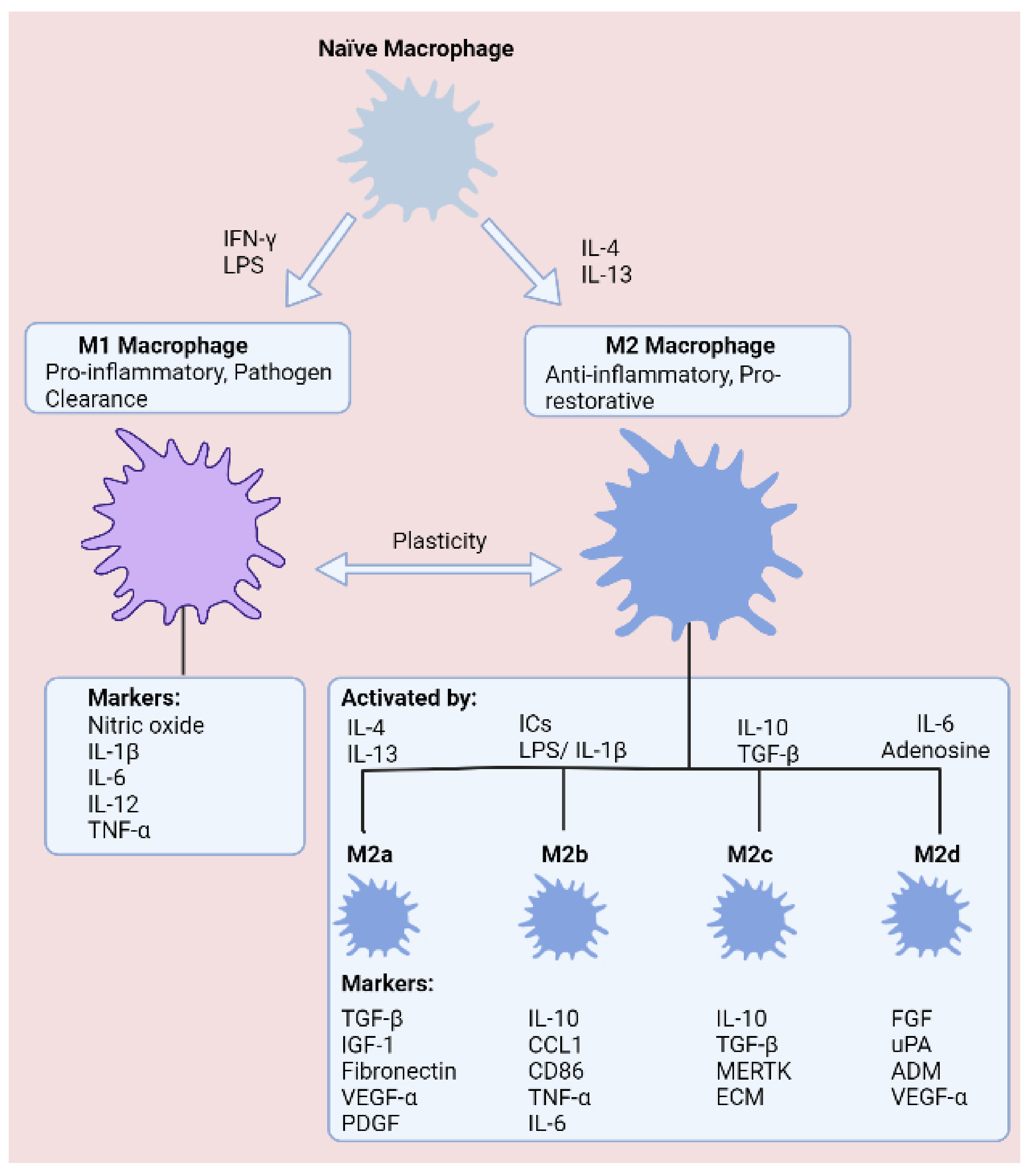
2.1. Macrophage Plasticity in Inflammatory Bowel Disease

Macrophages are multifaceted, highly dynamic cells that exhibit vast heterogeneity, reflecting both the tissue microenvironment that they occupy, along with the lineage that they are derived from [6]. In order to conduct specialised duties, macrophages exhibit a high degree of plasticity and display specific phenotypes, permitting the macrophage to adapt to the surrounding microenvironment. Intestinal-resident macrophages for instance, reside in the lamina propria, enabling the macrophage to sample extracellular surroundings and process antigens that they encounter. These surveillance duties facilitate a myriad of functions and defence mechanisms. Therefore, interactions of intestinal macrophages with microbes or foods could be viewed as critical events in shaping macrophage plasticity. For example, the tissue microenvironment could be expected to modulate cellular plasticity by virtue of a combination of a diverse cytokine milieu and environmental cues. This notion has been highlighted in murine studies, where pro-inflammatory macrophages that display high expression of Ly6C (LyC6hi) are reported to undergo in situ functional switches to pro-restorative LyC6low macrophages in response to the surrounding environment [13].
Macrophage plasticity exists as part of a spectrum, where M1 and M2 represent polar ends [11]. M1 macrophages exhibit several distinguishing features that help underpin a pro-inflammatory phenotype. First of all, pro-inflammatory M1 macrophages are induced through stimulation with lipopolysaccharide (LPS) and interferon gamma (IFN-γ), leading to secretion of tumour necrosis factor alpha (TNF-α), interleukin 1 beta (IL-1β), interleukin 6 (IL-6), and interleukin (IL-12) to encourage microbicidal activity and phagocytosis [3]. These robust immune responses are energy demanding, and in recent years, it has become evident that macrophages undergo metabolic reprogramming in response to infection, which dictates the ability of the macrophage to conduct rapid responses to microbial invasion [14].
Given the necessity for almost-instantaneous responses to prevent microbial dissemination within the body, M1 macrophages are understood to upregulate glycolysis during polarisation as a means of supporting vigorous immune responses and structural changes. In respect of recruiting immune cell populations and enhancing microbial killing, M1 macrophages accumulate succinate during pro-inflammatory conditions, which stabilises HIF-1α and promotes secretion of IL-1β [15]. At the other end of the spectrum, M2 macrophages are instrumental in suppressing immune responses, minimising tissue damage, and resolving tissue healing following injury. In an alternative manner to M1 macrophages, the anti-inflammatory programming of M2 macrophages is fuelled through oxidative phosphorylation (OXPHOS) and fatty acid oxidation (FAO). OXPHOS is preferred on account of the abundance of ATP molecules produced relative to glycolysis, providing additional energy for tissue resolution. Understandably, FAO is another crucial metabolic pathway, as FAO generates acetyl-coA, thus helping to ensure maximum ATP generation from the tricarboxylic acid (TCA) cycle and OXPHOS. M2 macrophages have been further branched into the M2a, M2b, M2c, and M2d subtypes, which are classified according to cytokine secretion, expression of cell surface markers, and transcriptome and biological activities (Figure 1).
Figure 1. States of macrophage polarisation. Macrophages are commonly defined as classically activated (LPS and IFN-γ) M1 macrophages or alternatively activated (IL-4 and IL-13) M2 macrophages, which comprise four subgroups (M2a–M2d). Macrophages exhibit high plasticity, enabling transitions between phenotypes according to the surrounding tissue microenvironment.

2.2. The cGAS-STING Signalling Pathway: An Emerging Regulator of Macrophage Plasticity

A driving force behind perpetuated inflammation and tissue injury in IBD involves an intricate link between the innate and adaptive immune systems—namely, recruitment of T lymphocytes to the site of injury through the release of chemokines by macrophages. Therefore, pathways involved in mediating these responses are of high importance to unravel. Activation of the cyclic GMP-AMP synthase (cGAS)—stimulator of interferon genes (STING) signalling pathway, which promotes macrophage cross-priming—is reported to be crucial for bridging this link [16].
cGAS is a cytosolic dsDNA sensor that detects intracellular DNA derived from pathogens or damaged host cells. Upon recognising dsDNA, cGAS generates the secondary messenger 2′3′-cGAMP from ATP and GTP, which in turn activates the endoplasmic reticulum-localised STING protein. These interactions initiate a downstream signalling cascade culminating in activation of transcription factors nuclear factor kappa B (NF-ĸB) and interferon regulatory factor 3 (IRF3), stimulating the release of interferon-beta (IFN-β) and pro-inflammatory cytokines. Activation of the cGAS-STING axis in macrophages is an important initial step in the recruitment and activation of T lymphocytes, setting in motion clonal expansion of these cell populations. Later, these T lymphocytes become embodied as indispensable arms of the adaptive immune system. Indeed, heightened activation of macrophage cGAS-STING signalling has previously been associated with a high-fat diet, demonstrating an intricate link between diet and systemic inflammation. Moreover, a functional role of the cGAS-STING signalling axis extends to intestinal epithelial cells, where the pathway mediates protective effects by promoting intestinal barrier integrity [17][18].

3. Dietary Approaches for Targeting Macrophage Plasticity

3.1. Short-Chain Fatty Acids

Co-evolution has given rise to the emergence of symbiotic relationships between the host and commensals. The microbiota in the gut are chief examples that acquire energy from the food that passes along the intestinal track of the host. Microbiota reciprocate to this gesture by promoting intestinal barrier homeostasis, dampening inflammation, strengthening the epithelial barrier, and preventing colonisation by pathogens [19]. A predominant mechanism of conducting protective duties is via production of SCFAs, which are fatty acids containing fewer than six carbon atoms, the most common in the mammalian gut being acetate (C2), propionate (C3), and butyrate (C4) [20]. Intriguingly, SCFAs ameliorate disease activity in IBD and promote healing of the colon [21][22].
In the mammalian gut, microbiota generate SCFAs through fermentation of indigestible fibres. Accordingly, several high-fibre foods have been reported to induce favourable outcomes for reducing risk of and disease state of IBD. Remarkably, fibre exhibits several clinical benefits in IBD, such as prolonging remission and reducing lesions in the intestinal mucosa [23]. Imbalances in consumption of fruit and vegetables, both abundant in fibre, were reported to be a risk factor for the emergence of IBD [24][25]. Oats are another rich source of dietary fibre and have been shown to prevent deterioration of gastrointestinal symptoms in UC [26]. Likewise, high-fibre legumes, such as navy and black beans, are capable of attenuating intestinal inflammation in murine models of IBD [27]. Taken together, it is evident that intake of high-fibre foods is advantageous for maintaining intestinal health. Indeed, interplay between fibre and microbiota, leading to generation of SCFAs, is a critical event in this process.
Following production by the microbiota, SCFAs are available to interact with macrophages stationed along the intestinal mucosa. Luminal SCFAs are taken up by the cell through various means, including passive diffusion and carrier-mediated transportation or otherwise through binding to G-protein coupled receptors (GPR41, GPR43, and GPR109a) [28]. Following uptake by the cell, SCFAs mediate immunomodulatory changes to immune cells through several mechanisms [29][30][31][32]. The most studied SCFA—butyrate—exerts anti-inflammatory effects in IBD through inhibition of NF-ĸB activation, suppressing release of pro-inflammatory cytokines (Figure 2) [33]. In accordance, acetate and propionate exhibit comparable inhibitory properties with butyrate at suppressing NF-κB activation [34]. Indeed, while SCFAs also activate GPCR signalling, immunomodulatory effects are mediated independently of these receptors [29].
Figure 2. Mechanisms that dietary metabolites modulate cells of the gastrointestinal tract. Fibre and tryptophan are metabolised by the microbiota to release short-chain fatty acids and indole compounds into the gut lumen. (1). Short-chain fatty acids enhance PPAR-γ activation and reduce IFN-γ production of intestinal epithelial cells, while inducing immunomodulatory effects on intestinal macrophages. (2). Indole suppresses activation of NF-kB and stimulates production of IL-22 by group 3 innate lymphoid cells, promoting epithelial barrier homeostasis. (3) Omega-3 polyunsaturated fats promote an anti-inflammatory macrophage phenotype through activation of PPAR-α and PPAR-γ, along with inhibition of NF-κB, culminating as decreased production of pro-inflammatory cytokines.
Anti-inflammatory effects mediated by butyrate have encouraged various laboratories to delineate molecular interactions underpinning these effects. Traditionally, inhibition of histone deacetylases (HDAC) has been believed to underlie the anti-inflammatory signature of butyrate [35]. Investigations by Chang et al., for example, determined that butyrate acts as an inhibitor of HDAC, which is an enzyme that regulates gene transcription by removing acetyl groups from histones. Butyrate downregulated NO, IL-6, and IL-12 in intestinal macrophages, likely through enhanced histone acetylation [29]. These findings build on prior data showing that butyrate attenuates LPS-induced secretion of TNF-α, IL-1β, IL-6, and NO, whilst prompting the release of anti-inflammatory IL-10 in RAW 264.7 monocytes [32]. On the basis that butyrate is an HDAC inhibitor, studies have speculated that the SCFA induces epigenetic reprogramming [30]. In line with this concept, bone marrow-derived macrophage (BMDM) polarisation toward an M2 phenotype was induced by butyrate through acetylation of H3K9, which showed to be a histone modification that enhances STAT6 activation [36]. The question remains whether butyrate can modulate macrophage plasticity. Production of ROS, which are key regulators of an M1 phenotype, can be attenuated in neutrophils following butyrate treatment [37][38]. Therefore, it is plausible that butyrate exerts similar effects on macrophages. Collectively, these observations represent a molecular basis to explain why a high-fibre diet has a protective effect against intestinal disorders [23].

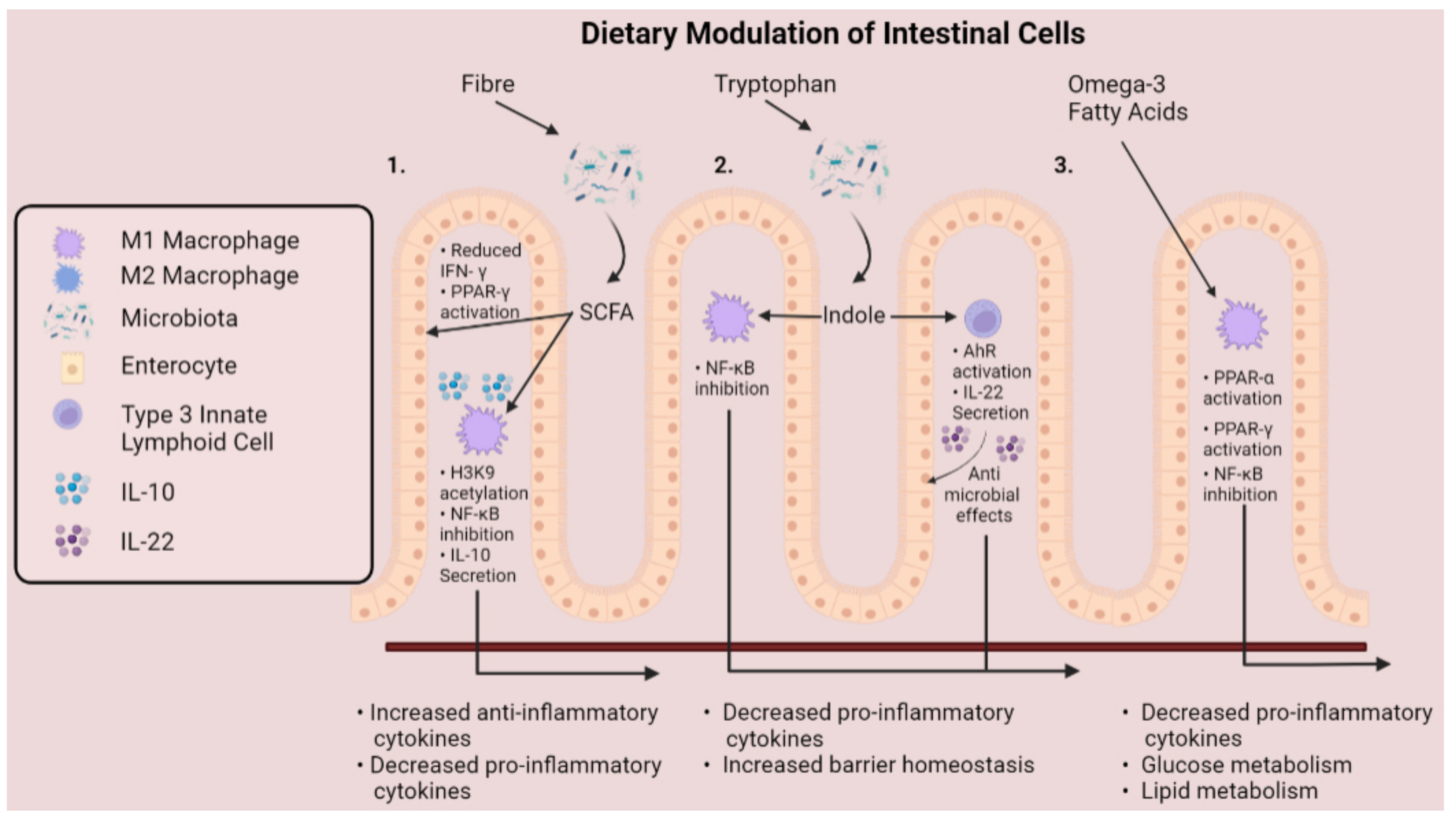
3.2. Tryptophan-Derived Metabolites

Tryptophan is an essential amino acid (humans cannot produce tryptophan endogenously), meaning instead that tryptophan must be supplemented in the diet. Many foods are good sources of tryptophan, including oats, milk, cheese, tuna fish, chicken, and turkey [39]. Amongst these tryptophan-rich foods, oats, lean meats, milk, and cheese are all associated with ameliorated intestinal inflammation in IBD [40].
Indeed, consumption of tryptophan in the diet is associated with intestinal homeostasis. Serum tryptophan levels, for example, are lower in individuals with IBD, denoting that tryptophan deficiency or degradation may exacerbate gut dysbiosis [41]. This notion extends to animal models of colitis, such as in mice, where mice deficient in tryptophan were recorded to display exacerbated colitis [42] (p. 2). From a medical standpoint, it is apparent that dietary tryptophan supplementation could represent a means of attenuating intestinal inflammation. This is supported by animal models, where dietary supplementation of tryptophan reduced the severity of DSS-induced colitis in pigs [43].
Again, intestinal microbiota play a fundamental role in the beneficial outcomes of dietary tryptophan. In the body, tryptophan is subject to biosynthetic manipulation by the host and microbes to generate various metabolites [44]. Specifically, ingested tryptophan is metabolised in the intestines by three distinct pathways; the first two of these pathways—the serotonin and kynurenine pathways—are conducted by host enzymes, whilst the third pathway, which produces the immunomodulatory indole compounds, is performed by microbiota [45].
A broad-spectrum of indole compounds is generated by the intestinal flora—the structure of each metabolite differing according to the biochemical transformations they are subjected to. Clostridium sporogenes, from the phylum Firmicutes for example, generates indole-propionic acid from tryptophan through induction of the enzyme phenyllactate dehydratase [46]. Moreover, Lactobacilli reuteri generates indole-3-aldehyde from tryptophan—a reaction catalysed by an aromatic amino acid aminotransferase [47]. Alternatively, indole may also be sourced directly from the diet from produce such as broccoli, which similarly has been shown to attenuate murine colitis [48].
Encouragingly, indole compounds have also been reported to promote mucosal homeostasis in the intestines [49]. The mechanistic basis for indole alleviating intestinal inflammation can be partly attributed to interactions with the Aryl hydrocarbon receptor (AHR), which various indole compounds display affinity for [50]. The AHR is a transcription factor that upon activation is responsible for several anti-inflammatory mechanisms, including regulating intestinal homeostasis. In macrophages, AHR dampens LPS-induced inflammation—as mice lacking macrophage-specific expression of AHR are more susceptible to LPS-induced septic shock [51]. Given that macrophages propel inflammatory signalling in IBD, whether interactions between indole and macrophages can modulate cellular plasticity is an avenue of interest.

References

  1. Alatab, S.; Sepanlou, S.G.; Ikuta, K.; Vahedi, H.; Bisignano, C.; Safiri, S.; Sadeghi, A.; Nixon, M.R.; Abdoli, A.; Abolhassani, H.; et al. The global, regional, and national burden of inflammatory bowel disease in 195 countries and territories, 1990–2017: A systematic analysis for the Global Burden of Disease Study 2017. Lancet Gastroenterol. Hepatol. 2020, 5, 17–30.
  2. Viola, M.F.; Boeckxstaens, G. Niche-specific functional heterogeneity of intestinal resident macrophages. Gut 2021, 70, 1383–1395.
  3. Verdeguer, F.; Aouadi, M. Macrophage heterogeneity and energy metabolism. Exp. Cell Res. 2017, 360, 35–40.
  4. Yoshida, N.; Frickel, E.-M.; Mostowy, S. Macrophage–microbe interactions: Lessons from the zebrafish model. Front. Immunol. 2017, 8, 1703.
  5. Mahida, Y.R. The key role of macrophages in the immunopathogenesis of inflammatory bowel disease. Inflamm. Bowel Dis. 2000, 6, 21–33.
  6. Italiani, P.; Boraschi, D. Development and functional differentiation of tissue-resident versus monocyte-derived macrophages in inflammatory reactions. In Macrophages; Springer: Berlin/Heidelberg, Germany, 2017; pp. 23–43.
  7. Honold, L.; Nahrendorf, M. Resident and monocyte-derived macrophages in cardiovascular disease. Circ. Res. 2018, 122, 113–127.
  8. Gomez Perdiguero, E.; Klapproth, K.; Schulz, C.; Busch, K.; Azzoni, E.; Crozet, L.; Garner, H.; Trouillet, C.; De Bruijn, M.F.; Geissmann, F.; et al. Tissue-resident macrophages originate from yolk-sac-derived erythro-myeloid progenitors. Nature 2015, 518, 547–551.
  9. Yona, S.; Kim, K.W.; Wolf, Y.; Mildner, A.; Varol, D.; Breker, M.; Strauss-Ayali, D.; Viukov, S.; Guilliams, M.; Misharin, A.; et al. Fate mapping reveals origins and dynamics of monocytes and tissue macrophages under homeostasis. Immunity 2013, 38, 79–91.
  10. Bain, C.C.; Scott, C.L.; Uronen-Hansson, H.; Gudjonsson, S.; Jansson, O.; Grip, O.; Guilliams, M.; Malissen, B.; Agace, W.W.; Mowat, A. Resident and pro-inflammatory macrophages in the colon represent alternative context-dependent fates of the same Ly6C hi monocyte precursors. Mucosal Immunol. 2013, 6, 498–510.
  11. Das, A.; Sinha, M.; Datta, S.; Abas, M.; Chaffee, S.; Sen, C.K.; Roy, S. Monocyte and macrophage plasticity in tissue repair and regeneration. Am. J. Pathol. 2015, 185, 2596–2606.
  12. Shaw, T.N.; Houston, S.A.; Wemyss, K.; Bridgeman, H.M.; Barbera, T.A.; Zangerle-Murray, T.; Strangward, P.; Ridley, A.J.; Wang, P.; Tamoutounour, S.; et al. Tissue-resident macrophages in the intestine are long lived and defined by Tim-4 and CD4 expression. J. Exp. Med. 2018, 215, 1507–1518.
  13. Ramachandran, P.; Pellicoro, A.; Vernon, M.A.; Boulter, L.; Aucott, R.L.; Ali, A.; Hartland, S.N.; Snowdon, V.K.; Cappon, A.; Gordon-Walker, T.T.; et al. Differential Ly-6C expression identifies the recruited macrophage phenotype, which orchestrates the regression of murine liver fibrosis. Proc. Natl. Acad. Sci. USA 2012, 109, E3186–E3195.
  14. Galván-Peña, S.; O’Neill, L.A. Metabolic reprograming in macrophage polarization. Front. Immunol. 2014, 5, 420.
  15. Tannahill, G.M.; Curtis, A.M.; Adamik, J.; Palsson-McDermott, E.M.; McGettrick, A.F.; Goel, G.; Frezza, C.; Bernard, N.J.; Kelly, B.; Foley, N.H.; et al. Succinate is an inflammatory signal that induces IL-1β through HIF-1α. Nature 2013, 96, 238–242.
  16. Ou, L.; Zhang, A.; Cheng, Y.; Chen, Y. The cGAS-STING pathway: A promising immunotherapy target. Front. Immunol. 2021, 12, 795048.
  17. Fischer, J.C.; Bscheider, M.; Eisenkolb, G.; Lin, C.C.; Wintges, A.; Otten, V.; Lindemans, C.A.; Heidegger, S.; Rudelius, M.; Monette, S.; et al. RIG-I/MAVS and STING signaling promote gut integrity during irradiation-and immune-mediated tissue injury. Sci. Transl. Med. 2017, 9, eaag2513.
  18. Hu, S.; Fang, Y.; Chen, X.; Cheng, T.; Zhao, M.; Du, M.; Li, T.; Li, M.; Zeng, Z.; Wei, Y.; et al. cGAS restricts colon cancer development by protecting intestinal barrier integrity. Proc. Natl. Acad. Sci. USA 2021, 118, e2105747118.
  19. Hiippala, K.; Jouhten, H.; Ronkainen, A.; Hartikainen, A.; Kainulainen, V.; Jalanka, J.; Satokari, R. The potential of gut commensals in reinforcing intestinal barrier function and alleviating inflammation. Nutrients 2018, 10, 988.
  20. Ríos-Covián, D.; Ruas-Madiedo, P.; Margolles, A.; Gueimonde, M.; de los Reyes-gavilán, C.G.; Salazar, N. Intestinal short chain fatty acids and their link with diet and human health. Front. Microbiol. 2016, 7, 185.
  21. Lührs, H.; Gerke, T.; Müller, J.G.; Melcher, R.; Schauber, J.; Boxberger, F.; Scheppach, W.; Menzel, T. Butyrate inhibits NF-κB activation in lamina propria macrophages of patients with ulcerative colitis. Scand. J. Gastroenterol. 2002, 37, 458–466.
  22. Van der Beek, C.M.; Dejong, C.H.; Troost, F.J.; Masclee, A.A.; Lenaerts, K. Role of short-chain fatty acids in colonic inflammation, carcinogenesis, and mucosal protection and healing. Nutr. Rev. 2017, 5, 286–305.
  23. Pituch-Zdanowska, A.; Banaszkiewicz, A.; Albrecht, P. The role of dietary fibre in inflammatory bowel disease. Przeglad Gastroenterologiczny 2015, 10, 135.
  24. Gilat, T.; Hacohen, D.; Lilos, P.; Langman, M. Childhood factors in ulcerative colitis and Crohn’s disease: An international cooperative study. Scand. J. Gastroenterol. 1987, 22, 1009–1024.
  25. Amre, D.K.; D’souza, S.; Morgan, K.; Seidman, G.; Lambrette, P.; Grimard, G.; Israel, D.; Mack, D.; Ghadirian, P.; Deslandres, C.; et al. Imbalances in dietary consumption of fatty acids, vegetables, and fruits are associated with risk for Crohn’s disease in children. Off. J. Am. Coll. Gastroenterol. ACG 2007, 102, 2016–2025.
  26. Nyman, M.; Nguyen, T.D.; Wikman, O.; Hjortswang, H.; Hallert, C. Oat bran increased fecal butyrate and prevented gastrointestinal symptoms in patients with quiescent ulcerative colitis—Randomized controlled trial. Crohns Colitis 2020, 2, otaa005.
  27. Zhang, C.; Monk, J.M.; Lu, J.T.; Zarepoor, L.; Wu, W.; Liu, R.; Pauls, K.P.; Wood, G.A.; Robinson, L.; Tsao, R.; et al. Cooked navy and black bean diets improve biomarkers of colon health and reduce inflammation during colitis. Br. J. Nutr. 2014, 111, 1549–1563.
  28. Sun, M.; Wu, W.; Liu, Z.; Cong, Y. Microbiota metabolite short chain fatty acids, GPCR, and inflammatory bowel diseases. J. Gastroenterol. 2017, 52, 1–8.
  29. Chang, P.V.; Hao, L.; Offermanns, S.; Medzhitov, R. The microbial metabolite butyrate regulates intestinal macrophage function via histone deacetylase inhibition. Proc. Natl. Acad. Sci. USA 2014, 111, 2247–2252.
  30. Flemming, A. Butyrate boosts microbicidal macrophages. Nat. Rev. Immunol. 2019, 19, 135.
  31. Ji, J.; Shu, D.; Zheng, M.; Wang, J.; Luo, C.; Wang, Y.; Guo, F.; Zou, X.; Lv, X.; Li, Y.; et al. Microbial metabolite butyrate facilitates M2 macrophage polarization and function. Sci. Rep. 2016, 6, 24838.
  32. Liu, T.; Li, J.; Liu, Y.; Xiao, N.; Suo, H.; Xie, K.; Yang, C.; Wu, C. Short-chain fatty acids suppress lipopolysaccharide-induced production of nitric oxide and proinflammatory cytokines through inhibition of NF-κB pathway in RAW264. 7 cells. Inflammation 2012, 35, 1676–1684.
  33. Segain, J.P.; De La Blétiere, D.R.; Bourreille, A.; Leray, V.; Gervois, N.; Rosales, C.; Ferrier, L.; Bonnet, C.; Blottiere, H.M.; Galmiche, J.P. Butyrate inhibits inflammatory responses through NFκB inhibition: Implications for Crohn’s disease. Gut 2000, 47, 397–403.
  34. Tedelind, S.; Westberg, F.; Kjerrulf, M.; Vidal, A. Anti-inflammatory properties of the short-chain fatty acids acetate and propionate: A study with relevance to inflammatory bowel disease. World J. Gastroenterol. WJG 2007, 13, 2826.
  35. Candido, E.P.M.; Reeves, R.; Davie, J.R. Sodium butyrate inhibits histone deacetylation in cultured cells. Cell 1978, 14, 105–113.
  36. Gujral, P.; Mahajan, V.; Lissaman, A.C.; Ponnampalam, A.P. Histone acetylation and the role of histone deacetylases in normal cyclic endometrium. Reprod. Biol. Endocrinol. 2020, 18, 84.
  37. Li, G.; Lin, J.; Zhang, C.; Gao, H.; Lu, H.; Gao, X.; Zhu, R.; Li, Z.; Li, M.; Liu, Z. Microbiota metabolite butyrate constrains neutrophil functions and ameliorates mucosal inflammation in inflammatory bowel disease. Gut Microbes 2021, 3, 1968257.
  38. Rendra, E.; Riabov, V.; Mossel, D.M.; Sevastyanova, T.; Harmsen, M.C.; Kzhyshkowska, J. Reactive oxygen species (ROS) in macrophage activation and function in diabetes. Immunobiology 2019, 224, 242–253.
  39. Richard, D.M.; Dawes, M.A.; Mathias, C.W.; Acheson, A.; Hill-Kapturczak, N.; Dougherty, D.M. L-tryptophan: Basic metabolic functions, behavioral research and therapeutic indications. Int. J. Tryptophan Res. 2009, 2, IJTR-S2129.
  40. Campmans-Kuijpers, M.J.; Dijkstra, G. Food and food groups in inflammatory bowel disease (Ibd): The design of the groningen anti-inflammatory diet (graid). Nutrients 2021, 13, 1067.
  41. Nikolaus, S.; Schulte, B.; Al-Massad, N.; Thieme, F.; Schulte, D.M.; Bethge, J.; Rehman, A.; Tran, F.; Aden, K.; Häsler, R.; et al. Increased tryptophan metabolism is associated with activity of inflammatory bowel diseases. Gastroenterology 2017, 153, 1504–1516.
  42. Hashimoto, T.; Perlot, T.; Rehman, A.; Trichereau, J.; Ishiguro, H.; Paolino, M.; Sigl, V.; Hanada, T.; Hanada, R.; Lipinski, S.; et al. ACE2 links amino acid malnutrition to microbial ecology and intestinal inflammation. Nature 2012, 487, 477–481.
  43. Kim, C.J.; Kovacs-Nolan, J.A.; Yang, C.; Archbold, T.; Fan, M.Z.; Mine, Y. l-Tryptophan exhibits therapeutic function in a porcine model of dextran sodium sulfate (DSS)-induced colitis. J. Nutr. Biochem. 2010, 21, 468–475.
  44. Alkhalaf, L.M.; Ryan, K.S. Biosynthetic manipulation of tryptophan in bacteria: Pathways and mechanisms. Chem. Biol. 2015, 22, 317–328.
  45. Agus, A.; Planchais, J.; Sokol, H. Gut microbiota regulation of tryptophan metabolism in health and disease. Cell Host Microbe 2018, 23, 716–724.
  46. Dodd, D.; Spitzer, M.H.; Van Treuren, W.; Merrill, B.D.; Hryckowian, A.J.; Higginbottom, S.K.; Le, A.; Cowan, T.M.; Nolan, G.P.; Fischbach, M.A.; et al. A gut bacterial pathway metabolizes aromatic amino acids into nine circulating metabolites. Nature 2017, 51, 648–652.
  47. Zelante, T.; Iannitti, R.G.; Cunha, C.; De Luca, A.; Giovannini, G.; Pieraccini, G.; Zecchi, R.; D’Angelo, C.; Massi-Benedetti, C.; Fallarino, F.; et al. Tryptophan catabolites from microbiota engage aryl hydrocarbon receptor and balance mucosal reactivity via interleukin-22. Immunity 2013, 39, 372–385.
  48. Hubbard, T.D.; Murray, I.A.; Nichols, R.G.; Cassel, K.; Podolsky, M.; Kuzu, G.; Tian, Y.; Smith, P.; Kennett, M.J.; Patterson, A.D.; et al. Dietary broccoli impacts microbial community structure and attenuates chemically induced colitis in mice in an Ah receptor dependent manner. J. Funct. Foods 2017, 37, 685–698.
  49. Shimada, Y.; Kinoshita, M.; Harada, K.; Mizutani, M.; Masahata, K.; Kayama, H.; Takeda, K. Commensal bacteria-dependent indole production enhances epithelial barrier function in the colon. PLoS ONE 2013, 8, e80604.
  50. Miller, C.A. Expression of the human aryl hydrocarbon receptor complex in yeast: Activation of transcription by indole compounds. J. Biol. Chem. 1997, 272, 32824–32829.
  51. Sekine, H.; Mimura, J.; Oshima, M.; Okawa, H.; Kanno, J.; Igarashi, K.; Gonzalez, F.J.; Ikuta, T.; Kawajiri, K.; Fujii-Kuriyama, Y. Hypersensitivity of aryl hydrocarbon receptor-deficient mice to lipopolysaccharide-induced septic shock. Mol. Cell. Biol. 2009, 29, 6391–6400.
More
Information
Contributors MDPI registered users' name will be linked to their SciProfiles pages. To register with us, please refer to https://encyclopedia.pub/register : , ,
View Times: 358
Revisions: 2 times (View History)
Update Date: 27 May 2022
1000/1000
Video Production Service