Submitted Successfully!
To reward your contribution, here is a gift for you: A free trial for our video production service.
Thank you for your contribution! You can also upload a video entry or images related to this topic.
Version Summary Created by Modification Content Size Created at Operation
1 -- 4099 2022-05-18 23:14:15 |
2 format corrected. + 1 word(s) 4100 2022-05-19 08:02:59 |

Video Upload Options

Do you have a full video?

Confirm

Are you sure to Delete?
Cite
If you have any further questions, please contact Encyclopedia Editorial Office.
Fathima, S.; Shanmugasundaram, R.; , .; Selvaraj, R. Gastrointestinal Microbiota for Growth and Performance in Chickens. Encyclopedia. Available online: https://encyclopedia.pub/entry/23093 (accessed on 25 April 2024).
Fathima S, Shanmugasundaram R,  , Selvaraj R. Gastrointestinal Microbiota for Growth and Performance in Chickens. Encyclopedia. Available at: https://encyclopedia.pub/entry/23093. Accessed April 25, 2024.
Fathima, Shahna, Revathi Shanmugasundaram,  , Ramesh Selvaraj. "Gastrointestinal Microbiota for Growth and Performance in Chickens" Encyclopedia, https://encyclopedia.pub/entry/23093 (accessed April 25, 2024).
Fathima, S., Shanmugasundaram, R., , ., & Selvaraj, R. (2022, May 18). Gastrointestinal Microbiota for Growth and Performance in Chickens. In Encyclopedia. https://encyclopedia.pub/entry/23093
Fathima, Shahna, et al. "Gastrointestinal Microbiota for Growth and Performance in Chickens." Encyclopedia. Web. 18 May, 2022.
Gastrointestinal Microbiota for Growth and Performance in Chickens
Edit

The gut barrier is comprised of intestinal microbiota and their metabolites, mucins secreted by goblet cells, host-derived antimicrobial peptides such as defensins, and cathelicidins, IgA, intestinal epithelium, microfold cells (M cells), Paneth cells, tuft cells and lymphoid tissues in the sub-epithelium and lamina propria. The gut barrier serves to contain the gut microbiota within the lumen while permitting the absorption of nutrients. Intestinal health, tolerance to food and microbial antigens, and homeostasis are achieved through complex interactions between the multiple components in the gut.

probiotic prebiotic synbiotic poultry microbiota

1. Introduction

The gut barrier is comprised of intestinal microbiota and their metabolites, mucins secreted by goblet cells, host-derived antimicrobial peptides such as defensins, and cathelicidins, IgA, intestinal epithelium, microfold cells (M cells), Paneth cells, tuft cells and lymphoid tissues in the sub-epithelium and lamina propria [1][2]. The gut barrier serves to contain the gut microbiota within the lumen while permitting the absorption of nutrients [1][3]. Intestinal health, tolerance to food and microbial antigens, and homeostasis are achieved through complex interactions between the multiple components in the gut [2].

2. Probiotics in Broiler Production

With the ban on the use of antibiotics by the European Union (EU) and the limited use of antibiotics in the United States (US) in chicken production, probiotics are emerging as a potential alternative. Probiotics or direct-fed microbials (DFM) are defined as “live microorganisms which when administered in adequate amounts confer a health benefit on the host”, by improving the gastrointestinal microbial balance and consequent enhancement in nutrient absorption, growth rate, feed efficiency, and immune competence against potential pathogens [4][5][6]. An ideal probiotic should be of host origin, have demonstrated a beneficial effect on the host, should be able to alter the gut microflora advantageous to the host, should be resistant to gastric acid and bile salts, should adhere to the gut mucosal epithelium, and should exhibit beneficial effects. Probiotics should also be non-pathogenic, free of adverse side effects, sensitive to antibiotics, exhibit antimicrobial properties against common pathogens, modulate intestinal functions, and immune responses of the host [7][8]. The probiotic should also be able to withstand the processing and storage conditions [9].
Ideally, probiotics should be host-derived because 1. The microbes have evolved and adapted to the ecology of the host gastrointestinal tract and hence, can readily proliferate and establish a stable population 2. Express beneficial effects better than probiotic strains obtained from other species [10]. It is therefore essential to develop host-specific probiotics for achieving optimal health and production. Probiotic bacteria should be typically isolated from the gut of healthy animals. Multiple species and strains of gut microbes must be evaluated for their probiotic potential and the one conferring maximum benefit to the host must be selected [11]. For example, only 6 out of 57 lactic acid bacteria (LAB) strains, isolated from the crop and intestine of healthy broilers, possess probiotic properties that included the ability to adhere to ileal epithelial cells, hydrophobicity, tolerance and survivability in gastric juice, bile salts, and phenol, susceptibility to antibiotics, ability to auto-aggregate and co-aggregate [12].
Poultry is a major source of foodborne pathogens of zoonotic significance [13] and foodborne pathogen infection cause economic loss to the poultry industry. Probiotics possess antimicrobial activity and can be applied to decrease the incidence of foodborne pathogens in poultry [14]. Probiotics secrete bacteriocins, hydrogen peroxide, alcohol, and organic acids and exhibit broad spectrum antimicrobial activity [15]. In a study conducted to evaluate the probiotic efficiency of LAB isolated from poultry, it was demonstrated that 77 strains out of the 296 strains screened inhibited the proliferation of E. coli and Salmonella enteritidis. Of the 77 strains, eight strains of their greater spectrum of pathogen antagonism and other desirable properties of probiotics. The strain with maximum probiotic potential was identified as L. salivarius [16].
The probiotic strains should be able to survive the acidic pH in the proventriculus and gizzard for the strains to successfully colonize the intestine [17]. The transit time of feed through the proventriculus and gizzard is approximately 2 h. L. acidophilus survives at pH 4 for 2 h [18]. pH tolerance is strain specific. For example, L. plantarum VJI21 exhibit 80% survivability whereas the isolate VJC1 exhibit only 64% survivability at low pH [19].
Potential probiotic strains should be able to withstand bile salts in the duodenum and cecum [20]. Probiotics of host origin will be able to tolerate the bile salt concentration of the host from which the probiotic strain was isolated. L. acidophilus can survive for 15 h in 2% bile. Lactobacillus strains possess bile salt hydrolase activity and therefore neutralize the antimicrobial activity of bile salts to survive in the gut [19].
The ability of probiotics to adhere to the intestinal epithelial cells and mucosal surfaces is essential for forming biofilms, which prevents the detachment of bacteria during gut contraction and peristaltic movement. This property of biofilm formation is termed auto-aggregation [21]. Co-aggregation enables the probiotic species to displace pathogenic bacteria [20]. Hydrophobicity enables probiotics to adhere to the gut epithelium [16]. Auto-aggregation, co-aggregation, and microbial hydrophobicity enables the probiotics to competitively exclude pathogenic species from the host gut [19][21][22]. L. acidophilus decreases the adhesion of E. coli and S. typhimurium to Caco-2 cells [18].
Antibiotic susceptibility of potential probiotics should be evaluated to ensure no antibiotic resistant genes are transferred from the probiotic species to the gut microbiota [19]. In a study evaluating antibiotic susceptibility of Lactobacillus strains isolated from chicken, 90% of the 88 isolates were resistant to tiamulin, 74% to tetracyclines, and 70% to lincomycin. Multidrug resistance was detected in 79.5% of isolates [23]. These results indicate that caution should be applied before choosing a particular probiotic species as the probiotic species might carry antibiotic resistant genes.
Probiotic organisms should be consumed at adequate doses to confer a desired health benefit. A minimum viable cell concentration of 1 × 10⁶ CFU g−1 is essential for successful colonization of the gut by probiotics [24]. Probiotic supplementation at 1 × 10⁸ to 1 × 10⁹ CFU g−1 is effective and can increase the number of beneficial bacteria [9][25]. In a study conducted in broiler chickens by Mountzouris et al., 2010 [25], it was demonstrated that there is a significant increase in the concentration of Lactobacillus and Bifidobacterium and a decrease in coliform counts in the caeca of birds fed probiotics. Microbiological factors such as the strain of the probiotic, water activity, pH, presence of salt, and molecular oxygen determine the survivability of probiotics during storage. Feed processing parameters such as heat treatment, cooling rate, storage length, and packaging materials also determine the survivability of probiotics [26][27][28]. The survivability of probiotic bacteria during the processing and storage of the product is dependent on the species and strain of probiotics [29]. Thermotolerance of Lactobacilli can be increased by subjecting the bacterium to heat shock at sublethal temperatures, which is 10 °C above the optimum temperature for growth, before exposure to lethal temperatures [30]. Bacillus spores can survive at high temperatures and are stable during feed processing steps such as pelleting [24].
Probiotics can be a single strain or a combination of different microorganisms such as bacteria of the species Lactobacillus, Bifidobacterium, Bacteroides, Bacillus, or Enterococcus. Saccharomyces spp., a yeast, is used as a probiotic either alone or in combination with other bacterial strains [31]. Probiotic preparations can also contain multiple strains of the same species such as Lactobacillus casei, L. acidophilus, L. bulgaricus, L. reuteri, L. salivarius, and L. animalis [9][32][33]. Probiotics can also be categorized as ‘colonizing’ species which consists of Lactobacillus, Streptococcus, and Enterococcus spp., and ‘non-colonizing’ species which consists of Bacillus spp. spores and the yeast Saccharomyces cerevisiae [34]. Bacteria of the colonizing species compete for potential binding sites on the intestinal epithelium or mucosa while non-colonizing species though viable in the intestinal content, will not colonize the intestinal epithelium [35]. Colonizing species resist the growth of pathogens by competitive exclusion. In contrast to the requirement for continuous administration of non-colonizing species, colonizing species requires to be administered once, though a supplementary dosage at a later age can be beneficial [36]. Gut microbiota is also classified as luminal and mucosal associated microbiota. The expression of adhesion molecules on the intestinal epithelial cells, rate of mucus production, and secretory immunoglobulins determine the composition of mucosal microbiota [37]. The composition of luminal microbiota is influenced by the presence of antimicrobial substances, feed passage rate, nutrient availability, and the interaction between the microbiota and the host immune system [13][29].
Among the different probiotics that are available, three major microorganisms, namely Lactic acid bacteria, Bifidobacteria, and Saccharomyces, dominate the probiotic market [38]. Other probiotics that are available for the poultry industry include Enterococcus, Pediococcus [39], Bacillus, E. coli, Aspergillus, Candida, and several other microbial species [40]. Although the probiotics industry is rapidly growing with several commercially available probiotics flooding the market, at any given time it is customary to supplement birds with a combination of a limited number of four or five microbial species [41]. It is not clear how the supplemented four to five different strains can contribute to the bacterial diversity in healthy chickens or even restore diversity in antibiotic treated birds. In humans treated with antibiotics, gut mucosal microbiome reconstitution is impaired by probiotics suggesting that probiotics might prevent the original gut population from recovering to the pre-antibiotic phase [42].
One alternative to applying limited numbers of probiotic species would be to transfer gut microbiota from healthy adult chickens to young chicks. The poultry industry will benefit from transferring mixed cultures of gut microbiota from healthy chickens rather than supplementing a limited number of probiotic species. Such an attempt has been extensively applied to control Clostridium difficile infection in humans. Fecal microbiota transplantation is the transfer of fecal matter from healthy donors into the gut of recipients to alter the gut microbiome of the recipient [43]. Fecal microbiota transplantation increases gut microbiota diversity and restores the gut microbiome post-antibiotic therapy. Post-transplantation, the fecal microbiota of fecal transplant recipients was more diverse and similar to that of the healthy donor [44]. Such an approach was attempted in early 1973, wherein transferring an undefined mixed culture from healthy chickens, but not a single Lactobacillus probiotic species, successfully inhibited Salmonella colonization in chicks [45]. This procedure was termed the Nurmi competitive exclusion concept [40] and has since been applied to control salmonellosis and campylobacteriosis in poultry [46].
It is necessary to understand the symbiotic relationship between the host and the microbiota. A nutrient-rich ambient environment of the host facilitates the colonization and proliferation of microbiota while the microbiota stimulates the digestive and immune systems of the host to improve the production performance [8][47]. In a meta-analysis study conducted by Blajman et al. (2014) [48], it was identified that probiotic supplementation improves BWG by 661 g and improves FCR with 281 g less feed consumed/kg of weight gain. The meta-analysis study also concluded that administration of probiotics in drinking water is more efficacious compared to supplementation in feed. The major limitations impeding the use of probiotics are the inconsistent effects and the incomplete understanding of the mode of action of probiotics.

3. Routes of Administration

Probiotics should be administered as early in life as possible to achieve desired beneficial effects [49]. Supplementation in feed or water on the day of hatch is the most common and convenient method to apply probiotics in poultry production [5]. Supplementation in feed or water is dependent on the individual consumption and thus the dose of probiotic consumed varies depending on the quantity of feed or water consumed. Supplementing probiotics in feed and water provide a mean for continuous supplementation of the probiotics to the bird, though the presence of antimicrobials in food or water can affect the viability of the probiotic bacteria [50][51].
Several studies have reported higher efficiency of probiotics when supplemented in drinking water [48][52]. This can be due to the shorter transit time of water, compared to the feed, in the upper gastrointestinal tract. A shorter transit time will reduce the duration for which probiotics are exposed to lower pH and bile salts in the upper gut. Water dilutes the acid, enzymes, and other digestive secretions and thereby reduces their negative effects on probiotics [51]. Bacteria require a period of acclimatization to the new environment. This period of time, termed the lag phase, will be longer when the conditions are less optimal. Supplementing probiotics in water mimics the reconstitution of lyophilized products and reduces the lag phase, thereby enabling the bacteria to adapt better and proliferate in the gut [51][53][54].
Probiotics can be injected into the incubating egg to achieve early colonization of the beneficial bacteria [55]. Delivery of probiotics to chicken embryos in ovo establishes a healthy gut microbiome [56][57]. In ovo probiotic inoculation can be applied either as in ovo stimulation [58] or in ovo feeding [59] which are inoculation on day 12 and day 17–18 of egg incubation respectively. In in ovo stimulation, prebiotic species are deposited on the air cell on day 12 to stimulate the development of innate gut microflora, which is ingested when the chicks start piping [60]. In in ovo feeding, probiotic species are injected into the amnion or embryo [61][62][63]. In a study conducted by Oliveira et al. [55], it was demonstrated that in ovo inoculation of probiotics significantly reduced mortality when challenged with Salmonella on day-4 post-hatch compared to the control group. In ovo inoculation of probiotics facilitates early colonization of beneficial bacteria and competitively excludes Salmonella. However, in ovo injection of one or a mixture containing only a few beneficial bacteria will likely not be effective in establishing a diverse intestinal microbiome comparable to that of a healthy adult chicken. On the other hand, inoculation of a mixed culture can introduce unknown species of bacteria that could be detrimental to the embryonated egg [64].
Spraying is another method of probiotic administration wherein the undiluted culture is sprayed on chicks when they are 50–70% hatched [65]. Cultures can also be sprayed in the environment or the litter material of the bird. Spraying eliminates the water quality concerns and other variables associated with probiotic administration in feed or water. Spraying is a cost-effective and suitable for mass applications [66][67]. Probiotics can also be delivered by oral gavage, but the high labor requirement makes the route of delivery unfeasible [62].

4. Factors to Be Considered during Probiotics Supplementation

The survivability of the probiotic organisms in the upper gastrointestinal tract is of paramount importance for the probiotic species to colonize the gut. Microencapsulation preserves the viability of probiotics in a feed matrix. However, in vitro tests with simulated gastric juices are typically applied to study the efficiency of microencapsulation in maintaining the viability of probiotics [68]. The high complexity of the gastrointestinal tract of live animals with variable pH, gastric and intestinal juices, peristalsis, and presence of gut microbiota makes in vitro studies to be of low predictive value for in vivo success. L. casei failed to colonize mice’s gut when supplemented with sodium caseinate capsules [69]. More studies have to be conducted using in vivo chicken models to assess the protective effect of microencapsulation on probiotic survival.
Even if the commercial probiotics remain viable on reaching the poultry gut, direct evidence to show the colonization of probiotic species post supplementation is lacking. The survival and colonization of a probiotic species are a function of several factors including bile salts and pH. Considering that the pH of the chicken intestine ranges from 3.1 to 6.6 [70], it is possible that the supplemented probiotics may even fail to colonize the gut. The classic approach to identifying the survival and colonization of the in-feed probiotic species is to isolate them by cultivation in species-specific agar plates [71]. But this technique has limitations in that the supplemented probiotic species have no antibiotic resistance genes and species-specific media is not available for most of the probiotic species. Advanced genome-based techniques though have been extensively used to determine the diversity and structure of the microbial population, they cannot quantify individual bacterial species. Because of these constraints, very rarely attempts have been made to quantify the survival and colonization of the supplemented probiotics. A recent article from the researchers' group identified that out of the five supplemented probiotics, only four species successfully colonized the gut post supplementation and were identified at levels ranging between 0.03 and 1.2% depending on the region of the intestine and the probiotic species studied [72]. Among the sparse articles that measured the survival of the supplemented probiotics in the poultry gut, it is not clear whether it can be stated with certainty that the probiotics, in fact, colonized the gut considering the fact that humans supplemented with probiotics only transiently increased the probiotic species concentration in their feces [73].
One of the major factors that can prevent the colonization of the gut by the supplemented probiotics is “Colonization resistance”. Colonization resistance is loosely defined as a mechanism by which the resident microbiota prevents colonization of exogenous microorganisms [74] where the existing microorganism prevents the probiotics from colonizing the gut. In mice colonization of the gut was limited by the colonization resistance of the existing microbiome [73]. Humans can be classified as “permissive” or “resistant” to the colonization of a particular strain of probiotics based on their genetic makeup [75] and the probiotic colonization pattern of individual humans can be predicted using the microbiome features of the individual [73]. The poultry industry might benefit from probiotics that are designed for specific breeds of birds so that the bacterial strains in the probiotics have a greater chance of colonization.
Another extension of these individualized breed-specific probiotics would be developing probiotics specific for age. Although humans [76], mice [77], and poultry [78] show age-specific differences in their gut microbiota composition, it is common to find that the same combination of four to five probiotic species is being fed to birds of all ages to counter all disease conditions. Developing age-based probiotic supplements should be one of the future approaches for developing new probiotics for the poultry industry.
Most probiotics are a normal inhabitants of the host gastrointestinal tract. The presence of antimicrobial resistance genes in bacteria such as Bifidobacteria and Lactobacilli by itself is not a matter of concern because of their lack of infectivity. Antibiotic resistance in probiotic bacteria is desirable to some extent as it can restore the gut microbiota after antibiotic therapy [79]. But the abundance of these bacteria in the gut presents the risk of transfer of antibiotic resistance genes from these probiotic species to pathogenic bacteria [80]. Transfer of these genes from animals or meat to humans through feces, soil, water, or food can result in the development of antibiotic resistance in human pathogens and treatment failure during infection [81]. Consumption of raw or undercooked meat can also result in the transfer of antibiotic resistance genes harboring zoonotic pathogens such as Salmonella and Campylobacter from poultry [82][83].
Generally, probiotic supplementation is associated with improved performance parameters including enhanced weight gain, superior carcass quality, increased carcass yield, improved meat sensory characteristics, and increased egg size, shell strength, shell thickness, and weight [84][85][86]. A commonly used bacterial species in probiotics is Lactobacillus spp., which is known to reduce gastrointestinal pathogen load, stimulate the immune system, and improve the growth and performance of the bird. However, direct exposure of rooster semen to Lactobacillus acidophilus was observed to be detrimental to semen quality. In a study conducted by Haines et al. [87], it was observed that exposure of rooster semen to Lactobacillus and Bifidobacterium virtually rendered the sperm immotile. The pH of semen was also found to be significantly reduced on exposure to these bacteria. This could be due to the direct attachment of the bacteria to the sperm, obstructing the movement of sperm by the non-motile bacteria, or reduction in pH due to the production of lactic acid. Therefore, long-term supplementation of Lactobacillus and Bifidobacterium containing probiotics to breeder stock should be done with caution.

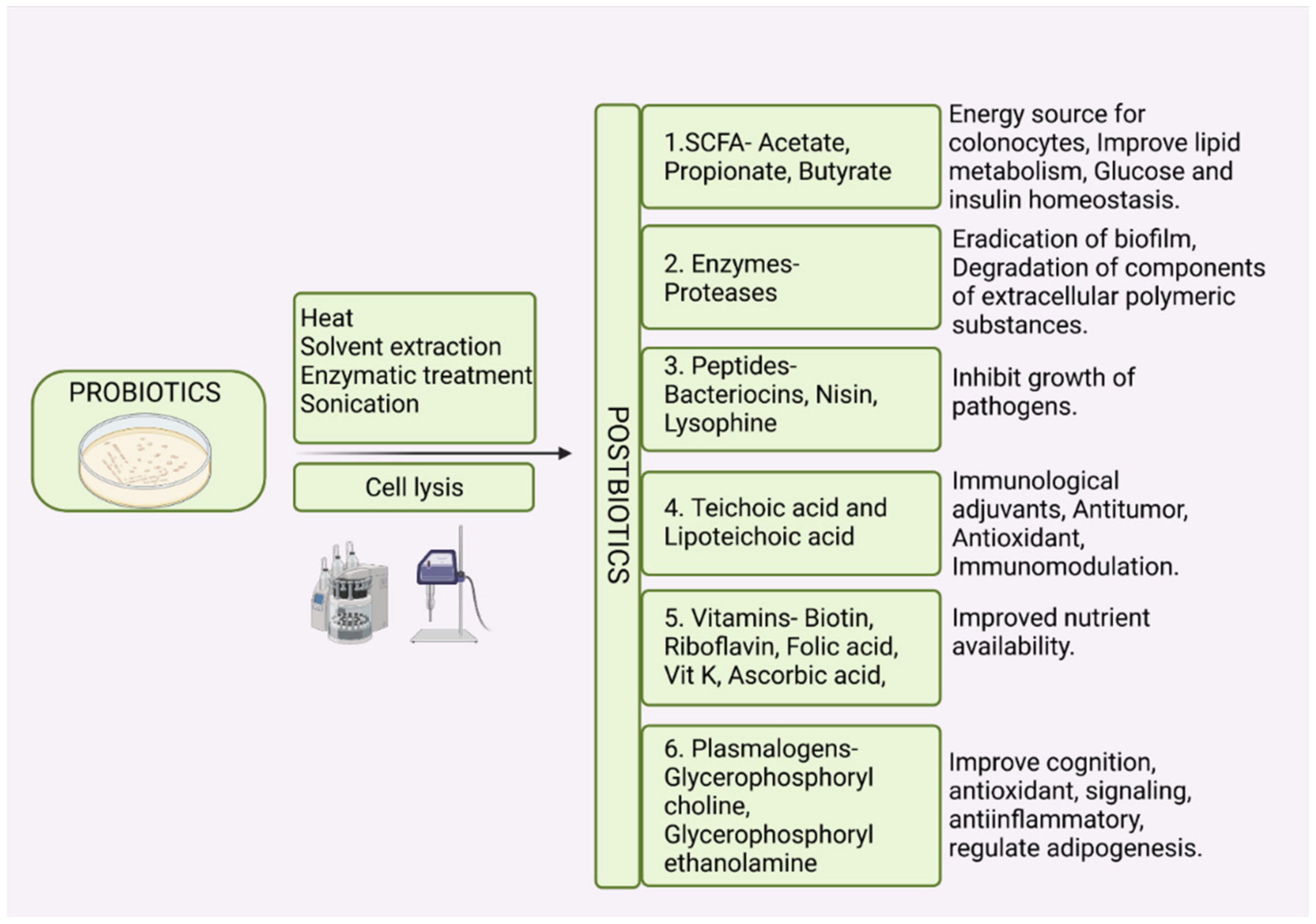
5. Postbiotics and Paraprobiotics

The efficacy of probiotics is associated with the viability of the microorganisms used. However, there is evidence that the viability of the microbes is not a requisite for conferring the beneficial effects of probiotics to the host [88]. The emergence of terms such as ‘postbiotics’, ‘paraprobiotics’, ‘metabiotics’, ‘inactivated probiotics’, and ‘ghost probiotics’ indicates that supplementing non-viable microbes or microbial products can also confer health benefits to the host. Postbiotics are low molecular weight soluble factors that are either secreted by live bacteria or released after bacterial cell lysis and when administered in sufficient amounts confer health benefits to the host [88]. Postbiotics are obtained by disrupting the microbial cell structure through heat treatment [89], enzymatic treatment [90], solvent extraction [91], or sonication [92]. Postbiotics are further purified through centrifugation, column purification, freeze-drying, microfiltration, and dialysis [93][94].
Paraprobiotics also termed as “inactivated probiotics” or “ghost probiotics”, are non-viable probiotic or non-probiotic microbial cells or cell fractions which when administered in sufficient amounts confer beneficial effects to the host [95][96]. Paraprobiotics are essentially non-culturable, but immunologically active microbial cells that benefit the host. Thermal treatment, high pressure, irradiation, and sonication, which induce cell death without membrane degradation, are applied in the production of paraprobiotics [95][96][97][98][99]. Flow cytometry using fluorescent dyes is used to assess the functional state of inactivated cells [100].
Postbiotics (Nonbiotics) are low molecular weight non-viable factors such as metabolic products, byproducts, or cell wall components (metabiotics, cell-free supernatants, secretions, cell lysates, or biogenic metabolites) derived from probiotic microorganisms which when administered in sufficient amounts confer health benefits to the consumer [88][96]. Postbiotics can be soluble substances secreted by the live bacteria or the products of bacterial cell lysis, as summarized in Figure 1. This soluble fraction contains SCFAs, bacteriocins, vitamins, peptides, organic acids, hydrogen peroxide, enzymes, cell surface proteins, plasmalogens, peptidoglycan-derived muropeptides, teichoic acids, exopolysaccharides and endopolysaccharides [96][101][102]. The exact mechanism of action of postbiotics is not completely elucidated. In a study conducted by Yan et al. [103], it was demonstrated that Lactobacillus rhamnosus derived protein p40 activates EGFR and prevents the cytokine-induced apoptosis of intestinal epithelial cells in inflammatory conditions such as inflammatory bowel disease (IBD). In a study conducted by Humam et al. [104], it was demonstrated that the birds fed postbiotics derived from different strains of L. plantarum exhibited significantly improved FCR values compared to the control group. Postbiotic supplementation increased the expression of IGF-1 and growth hormone receptors during heat stress [104].
Figure 1. Postbiotics are soluble low molecular weight metabolites or cell lysis products derived from live or inactivated probiotic bacteria which when administered in adequate quantities demonstrate beneficial effects on host health. Created with Biorender.com (4 April 2022).
Although the mechanism of postbiotic action is not completely explained, scientific data provide evidence for the role of postbiotics in several physiological functions. Postbiotics can be classified based on their structural composition or their physiological functions. Based on their role in host physiology, postbiotics can be 1. antimicrobials such as cell-free supernatants of L. plantarum [105], 2. immunomodulatory substances such as cell wall components of Bifidobacterium and Lactobacillus, lipoteichoic acid of L. plantarum [91], 3. anti-inflammatory factors such as cell free supernatant of L. paracasei and L. rhamnosus [103][106], 4. anti-proliferative substances such as sonicated cell suspension of L. casei [107], 5. anti-obesogenic compounds such as fragmented cells of L. amylovorus [108], 6. Hypocholesterolemic substances [98], 7. anti-hypertensives such as polysaccharide glycopeptide complexes of L. casei [94], and 8. antioxidants such as intracellular contents of Streptococcus salivarius, L. acidophilus, and L. casei [103][107]. Supplementation of bacteriocin from L. plantarum improves growth rate, increases the fecal LAB population, and reduces the abundance of Enterobacteriaceae in broilers [109]. Thus, the concept of feeding live microbials is being replaced by postbiotics which have been demonstrated to confer similar health benefits to the host.
Based on the composition, postbiotics can be 1. carbohydrates such as galactose-rich polysaccharides and teichoic acids 2. lipids such as propionate and butyrate 3. proteins such as p40 molecule and lactocepins 4. organic acids such as propionic acid and 3-phenyl lactic acid 5. vitamins such as B-complex vitamins 6. complex molecules such as lipoteichoic acids and peptidoglycan derived muropeptides [101][110][111]. In a study conducted by Abd El-Ghany et al. [112], it was reported that supplementation of inactivated Lactobacillus to chickens improved the immune response to Newcastle disease virus vaccines. The improved immune response of the birds is due to the lipopolysaccharides, lipoteichoic acid, and teichoic acid present in the bacterial cell wall which acts as adjuvants to stimulate the host immune response. Lysis of the inactivated bacteria in the host gut releases nuclear antigens which might stimulate the host humoral and cell-mediated immune responses.
One of the advantages of postbiotics and paraprobiotics over probiotics is the possibility of postbiotics and paraprobiotics applications in immunocompromised patients. Administration of live microbes presents a risk to immunocompromised patients because of the possibility of occurrence of opportunistic infections and metastasis of probiotic microbes. For instance, translocation of the Streptococcus gallolyticus from the gut to the bloodstream has been implicated in colorectal cancer in humans [113]. Paraprobiotics or postbiotics such as SCFA have antimutagenic activities and selectively target cancer cells and can be applied to treat cancer patients [111]. Postbiotics have a clear chemical structure, longer shelf life, and safety dose parameters. In a study conducted by Shigwedha et al. [114], it was observed that probiotic cell fragments, the structural components of probiotic cell lysates, exert a broad spectrum of immunomodulatory functions. The shelf stability of probiotic cell fragments can be up to 5 years. The use of postbiotics can also eliminate the risk of prevalence and transfer of antibiotic resistance genes from probiotic species to the consumer [115].

References

  1. Broom, L.J. Gut barrier function: Effects of (antibiotic) growth promoters on key barrier components and associations with growth performance. Poult. Sci. 2018, 97, 1572–1578.
  2. Broom, L.J.; Kogut, M.H. The role of the gut microbiome in shaping the immune system of chickens. Vet. Immunol. Immunopathol. 2018, 204, 44–51.
  3. Befus, A.D.; Johnston, N.; Leslie, G.A.; Bienenstock, J. Gut-associated lymphoid tissue in the chicken. I. Morphology, ontogeny, and some functional characteristics of Peyer’s patches. J. Immunol. 1980, 125, 2626–2632.
  4. Alagawany, M.; Abd El-Hack, M.E.; Farag, M.R.; Sachan, S.; Karthik, K.; Dhama, K. The use of probiotics as eco-friendly alternatives for antibiotics in poultry nutrition. Environ. Sci. Pollut. Res. 2018, 25, 10611–10618.
  5. Vila, B.; Esteve-Garcia, E.; Brufau, J. Probiotic micro-organisms: 100 years of innovation and efficacy; modes of action. Worlds Poult. Sci. J. 2010, 66, 369–380.
  6. Joint FAO/WHO Working Group. Guidelines for the Evaluation of Probiotics in Food. In Food and Agriculture Organization; World Health Organization: London, UK, 2002.
  7. Fidanza, M.; Panigrahi, P.; Kollmann, T.R. Lactiplantibacillus plantarum–Nomad and Ideal Probiotic. Front. Microbiol. 2021, 12, 2911.
  8. Ma, T.; Suzuki, Y. Dissect the mode of action of probiotics in affecting host-microbial interactions and immunity in food producing animals. Vet. Immunol. Immunopathol. 2018, 205, 35–48.
  9. Patterson, J.A.; Burkholder, K.M. Application of prebiotics and probiotics in poultry production. Poult. Sci. 2003, 82, 627–631.
  10. Dowarah, R.; Verma, A.K.; Agarwal, N.; Singh, P.; Singh, B.R. Selection and characterization of probiotic lactic acid bacteria and its impact on growth, nutrient digestibility, health and antioxidant status in weaned piglets. PLoS ONE 2018, 13, e0192978.
  11. Ehrmann, M.A.; Kurzak, P.; Bauer, J.; Vogel, R.F. Characterization of lactobacilli towards their use as probiotic adjuncts in poultry. J. Appl. Microbiol. 2002, 92, 966–975.
  12. Reuben, R.C.; Roy, P.C.; Sarkar, S.L.; Alam, R.; Jahid, I.K. Isolation, characterization, and assessment of lactic acid bacteria toward their selection as poultry probiotics. BMC Microbiol. 2019, 19, 253.
  13. European Food Safety Authority and European Centre for Disease Prevention and Control (EFSA and ECDC). The European Union summary report on trends and sources of zoonoses, zoonotic agents and food-borne outbreaks in 2017. EFSa J. 2018, 16, e05077.
  14. Hesari, M.R.; Darsanaki, R.K.; Salehzadeh, A. Antagonistic activity of probiotic bacteria isolated from traditional dairy products against E. coli O157: H7. J. Med. Bacteriol. 2017, 6, 23–30.
  15. Liao, S.F.; Nyachoti, M. Using probiotics to improve swine gut health and nutrient utilization. Anim. Nutr. 2017, 3, 331–343.
  16. Garriga, M.; Pascual, M.; Monfort, J.M.; Hugas, M. Selection of lactobacilli for chicken probiotic adjuncts. J. Appl. Microbiol. 1998, 84, 125–132.
  17. Farner, D.S. The hydrogen ion concentration in avian digestive tracts. Poult. Sci. 1942, 21, 445–450.
  18. Pan, X.; Chen, F.; Wu, T.; Tang, H.; Zhao, Z. The acid, bile tolerance and antimicrobial property of Lactobacillus acidophilus NIT. Food Control 2009, 20, 598–602.
  19. Nallala, V.; Sadishkumar, V.; Jeevaratnam, K. Molecular characterization of antimicrobial Lactobacillus isolates and evaluation of their probiotic characteristics in vitro for use in poultry. Food Biotechnol. 2017, 31, 20–41.
  20. Lin, J.; Sahin, O.; Michel, L.O.; Zhang, Q. Critical role of multidrug efflux pump CmeABC in bile resistance and in vivo colonization of Campylobacter jejuni. Infect. Immun. 2003, 71, 4250–4259.
  21. Taheri, H.R.; Moravej, H.; Tabandeh, F.; Zaghari, M.; Shivazad, M. Screening of lactic acid bacteria toward their selection as a source of chicken probiotic. Poult. Sci. 2009, 88, 1586–1593.
  22. Collado, M.C.; Meriluoto, J.; Salminen, S. Adhesion and aggregation properties of probiotic and pathogen strains. Eur. Food Res. Technol. 2008, 226, 1065–1073.
  23. Dec, M.; Urban-Chmiel, R.; Stępień-Pyśniak, D.; Wernicki, A. Assessment of antibiotic susceptibility in Lactobacillus isolates from chickens. Gut Pathog. 2017, 9, 1–16.
  24. Ramlucken, U.; Ramchuran, S.O.; Moonsamy, G.; van Rensburg, C.J.; Thantsha, M.S.; Lalloo, R. Production and stability of a multi-strain Bacillus based probiotic product for commercial use in poultry. Biotechnol. Rep. 2021, 29, e00575.
  25. Mountzouris, K.C.; Tsitrsikos, P.; Palamidi, I.; Arvaniti, A.; Mohnl, M.; Schatzmayr, G.; Fegeros, K. Effects of probiotic inclusion levels in broiler nutrition on growth performance, nutrient digestibility, plasma immunoglobulins, and cecal microflora composition. Poult. Sci. 2010, 89, 58–67.
  26. Tripathi, M.K.; Giri, S.K. Probiotic functional foods: Survival of probiotics during processing and storage. J. Funct. Foods 2014, 9, 225–241.
  27. Lee, Y.K.; Salminen, S. Handbook of Probiotics and Prebiotics; John Wiley & Sons: Hoboken, NJ, USA, 2009.
  28. Teixeira, P.C.; Castro, M.H.; Malcata, F.X.; Kirby, R.M. Survival of Lactobacillus delbrueckii ssp. bulgaricus following spray-drying. J. Dairy Sci. 1995, 78, 1025–1031.
  29. Tamime, A.Y.; Saarela, M.; Sondergaard, A.K.; Mistry, V.V.; Shah, N.P. Production and maintenance of viability of probiotic microorganisms in dairy products. Probiotic Dairy Prod. 2005, 3, 39–63.
  30. Teixeira, P.; Castro, H.; Kirby, R. Inducible thermotolerance in Lactobacillus bulgaricus. Lett. Appl. Microbiol. 1994, 18, 218–221.
  31. Park, Y.H.; Hamidon, F.; Rajangan, C.; Soh, K.P.; Gan, C.Y.; Lim, T.S.; Abdullah, W.N.W.; Liong, M.T. Application of probiotics for the production of safe and high-quality poultry meat. Korean J. Food Sci. Anim. Resour. 2016, 36, 567.
  32. Thomke, S.; Elwinger, K. Growth Promotants in Feeding Pigs and Poultry. III. Alternatives to Antibiotic Growth Promotants. Ann. Zootech. INRA/EDP Sci. 1998, 47, 245–271. Available online: https://hal.archives-ouvertes.fr/hal-00889716 (accessed on 1 March 2022).
  33. Gadde, U.; Kim, W.H.; Oh, S.T.; Lillehoj, H.S. Alternatives to antibiotics for maximizing growth performance and feed efficiency in poultry: A review. Anim. Health Res. Rev. 2017, 18, 26–45.
  34. Huyghebaert, G.; Ducatelle, R.; Van Immerseel, F. An update on alternatives to antimicrobial growth promoters for broilers. Vet. J. 2011, 187, 182–188.
  35. Gauthier, R. Defining the Alternatives. Canadian Poultry, 10 January 2008.
  36. Huyghebaert, G. Alternatives for Antibiotic in Poultry; CONFERENCE SPONSORS: Timonium, MD, USA, 2005; p. 38.
  37. Jeurissen, S.H.; Lewis, F.; van der Klis, J.D.; Mroz, Z.; Rebel, J.M.; Ter Huurne, A.A. Parameters and techniques to determine intestinal health of poultry as constituted by immunity, integrity, and functionality. Curr. Issues Intest. Microbiol. 2002, 3, 1–14.
  38. Figueroa-González, I.; Quijano, G.; Ramirez, G.; Cruz-Guerrero, A. Probiotics and prebiotics—Perspectives and challenges. J. Sci. Food Agric. 2011, 91, 1341–1348.
  39. Markazi, A.; Luoma, A.; Shanmugasundaram, R.; Mohnl, M.; Murugesan, G.R.; Selvaraj, R. Effects of drinking water synbiotic supplementation in laying hens challenged with Salmonella. Poult. Sci. 2018, 97, 3510–3518.
  40. Kabir, S.M. The role of probiotics in the poultry industry. Int. J. Mol. Sci. 2009, 10, 3531–3546.
  41. Wei, S.; Morrison, M.; Yu, Z. Bacterial census of poultry intestinal microbiome. Poult. Sci. 2013, 92, 671–683.
  42. Suez, J.; Zmora, N.; Zilberman-Schapira, G.; Mor, U.; Dori-Bachash, M.; Bashiardes, S.; Zur, M.; Regev-Lehavi, D.; Brik, R.B.; Federici, S. Post-antibiotic gut mucosal microbiome reconstitution is impaired by probiotics and improved by autologous FMT. Cell 2018, 174, 1406–1423.e16.
  43. Gupta, S.; Allen-Vercoe, E.; Petrof, E.O. Fecal Microbiota Transplantation: In perspective. Ther. Adv. Gastroenterol. 2016, 9, 229–239.
  44. Seekatz, A.M.; Aas, J.; Gessert, C.E.; Rubin, T.A.; Saman, D.M.; Bakken, J.S.; Young, V.B. Recovery of the gut microbiome following fecal microbiota transplantation. mBio 2014, 5, 893.
  45. Nurmi, E.; Rantala, M. New aspects of Salmonella infection in broiler production. Nature 1973, 241, 210–211.
  46. Stern, N.J.; Cox, N.A.; Bailey, J.S.; Berrang, M.E.; Musgrove, M.T. Comparison of mucosal competitive exclusion and competitive exclusion treatment to reduce Salmonella and Campylobacter spp. colonization in broiler chickens. Poult. Sci. 2001, 80, 156–160.
  47. Lee, Y.K.; Mazmanian, S.K. Has the microbiota played a critical role in the evolution of the adaptive immune system? Science 2010, 330, 1768–1773.
  48. Blajman, J.E.; Frizzo, L.S.; Zbrun, M.V.; Astesana, D.M.; Fusari, M.L.; Soto, L.P.; Rosmini, M.R.; Signorini, M.L. Probiotics and broiler growth performance: A meta-analysis of randomised controlled trials. Br. Poult. Sci. 2014, 55, 483–494.
  49. Edens, F.W. An alternative for antibiotic se in poultry: Probiotics. Braz. J. Poult. Sci. 2003, 5, 75–97.
  50. Olnood, C.G.; Beski, S.S.; Iji, P.A.; Choct, M. Delivery routes for probiotics: Effects on broiler performance, intestinal morphology and gut microflora. Anim. Nutr. 2015, 1, 192–202.
  51. Karimi Torshizi, M.A.; Moghaddam, A.R.; Rahimi, S.H.; Mojgani, N. Assessing the effect of administering probiotics in water or as a feed supplement on broiler performance and immune response. Br. Poult. Sci. 2010, 51, 178–184.
  52. Eckert, N.H.; Lee, J.T.; Hyatt, D.; Stevens, S.M.; Anderson, S.; Anderson, P.N.; Beltran, R.; Schatzmayr, G.; Mohnl, M.; Caldwell, D.J. Influence of probiotic administration by feed or water on growth parameters of broilers reared on medicated and nonmedicated diets. J. Appl. Poult. Res. 2010, 19, 59–67.
  53. Hogg, S. Essential Microbiology; John Wiley & Sons: West Sussex, UK; Hoboken, NJ, USA, 2013.
  54. Higgins, J.P.; Higgins, S.E.; Vicente, J.L.; Wolfenden, A.D.; Tellez, G.; Hargis, B. Temporal effects of lactic acid bacteria probiotic culture on Salmonella in neonatal broilers. Poult. Sci. 2007, 86, 1662–1666.
  55. De Oliveira, J.E.; Van der Hoeven-Hangoor, E.; Van de Linde, I.B.; Montijn, R.C.; Van der Vossen, J. In ovo inoculation of chicken embryos with probiotic bacteria and its effect on posthatch Salmonella susceptibility. Poult. Sci. 2014, 93, 818–829.
  56. Roto, S.M.; Kwon, Y.M.; Ricke, S.C. Applications of in ovo technique for the optimal development of the gastrointestinal tract and the potential influence on the establishment of its microbiome in poultry. Front. Vet. Sci. 2016, 3, 63.
  57. Ferket, P.R. Embryo Epigenomic Response to Breeder Management and Nutrition; World’s Poultry Congress (Abstr.); Semantic Scholar: Seattle, WA, USA, 2012.
  58. Gulewicz, K.; Bednarczyk, M. Method for Stimulating Favourable Bacteria Profile in Hatched Chicks. Sposób Stymulacji Korzystnego Profilu Bakteryjnego Wylężonych Piskląt. Poland Application PL20030364037, 12 December 2003.
  59. Uni, Z.; Ferket, P. Enhancement of Development of Oviparous Species by in OVO Feeding. U.S. Patent 6,592,878 B2, 15 July 2003.
  60. Sławińska, A.; Siwek, M.; Żylińska, J.; Bardowski, J.; Brzezińska, J.; Gulewicz, K.A.; Nowak, M.; Urbanowski, M.; Płowiec, A.; Bednarczyk, M. Influence of synbiotics delivered in ovo on immune organs development and structure. Folia Biol. 2014, 62, 277–285.
  61. Bednarczyk, M.; Stadnicka, K.; Kozłowska, I.; Abiuso, C.; Tavaniello, S.; Dankowiakowska, A.; Sławińska, A.; Maiorano, G. Influence of different prebiotics and mode of their administration on broiler chicken performance. Animal 2016, 10, 1271–1279.
  62. Li, T.; Castañeda, C.D.; Miotto, J.; McDaniel, C.; Kiess, A.S.; Zhang, L. Effects of in ovo probiotic administration on the incidence of avian pathogenic Escherichia coli in broilers and an evaluation on its virulence and antimicrobial resistance properties. Poult. Sci. 2021, 100, 100903.
  63. Siwek, M.; Slawinska, A.; Stadnicka, K.; Bogucka, J.; Dunislawska, A.; Bednarczyk, M. Prebiotics and synbiotics–in ovo delivery for improved lifespan condition in chicken. BMC Vet. Res. 2018, 14, 1–17.
  64. Villaluenga, C.M.; Wardeńska, M.; Pilarski, R.; Bednarczyk, M.; Gulewicz, K. Utilization of the chicken embryo model for assessment of biological activity of different oligosaccharides. Folia Biol. 2004, 52, 135–142.
  65. Blankenship, L.C.; Bailey, J.S.; Cox, N.A.; Stern, N.J.; Brewer, R.; Williams, O. Two-step mucosal competitive exclusion flora treatment to diminish salmonellae in commercial broiler chickens. Poult. Sci. 1993, 72, 1667–1672.
  66. Wolfenden, A.D.; Pixley, C.M.; Higgins, J.P.; Higgins, S.E.; Vicente, J.; Torres-Rodriguez, A.; Hargis, B.M.; Tellez, G. Evaluation of spray application of a Lactobacillus-based probiotic on Salmonella enteritidis colonization in broiler chickens. Int. J. Poult. Sci. 2007, 6, 493–496.
  67. Cox, N.A.; Bailey, J.S.; Blankenship, L.C. Alternative administration of competitive exclusion treatment. In Colonization Control of Human Bacterial Enteropathologens in Poultry; Academic Press: Cambridge, MA, USA, 1991; pp. 105–118.
  68. Nicetic, M.; Kailasapathy, K.; Tarasoff, L. Mechanical Stability of Food Gum Gels for Immobilization of Probiotic Bacteria. In Proceedings of the 8th International Workshop on Bioencapsulation, Recent Progress in Research and Technology, Trondheim, Norway, 13–15 September 1999; Abstract, P11. pp. 13–15.
  69. Würth, R.; Hörmannsperger, G.; Wilke, J.; Foerst, P.; Haller, D.; Kulozik, U. Protective effect of milk protein based microencapsulation on bacterial survival in simulated gastric juice versus the murine gastrointestinal system. J. Funct. Foods 2015, 15, 116–125.
  70. Cottyn, B.; Heylen, K.; Heyrman, J.; Vanhouteghem, K.; Pauwelyn, E.; Bleyaert, P.; Van Vaerenbergh, J.; Höfte, M.; De Vos, P.; Maes, M. Pseudomonas cichorii as the causal agent of midrib rot, an emerging disease of greenhouse-grown butterhead lettuce in Flanders. Syst. Appl. Microbiol. 2009, 32, 211–225.
  71. Nkukwana, T.T.; Muchenje, V.; Masika, P.J.; Mushonga, B. Intestinal morphology, digestive organ size and digesta pH of broiler chickens fed diets supplemented with or without Moringa oleifera leaf meal. S. Afr. J. Anim. Sci. 2015, 45, 362–370.
  72. Shanmugasundaram, R.; Mortada, M.; Murugesan, G.R.; Selvaraj, R.K. In vitro characterization and analysis of probiotic species in the chicken intestine by real-time polymerase chain reaction. Poult. Sci. 2019, 98, 5840–5846.
  73. Zmora, N.; Zilberman-Schapira, G.; Suez, J.; Mor, U.; Dori-Bachash, M.; Bashiardes, S.; Kotler, E.; Zur, M.; Regev-Lehavi, D.; Brik, R.B. Personalized gut mucosal colonization resistance to empiric probiotics is associated with unique host and microbiome features. Cell 2018, 174, 1388–1405.e21.
  74. Mullineaux-Sanders, C.; Suez, J.; Elinav, E.; Frankel, G. Sieving through gut models of colonization resistance. Nat. Microbiol. 2018, 3, 132–140.
  75. Ferreiro, A.; Dantas, G.; Ciorba, M.A. Insights into how probiotics colonize the healthy human gut. Gastroenterology 2019, 156, 820–822.
  76. Yatsunenko, T.; Rey, F.E.; Manary, M.J.; Trehan, I.; Dominguez-Bello, M.G.; Contreras, M.; Magris, M.; Hidalgo, G.; Baldassano, R.N.; Anokhin, A.P. Human gut microbiome viewed across age and geography. Nature 2012, 486, 222–227.
  77. Murphy, E.F.; Cotter, P.D.; Healy, S.; Marques, T.M.; O’sullivan, O.; Fouhy, F.; Clarke, S.F.; O’toole, P.W.; Quigley, E.M.; Stanton, C. Composition and energy harvesting capacity of the gut microbiota: Relationship to diet, obesity and time in mouse models. Gut 2010, 59, 1635–1642.
  78. Oakley, B.B.; Lillehoj, H.S.; Kogut, M.H.; Kim, W.K.; Maurer, J.J.; Pedroso, A.; Lee, M.D.; Collett, S.R.; Johnson, T.J.; Cox, N.A. The chicken gastrointestinal microbiome. FEMS Microbiol. Lett. 2014, 360, 100–112.
  79. Bozdogan, B.; Galopin, S.; Leclercq, R. Characterization of a new erm-related macrolide resistance gene present in probiotic strains of Bacillus clausii. Appl. Environ. Microbiol. 2004, 70, 280–284.
  80. Gueimonde, M.; Sánchez, B.; de Los Reyes-Gavilán Clara, G.; Margolles, A. Antibiotic resistance in probiotic bacteria. Front. Microbiol. 2013, 4, 202.
  81. Feld, L.; Schjørring, S.; Hammer, K.; Licht, T.R.; Danielsen, M.; Krogfelt, K.; Wilcks, A. Selective pressure affects transfer and establishment of a Lactobacillus plantarum resistance plasmid in the gastrointestinal environment. J. Antimicrob. Chemother. 2008, 61, 845–852.
  82. Witte, W. Ecological impact of antibiotic use in animals on different complex microflora: Environment. Int. J. Antimicrob. Agents 2000, 14, 321–325.
  83. Schjørring, S.; Krogfelt, K.A. Assessment of bacterial antibiotic resistance transfer in the gut. Int. J. Microbiol. 2011, 2011, 312956.
  84. Hidayat, M.N.; Malaka, R.; Agustina, L.; Pakiding, W. Abdominal fat percentage and carcass quality of broiler given probiotics Bacillus spp. Metab. Clin. Exp. 2016, 22, 3–60.
  85. Haines, M.D.; Parker, H.M.; McDaniel, C.D.; Kiess, A.S. When rooster semen is exposed to Lactobacillus fertility is reduced. Int. J. Poult. Sci. 2015, 14, 541–547.
  86. Peralta-Sánchez, J.M.; Martín-Platero, A.M.; Ariza-Romero, J.J.; Rabelo-Ruiz, M.; Zurita-González, M.J.; Baños, A.; Rodríguez-Ruano, S.M.; Maqueda, M.; Valdivia, E.; Martínez-Bueno, M. Egg production in poultry farming is improved by probiotic bacteria. Front. Microbiol. 2019, 10, 1042.
  87. Haines, M.D.; Parker, H.M.; McDaniel, C.D.; Kiess, A.S. Impact of 6 different intestinal bacteria on broiler breeder sperm motility in vitro. Poult. Sci. 2013, 92, 2174–2181.
  88. Aguilar-Toalá, J.E.; Garcia-Varela, R.; Garcia, H.S.; Mata-Haro, V.; González-Córdova, A.F.; Vallejo-Cordoba, B.; Hernández-Mendoza, A. Postbiotics: An evolving term within the functional foods field. Trends Food Sci. Technol. 2018, 75, 105–114.
  89. Lee, M.; Zang, Z.; Choi, E.; Shin, H.; Ji, G. Cytoskeleton reorganization and cytokine production of macrophages by bifidobacterial cells and cell-free extracts. J. Microbiol. Biotechnol. 2002, 12, 398–405.
  90. Li, S.; Zhao, Y.; Zhang, L.; Zhang, X.; Huang, L.; Li, D.; Niu, C.; Yang, Z.; Wang, Q. Antioxidant activity of Lactobacillus plantarum strains isolated from traditional Chinese fermented foods. Food Chem. 2012, 135, 1914–1919.
  91. Kim, H.G.; Lee, S.Y.; Kim, N.R.; Lee, H.Y.; Ko, M.Y.; Jung, B.J.; Kim, C.M.; Lee, J.M.; Park, J.H.; Han, S.H. Lactobacillus plantarum lipoteichoic acid down-regulated Shigella flexneri peptidoglycan-induced inflammation. Mol. Immunol. 2011, 48, 382–391.
  92. Tiptiri-Kourpeti, A.; Spyridopoulou, K.; Santarmaki, V.; Aindelis, G.; Tompoulidou, E.; Lamprianidou, E.E.; Saxami, G.; Ypsilantis, P.; Lampri, E.S.; Simopoulos, C. Lactobacillus casei exerts anti-proliferative effects accompanied by apoptotic cell death and up-regulation of TRAIL in colon carcinoma cells. PLoS ONE 2016, 11, e0147960.
  93. Matsuguchi, T.; Takagi, A.; Matsuzaki, T.; Nagaoka, M.; Ishikawa, K.; Yokokura, T.; Yoshikai, Y. Lipoteichoic acids from Lactobacillus strains elicit strong tumor necrosis factor alpha-inducing activities in macrophages through Toll-like receptor 2. Clin. Vaccine Immunol. 2003, 10, 259–266.
  94. Sawada, H.; Furushiro, M.; Hirai, K.; Motoike, M.; Watanabe, T.; Yokokura, T. Purification and Characterization of an Antihypertensive Compound from Lactohacillus casei. Agric. Biol. Chem. 1990, 54, 3211–3219.
  95. De Almada, C.N.; Almada, C.N.; Martinez, R.C.; Sant’Ana, A.S. Paraprobiotics: Evidences on their ability to modify biological responses, inactivation methods and perspectives on their application in foods. Trends Food Sci. Technol. 2016, 58, 96–114.
  96. Taverniti, V.; Guglielmetti, S. The immunomodulatory properties of probiotic microorganisms beyond their viability (ghost probiotics: Proposal of paraprobiotic concept). Genes Nutr. 2011, 6, 261–274.
  97. Ou, C.; Lin, S.; Tsai, J.; Lin, M. Heat-killed lactic acid bacteria enhance immunomodulatory potential by skewing the immune response toward Th1 polarization. J. Food Sci. 2011, 76, M260–M267.
  98. Shin, H.S.; Park, S.Y.; Lee, D.K.; Kim, S.; An, H.M.; Kim, J.R.; Kim, M.J.; Cha, M.G.; Lee, S.W.; Kim, K.J. Hypocholesterolemic effect of sonication-killed Bifidobacterium longum isolated from healthy adult Koreans in high cholesterol fed rats. Arch. Pharm. Res. 2010, 33, 1425–1431.
  99. Ananta, E.; Knorr, D. Comparison of inactivation pathways of thermal or high pressure inactivated Lactobacillus rhamnosus ATCC 53103 by flow cytometry analysis. Food Microbiol. 2009, 26, 542–546.
  100. Gandhi, A.; Shah, N.P. Effect of salt on cell viability and membrane integrity of Lactobacillus acidophilus, Lactobacillus casei and Bifidobacterium longum as observed by flow cytometry. Food Microbiol. 2015, 49, 197–202.
  101. Tsilingiri, K.; Rescigno, M. Postbiotics: What else? Benef. Microb. 2013, 4, 101–107.
  102. Konstantinov, S.R.; Kuipers, E.J.; Peppelenbosch, M.P. Functional genomic analyses of the gut microbiota for CRC screening. Nat. Rev. Gastroenterol. Hepatol. 2013, 10, 741–745.
  103. Yan, F.; Cao, H.; Cover, T.L.; Washington, M.K.; Shi, Y.; Liu, L.; Chaturvedi, R.; Peek, R.M.; Wilson, K.T.; Polk, D.B. Colon-specific delivery of a probiotic-derived soluble protein ameliorates intestinal inflammation in mice through an EGFR-dependent mechanism. J. Clin. Investig. 2011, 121, 2242–2253.
  104. Humam, A.M.; Loh, T.C.; Foo, H.L.; Samsudin, A.A.; Mustapha, N.M.; Zulkifli, I.; Izuddin, W.I. Effects of feeding different postbiotics produced by Lactobacillus plantarum on growth performance, carcass yield, intestinal morphology, gut microbiota composition, immune status, and growth gene expression in broilers under heat stress. Animals 2019, 9, 644.
  105. Kareem, K.Y.; Hooi Ling, F.; Teck Chwen, L.; May Foong, O.; Anjas Asmara, S. Inhibitory activity of postbiotic produced by strains of Lactobacillus plantarum using reconstituted media supplemented with inulin. Gut Pathog. 2014, 6, 23.
  106. Tsilingiri, K.; Barbosa, T.; Penna, G.; Caprioli, F.; Sonzogni, A.; Viale, G.; Rescigno, M. Probiotic and postbiotic activity in health and disease: Comparison on a novel polarised ex-vivo organ culture model. Gut 2012, 61, 1007–1015.
  107. Choi, S.S.; Kim, Y.; Han, K.S.; You, S.; Oh, S.; Kim, S.H. Effects of Lactobacillus strains on cancer cell proliferation and oxidative stress in vitro. Lett. Appl. Microbiol. 2006, 42, 452–458.
  108. Nakamura, F.; Ishida, Y.; Sawada, D.; Ashida, N.; Sugawara, T.; Sakai, M.; Goto, T.; Kawada, T.; Fujiwara, S. Fragmented lactic acid bacterial cells activate peroxisome proliferator-activated receptors and ameliorate dyslipidemia in obese mice. J. Agric. Food Chem. 2016, 64, 2549–2559.
  109. Thanh, N.T.; Loh, T.C.; Foo, H.L.; Hair-Bejo, M.; Azhar, B.K. Effects of feeding metabolite combinations produced by Lactobacillus plantarum on growth performance, faecal microbial population, small intestine villus height and faecal volatile fatty acids in broilers. Br. Poult. Sci. 2009, 50, 298–306.
  110. Compare, D.; Rocco, A.; Coccoli, P.; Angrisani, D.; Sgamato, C.; Iovine, B.; Salvatore, U.; Nardone, G. Lactobacillus casei DG and its postbiotic reduce the inflammatory mucosal response: An ex-vivo organ culture model of post-infectious irritable bowel syndrome. BMC Gastroenterol. 2017, 17, 53.
  111. Verma, A.; Shukla, G. Synbiotic (Lactobacillus rhamnosus Lactobacillus acidophilus inulin) attenuates oxidative stress and colonic damage in 1, 2 dimethylhydrazine dihydrochloride-induced colon carcinogenesis in Sprague–Dawley rats. Eur. J. Cancer Prev. 2014, 23, 550–559.
  112. Abd El-Ghany, W.; Hosny, F.; Quesnell, R.; Sakai, L. The Effect of a Postbiotic Produced by Stabilized Non-Viable Lactobacilli on the Health, Growth Performance, Immunity, and Gut Status of Colisepticaemic Broiler Chickens; Research Square: Durham, NC, USA, 2022.
  113. Abdulamir, A.S.; Hafidh, R.R.; Bakar, F.A. Molecular detection, quantification, and isolation of Streptococcus gallolyticus bacteria colonizing colorectal tumors: Inflammation-driven potential of carcinogenesis via IL-1, COX-2, and IL-8. Mol. Cancer 2010, 9, 249.
  114. Shigwedha, N. Probiotical cell fragments (PCFs) as “novel nutraceutical ingredients”. J. Biosci. Med. 2014, 2, 43–55.
  115. Lin, C.; Fung, Z.; Wu, C.; Chung, T. Molecular Characterization of a Plasmid-Borne (pTC82) Chloramphenicol Resistance Determinant (cat-TC) fromLactobacillus reuteriG4. Plasmid 1996, 36, 116–124.
More
Information
Contributors MDPI registered users' name will be linked to their SciProfiles pages. To register with us, please refer to https://encyclopedia.pub/register : , , ,
View Times: 446
Entry Collection: Gastrointestinal Disease
Revisions: 2 times (View History)
Update Date: 19 May 2022
1000/1000